Carbon Nanotube Composites: Advanced Properties for ... · 1.4.2 Template-Assisted Synthesis of...
27
i Carbon Nanotube Composites: Advanced Properties for Emerging Applications A thesis submitted in fulfillment of the requirements for the degree of Doctor of Philosophy Engineering (Nanotechnology) By TARIQ ATEEQ ALTALHI January 2014 School of Chemical Engineering Faculty of Engineering, Computer and Mathematical Sciences The University of Adelaide
Transcript of Carbon Nanotube Composites: Advanced Properties for ... · 1.4.2 Template-Assisted Synthesis of...
i
Carbon Nanotube Composites:
Advanced Properties for Emerging Applications
A thesis submitted in fulfillment of
the requirements for the degree of
Doctor of Philosophy
Engineering (Nanotechnology)
By TARIQ ATEEQ ALTALHI
January 2014
School of Chemical Engineering
Faculty of Engineering, Computer and Mathematical Sciences
The University of Adelaide
ii
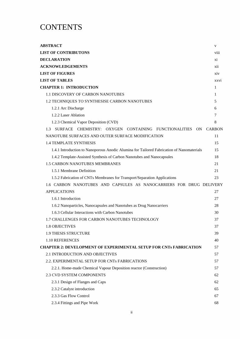
CONTENTS
ABSTRACT v
LIST OF CONTRIBUTONS viii
DECLARATION xi
ACKNOWLEDGEMENTS xii
LIST OF FIGURES xiv
LIST OF TABLES xxvi
CHAPTER 1: INTRODUCTION 1
1.1 DISCOVERY OF CARBON NANOTUBES 1
1.2 TECHNIQUES TO SYNTHESISE CARBON NANOTUBES 5
1.2.1 Arc Discharge 6
1.2.2 Laser Ablation 7
1.2.3 Chemical Vapor Deposition (CVD) 8
1.3 SURFACE CHEMISTRY: OXYGEN CONTAINING FUNCTIONALITIES ON CARBON
NANOTUBE SURFACES AND OUTER SURFACE MODIFICATION 11
1.4 TEMPLATE SYNTHESIS 15
1.4.1 Introduction to Nanoporous Anodic Alumina for Tailored Fabrication of Nanomaterials 15
1.4.2 Template-Assisted Synthesis of Carbon Nanotubes and Nanocapsules 18
1.5 CARBON NANOTUBES MEMBRANES 21
1.5.1 Membrane Definition 21
1.5.2 Fabrication of CNTs Membranes for Transport/Separation Applications 23
1.6 CARBON NANOTUBES AND CAPSULES AS NANOCARRIERS FOR DRUG DELIVERY
APPLICATIONS 27
1.6.1 Introduction 27
1.6.2 Nanoparticles, Nanocapsules and Nanotubes as Drug Nanocarriers 28
1.6.3 Cellular Interactions with Carbon Nanotubes 30
1.7 CHALLENGES FOR CARBON NANOTUBES TECHNOLOGY 37
1.8 OBJECTIVES 37
1.9 THESIS STRUCTURE 39
1.10 REFERENCES 40
CHAPTER 2: DEVELOPMENT OF EXPERIMENTAL SETUP FOR CNTs FABRICATION 57
2.1 INTRODUCTION AND OBJECTIVES 57
2.2. EXPERIMENTAL SETUP FOR CNTs FABRICATIONS 57
2.2.1. Home-made Chemical Vapour Deposition reactor (Construction) 57
2.3 CVD SYSTEM COMPONENTS 62
2.3.1 Design of Flanges and Caps 62
2.3.2 Catalyst introduction 65
2.3.3 Gas Flow Control 67
2.3.4 Fittings and Pipe Work 68

iii
2.4 CONCLUSIONS 69
2.5 REFERENCES 69
CHAPTER 3: FABRICATION AND CHARACTERISATION OF CNTS MEMBRANES 71
3.1. INTRODUCTION AND OBJECTIVES 71
3.2. EXPERIMENTAL SECTION 74
3.2.1 Materials 74
3.2.2 Synthesis of Nanoporous Anodic Alumina Membranes (NAAMs) 74
3.2.3 Synthesis of Titania Nanotubes (TNTs) 75
3.2.4 Synthesis of CNT/NAAMs Composite Membranes 76
3.2.5 Characterisation 76
3.3 RESULTS AND DISCUSSIONS 77
3.3.1 Structural Characterisation of Nanoporous Anodic Alumina Membranes (NAAMs) 77
3.3.2 Structural Characterisation of CNT/NAA Composites Fabricated using Catalyst and Catalyst-Free
CVD Process 78
3.3.3 Free-Catalyst CNTs Grown inside TNTs and their Comparison with CNTs Grown inside NAA
Templates 83
3.3.4 Chemical Analysis of Catalyst and Catalyst-Free CNTs from NAA and TNTs Template 87
3.3.4.1 XPS Analysis 87
3.3.4.2 Raman Analysis 89
3.4 CONCLUSIONS 91
3.5. FABRICATION AND STRUCTURAL CHARACTERISATION OF CNTS-NAAMS AND FREE-
STANDING CNTS FROM LOW COST WASTE PLASTIC MATERIAL 92
3.5.1 Introduction and Aims 92
3.5.2 Experimental 94
3.5.2.1 Materials 94
3.5.2.2 Commercially available LLDPE plastic bag 95
3.5.2.3 Synthesis of CNTs/NAAMs 95
3.5.2.4 Characterisation 96
3.5.3 Results and Discussion 97
3.5.3.1 Structural Characterisation of CNTs/NAAMs and Liberated CNTs 97
3.5.3.2 Chemical Analysis of CNT/-NAAMs 99
3.5.3.3 Chemical Characterisation of Liberated CNTs 102
3.5.4 Conclusions 102
3.6 REFERENCES 103
CHAPTER 4: CONTROLLING DIMENSIONS AND SHAPE OF CNTS 108
4.1 INTRODUCTION AND AIMS 108
4.2 EXPERIMENTAL 109
4.2.1Materials 109
4.2.2 Fabrication of nanoporous anodic alumina (NAA) 109
4.2.2.1 Fabrication of Nanoporous Anodic Alumina in Oxalic Acid with Different Pore Diameter 110
4.2.2.2 Two-Step Anodization Process in Sulfuric Acid 110

iv
4.2.2.3 Preparation NAA Templates of Different Lengths using Oxalic Acid 111
4.2.2.4 NAA Templates of Different Lengths in Sulfuric Acid with Smaller Pore Diameter 111
4.2.3 Typical Synthesis of CNTs/NAA Composites 111
4.2.4 Synthesis of CNTs/NAA Composites Membranes for Long Time Deposition 111
4.3 RESULTS AND DISCUSSION 112
4.3.1 Structural Characterisation of CNTs/NAA and Free-Standing CNTs with Different Diameters 112
4.3.1.1 Structural Characterisation of CNTs/NAA and Free-Standing CNTs from NAA Prepared in
Oxalic Acid 112
4.3.1.2 Structural Characterisation of CNTs/NAA Composites and Free-Standing CNTs from
Commercial Nanoporous Anodic Alumina 114
4.3.1.3. Structural Characterisation of CNTs-NAA and Free-Standing CNTs from Prepared from
Sulfuric Acid NAA Template 116
4.3.2 Structural Characterisation of CNTs/NAA with Different Thickness Based on Thickness of NAA
Template Prepared in both Oxalic and Sulfuric Acid under Mild Anodization 117
4.3.3 Controlling CNTs Diameter by CVD Deposition Time 120
4.4 FABRICATION AND CHARACTERISATION OF CNTS WITH PERIODICALLY SHAPED
MORPHOLOGY 123
4.5 CONCLUSIONS 129
4.6 REFERENCES 130
CHAPTER 5: FABRICATION AND CHARACTERISATION OF DOPED CNTs 132
5.1 INTRODUCTION AND OBJECTIVES 132
5.2 EXPERIMENTAL 134
5.2.1 Materials 134
5.2.2 Synthesis of Nanoporous Anodic Alumina Templates 134
5.2.3 Catalyst-Free Fabrication of Doped CNTs/NAA Composites 134
5.2.4 Characterisation 135
5.3 RESULTS AND DISCUSSION 135
5.3.1 Structural and Chemical Characterisation of Doped CNTs/NAA Composites 135
5.4 CONCLUSIONS 140
5.5 REFERENCES 141
CHAPTER 6: CHEMICAL FUNCTIONALIZATION OF INNER WALLS OF CNTS WITH LONG-
CHAIN ALIPHATIC AMINES 142
6.1 INTRODUCTION AND OBJECTIVES 142
6.2 EXPERIMENTAL DETAILS 147
6.2.1 Materials and Functionalization 147
6.3 RESULTS AND DISCUSSION 147
6.3.1 Morphology and Structure 147
6.3.2 Characterisation of Surface and Chemical Composition 150
6.3.2.1 Inner Functionalization of CNTs 150
6.3.2.2 Nitrogen Atoms Concentration and Distribution 151
6.4 CONCLUSIONS 157

v
6.4 REFERENCES 158
CHAPTER 7: SYNTHESIS OF WELL-ORGANISED CARBON NANOTUBE MEMBRANES WITH
TUNEABLE MOLECULAR TRANSPORT 163
7.1 INTRODUCTION AND OBJECTIVES 163
7.2 EXPERIMENTAL 166
7.2.1 Materials 166
7.2.2 Fabrication of Nanoporous Anodic Alumina Membranes (NAAMs) 166
7.2.3 Synthesis and Surface Chemistry of CNTs-NAAMs 166
7.2.4 Experimental Set-up for Molecular Transport Studies 167
7.2.4.1 Calibration Curves for Ionic Transport Analysis 168
7.2.5 Characterisation of CNTs/NAAMs 169
7.2.5.1 Structural Characterisation of CNTs/NAAMs 169
7.2.5.2 Surface Characterizatio of CNTs/NAAMs 169
7.3. RESULT AND DISCUSSION 169
7.3.1. Structural Characterisation 169
7.3.2. Surface Characterisation of Liberated CNTs by XPS 169
7.3.3. Molecular Transport Performance of CNTs-NAAMs 173
7.4 CONCLUSIONS 177
7.5 REFERENCES 178
CHAPTER 8: HIGHLY BIOCOMPATIBLE CARBON NANOCAPSULES DERIVED FROM PLASTIC
WASTE: RECYCLING TOWARDS ADVANCED CANCER THERAPIES 183
8.1 INTRODUCTION 183
8.2. EXPERIMENTAL 185
8.2.1 NAA Template Fabrication 185
8.2.2. Synthesis of Carbon Nanocapsules (CNC) 186
8.2.3 Drug Loading in Carbon Nanocapsules and Release Study 186
8.2.4 Characterisation 187
8.2.4.1 Structural Characterisation 187
8.2.5 Cell Culture 187
8.2.5.1 Treatment of Cancer Cells with Nanocapsules 187
8.2.5.2 Toxicity Study of Carbon Nanocapsules 188
8.2.5.3 Cell Viability Study 188
8.3. RESULTS AND DISCUSSION 188
8.3.1 Structural Morphology of NAA Template and CNTs Fabricated inside NAA Templates 188
8.3.2 Characterisation of Drug Loading and Drug Release 189
8.3.3 Cell Viability 192
8.4 CONCLUSIONS 198
8.5 REFERENCES 198
CHAPTER 9: CONCLUSIONS AND FUTURE WORK 202
9.1. CONCLUSIONS 202
9.2. FUTURE WORK 205

vi
Abstract
Carbon nanotubes (CNTs) have been considered as an outstanding nanomaterial, envisaged
for developing a new generation of membranes for advanced molecular separation as a result
of their unique transport properties and ability to mimic biological protein channels.
Nevertheless, the excellent physical and chemical properties of CNTs make this material
attractive for other potential applications. For example, free-standing or liberated CNTs are
nanostructures with excellent properties to develop smart nanocarriers for targeted and
localized drug delivery. Before these applications become feasible, however, the fabrication
process of CNTs must be entirely understood in order to produce nanostructures with totally
controlled dimensions and properties. So far, some approaches have been used to synthesise
CNTs, the most representative of which are arc discharge, laser ablation and catalytic
chemical vapor deposition (C-CVD). However, these fabrication methods present many
fundamental disadvantages (e.g. expensive equipment, high temperature of synthesis, use of
toxic and hazardous materials, impurities/contaminations, etc.). Therefore, the physical and
chemical properties of the resulting CNTs rely both on fabrication method and manufacturer,
thus preventing the production of standardized CNTs.
In this scenario, this thesis puts forward a catalyst-free CVD approach for fabricating CNTs
with totally controlled properties (e.g. geometry, shape, chemical composition, surface
chemistry, etc.) by using nanoporous templates with well-defined chemistry and geometry. As
a result of its simplicity, versatility, scalability and cost-competitive fabrication process, this
approach is envisaged for producing CNTs featuring standardized properties, which are
required for a broad range of applications (e.g. separations, drug delivery, etc.). To develop
this CVD approach, the optimal conditions for the fabrication of catalyst-free CNTs were
determined by varying such parameters as temperature, reaction path length, absence or

vii
presence of catalyst, type of nanoporous template (i.e. nanoporous anodic alumina (NAA) or
titania nanotubes (TNTs)) and type of carbon source. The most relevant aspects of this study
were:
1 – Carbon Source: Two unconventional carbon sources were explored: namely, a mixed
solution of toluene and ethanol and non-degradable grocery plastic bags.
2 – Nanoporous Template: To understand the mechanism of this catalyst-free CVD approach
using nanoporous templates, a set of experiments comparing the growth of CNTs inside NAA
and TNTs were performed. This made it possible to understand the role of the nanoporous
template in the growth of CNTs as well as to establish of the mechanism of growth of CNTs
inside these nanoporous templates.
3 – Geometry and Shape: CNTs with different geometries and shapes (e.g. periodically
modulated diameters) were fabricated by using NAA templates featuring different geometries
and shapes. This confirmed the capability of the proposed CVD approach to synthesise CNTs
with desired shapes and geometries, offering new opportunities to develop innovative
nanostructures for emerging applications.
4 – Chemical Composition: The presence of heteroatoms has a direct impact over the
synthesis of CNTs. To throw light on this question, the effect of such heteroatoms as nitrogen
(N), sulfur (S), phosphorus (P) and co-doped sulfur/phosphorus (S/P) on the quality of the
resulting CNTs was investigated.
5 – Surface Chemistry Functionalization: Chemical modification of the inner surface of CNTs
was achieved through gas-phase and solvent-free functionalization with different functional

viii
compounds (i.e. 1-octadecylamine (ODA), 1,8-diaminooctane (DO) and polyethyleneimine
(PEI)).
6 – Applications: Finally, CNT membranes and free-standing CNTs obtained by the above-
mentioned CVD approach were used in two significant applications:
Sophisticated separation nanodevices (separation): To demonstrate the capability of
these membranes to selectively tune molecular transport as a function of the interaction
between molecules and inner surface of CNTs, the transport performance of these
membranes was analyzed when transporting several dye molecules with positive,
negative and neutral charge.
Smart nano-carriers for delivering chemotherapeutic drugs (drug delivery): Free standing
CNTs with hydrophilic core were used as nanocontainers for delivering anti-cancer drug.
These CNTs were loaded with doxorubicin (Dox) and its external surface was
chemically functionalized with a biodegradable polymer (chitosan) by anchoring its
polymeric chains to functional groups on the external surface of CNTs.
The presented results are expected to be the starting point of the development of new
nanodevices based on innovative CNTs featuring totally controlled properties (i.e.
standardized product), which could be used in a broad range of research fields and
commercial applications.

ix
List of Contributions
Journal Papers Published:
1. T. Altalhi, T. Kumeria, A. Santos, D. Losic, Synthesis of well-organised carbon
nanotube membranes from non-degradable plastic bags with tuneable molecular
transport: Towards smart nanotechnological recycling, Carbon, 2013, 63, 423-433.
2. T. Kumeria, M. Bariana, T. Altalhi, M. Kurkuri, C. T. Gibson, W. Yang, D. Losic,
Graphene oxide decorated diatom silica particles as a new nano-hybrid: toward smart
natural drug carriers, Journal of Materials Chemistry B, 2013, 1, 6302-6311.
3. T. Altalhi, M. Ginic-Markovic, N. Han, S. Clarke, D. Losic, Synthesis of Carbon
Nanotube (CNT) Composite Membranes, Membranes, 2010, 1, 37-47.
Journal Papers (in preparations):
4. M. Alsawat, T. Altalhi,, J. Shapter, D. Losic, Influence of dimensions, inter-distance
and crystallinity of titania nanotubes (TNTs) on their photocatalytic activity, Catalysis
Science and Technology (RSC), 2014 (under review)
5. T. Altalhi, T. Kumeria, A. Santos, D. Losic: Fabrication of CNTs with periodically
shaped nanostructure and characterisation of their properties, Advanced Functional
Materials, 2014 (in final preparation, to be submitted).
6. T. Altalhi, A. Santos, T. Kumeria, D. Losic, Doping carbon nanotubes for improved
biocompatibility and degradation properties, Biomaterials, 2014 (in preparation, study
of degradation properties in progress)
7. T. Altalhi, E. V. Basiuk, J. Rizo, V. A. Basiuk, M. A. T. Kumeria, A. Santos, D,
Losic, Functionalization of innerwals of crabon nanotubes with long-chain amines,
Journal of Materials Chemistry, 2014 (in final preparation).

x
8. T. Altalhi, R. Bhavesh, T. Kumeria, A. Santos, D. Losic, Synthesis of catalyst free-
biocompatible carbon nanotubes from grocery plastic, as intracellular drug delivery
vehicle for enhanced mitochondrial dysfunction and cell death in breast cancer,
Journal of Materials Chemistry, 2013 (in final prep).
Conference Papers (peer reviewed)
1. T. Altalhi, E.V. Basiuk, J. Rizo, V. A. Basiuk, M. Ginic-Markovic, S. Clarke, D.
Losic, Chemical Functionalization of inner walls of carbon nanotubes with long chain
amine, Chemeca 2012, Proceedings, paper 0132 (23-26 Sept. Wellington, New
Zealand).
2. T. Kumeria, T. Altalhi, D. Losic, Reflective interfernce study of binding nickel ions on
nitrilotriacetic -NTA) nanoporous alumina chips for detection of His-Tagged proteins,
Chemeca 2012 Proceedings, paper 0206 (23-26 Sept 2012, Wellington, New Zealand).
3. T. Altalhi, M. Ginic-Markovic, S. Clarke, D. Losic, Fabrication of doped carbon
nanotubes by template synthesis using nanoprous alumina as a template, ICONN 2012
International conference on Nanoscience and Nanotechnology (5-9 Feb 2012 Perth,
WA) paper 712 (4 pages)
4. T. Altalhi, M. Ginic-Markovic, S. Clarke, D, Losic, Carbon nanotubes with improved
biocompatibility prepared by template synthesis, which combines catalyst-free
chemical vapor deposition (CVD) and chemical doping process, Chemeca 2011, paper
0359 (18-21 Sept. 2011, Sydney, Australia).
Conference Presentations (Abstracts)
1. R. Bhavesh, T. Altalhi, A. Santos, S. Hay, A. Evdokiou, D. Losic, Synthesis of catalyst
free-biocompatible nanocapsules as intracellular drug delivery vehicle for enhanced
mitochondrial dysfunction and cell death in breast cancer, (poster presentation), 22nd
Annual Conference of the Australasian Society for Biomaterials and Tissue
Engineering (ASBTE) 2-5 April 2013 Barossa Valley, Adelaide, Australia
xi
2. T. Kumeria, T. Altalhi, M. Bariana, S. Chen, M. Kurkuri, W. Yang, D. Losic,
Graphene modifications of diatom silica particles, (poster presentation). Oz
Carbon 1-3 July 2013, Adelaide, Australia
3. K. Gulati, T. Altalhi, D. Findlay, D. Losic, Advanced drug releasing implants for bone
therapies composed of carbon nanotubes and titania nanotube arrays, (poster
presentation). Oz Carbon 1-3 July 2013, Adelaide, Australia
4. T. Altalhi, M. Ginic-Markovic, S. Clarke, D. Losic, Carbon nanotube membranes for
advanced molecular separations, (poster presentation). Thirteenth International
Conference on the Science and Application of Nanotubes, 24-29 June 2012, Brisbane
Convention & Exhibition Centre, Brisbane, Australia
5. T. Altalhi, E.V. Basiuk, J. Rizo, V.A. Basiuk, D. Losic, Carbon nanotube membranas
with chemically functionalized inner walls with long chain aliphatic amines, (poster
presentation). Thirteenth International Conference on the Science and Application of
Nanotubes, 24-29 June 2012, Brisbane Convention & Exhibition Centre, Brisbane,
Australia
6. T. Altalhi, H. Han, M. Ginic-Markovic, S. Clark, D. Losic, Synthesis of Multi-Walled
Carbon Nanotube Framework Membranes for Emerging Applications (poster
presentation) ARNAM/ARCNN Joint Workshop, Flinders University, 19-23 July 2010,
Adelaide, Australia
7. M. Ginic-Markovic, T. Altalhi, N. Han, S. Clarke, D. Losic, Synthesis of Multi-
Walled Carbon Nanotube (MWCNT) Composite Membranes (poster presentation)
AMS6/IMSTEC10, 6th conference of the Asian Membrane Society and the 7th
International Membrane Science and Technology Conference, 22-26 Nov. 2010,
Sydney, Australia (Selected for journal publications)

xii
Declaration
I certify that this work contains no material which has been accepted for the award of any
other degree or diploma in any university or other tertiary institution and, to the best of my
knowledge and belief, contains no material previously published or written by another person,
except where due. In addition, I certify that no part of this work will, in the future, be used in
a submission for any other degree or diploma in any university or other tertiary institution
without the prior approval of the University of Adelaide and where applicable, any partner
institution responsible for the joint-award of this degree. I give consent to this copy of my
thesis when deposited in the University Library, being made available for loan and
photocopying, subject to the provisions of the Copyright Act 1968. The author acknowledges
that copyright of published works contained within this thesis resides with the copyright
holder(s) of those works. I also give permission for the digital version of my thesis to be made
available on the web, via the University’s digital research repository, the Library catalogue
and also through web search engines, unless permission has been granted by the University to
restrict access for a period of time.
TARIQ ALTALHI

xiii
Acknowledgements
This thesis is not only a result of my passion for science but also of the help and support from
many people, without their influential support and guidance this thesis would have never been
accomplished.
I would like to thank my supervisors prof. Dusan Losic and Dr. Abel Santos for their support
throughout. Prof. Losic, who gave me his priceless trust and total freedom to explore the
exciting world of nanotechnology under his guidance. I will never forget Dr. Santos’ words of
support during the final stage of my PhD study about Chemistry. He is a brilliant scientist and
a continuous source of scientific inspiration for me.
I would like to thank my brother and colleague Mohammed Alsawat, who I have known
throughout my life. He is a great man with a very generous and kind heart and has always
supported me during the hard times of this PhD study (اخي وصديقي الي االبد باذن هللا عز وجل). For
this PhD work, I would like to extend my gratitude to: Dr. Benjamin Wade and Dr. Ken
Neubauer who taught me to operate the TEM, SEM. I have always received great support
from all the Losic group’s member of the Nanomaterials team including Dr. Mahaveer D.
Kurkuri, Shervin Kabiri, Karan Gulati, and Krishna Kant in particular. Also Special thanks to
Riju Bhavesh who taught me about the drug delivery, which helped in further extending the
application of my material (CNTs) to this research field. Thanks to my friend and sister
Manpreet Bariana for all her funny jokes and sense of humornaife. Also, I would like to thanks
all my brothers and sisters.. Help from S. Hay from Queen Elizabeth Hospital for cell studies
are greatly acknowledged. Help and support from my great teacher christopher Price for build
CVD reactor in generosity collaboration from Flinders university workshop is greatly
appreciated and acknowledged.

xiv
I want to extend a special acknowledgement to Tushar Kumeria for his support and friendship
without his help I will never ever be able to succeed and I want to tell him how much your
brotherhood meant to me (ستظل اخي وسندي الي االبد باذن هللا عز وجل).
Finally I would like to acknowledge my great wife for her support through all the rough
moments of my life in the Australia. She is one in a billion. Special thanks to my daughter for
cheering me up all the time. And last but not least, I thank my family, Dalal, Basmmah
,Afrah, Raghad, Rana, Lubna, Mlak, Maria, Rima, Rose, Abdul Rahman and above all my
dearest parents. Lastly, I want to thank Allah for maintaining his grace upon me at all times.
(الي امي ياأغلى من سكن قلبي اليك كل الحب والتقدير)
I dedicate this painstakingly written thesis to these very special people in my life:
My parents
My lovely wife, Hajer Alharbi
My daughters, Hala and Rosa.
My brothers, Maher, Mohammed, Talal, Bader, Abdullah, Hemid Alshreef.
And both my uncle and dearest friend, Saleh Marzooq & Aidah Alshreef, who have been &
still are, most sorely missed.

xv
List of Figures
Chapter 1
Figure 1.1. Multi-wall carbon nanotubes discovered in 1991 by S. Ijima.
Figure 1.2. Schematic diagrams of single and multi-wall carbon nanotubes. a) Single-wall
carbon nanotubes (SWCNTs). b) Multi-wall carbon nanotubes (MWCNTs).
Figure 1.3. Schematic diagram showing the electronic properties of two different carbon
nanotubes. a) The armchair nanotube (5,5) represents the behavior of metallic CNTs (finite
value of charge carriers in the density of states (DOS) at the Fermi energy level). b) The
zigzag nanotube (7,0) represents a small gap semiconductor (no charge carriers in the DOS at
the Fermi energy level). Sharp spikes in the DOS are van Hove singularities for both tubes.
Figure 1.4. Chirality in CNTs. a) Graphite sheet describing the chiral vector. b) From left to
right: armchair, zigzag and helical carbon nanotube structures.
Figure 1.5. Schematic illustration of the arc discharge technique for synthesizing CNTs,
which consists of two graphite electrodes to produce direct electrical current in an inert gas
atmosphere.
Figure 1.6. Schematic diagram of a laser ablation system. Laser beam vaporizes a target of
graphite placed in a horizontal tube under flow of inert gas at 1200 °C. The resulting CNTs
are deposited and cooled on the walls of the quartz tube by a water-cooled collector.
Figure 1.7. Schematic diagram of a chemical vapor deposition reactor for carbon nanotube
synthesis.
Figure 1.8. A scanning electron microscope (SEM) image of aligned CNTs featuring
diameters of 40-50 nm grown by CVD.

xvi
Figure 1.9. Schematic diagram showing various labeling strategies for SWCNTs. a) EDC, 5-
(5-aminopenty) thioureidyl fluorescein. b) EDC, biotin- LC-PEO-amine. c) fluorescein
modified streptavidin.
Figure 1.10. Schematic representation of the absorption of surfactants onto the surface of
CNTs by non-covalent functionalization . (SDBS = sodium dodecylbenzenesulfonate, SDS =
sodium dodecylsulfate).
Figure 1.11. General scheme summarizing surface modifications of carbon nanotubes .
Figure 1.12. Schematic diagram showing the basic structure of NAA
Figure 1.13. Synthesis of conventional and N-doped MWCNTs with double coaxial structure
Figure 1.14: Formation route of CNT “test tubes” by the template process
Figure 1.15: Fabrication schematic of Holt's CNTs using multiple lithographic steps
Figure 1.16. Schematic of CNT membrane fabrication by embedding CNTs in polymer
matrix
Figure 1.17. TEM images of catalytically synthesised carbon nanotubes: a) CNTs produced
by catalytic CVD with a curly and irregular structure, b) CNTs with internal closures, c)
CNTs grown with the metal catalyst embedded in the carbon, d) CNTs with small diameter
(~2nm) filled with water in liquid and vapor phase trapped inside the core .
Figure 1.18. Interaction mechanisms of CNTs with cells based on two major process either
endocytosis & nanopenetration which are energy dependent.
Figure 1.19. Schematic illustration of a PEGylated drug-CNTs conjugate for PTX delivery.
Figure 1.20. Sketch of available absorption sites on nanotube bundles.
Chapter 2
Figure 2.1. Schematic diagram showing the different parts of our CVD systems used to
fabricate CNTs.

xvii
Figure 2.2. XD-1200NT horizontal furnace used in this project.
Figure 2.3. HTF tubular furnace used in this project.
Figure 2.4. Single let flange ends.
Figure 2.5. Single let flanges from the company for tubular furnace.
Figure 2.6. Quartz tube used for tubular furnace.
Figure 2.7. a) Female flanges of different sizes based on the used quartz tube and b) bolts for
connections.
Figure 2.8. a) Inlet male flanges with different sizes based on the used quartz tube and b)
outlet male flanges featuring different sizes based on the used quartz tube.
Figure 2.9. a) Metallic and b) non-metallic gaskets.
Figure 2.10. Arrangement of CVD joints components.
Figure 2.11. Particle generator (Model 241PG) fitted to developed CVD system.
Figure 2.12. Gas outlets fittings.
Figure 2.13. Detailed view of gas outlets fittings
Figure 2.14. Custom fabricated Teflon reservoir with O-ring groove (indicated by red arrow).
Figure 2.15. Customized particle generator along with the branched tee fitted to the particle
generator.
Figure 2.16. Customized bobbin gas flow controller.
Figure 2.17. The complete CVD setup.
Chapter 3
Figure 3.1. Schematic of fabrication CNTs/NAA composite membrane by CVD growth of
carbon nanotubes inside of porous alumina oxide pores.
Figure 3.2. Schematic of fabrication CNTs/TNTs composite by CVD growth of carbon
nanotubes inside of titanium oxide pores.

xviii
Figure 3.3. SEM images of NAAMs prepared by electrochemical anodization of Al foils. a)
Cross-section of the whole membrane structure. b-c) Top surface of pores. d) High-resolution
cross-sectional image of NAAMs showing straight cylindrical pores. e) Bottom surface with
closed pores before pore opening. f) Bottom surface after pore opening.
Figure 3.4. SEM images of CNTs synthetized inside the pores of NAAMs. a) Cross-sectional
image of prepared CNTs/NAA composite membrane showing CNTs inside NAA pores. b) A
cross-sectional image of closed NAA pores with inset showing scheme of closed pores. c)
CNTs structure inside the pores obtained from fractured membranes with inset showing CNTs
after partial removal of oxide. d-e) High resolution images of CNTs inside the pores showing
that CNTs have identical shape as the porous structure.
Figure 3.5. CNTs grown inside NAA templates by catalyst-based and catalyst-free CVD
approaches. a-b) Overgrowth of CNTs on the top of NAATs caused by catalytic reaction on
the NAA surface. c) Overgrowth of CNTs caused by ferrocene catalytic reaction from the
pores. d) TEM image of CNTs prepared after liberation of nanotubes from template showing
two different types of CNTs (1) iron catalyst induced growth on surface and (2) CNTs grown
inside pores. e) High resolution TEM image of catalyst growth CNTs. f) High resolution TEM
image of CNTs grown inside NAA pores.
Figure 3.6. CNTs/NAA composite membranes prepared by catalyst-free carbon precursor
(toluene/ethanol). a) Cross-sectional SEM image of the whole membrane. b) SEM image from
the top surface showing that the growth of CNTs is terminated on the top without overgrowth
with a high resolution image in the inset. c) TEM image of liberated CNTs grown inside NAA
pores.
Figure 3.7. Top surface of CNTs/NAA composite prepared by catalyst-free carbon precursor
(toluene/ethanol) at different temperatures. a) 650 0C. b) 750
0C. c) 850
0C.
Figure 3.8. EDX analysis of CNTs synthesised in NAAMs with catalyst carbon precursor

xix
(ferrocene/toluene) taken from a) the top of the NAAMs surface and b) the inside of NAAMs
pores.
Figure 3.9. CNTs fabrication inside TNTs. a) SEM cross-section image with self-ordered
TNTs. b) Cross-section SEM image with CNTs inside TNTs. c) TEM image of CNTs
liberated from TNTs by chemical etching. d) Magnified TEM image of individual CNTs.
Figure 3.10: Surface morphology of free-standing CNTs produced inside NAA and TNTs as
templates. a) CNTs produced by the proposed CVD method using NAA template for 15 min
of deposition time. b) TEM image of a CNTs produced by the proposed CVD method using
NAA template for 45 min of deposition time. c) CNTs produced by the proposed CVD
method using TNTs template for 15 min of deposition time. d) TEM image of a CNT
produced by the proposed CVD method using TNTs template for 45 min of deposition time
(Scale bars 50 nm).
Figure 3.11. Overview of the experimental procedure for investigating the catalytic role of
the graphitic layers of the CNTs in the growth of multiple graphitic layers
Figure 3.12. TEM images of liberated CNTs fabricated at different CVD times and
conditions. a) TEM image of CNTs after dissolution of NAAMs. b) TEM image of liberated
CNTs after CVD deposition for 45 min. c) TEM image of a CNT produced by the proposed
CVD for 45 min inside NAAMs featuring rough surface as indication of increasing of the
wall thickness.
Figure 3.13. High resolution XPS spectra of wide-range and C 1s scan spectrum of catalyst-
free CNTs from NAA and TNTs templates and catalyst-based CNTs. a) Wide-range survey
spectrum of CNTs produced inside NAA template. b) High resolution XPS spectra of C 1s
spectrum of catalyst-free CNTs liberated from NAA templates. c) Wide-range survey
spectrum of CNTs produced inside TNTs template. d) High resolution XPS spectra of C 1s
spectrum of catalyst-free CNTs liberated from TNTs. e) Wide-range survey spectrum of

xx
CNTs produced by catalyst-based CVD process. f) High resolution XPS spectra of C 1s
spectrum of catalyst based CVD liberated CNTs.
Figure 3.14. Raman spectra of catalyst-free and catalyst-based CNTs. a) Raman spectra of
catalyst-free CNTs liberated from NAA. b) Raman spectra of catalyst-free CNTs liberated
from TNTs. c) Raman spectra of catalyst-based CNTs liberated from NAA.
Figure 3.15. Schematic diagram showing the fabrication process of CNTs-NAAMs and
liberated CNTs by the proposed catalyst-free CVD process using non-degradable plastic bags
as a carbon source. a) As-produced NAAM prepared by electrochemical anodization of Al
chips. b) Prepared CNTs/NAAM with CNTs embedded in the alumina matrix after the CVD
process. c) Liberated CNTs after dissolution of the alumina matrix by wet chemical etching as
an outcome for other applications.
Figure 3.16. Digital photography of a commercial LLDPE plastic bag.
Figure 3.17. Set of SEM images of as-produced NAAMs and CNTs-NAAMs fabricated by
CVD synthesis from commercially available non-degradable plastic bags. a) NAAM template
top view (scale bar = 500 nm). b) Detail of CNTs embedded in a NAAM template (scale bar =
100 nm). c) NAAM template cross-section view (scale bar = 25 µm). d) CNTs-NAAM cross-
section view (scale bar = 25 µm). e-f) Magnified views of white squares shown in (c) and (d),
respectively (scale bars = 250 nm and 2 µm, respectively). g-h) Digital photographs of a
NAAM before and after CVD of CNTs, respectively (scale bar = 0.5 cm).
Figure 3.18. EDX analysis of non-annealed CNTs-NAAMs.
Figure 3.19. Set of comparative SEM images of CNTs-NAAMs fabricated at different CVD
times by the proposed catalyst-free CVD process using non-degradable plastic bags as a
carbon source. a) 30 min (scale bar = 100 nm – wall thickness = 4 ± 2 nm). b) 60 min (scale
bar = 100 nm – wall thickness = 9 ± 3 nm).
Figure 3.20. Set of SEM and TEM images of CNTs fabricated at different CVD times. a)
SEM image of CNTs after dissolution of NAAM (i.e. liberated CNTs). b) TEM image of a

xxi
CNT produced by the proposed CVD for 30 min (scale bar = 10 nm) and c) 60 min (scale bars
= 20 nm).
Figure 3.21. Raman spectrum of liberated CNTs prepared from plastic bags.
Chapter 4
Figure 4.1. Scheme of fabrication of NAA template and carbon nanotubes with and without
periodically shaped pore structures using cyclic and normal anodization.
Figure 4.2. NAA prepared by electrochemical anodization of Al foils in oxalic electrolyte
under mild anodization conditions and CNTs/NAA composites prepared by catalyst-free
carbon precursor (toluene/ethanol). a) SEM image of the surface of NAA pores before CNTs
deposition. b) CNTs structure inside NAA pores. c) Liberated CNTs after removal of NAA
template. d) HRTEM image of liberated CNTs grown inside NAA pores.
Figure 4.3. NAA prepared by electrochemical anodization of Al foils in oxalic electrolyte
under hard anodization conditions and CNTs/NAA composites prepared by catalyst-free
carbon precursor (toluene/ethanol). a) SEM image of the surface of NAA pores before CNTs
deposition. b) CNTs structure inside NAA pores. c) Liberated CNTs after removal of NAA
template. d) HRTEM image of liberated CNTs grown inside NAA pores.
Figure 4.4. Commercial NAA membrane and CNTs/NAAMs composites prepared by
catalyst-free carbon precursor (toluene/ethanol). a) SEM image of the surface of pores before
CNTs deposition. b) CNTs structure inside NAA pores. c) Liberated CNTs after removal of
NAA template. d) HRTEM image of liberated CNTs grown inside pores of a commercial
NAA template.
Figure 4.5. NAA prepared by electrochemical anodization of Al foil in sulfuric acid
electrolyte and CNTs/NAA composite prepared by catalyst-free carbon precursor
(toluene/ethanol). a) SEM image of the surface of pores before CNTs deposition. b) CNTs

xxii
structure inside pores. c) Liberated CNTs after removal of NAA template. d) HRTEM image
of liberated CNTs grown inside NAA pores.
Figure 4.6. SEM images of CNTs/NAA composites with different lengths produced in oxalic
acid. a) 1 μm. b) 10 μm. c) 30 μm. d) 40 μm. e) 50 μm. f) 60 μm.
Figure 4.7. SEM images of CNTs/NAA composites with different lengths produced in
sulphuric acid. a) 50 μm. b) 60 μm. c) 80 μm. d) 110 μm.
Figure 4.8. SEM and TEM images of CNTs grown by catalyst-based CVD method. a) Cross-
sectional SEM image of NAA revealing the growth of CNTs. b) TEM images showing
liberated CNTs with irregular structure. c-f) Details of Fe nanoparticles embedded inside
CNTs with different shapes blocking channels.
Figure 4.9. High resolution SEM images of CNTs prepared at different carbon deposition
periods by CVD in NAA templates fabricated under hard anodization condition (scale bar =
100 nm).
Figure 4.10. SEM and TEM images of CNTs prepared in mild anodized NAA templates
under different carbon deposition periods. a-c) Deposition time 15 min. b-d) Deposition time
75 min.
Figure 4.11. SEM images of CNTs prepared in mild anodized NAA templates for 75min
under a) discontinuous and b) continuous deposition processes.
Figure 4.12. SEM images of CNTs prepared in mild anodized NAA templates for 15 min
under two different temperatures. a) 850 °C. b) 1000 °C.
Figure 4. 13. NAA templates with pore diameter modulations. a-c) Cross-sectional SEM
images of NAA templates showing two different patterns of pore shapes with long and short
pore segment.
Figure 4.14. CNTs grown inside NAA featuring modulated pore diameter. a) SEM image
showing CNTs structures inside the NAA pores. d) Cross-sectional image of CNTs with
periodically shaped structures from the bottom of NAA in case of closed pores. c) Details of

xxiii
CNTs structures obtained from fractured CNTs/NAA composite. d-e) CNTs inside NAA
nanopores after partial removal of oxide showing periodically shaped CNTs outside pores.
Figure 4.15. CNTs with periodically shaped morphology with a) long, b) middle and c) short
segments prepared using shaped NAA template. Inset in b) shows the shape of pore and
corresponding CNTs structure. d) Liberated CNTs with shaped structure and closed bottom
after removal of NAA template. e-f) TEM image of CNTs.
Figure 4.16. Set of cross-sectional images of CNTs with periodically shaped morphology
prepared using different NAA templates with a-b) very long (2 µm), c-d) middle (1 µm) and
e-f) very short segments (150 nm).
Figure 4.17. CNTs with shaped morphology, which combines: flat, short and long periodic
segments in one NAA templates. The scheme of CNTs structure is presented at the center.
Figure 4.18.TEM image of liberated CNTs with shaped structure and closed bottom after
removal the NAA template prepared in sulfuric acid electrolyte.
Chapter 5
Figure 5.1. Schematic diagram showing the fabrication of doped CNTs/NAA composites
Figure 5.2. SEM images of doped CNTs synthetised inside NAATs. a) Undoped CNTs (0-
CNTs). b) Sulphur doped CNTs (S-CNTs) synthesised form Ph2S2 for 30 min. c) Phosphorous
CNTs (P-CNTs) synthesised form TPP for 30 min. d) Cross-sectional image of prepared P-
CNT/NAATs composite showing CNTs inside pores with identical length to the template
thickness from TPP for 30min. e) Magnified view of P-CNT/NAATs structure.
Figure 5.3. SEM images and EDXS analysis of free-standing doped CNTs after liberation of
nanotubes from NAA template. a) Undoped CNTs (0-CNTs). b) Phosphorous doped. c)
Sulphur doped CNTs. d) Sulphur doped CNTs. e) Sulphur/phosphorus doped CNTs (1:1).
xxiv
Figure 5.4. TEM images of doped CNTs after liberation from template showing different
walls thicknesses. a) 0-CNTs. b) P-CNTs. c) S-CNTs (Ph2S2 100%). d) S-CNTs (Ph2S2 50%).
e) P/S-CNTs (1:1).
Chapter 6
Figure 6.1. Schematic of fabrication CNT/NAA composites by CVD growth of carbon
nanotubes by chemical attachment of long-chain aliphatic amines to the inner surface of
pristine CNTs: octadecylamine (ODA), 1,8-diaminooctane (DO) and branched
polyethylenimine (PEI).
Figure 6.2. SEM images of a) 0-CNTs. b) Magnified view of 0-CNTs. c) DO-CNTs. d)
Magnified view of DO-CNTs
Figure 6.3. Low and high resolution TEM images of a-b) 0-CNTs. c-d) PEI-CNTs. e-f) DO-
CNTs.
Figure 6.4. Comparative XPS C1s spectra of a) DO-CNTs/NAA composite (inner surface)
and b) DO-CNTs (outer surface) liberated from NAA template. Pristine CNTs (0-CNTs) were
used as a control.
Figure 6.5. XPS survey scan spectrums of the 0-CNTs, PEI-CNTs, DO-CNTs and ODA-
CNTs as a function of the amount of N.
Figure. 6.6. a-b)High resolution C 1s of 0-CNTs , and b-c) High resolution C 1s and N 1s of
PEI-CNTs, d-e and g-f) shows high resolution N 1s,C1s of DO-CNTs and ODA-CNTs
respectively.
Figure. 6.7. FTIR spectra of CNTs+PEI, and CNTs+PEI+AcOH samples, in comparison
with the spectrum of CNTs before the functionalization.

xxv
Chapter 7
Figure 7.1. Schematic diagram of fabrication CNT/NAA composites by CVD growth of
carbon nanotubes inside of NAA pores.
Figure 7.2. U-tube permeation cell used to perform our study about ionic transport of dye
molecules through CNTs-NAAMs.
Figure 7.3. Calibration curves relating the absorbance value with the concentration of each
dye in the permeate cell (absorbance was measured at wavelengths 553, 543 and 455 nm for
(RosB)2-
, (Ru(BPY)3)3+
and RhoB, respectively). a) Calibration curves and linear fittings. b)
Chemical structure of molecular dyes used in our diffusion studies.
Figure 7.4. XPS analysis of liberated CNTs. a) XPS spectrum of non-annealed CNTs. b) XPS
spectrum of annealed CNTs.
Figure 7.5. TEM magnified views of liberated CNTs before and after annealing treatment. a)
TEM magnified view of wall structure of non-annealed CNTs (scale bar = 5 nm). b) TEM
magnified view of wall structure of annealed CNTs (scale bar = 5 nm).
Figure 7.6. Molecular transport study through CNTs-NAAMs produced by the proposed
CVD process. a) Diffusion of (RosB)2-
through as-produced NAAMs and CNTs-NAAMs. b)
Diffusion of (RosB)2-
, Ru(BPY)3)3+
and RhoB through CNTs-NAAMs. c) Diffusion of
Ru(BPY)3)3+
through non-annealed and annealed CNTs-NAAMs. d) Diffusion of a mixture of
RhoB and Ru(BPY)3)3+
through CNTs-NAAMs showing the potential of these membranes to
perform selective separations of molecular mixtures.
Chapter 8
Figure 8.1. Schematic showing application of liberated CNTs for developing smart
nanocarriers for localized drug release over cancer cells.

xxvi
Figure 8.2. SEM images of CNTs or CNCs. a) without chitosan coating (high resolution
image in inset, sale bar 1 µm) and b) with chitosan coating.
Figure 8.3. Drug (Dox) release plots for uncoated and chitosan coated CNTs or CNCs in a
buffer of pH 7.0 and at a temperature of 37 0C for different time periods. a) Burst release of
360 min. b) Complete release including burst and sustained release for 9 days.
Figure 8.4. Light micrograph of TXSA cancer cells treated with drug loaded CNCs, empty
CNTs and Dox, in two different concentrations of 100 and 200 µL. The volume of the dosage
in each case was maintained to 1mL in PBS. Measurements were taken after 24 h of
incubation with the Dox-loaded CNTs/CNCs, empty CNTs and pure Dox.
Figure 8.5. Light micrograph of TXSA cancer cells stained in crystal violet after 5 days of
incubation with the Dox-loaded CNTs/CNCs, empty CNTs and pure Dox.
Figure 8.6. Light micrograph of empty CNT treated human foreskin fibroblasts (HFF),
stained in crystal violet after 5 days of incubation with the CNTs or CNCs. The two different
concentration of dosage is compared with control. The dosage volume was maintained at 1
mL in each case.
Figure 8.7. Comparative cell viability analysis on the 5th
day. a) Drug loaded carbon
nanocapsules (CNCs), empty carbon nanotubes (CNTs) and pure drug (Dox) when treated up
on TXSA cancer cells; studied against control (treated with equal volume of PBS). b) Empty
carbon nanotubes (CNTs) on human foreskin fibroblasts (HFF) as an attempt to study CNT
mediated cytotoxicity, if any.

xxvii
List of Tables
Chapter 1
Table 1.1. Typical anodization conditions and pore geometry in NAA produced by
anodization of aluminum.
Table 1.2. Comparisons of experimental data of the air flow rates examined for several CNTs
membranes with Knudsen model predictions, and the experimental water flow rates with
continuum flow model predictions.
Table 1.3. Biocompatibility of CNTs prepared by CVD, chemical vapor deposition; EAD,
electric-arc deposition; HiPCO, high-pressure CO disproportionation process; MWCNOs,
multi-walled carbon nano-onions; MWNT, multi-walled carbon nanotube; SWNT, single-
walled carbon nanotube; N/A, not available
Chapter 5
Table 5.1. Content analysis of doped CNTs for different precursors natures and
concentrations
Chapter 7
Table 7.1. Summary of high-resolution C 1s spectra after deconvolution for non-annealed and
annealed CNTs. Free-standing CNTs were analysed by X-ray photoelectron spectroscopy
(XPS Kratos Axis Ultra) by using the Al Kα (1486.7 eV) monochromatic line.
Table 7.2. Diffusion fluxes obtained for each molecular transport experiment performed in
this study