Carbohydrate nanocarriers in biomedical application...
202
Aus dem Max-Planck-Institut für Polymerforschung in Mainz Carbohydrate nanocarriers in biomedical application: construction and surface modification Dissertation zur Erlangung des Grades „Doktor der Naturwissenschaften“ Im Promotionsfach Physikalische Chemie eingereicht am Fachbereich Chemie, Pharmazie und Geowissenschaften der Johannes Gutenberg‐Universität in Mainz Biao Kang geboren in Shanxi, China Mainz, 2015
Transcript of Carbohydrate nanocarriers in biomedical application...
Aus dem Max-Planck-Institut für Polymerforschung in Mainz
Carbohydrate nanocarriers in biomedical
application: construction and surface modification
Dissertation
zur Erlangung des Grades
„Doktor der Naturwissenschaften“
Im Promotionsfach Physikalische Chemie
eingereicht am
Fachbereich Chemie, Pharmazie und Geowissenschaften
der Johannes Gutenberg‐Universität in Mainz
Biao Kang
geboren in Shanxi, China
Mainz, 2015

Die vorliegende Arbeit wurde im Zeitraum von Dezember 2011 bis November 2015 am
Max-Planck-Institut für Polymerforschung in Mainz im Arbeitskreis von Prof. Dr.
Katharina Landfester angefertigt.
Dekan: Prof. Dr. Holger Frey
1. Gutachter: Prof. Dr. Katharina Landfester
2. Gutachter: Prof. Dr. Till Opatz
Tag der mündlichen Prüfung: 26.11.2015


"The things we love destroy us every time, but everything
that kills me also makes me feel alive."
George R. R. Martin & Ryan Tedder

Acknowledgments
Numerous people has directly contributed to this work, or helped me in the process
to accomplish it, to whom I would like to take this opportunity to express my greatest
gratitude.
Firstly, I want to thank my supervisor Prof. Katharina Landfester for enrolling me to
study for my Ph.D. in her group, giving me every interesting and important projects,
and being supportive throughout my study, all of which makes my time in MPIP very
fruitful and rewarding.
Many thanks also go to my group leader, Frederik R. Wurm. He has enlightened me
in different aspects during research, especially concerning the organic synthesis,
interpretation of different data, and different polymerization techniques. You are the
best group leader I can ever imagine. Very fruitful discussions with Volker Mailänder
and Kristin Mohr also helped me a lot in their specialties.
I also own my appreciation to all the partners, without whom this work cannot be
accomplished, including Patricia Okwieka, Susanne Schöttler, Svenja Winzen and
Christine Rosenauer in our group; Jens Langhanki from Prof. Till Opatz's group;
Prof. Klaus Pfizenmaier, Porf. Roland E. Kontermann and Oliver Seifert from
University of Stuttgart; Ralf Meyer from the University Medical Center Mainz;
Mustafa Diken, Martin Daneschdar, J. Wüstehube-Lausch and Ugur Sahin from
BioNtech AG.
All the colleges and friends in our group, who come from different countries or
regions, have created a friendly working environment together, through which I
learned more about the different cultures and really broadened my horizon. Special
thanks to many colleges in our group, who are nice and patient to practice German
language with me, among them I want to especially thanks friends in my office 0.305
and in my lab 0.311: Laura, Marleen, Patricia, Sabrina and Sarah.
My parents and my brother also supported me in their unique ways in my Ph.D.
study. Although you were thousands of kilometers away, I feel like you are standing
by my side all the time.

At last, I would like to thank my future girlfriend/wife. I think your strategy of avoiding
meeting me too early, so that I can focus on my Ph.D. study and get the "Dr." title,
has worked. Looking forward to meeting you very soon.
I
Table of Contents
Abstract .................................................................................................................................................. 1
1. Introduction ....................................................................................................................................... 3
2. Theoretical background .................................................................................................................. 7
2.1 Recognition of saccharides by cell surface receptors and the use for targeting of
specific cells. ..................................................................................................................................... 7
2.2 Protein repellent properties of carbohydrates ....................................................................... 9
2.3 Glycoproteins: how nature uses carbohydrates ................................................................. 11
2.4 Carbohydrate-functionalized Nanocarriers .......................................................................... 12
2.5 Carbohydrate-constructed Nanocarriers .............................................................................. 31
3. Tailoring the stealth properties of biocompatible polysaccharide nanocapsules ................. 48
3.1 Results and discussion ........................................................................................................... 51
Synthesis and Characterization of HES-NCs. ....................................................................... 51
PEGylation of HES-NCs in water. ........................................................................................... 57
PEGylation of HES-NCs in cyclohexane. ............................................................................... 60
In vivo plasma half-life. .............................................................................................................. 62
Protein adsorption. ..................................................................................................................... 69
In-vivo biodistribution ................................................................................................................. 70
3.2 Experimental Part ................................................................................................................... 75
Materials ...................................................................................................................................... 75
Characterization methods ......................................................................................................... 76
4. Surface modification of HES-NCs for cell-specific targeting ................................................... 84
4.1 Results and discussion ........................................................................................................... 86
Surface functionalization of HES-NCs with mannose .......................................................... 86
Surface functionalization of HES-NCs with different antibodies ......................................... 93
Study of the interaction of the nanocapsules with blood plasma proteins ...................... 110
Influence of the protein corona on the targeting efficient ................................................... 115
Experimental Part......................................................................................................................... 118
5. Bioorthogonal synthesis of HES-NCs by catalyst free click chemistry ................................ 138
5.1 Results and discussion ......................................................................................................... 141
Synthesis of azide HES ........................................................................................................... 141
Nanocapsules by click chemistry ........................................................................................... 160
5.2 Experimental Part .................................................................................................................. 164

II
Summary and Outlook ..................................................................................................................... 168
Abbreviations .................................................................................................................................... 172
Erklärung ........................................................................................................................................... 176
Curriculum Vitae ............................................................................................................................... 177
References ........................................................................................................................................ 178
Abstract
1
Abstract Nanomedicine is a key technology for the 21st century. The specific targeting of
either tumor cells or immune cells in vivo by carefully designed and properly surface-
functionalized nanocarriers may become effective therapeutics for the treatment of a
variety of diseases. Carbohydrates, as prominent biomolecules, have shown their
outstanding ability in balancing the biocompatibility, stability, biodegradability, and
functionality of nanocarriers.
As a derivative of starch, hydroxyethyl starch (HES) possesses both high
biocompatibility and improved stability against enzymatic degradation, but
guarantees final body clearance due to its biodegradability. This thesis investigated
the stealth properties of novel HES nanocontainers in vitro and in vivo. The HES
nanocontainers were prepared via interfacial polyaddition in a water-in-oil
miniemulsion. The synthesized hollow nanocapsules can be loaded with hydrophilic
guests and tuned in size, chemically-functionalized in various pathways, and show
high shelf life stability. The surface of the HES-NCs is further functionalized with
poly(ethylene glycol) via different chemistries, which substantially enhanced blood
half-life time (Chapter 3). Importantly, methods for precise and reliable quantification
of the degree of functionalization are also introduced, which enable the precise
control of the chemistry on the capsules’ surface. The functionalized nanocapsules
serve as a modular platform for specific cell targeting, as they show no unspecific
up-taken by different cell types and show very long circulating time in blood (up to
72 h, chapter 3).
Subsequently, the PEGylated HES-NCs was further functionalized by mannose, and
the targeting effect of them in both the absence and presence of protein corona was
compared, while the influence of the additional mannose on the protein corona
composition around the PEGylated HES-NCs was studied (Chapter 4). Whenever
nanoparticles encounter biological fluids like blood, proteins adsorb on their surface,
which consequently form a so called protein corona. As its importance is widely
accepted, the information on the influence of surface functionalization of nanocarrier
on the protein corona is still sparse, especially on the topic that how the
functionalization of PEGylated nanocarrier with targeting agent will affect its protein
corona formation, and how this protein corona may in turn influence the targeting
Abstract
2
effect. In chapter 4, hydroxyethyl starch nanocarriers (HES NCs) were prepared,
PEGylated, and modified “on top” with mannose to target dendritic cells (DCs). Their
interaction with human plasma was studied: low overall protein adsorption with a
distinct protein pattern and a high specific DCs binding affinity prove an efficient
combination of “stealth” and targeting behavior.
Despite the long plasma halftime and specific targeting effect, encapsulation of
complex molecules into the above described HES-NCs is difficult as nucleophiles
like amines, thiols, or alcohols, and consequently will participate in the
polycondensation reaction with the diisocyanate electrophile. New strategies have to
be developed, which use bio-orthogonal reactions to generate the nanocarriers
allowing the encapsulation of more complex pharmaceutical agents. In chapter 5,
azide functionalized HES was prepared, so that capsules can be prepared by the
bio-orthogonal copper free click reaction can be used to encapsulate any water-
soluble drug or potential therapeutic agent.
1. Introduction
3
1. Introduction Since Paul Ehrlich has coined the term of the "magic bullet" for modern medicine in
the beginning of 20th century, the development of targeted drug delivery has
received immense interdisciplinary attention, ranging from chemistry over biology to
medicine.1 In the last decades, the idea has gradually evolved to the application of
nanometer-sized vehicles for the delivery of drugs, due to their advantages including
i) to protect the payload from degradation in vivo, ii) to allow specific targeting to the
diseased tissue and thus iii) to reduce the risk of systemic toxicity, and, finally, iv) to
release the drug, while the carrier is eliminated from the body without trace. All these
properties have been realized partly in today’s nanomedicine, however, have still not
been accomplished completely. The innovative design and chemical
functionalization of suitable nanocarriers is thus still the challenge to finally generate
“magic bullets”, selective drug delivery systems, of the 21st century.
The early-stage nanocarriers were mostly prepared from artificial polymeric2-5 or
inorganic materials.4,6-9 To increase their blood circulation times poly(ethylene glycol)
(PEG) is often attached to their surface as the so called “stealth layer” decreasing
protein adsorption.4-6,10 These early nanocarriers suffered several intrinsic
drawbacks, especially regarding their biocompatibility and biodegradability. More
recently, the research focus shifts to use natural materials for the fabrication of
nanocarriers, which are inherently compatible with the metabolic system and have
great potential in their biological and biomimetic effects.
Together with lipids, proteins, and nucleic acids, carbohydrates (or saccharides) are
one of the four major classes of biomolecules. The combination of several
advantages of carbohydrates makes them unique candidates for application in nano-
medicine:
i) chemically well-defined structure
ii) biocompatible/ biodegradable
iii) available on large scale
iv) protein-repellent
v) high water solubility
vi) no aggregation
1. Introduction
4
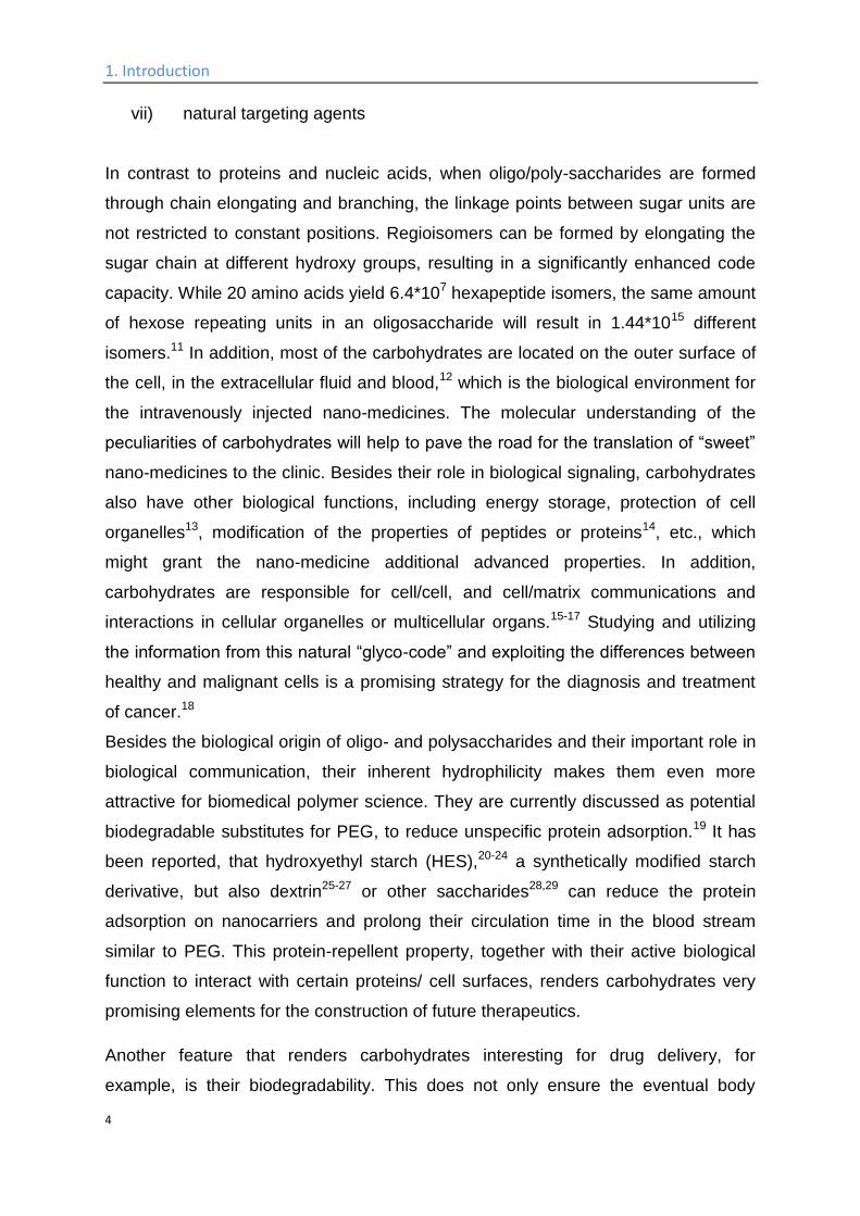
vii) natural targeting agents
In contrast to proteins and nucleic acids, when oligo/poly-saccharides are formed
through chain elongating and branching, the linkage points between sugar units are
not restricted to constant positions. Regioisomers can be formed by elongating the
sugar chain at different hydroxy groups, resulting in a significantly enhanced code
capacity. While 20 amino acids yield 6.4*107 hexapeptide isomers, the same amount
of hexose repeating units in an oligosaccharide will result in 1.44*1015 different
isomers.11 In addition, most of the carbohydrates are located on the outer surface of
the cell, in the extracellular fluid and blood,12 which is the biological environment for
the intravenously injected nano-medicines. The molecular understanding of the
peculiarities of carbohydrates will help to pave the road for the translation of “sweet”
nano-medicines to the clinic. Besides their role in biological signaling, carbohydrates
also have other biological functions, including energy storage, protection of cell
organelles13, modification of the properties of peptides or proteins14, etc., which
might grant the nano-medicine additional advanced properties. In addition,
carbohydrates are responsible for cell/cell, and cell/matrix communications and
interactions in cellular organelles or multicellular organs.15-17 Studying and utilizing
the information from this natural “glyco-code” and exploiting the differences between
healthy and malignant cells is a promising strategy for the diagnosis and treatment
of cancer.18
Besides the biological origin of oligo- and polysaccharides and their important role in
biological communication, their inherent hydrophilicity makes them even more
attractive for biomedical polymer science. They are currently discussed as potential
biodegradable substitutes for PEG, to reduce unspecific protein adsorption.19 It has
been reported, that hydroxyethyl starch (HES),20-24 a synthetically modified starch
derivative, but also dextrin25-27 or other saccharides28,29 can reduce the protein
adsorption on nanocarriers and prolong their circulation time in the blood stream
similar to PEG. This protein-repellent property, together with their active biological
function to interact with certain proteins/ cell surfaces, renders carbohydrates very
promising elements for the construction of future therapeutics.
Another feature that renders carbohydrates interesting for drug delivery, for
example, is their biodegradability. This does not only ensure the eventual body
1. Introduction
5
clearance of the materials, but is an additional handle to trigger drug release or
activation by certain enzymes.23-27 HES, for example, allows adjusting degradation
kinetics, depending on the degree of hydroxyethylation.23,24 In summary, the i)
biological activity, combined with ii) the potential stealth properties, and iii) the
enzymatic stimulus makes carbohydrates interesting materials for the design of
nanocarriers for biomedical applications. Both, surface-modification of preformed
nanoparticles with carbohydrates or the direct construction of the nanocarriers from
mono-, oligo-, or polysaccharides have attracted considerable attention during the
last decade over the borders of single disciplines, as can be seen in Scheme 1.
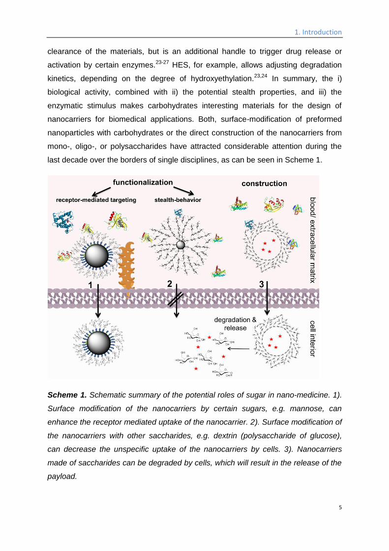
Scheme 1. Schematic summary of the potential roles of sugar in nano-medicine. 1).
Surface modification of the nanocarriers by certain sugars, e.g. mannose, can
enhance the receptor mediated uptake of the nanocarrier. 2). Surface modification of
the nanocarriers with other saccharides, e.g. dextrin (polysaccharide of glucose),
can decrease the unspecific uptake of the nanocarriers by cells. 3). Nanocarriers
made of saccharides can be degraded by cells, which will result in the release of the
payload.
1. Introduction
6
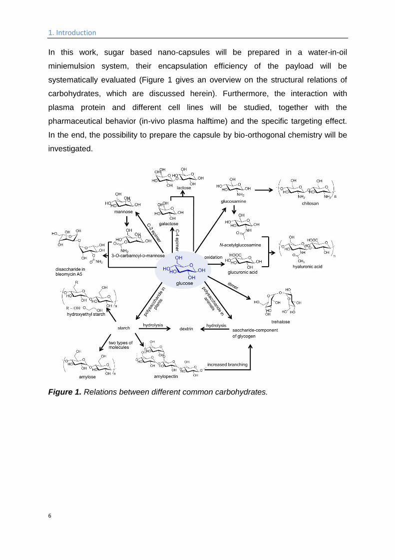
In this work, sugar based nano-capsules will be prepared in a water-in-oil
miniemulsion system, their encapsulation efficiency of the payload will be
systematically evaluated (Figure 1 gives an overview on the structural relations of
carbohydrates, which are discussed herein). Furthermore, the interaction with
plasma protein and different cell lines will be studied, together with the
pharmaceutical behavior (in-vivo plasma halftime) and the specific targeting effect.
In the end, the possibility to prepare the capsule by bio-orthogonal chemistry will be
investigated.
Figure 1. Relations between different common carbohydrates.
2. Theoretical background; 2.1 Recognition of saccharides by cell surface
7
2. Theoretical background The work in this chapter has been accepted for publication in Chemical Society
Reviews (Kang, B.; Opatz, T.; Landfester, K.; Wurm, F. R.; DOI:
10.1039/C5CS00092K)30 and has been altered for this thesis with permission.
2.1 Recognition of saccharides by cell surface receptors
and the use for targeting of specific cells. Cells of higher organisms are in constant communication and interaction with their
environment. In order to survive and maintain the appropriate functions, external
signals must be received by the cell-surface, and subsequently delivered into the
cell’s interior.31 While many of these different types of biological information are
encoded and delivered by protein-protein interactions, carbohydrates also play a
significant role.18,32,33 Carbohydrates act as recognition markers in different
pathological and physiological processes, most of them occurring on the surfaces of
cells. Three classes of proteins serve as receptors for the carbohydrate ligands:
enzymes (for the synthesis, remodeling and degradation of carbohydrate),
immunoglobulins and, most importantly, lectins34 which are membrane-bound
receptors and assist during the process of endocytosis.35
Through the binding with these receptors, many types of carbohydrates, including
mono-, oligo-, and polysaccharides have been found to specifically bind to certain
cell types. Mono/oligosaccharides like mannose derivatives exhibit strong binding to
the C-type lectin DC-SIGN on the surface of dendritic cells,36 C-type lectin receptors
on alveolar macrophages,37 and the plant lectin concanavalin A.38 Galactose can
also bind selectively to C-type lectin receptors on alveolar macrophages37 and
carbohydrate receptors on E. coli cells.39 Lactobionic acid can bind to
asialoglycoprotein receptors (ASGP-R) of hepatic tumor cells.40 For rhamnose, a
specific targeting effect to human skin cells was demonstrated.41 Polysaccharides
like hyaluronic acid or chitosan have been found to specifically bind to ocular
mucosa.42-44 Functionalized dextran has proven to specifically target vascular
smooth muscle cells45 and human endothelial cells.46 Many cellular events are
regulated by these sugar codes, including cell adhesion, proliferation, and cell
death.47-50
2. Theoretical background; 2.1 Recognition of saccharides by cell surface
8
Cancer still is one of the most prevalent deadly diseases worldwide and constitutes
one of the two major causes of death in industrialized countries. While the complete
eradication of malignant tumor is severely complicated by a tendency to form
metastases, all malignant cells have special biological signatures which distinguish
them from their healthy counterparts.
Carbohydrates, in particular glycoconjugates, play an essential role for cancer
metastasis and communication, through the interaction with endogenous lectins
present on the cancer cells.51-53 Presumably due to the fast metabolism of the tumor
tissue, some of these lectins, e. g. galectins, are found expressed at an elevated
level on malignant cells while they are not expressed detectably by their healthy
counterparts.12,54 Defined by their role as β-galactose receptors,55 galectins have
been reported as indicator for malignancies in stomach,56 liver,57 and the
corresponding colon cancer.58-60 A high galectin-1 level was reported in papillary
carcinomas, but not in the healthy tissues.61,62 A significant increase in the galecin-1
expression in adenocarcinoma cells was also reported, in contrast to the adjacent
normal endometrium.63 In addition, other carbohydrates, like hyaluronic acid also
shows specific binding to CD44 receptors64,65 which are expressed at low levels on
hematopoietic, epithelial, and neuronal cells but at much higher levels in various
tumor cells like lymphomas, melanomas, colorectal, and lung tumor cells.66,67 Thus,
many carbohydrate-related biomarkers have been developed which individually
exhibit specific binding to different cancer cells,68 and may open up possibilities to
specifically target cancer cells by an appropriate carbohydrate functionalization of
nanocarriers.
2. Theoretical background; 2.2 Protein repellent properties of carbohydrates
9
2.2 Protein repellent properties of carbohydrates When a nanocarrier enters a biology fluid, e.g. is intravenously injected into the
bloodstream, it will adsorb proteins on its surface, due to hydrophobic interactions
and the high surface energy of most types of nanocarriers.69,70 This procedure,
known as opsonization, can lead to phagocytosis of the nanocarrier by the
Mononuclear phagocyte system (MPS). The adsorbed proteins will determine the
fate of the nanocarrier in vivo (this process is often called the formation of a
“biological identity”),69 typically resulting in the fast clearance of the nanocarriers
from the blood. This makes any in vivo specific targeting a challenging task.69 In
order to prolong the in vivo plasma half-life times of the nanocarriers, the
opsonization needs to be reduced, either by the material of the nanocarrier itself, or
by surface modification (the dress of the nanocarriers). Currently, PEGylation is the
“gold standard” to achieve long blood circulation times and reduced unspecific
cellular uptake due to the hydrophilicity and the steric repulsion by PEG-modified
surfaces and proteins.71 PEGylation has achieved numerous successes in the past
decades, and many PEG-related products both in consumer care and biomedical
applications have improved the quality of life.71,72 In spite of these achievements,
recent studies reported several drawbacks of PEG. The occurrence of renal tubular
vacuolization in animal models have raised concerns that a prolonged therapy with
PEGylated drugs may lead to an accumulation of PEG in the cytoplasm of kidney
cells as the polymer is not biodegradable.73,74 In addition, PEG potentially forms
toxic degradation products upon storage which could provoke adverse effects.71
These setbacks of PEG could be circumvented by using polysaccharides as
substitutes which often show low hypersensitivity even after chemical
functionalization.75 The structural similarity of many polysaccharides, for example
HES or dextran, to the sugar component of glycogen, which is the form for the
storage of sugar in animals, is a probable explanation why they lack immunogenicity.
Moreover, the biodegradability of polysaccharides is advantageous over many other
synthetic polymers that are currently discussed as alternatives for PEG.76 Not only
the post-injection clearance of the nanocarriers is enhanced by its biodegradability,
but also enzymatic induced masking-unmasking or encapsulation-release cascades
of the payload are possible. 23,25,26,38,77,78 However, care has to be taken depending
2. Theoretical background; 2.2 Protein repellent properties of carbohydrates
10
on the chemical modification, e.g. anchoring or polymerizable groups that may alter
both the degradation process and the cytotoxicity of the carbohydrates.
Numerous studies have already proven that polysaccharide or their derivatives like
HES22,79-81 or dextran exhibit a low protein affinity.82,83 Furthermore, the microbial
polysaccharide pullulan, glycolipids, and dextran have shown their ability to
decrease the uptake of nanocarriers into the MPS,84,85 while HES has been proven
to suppress the unspecific uptake of the nanocarriers in vitro,86 and prolong the
plasma halftime in vivo, the process of its attachment also being called
HESylation.23,77 While several mono- or oligosaccharides are responsible for the
communication of biological information in the organism, some of them are capable
of impeding the phagocytosis of native cells by the MPS. Sialic acid is one example
of these saccharides and red blood cells without surface sialic acid are immediately
removed from the blood by the MPS.87 It has been proven, that when sialic acid is
coupled to the surface of quantum dots, the in vivo plasma half-life time of the latter
is prolonged.88
2. Theoretical background; 2.3 Glycoproteins: how nature uses carbohydrates
11
2.3 Glycoproteins: how nature uses carbohydrates In nature, glycoproteins, i.e. glycosylated polypeptides, are of high importance and
function as hormones,89 antibodies,90 antifreeze proteins,91 and proteins in the cell
membrane.14 After glycosylation, the attached (oligo)saccharides provide additional
properties for the protein, such as facilitating the protein folding and stabilizing the
conformation of the peptidic backbone,92 protection,93 elongation of the in vivo
plasma halftime,94 communication with the immune system,95 and adhesion to
cognate receptors on other cell surfaces.96,97
Inspired by these natural strategies, various researchers have prepared
neoglycoproteins for diverse applications. Pharmacologically active peptides have
been used for the treatment of various diseases.72,98 A major drawback, however, is
their usually rapid degradation in vivo. To optimize the pharmacokinetic properties of
such drugs, artificial polymers are frequently coupled to their surface. Typically PEG
is used for this purpose but in modern literature, an increasing percentage of
biodegradable biopolymers are coupled to proteins to optimize their therapeutic
performance. For example, hyaluronan-functionalized insulin showed a prolonged
and enhanced hypoglycemic effect, demonstrating the potential of hyaluronan for
increasing the plasma half-life of peptides.99
Anakinra, a synthetically generated interleukin-1 antagonist, is used for the
treatment of rheumatic arthritis, but has a plasma halftime of only 108 min; after
conjugation with HES its blood circulation time was increased by a factor of 6.5.100
Dextrin (a glucose polymer) with a molecular weight of 7,700 and 47,200 g/mol and
degree of succinoylation of 9–32 mol % was used to functionalize trypsin (a serine
protease) and thus masking its activity. The activity of the enzyme can be restored
after degradation of the polysaccharide by α-amylase.25-27 Also hyaluronic acid was
used for the functionalization of trypsin, resulting in an increase of its activity to
145% over the native protein, while exhibiting a 52% higher stability in the presence
of elastase (a protease).101 Although these works are beyond the scope of this work,
the idea of mimicking nature to utilize the advantageous properties of different
sugars is identical.
2. Theoretical background; 2.4 Carbohydrate-functionalized Nanocarriers
12
2.4 Carbohydrate-functionalized Nanocarriers Being a C-2-epimer of glucose, mannose is an important monosaccharide for the
glycosylation of proteins. Mannose-containing glycoproteins are produced in the liver
and secreted into the blood, hence mannose is distributed throughout the body.102
Many mannose-binding proteins, like the C-type lectins, are crucial for cell-surface
recognition and other communication events.103 Recently, mannose has been
applied to functionalize mesoporous silica nanoparticles,38 magnetic nanoparticles,18
gold nanoparticles,36 and polyanhydride nanoparticles37 (Table 1, entry 1) to
specifically target cells; distinct biological functionalities have been achieved in each
case.
When thiol-functionalized mannose is reacted with alkenyl-terminated silanes in a
radical thiol-ene addition (Table 1, entry 1, a), surface functionalization of
mesoporous silica nanoparticles can be achieved, whose pores can be sealed by
adding concanavalin A, a carbohydrate-binding protein, to the dispersion. The pores
can be re-opened under acidic conditions (pH < 5.5) or in a glucose-rich
environment. Release of the payload in the tumor tissue, where the pH value is
typically lower than that in healthy tissue, or under high blood sugar level is thus
possible.38 In another work, mannose-functionalized silica nanoparticles have been
prepared, which showed specific binding to MCF-7 human breast cancer cells.104
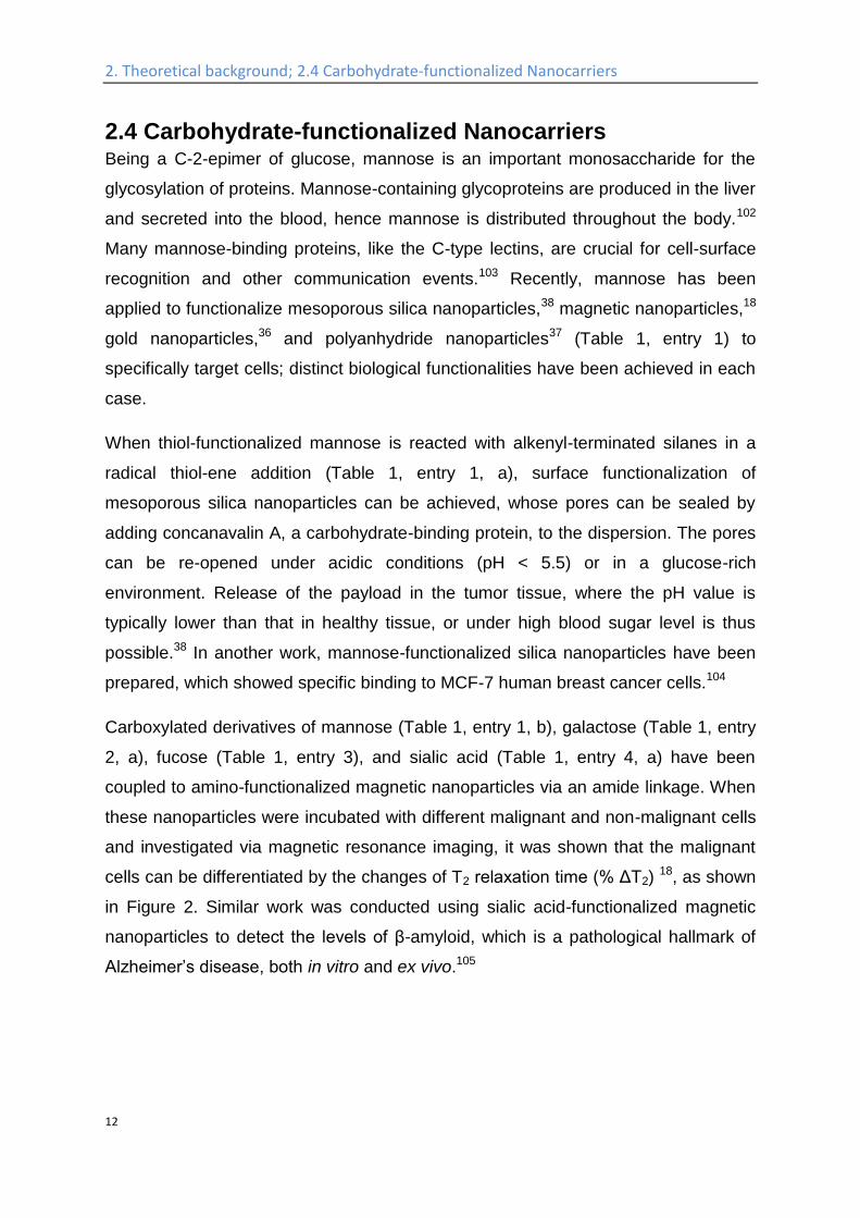
Carboxylated derivatives of mannose (Table 1, entry 1, b), galactose (Table 1, entry
2, a), fucose (Table 1, entry 3), and sialic acid (Table 1, entry 4, a) have been
coupled to amino-functionalized magnetic nanoparticles via an amide linkage. When
these nanoparticles were incubated with different malignant and non-malignant cells
and investigated via magnetic resonance imaging, it was shown that the malignant
cells can be differentiated by the changes of T2 relaxation time (% ΔT2) 18, as shown
in Figure 2. Similar work was conducted using sialic acid-functionalized magnetic
nanoparticles to detect the levels of β-amyloid, which is a pathological hallmark of
Alzheimer’s disease, both in vitro and ex vivo.105
2. Theoretical background; 2.4 Carbohydrate-functionalized Nanocarriers
13
Figure 2. Discrimination of the breast cancer cells from their healthy counterparts by
the changes of T2 relaxation time (% ΔT2) in magnetic resonance imaging, by
magnetic particles functionalized with: mannose (green), galactose (violet), fucose
(dark blue), sialic acid (red), glucose (light blue), compared to unmodified magnetic
particles (orange), (adapted with permission from reference18. Copyright 2010
American Chemical Society).
Different (oligo)mannosides have also been functionalized with thiols and coupled to
gold nanoparticles.36 The obtained glycosylated gold nanoparticles show stronger
binding to DC-SIGN (a C-type lectin) on the surface of dendritic cells compared to
gp120, which is a protein essential for the entry of HI virus into cells, and thus could
serve as a potential carbohydrate-based drug against HIV (Table 1, entry 1, c).
Similar glycosylated gold nanoparticles have also been prepared in another work
and observed to cross the blood−brain barrier (BBB) nearly 3-fold faster / more
efficiently than unmodified gold nanoparticles.106
Both, α-1,2-linked dimannose (Table 1, entry 1, d) and galactose (Table 1, entry 2,
g) have been coupled to polyanhydride nanoparticles through an amidation reaction
via 1-ethyl-3-(3-dimethylaminopropyl)-carbodiimide hydrochloride (EDC) as the
coupling agent. The obtained mannose surface-functionalized particles, which were
termed “pathogen-like” nanocarriers, exhibited specific binding to alveolar
macrophages through the surface C-type lectin and enhanced the expression of the
macrophage mannose receptor.37
It is the concern of some recent publications that the protein adsorption after contact
2. Theoretical background; 2.4 Carbohydrate-functionalized Nanocarriers
14
with blood will hamper all specific targeting of nanocarriers due to shielding of
targeting groups, which might reduce the efficiency of “targeted” drug delivery
systems remarkably.107-114 A current challenge is to understand the interaction of
blood proteins with nanocarriers which carry additional targeting groups. The
adsorption of plasma proteins onto the targeting agent could hinder the recognition
of the targeting agent by the respective cells and hence could make any in vivo
targeting impossible.69 The interactions of mannose-functionalized nanocarriers with
plasma proteins have been studied to address this problem. It turned out that, in
comparison to a PEGylated nano-carrier, additional functionalization of the
PEGylated nanocarrier with mannose did not significantly change its protein corona
formation. Furthermore, these mannose functionalized nanocarriers showed the
same binding affinity to dendritic cells (DCs) both in the presence and absence of
the plasma protein corona.115
Galactose is the C-4 epimer of glucose and is for example essential for the antigen
structure of red blood cells which is the determinant of the blood type. For O and A
antigens, two galactose units are contained in the saccharide portion while for the B
antigen, three galactose units are contained.116 Galactose functionalized with an
azide group at the C1-position, was coupled to pillar[5]arene by a Huisgen-type
cycloaddition, while the latter is self-assembled into nanorods (Table 1, entry 2, c),
which have proven a high affinity for the carbohydrate receptors on E. coli. as well
as low toxicity, and can be utilized as excellent cell glues to agglutinate these
bacteria.39 In another work, different statistical glycol-dithiocarbamate copolymers
were prepared and used to functionalize gold nanoparticles on the surface, which
were further coupled with gold(I) triphenylphosphine as an anticancer agent. Among
these glyconanoparticles, the galactose-functionalized ones were found to be 4-fold
more cytotoxic to HepG2 cells, in comparison with glucose and lactose
functionalized particles.117
Sialic acid is a monosaccharide, which is widely distributed in animal tissues and
mostly bound in form of glycoproteins.118 It plays an important role in recognition and
communication with the immune system,119-121 which is also proven by the fact that
red blood cells without sialic acid on the surface are immediately removed from

2. Theoretical background; 2.4 Carbohydrate-functionalized Nanocarriers
15
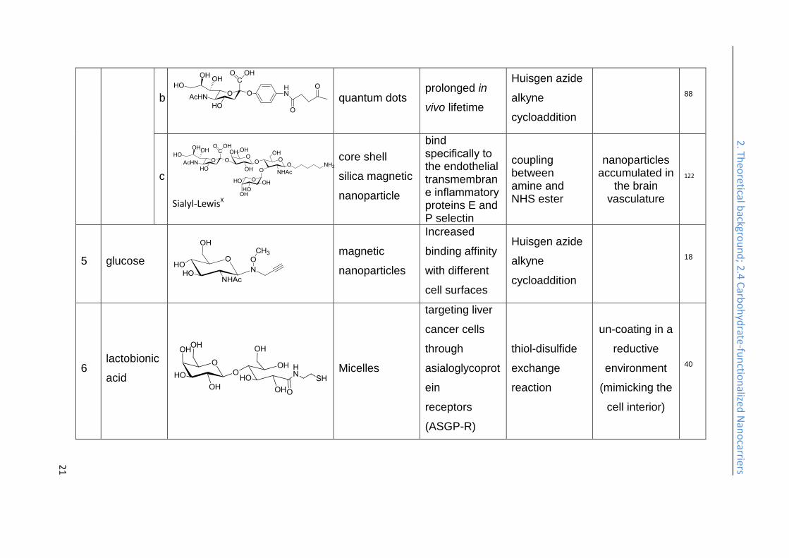
blood by the MPS.87 Ketone-functionalized sialic acid is reacted with aminooxy-
functionalized quantum dots, namely phosphorylcholine self-assembled monolayer-
coated quantum dots (PC-QDs), and their in vivo half-life times are extended
compared to quantum dots functionalized by other monosaccharides (Table 1, entry
4, b; and Figure 3).88
Figure 3. Images of major organs isolated from three tested mice, 2 h after the
administration of phosphorylcholine quantum dots (PC-QDs), lactose- functionalized
quantum dots (Lac-PC-QDs), and sialic acid-functionalized quantum dots (Nue5Ac-
PC-QDs). (Reprinted with permission from reference88. Copyright 2011 American
Chemical Society).
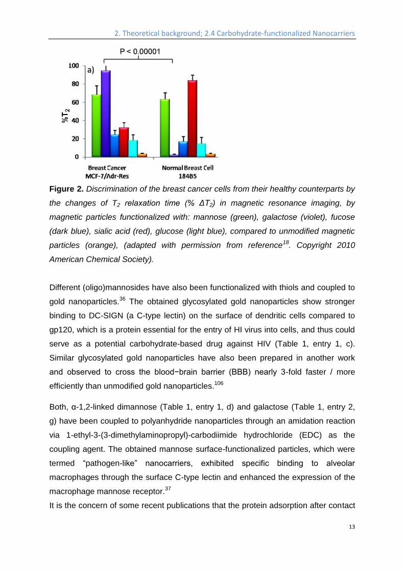
Sialyl-Lewisx is very important antigen for blood groups, which displays on the
terminus of glycolipids that are present on the cell surface, has been used to
functionalize superparamagnetic silica nanoparticles (Table 1, entry 4, c), with the
functionalization strategy shown in Figure 4. These nanoparticles have diameters of
around 18 nm and carry NH2-groups.122 Subsequent functionalization of these
particles with an NHS-ester allows coupling to amino-functionalized Sialyl-LewisX.
The obtained glycosylated nanoparticles bind specifically to the inflammation-
associated endothelial transmembrane proteins E and P selectin, both cell adhesion
molecules. In vivo studies have shown an accumulation in the brain vasculature by
measuring the relaxing time of the nanoparticles via MRI.122
2. Theoretical background; 2.4 Carbohydrate-functionalized Nanocarriers
16
Figure 4. Functionalization of superparamagnetic nanoparticles with a silica core by
Sialyl-LewisX. (Reprinted with permission from reference122. Copyright 2014
American Chemical Society).
Lactobionic acid (4-O-β-D-galactopyranosyl-D-gluconic acid, (Table 1, entry 6))
specifically bind to hepatocytes.123 Thiolated lactobionic acid was used to
functionalize block copolymers, which were prepared by the ring-opening
copolymerization of ε-caprolactone and a pyridyl disulfide containing cyclic
carbonate, followed by post polymerization modification with thiolated lactobionic
acid via the thiol-disulfide exchange reaction. The post-modified block copolymers
then self-assembled into micelles with lactobionic acid on the surface. These
micelles were shown to target liver cancer through asialoglycoprotein receptors
(ASGP-R), furthermore, the saccharide shells is cleavable under a reductive
environment mimicking the interior of a cell.40
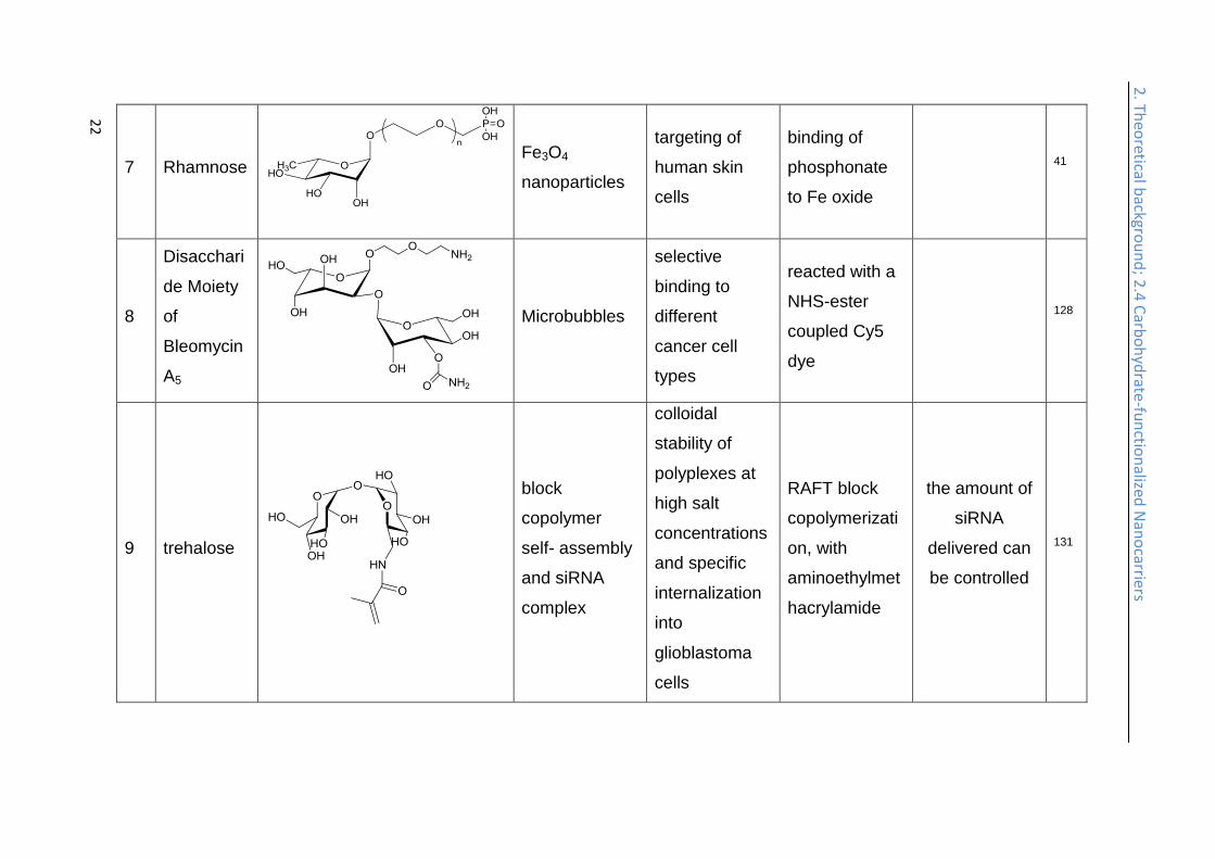
Rhamnose is a mannose-related 6-deoxy hexose which naturally occurs in the L-
form. It is found mainly in bacteria and plants and is often present in the cell walls
and is essential for the survival of bacteria.124 Phosphonated rhamnose has been
prepared (Table 1, entry 7) and anchored to magnetic nanoparticles through the
strong binding of phosphonates groups to metals. The rhamnose-functionalized
magnetic nanoparticles exhibited targeting effect to human skin cells. Since the iron
2. Theoretical background; 2.4 Carbohydrate-functionalized Nanocarriers
17
oxide nanoparticles are superparamagnetic, they can be used as MRI contrast agent
with specific cell targeting.41
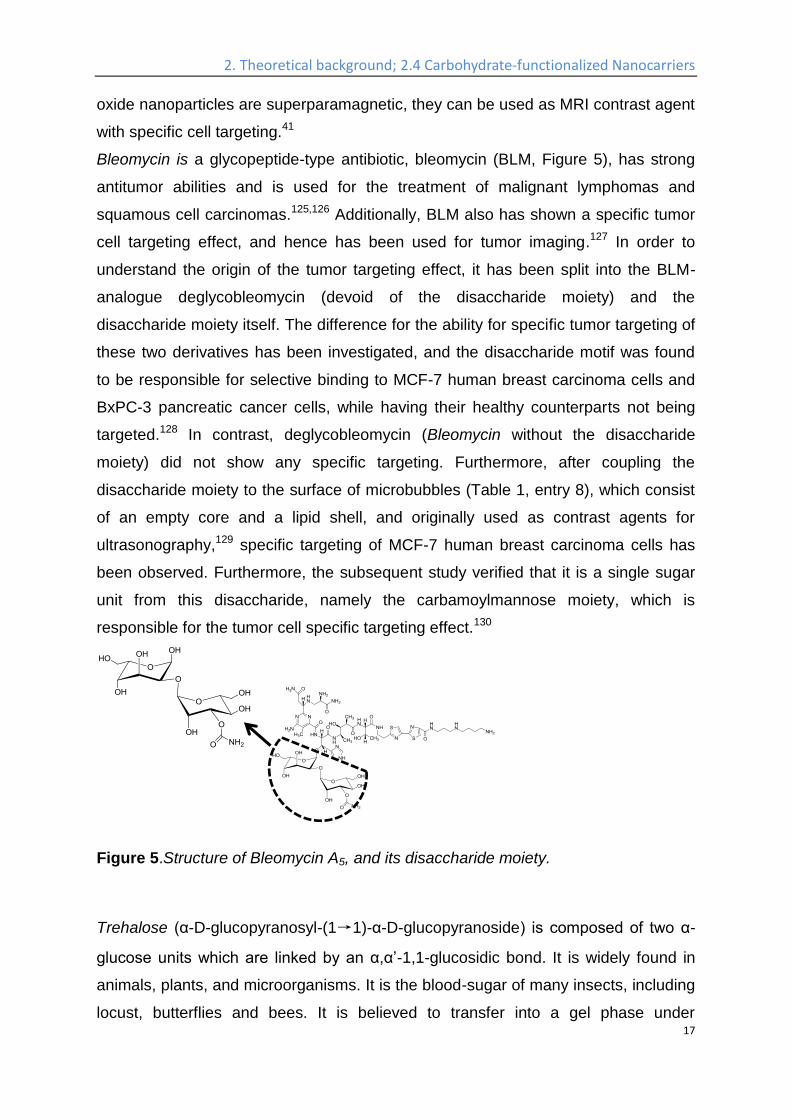
Bleomycin is a glycopeptide-type antibiotic, bleomycin (BLM, Figure 5), has strong
antitumor abilities and is used for the treatment of malignant lymphomas and
squamous cell carcinomas.125,126 Additionally, BLM also has shown a specific tumor
cell targeting effect, and hence has been used for tumor imaging.127 In order to
understand the origin of the tumor targeting effect, it has been split into the BLM-
analogue deglycobleomycin (devoid of the disaccharide moiety) and the
disaccharide moiety itself. The difference for the ability for specific tumor targeting of
these two derivatives has been investigated, and the disaccharide motif was found
to be responsible for selective binding to MCF-7 human breast carcinoma cells and
BxPC-3 pancreatic cancer cells, while having their healthy counterparts not being
targeted.128 In contrast, deglycobleomycin (Bleomycin without the disaccharide
moiety) did not show any specific targeting. Furthermore, after coupling the
disaccharide moiety to the surface of microbubbles (Table 1, entry 8), which consist
of an empty core and a lipid shell, and originally used as contrast agents for
ultrasonography,129 specific targeting of MCF-7 human breast carcinoma cells has
been observed. Furthermore, the subsequent study verified that it is a single sugar
unit from this disaccharide, namely the carbamoylmannose moiety, which is
responsible for the tumor cell specific targeting effect.130
Figure 5.Structure of Bleomycin A5, and its disaccharide moiety.
Trehalose (α-D-glucopyranosyl-(1→1)-α-D-glucopyranoside) is composed of two α-
glucose units which are linked by an α,α’-1,1-glucosidic bond. It is widely found in
animals, plants, and microorganisms. It is the blood-sugar of many insects, including
locust, butterflies and bees. It is believed to transfer into a gel phase under
2. Theoretical background; 2.4 Carbohydrate-functionalized Nanocarriers
18
dehydrating condition, protecting the cell internal organelles and hence the whole
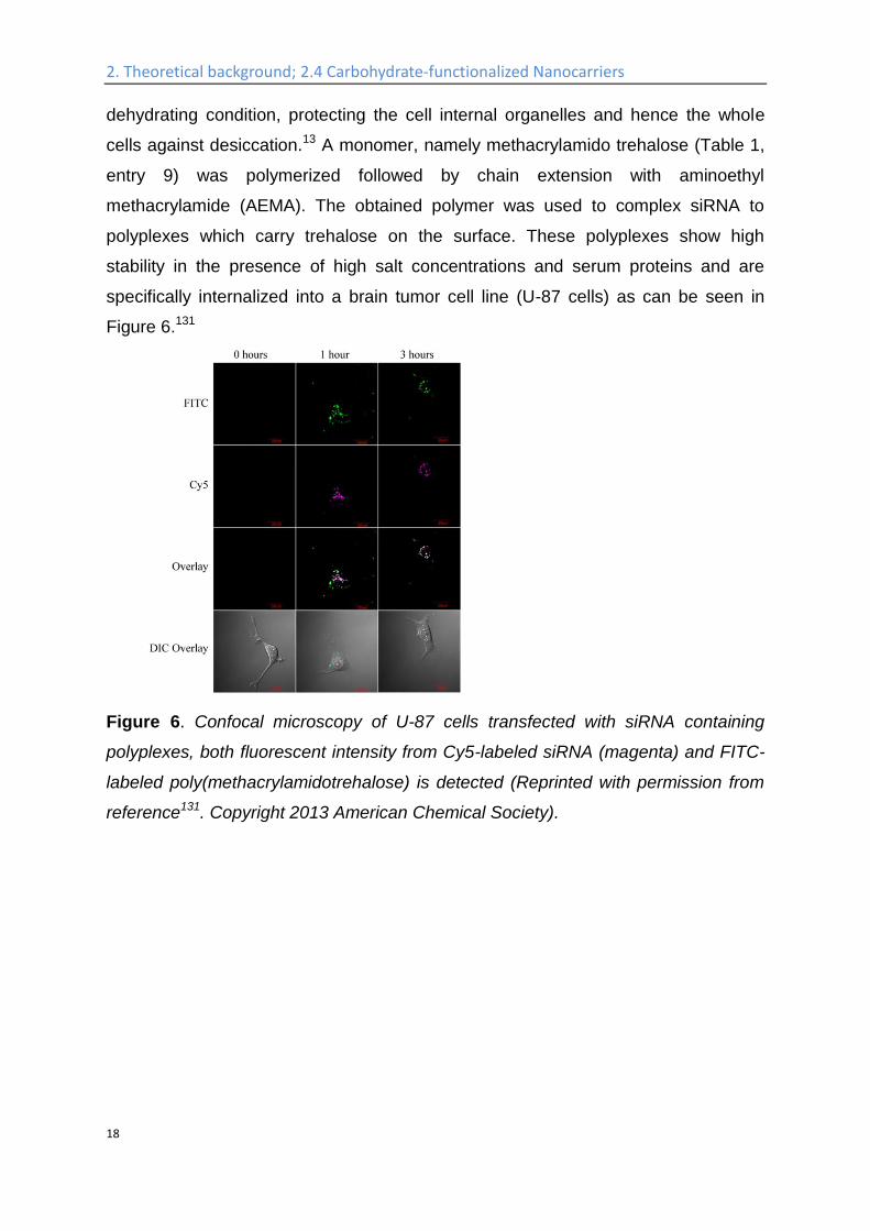
cells against desiccation.13 A monomer, namely methacrylamido trehalose (Table 1,
entry 9) was polymerized followed by chain extension with aminoethyl
methacrylamide (AEMA). The obtained polymer was used to complex siRNA to
polyplexes which carry trehalose on the surface. These polyplexes show high
stability in the presence of high salt concentrations and serum proteins and are
specifically internalized into a brain tumor cell line (U-87 cells) as can be seen in
Figure 6.131
Figure 6. Confocal microscopy of U-87 cells transfected with siRNA containing
polyplexes, both fluorescent intensity from Cy5-labeled siRNA (magenta) and FITC-
labeled poly(methacrylamidotrehalose) is detected (Reprinted with permission from
reference131. Copyright 2013 American Chemical Society).
2. Th
eoretical b
ackgrou
nd
; 2.4 C
arbo
hyd
rate-fu
nctio
nalized
Nan
ocarriers
19
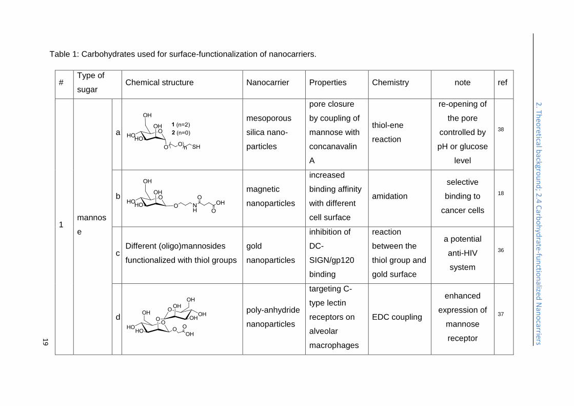
Table 1: Carbohydrates used for surface-functionalization of nanocarriers.
# Type of
sugar Chemical structure Nanocarrier Properties Chemistry note ref
1 mannos
e
a
mesoporous
silica nano-
particles
pore closure
by coupling of
mannose with
concanavalin
A
thiol-ene
reaction
re-opening of
the pore
controlled by
pH or glucose
level
38
b
magnetic
nanoparticles
increased
binding affinity
with different
cell surface
amidation
selective
binding to
cancer cells
18
c Different (oligo)mannosides
functionalized with thiol groups
gold
nanoparticles
inhibition of
DC-
SIGN/gp120
binding
reaction
between the
thiol group and
gold surface
a potential
anti-HIV
system
36
d
poly-anhydride
nanoparticles
targeting C-
type lectin
receptors on
alveolar
macrophages
EDC coupling
enhanced
expression of
mannose
receptor
37
2. Th
eoretical b
ackgrou
nd
; 2.4 C
arbo
hyd
rate-fu
nctio
nalized
Nan
ocarriers
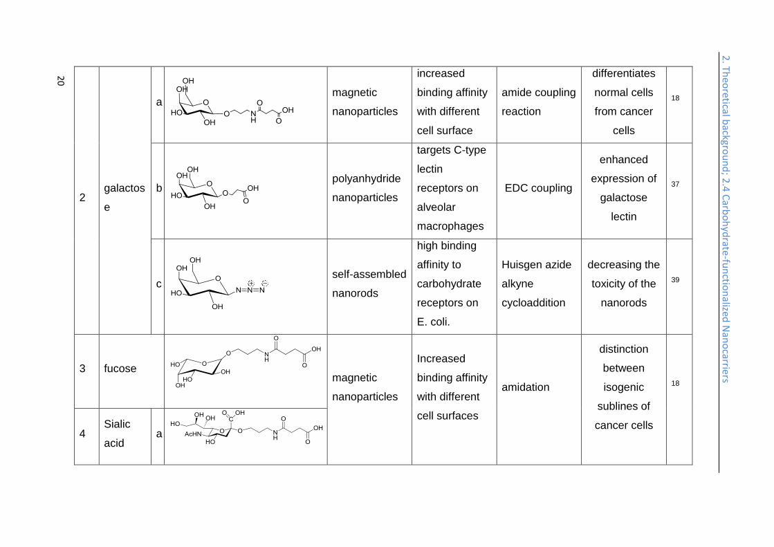
20
2 galactos
e
a
magnetic
nanoparticles
increased
binding affinity
with different
cell surface
amide coupling
reaction
differentiates
normal cells
from cancer
cells
18
b
polyanhydride
nanoparticles
targets C-type
lectin
receptors on
alveolar
macrophages
EDC coupling
enhanced
expression of
galactose
lectin
37
c
self-assembled
nanorods
high binding
affinity to
carbohydrate
receptors on
E. coli.
Huisgen azide
alkyne
cycloaddition
decreasing the
toxicity of the
nanorods
39
3 fucose
magnetic
nanoparticles
Increased
binding affinity
with different
cell surfaces
amidation
distinction
between
isogenic
sublines of
cancer cells
18
4 Sialic
acid a
2. Th
eoretical b
ackgrou
nd
; 2.4 C
arbo
hyd
rate-fu
nctio
nalized
Nan
ocarriers
21
b
quantum dots prolonged in
vivo lifetime
Huisgen azide
alkyne
cycloaddition
88
c
Sialyl-LewisX
core shell
silica magnetic
nanoparticle
bind specifically to the endothelial transmembrane inflammatory proteins E and P selectin
coupling between amine and NHS ester
nanoparticles accumulated in
the brain vasculature
122
5 glucose
magnetic
nanoparticles
Increased
binding affinity
with different
cell surfaces
Huisgen azide
alkyne
cycloaddition
18
6 lactobionic
acid
Micelles
targeting liver
cancer cells
through
asialoglycoprot
ein
receptors
(ASGP-R)
thiol-disulfide
exchange
reaction
un-coating in a
reductive
environment
(mimicking the
cell interior)
40
2. Th
eoretical b
ackgrou
nd
; 2.4 C
arbo
hyd
rate-fu
nctio
nalized
Nan
ocarriers
22
7 Rhamnose
Fe3O4
nanoparticles
targeting of
human skin
cells
binding of
phosphonate
to Fe oxide
41
8
Disacchari
de Moiety
of
Bleomycin
A5
Microbubbles
selective
binding to
different
cancer cell
types
reacted with a
NHS-ester
coupled Cy5
dye
128
9 trehalose
block
copolymer
self- assembly
and siRNA
complex
colloidal
stability of
polyplexes at
high salt
concentrations
and specific
internalization
into
glioblastoma
cells
RAFT block
copolymerizati
on, with
aminoethylmet
hacrylamide
the amount of
siRNA
delivered can
be controlled
131

2. Th
eoretical b
ackgrou
nd
; 2.4 C
arbo
hyd
rate-fu
nctio
nalized
Nan
ocarriers
23
10 starch
copper
nanoparticles lower toxicity
reduction of copper nitrate solution by ascorbic acid, starch as stabilizer for the nano-particle
excellent
bactericidal
action
132
11
Hydroxyet
hyl starch
(HES)
DNA-
polyplexes
micelles
reduced
unspecific cell
uptake
Schiff base
formation and
reductive
amination
deshielding of
the nanocarrier
possible
23,7
7
12 chitosan
a
Mw ~ 47kDa
poly lactic-co-
glycolic acid
nanoparticle
significantly
increased (>5-
fold) uptake by
MCF-7 cells
electrostatic
interactions 133
b
Molecular weight of 100,000
g/mol
hyaluronic-
paclitaxel
nanoparticle
protection of
the payload
electrostatic
interactions
pH responsive
release of
paclitaxel
134

2. Th
eoretical b
ackgrou
nd
; 2.4 C
arbo
hyd
rate-fu
nctio
nalized
Nan
ocarriers
24
c
> 75% deacetylated
silver
nanoparticle lower toxicity
chitosan as a
stabilizer
during
preparation
higher rate of
killing cancer
cell compared
to PEGylated
gold nanorod
135
d
degree of
deacetylation=82.7%; MW =
250,000
gold
nanoparticles
low unspecific
cell uptake,
enhanced
stability and
tumor targeting
ability
glycol-modified
chitosan is
used as
reducing agent
for Gold(III)
chloride in situ
tomography of
liver tissues
with metastatic
cancer
136
13 Hyaluro
nic Acid
a
micelles
prepared from
branched poly
(ethylene
imine)
Increased
transfection
efficiency and
decreased
cytotoxicity
reductive
amination 137
b
re-constituted
high density
lipoprotein
loaded with
lower
accumulation
in liver and
higher
atherosclerotic
electrostatic
interactions
efficiently
suppressed
the
advancement
138
2. Th
eoretical b
ackgrou
nd
; 2.4 C
arbo
hyd
rate-fu
nctio
nalized
Nan
ocarriers
25
lovastatin lesions
targeting
efficiency
of
atherosclerosis
c
gold nanocage
specific
binding to
cancer cells
via interaction
with CD44,
release in
lysosome
Au-catechol
bonds
near-infrared
irradiation
accelerates
the release
139
2. Theoretical background; 2.4 Carbohydrate-functionalized Nanocarriers
26
2. Th
eoretical b
ackgrou
nd
; 2.5
Carb
oh
ydrate
-fun
ction
alized N
ano
carriers
Starch is a polysaccharide based on glucose as the monomer, which is coupled via
glycosidic linkages. Two forms of starch are found in nature: amylose, a linear and
helical polysaccharide with α-1,4-glycosidic bonds, and amylopectin, a branched
poly(glucose) with 1,4- and 1,6- glycosidic bonds. It is the energy storage medium of
green plants and the most common carbohydrate in human diets.140 The sugar part
of glycogen, as another glucose polymer, is used to store glucose in animals with a
similar structure as amylopectin, but with a higher degree of branching. Starch is
used as the stabilizer during preparation of copper nanoparticles, while ascorbic acid
is used as reducing agent, and copper nitrate as the source of copper (Table 1, entry
10), which will result in starch-functionalized copper nanoparticles with a reduced
toxicity, while retaining high antibacterial potential against both gram negative and
gram positive strains.132
Hydroxyethyl starch. For some applications, the degradation kinetics of starch is too
fast; starch is rapidly hydrolyzed by plasma amylases. In order to balance the
biodegradability and stability, hydroxyethyl starch (HES) was introduced. It is
prepared by ethoxylation of the hydroxyl groups with ethylene oxide, resulting in
decreased biodegradation kinetics. The degree of hydroxyethyl-substitution is
expressed by the molar substitution, which is the mean number of hydroxyethyl
groups per glucose unit, and ranges between 0 and 3. The higher the molar
substitution, or the higher the C2/C6 ratio of hydroxyethylation, the lower the rate of
metabolization.75 Moreover, HES exhibits low hypersensitivity75 and depressed
protein adsorption22,79-81 rendering it an interesting substitute for PEG for the
preparation of stealth nanocarriers. HESylation of proteins and nanocarriers is of
high potential for future drug delivery vehicles as it combines adjustable degradation
with stealth properties.141 HES with different molecular weights and degrees of
substitution were coupled to poly(ethylene imine) via Schiff base formation and
reductive amination (Table 1, entry 11). Subsequent complexation of the polymer
with DNA generated so called DNA-polyplexes, which presented HES on their
surface. These polyplexes proved to exhibit stealth properties, as the nanocarrier is
protected against α-amylase. The effect of deshielding is also affected by the degree
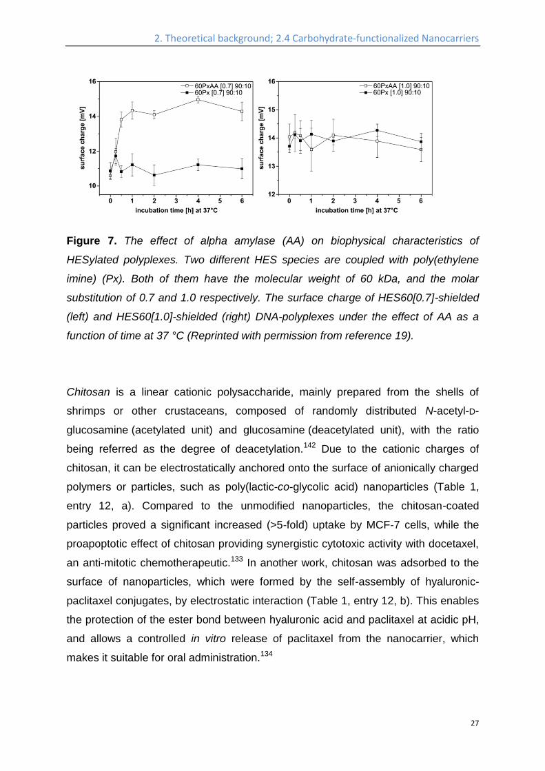
of substitution of HES, as can be seen in Figure 7.23,77
2. Theoretical background; 2.4 Carbohydrate-functionalized Nanocarriers
27
Figure 7. The effect of alpha amylase (AA) on biophysical characteristics of
HESylated polyplexes. Two different HES species are coupled with poly(ethylene
imine) (Px). Both of them have the molecular weight of 60 kDa, and the molar
substitution of 0.7 and 1.0 respectively. The surface charge of HES60[0.7]-shielded
(left) and HES60[1.0]-shielded (right) DNA-polyplexes under the effect of AA as a
function of time at 37 °C (Reprinted with permission from reference 19).
Chitosan is a linear cationic polysaccharide, mainly prepared from the shells of
shrimps or other crustaceans, composed of randomly distributed N-acetyl-D-
glucosamine (acetylated unit) and glucosamine (deacetylated unit), with the ratio
being referred as the degree of deacetylation.142 Due to the cationic charges of
chitosan, it can be electrostatically anchored onto the surface of anionically charged
polymers or particles, such as poly(lactic-co-glycolic acid) nanoparticles (Table 1,
entry 12, a). Compared to the unmodified nanoparticles, the chitosan-coated
particles proved a significant increased (>5-fold) uptake by MCF-7 cells, while the
proapoptotic effect of chitosan providing synergistic cytotoxic activity with docetaxel,
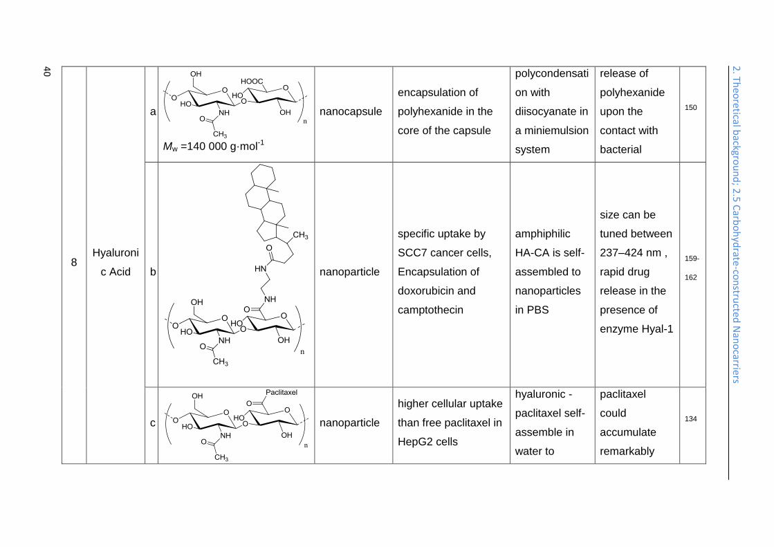
an anti-mitotic chemotherapeutic.133 In another work, chitosan was adsorbed to the
surface of nanoparticles, which were formed by the self-assembly of hyaluronic-
paclitaxel conjugates, by electrostatic interaction (Table 1, entry 12, b). This enables
the protection of the ester bond between hyaluronic acid and paclitaxel at acidic pH,
and allows a controlled in vitro release of paclitaxel from the nanocarrier, which
makes it suitable for oral administration.134
2. Theoretical background; 2.4 Carbohydrate-functionalized Nanocarriers
28
2. Th
eoretical b
ackgrou
nd
; 2.5
Carb
oh
ydrate
-fun
ction
alized N
ano
carriers
Nanoparticles based on effective Au and Ag photothermal transducers can be used
to trigger localized hyperthermia of tumors. Chitosan has been used for the surface
functionalization of silver nanoparticles (Table 1, entry 12, c), and gold nanoparticles
(Table 1, entry 12, d). To a mixture of aqueous solutions of trisodium citrate,
ascorbic acid, chitosan, and preformed Ag nanoparticles, a solution of AgNO3 was
added dropwise, and chitosan surface functionalized Ag nanoparticles are obtained.
These Ag nanoparticles show a lower toxicity compared to PEGylated gold nanorods,
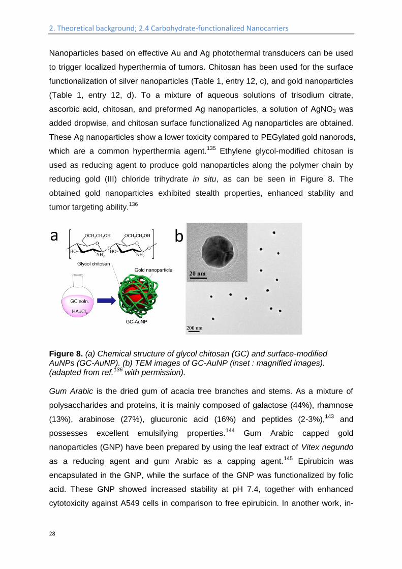
which are a common hyperthermia agent.135 Ethylene glycol-modified chitosan is
used as reducing agent to produce gold nanoparticles along the polymer chain by
reducing gold (III) chloride trihydrate in situ, as can be seen in Figure 8. The
obtained gold nanoparticles exhibited stealth properties, enhanced stability and
tumor targeting ability.136
Figure 8. (a) Chemical structure of glycol chitosan (GC) and surface-modified AuNPs (GC-AuNP). (b) TEM images of GC-AuNP (inset : magnified images). (adapted from ref.136 with permission). Gum Arabic is the dried gum of acacia tree branches and stems. As a mixture of
polysaccharides and proteins, it is mainly composed of galactose (44%), rhamnose
(13%), arabinose (27%), glucuronic acid (16%) and peptides (2-3%),143 and
possesses excellent emulsifying properties.144 Gum Arabic capped gold
nanoparticles (GNP) have been prepared by using the leaf extract of Vitex negundo
as a reducing agent and gum Arabic as a capping agent.145 Epirubicin was
encapsulated in the GNP, while the surface of the GNP was functionalized by folic
acid. These GNP showed increased stability at pH 7.4, together with enhanced
cytotoxicity against A549 cells in comparison to free epirubicin. In another work, in-
2. Theoretical background; 2.4 Carbohydrate-functionalized Nanocarriers
29
vivo studies of gum Arabic functionalized GNP resulted in significant alterations in
lung tumors in mice upon laser irradiation, including cyto-toxicity, apoptosis,
decreased inflammation and angiogenesis, and enhanced lipid peroxidation.146
Hyaluronic acid is a polysaccharide distributed widely in all tissues and body fluids of
vertebrates and is most abundantly found in the connective tissues, serves many
physiological functions, including lubrication, filtering, water homeostasis, and
regulation of plasma protein distribution. It is metabolized by receptor-mediated
endocytosis, and subsequent lysosomal degradation.147 Hyaluronic acid was
conjugated with branched poly(ethylene imine) via reductive amination (Table 1,
entry 13, a). Then, the polymer was self-assembled into micelles which were
surface-modified by hyaluronic acid and proved increased transfection efficiency and
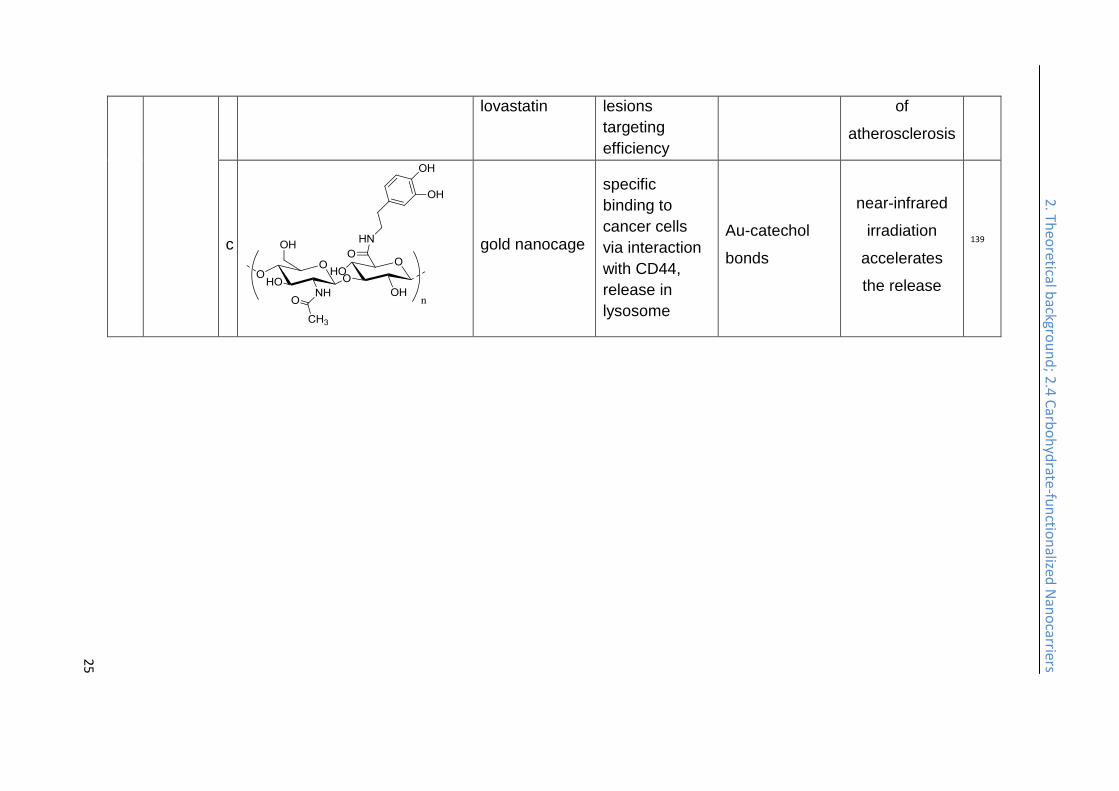
decreased cytotoxicity.137 A reconstituted high density lipoprotein loaded with
lovastatin (a statin which blocks the de novo-synthesis of cholesterol) was
functionalized by hyaluronic acid (Table 1, entry 13, b), through electrostatic
adsorption of hyaluronic acid to a cationic lipid core of the nanoparticle. After
surface-modification, the nanocarrier has lower accumulation in liver and better
atherosclerotic lesions targeting efficiency, and efficiently suppressed advancement
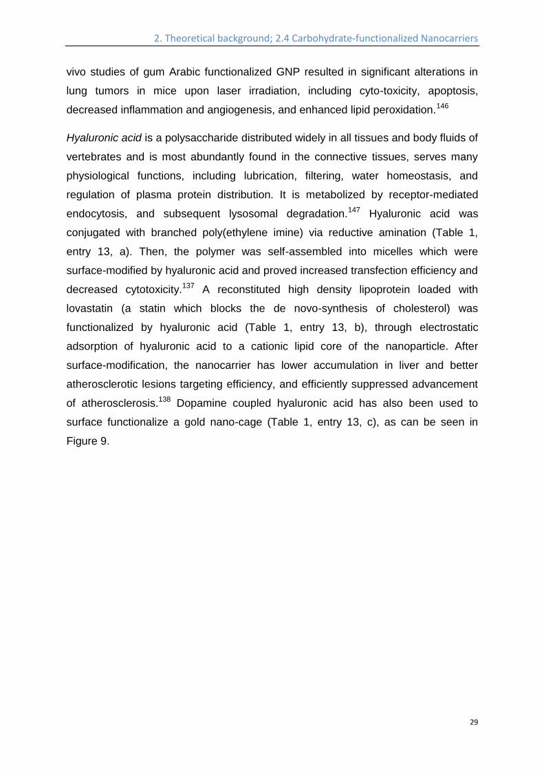
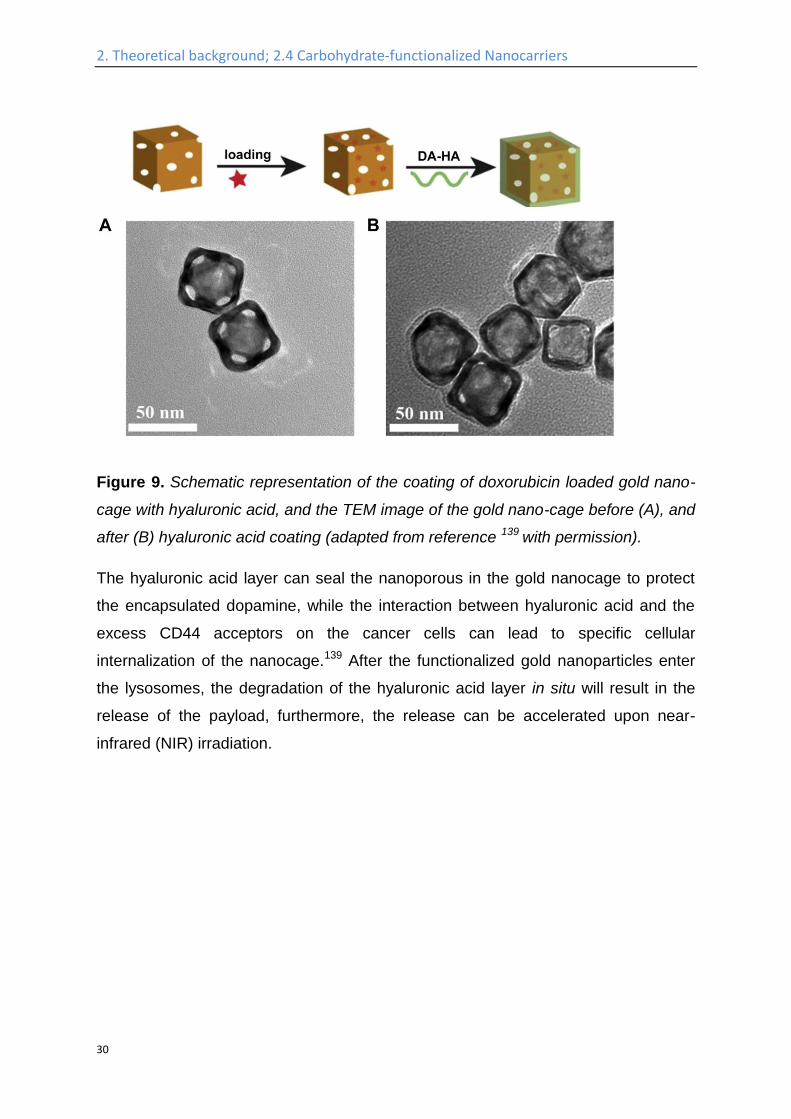
of atherosclerosis.138 Dopamine coupled hyaluronic acid has also been used to
surface functionalize a gold nano-cage (Table 1, entry 13, c), as can be seen in
Figure 9.
2. Theoretical background; 2.4 Carbohydrate-functionalized Nanocarriers
30
2. Th
eoretical b
ackgrou
nd
; 2.5
Carb
oh
ydrate
-fun
ction
alized N
ano
carriers
Figure 9. Schematic representation of the coating of doxorubicin loaded gold nano-
cage with hyaluronic acid, and the TEM image of the gold nano-cage before (A), and
after (B) hyaluronic acid coating (adapted from reference 139 with permission).
The hyaluronic acid layer can seal the nanoporous in the gold nanocage to protect
the encapsulated dopamine, while the interaction between hyaluronic acid and the
excess CD44 acceptors on the cancer cells can lead to specific cellular
internalization of the nanocage.139 After the functionalized gold nanoparticles enter
the lysosomes, the degradation of the hyaluronic acid layer in situ will result in the
release of the payload, furthermore, the release can be accelerated upon near-
infrared (NIR) irradiation.
2. Theoretical background; 2.5 Carbohydrate-constructed Nanocarriers
31
2.5 Carbohydrate-constructed Nanocarriers
Due to their outstanding biocompatibility, biodegradability, high diversity of chemical
functionalities, and versatile biological functions, carbohydrates are also useful for
the construction of nanocarriers for biomedical applications.
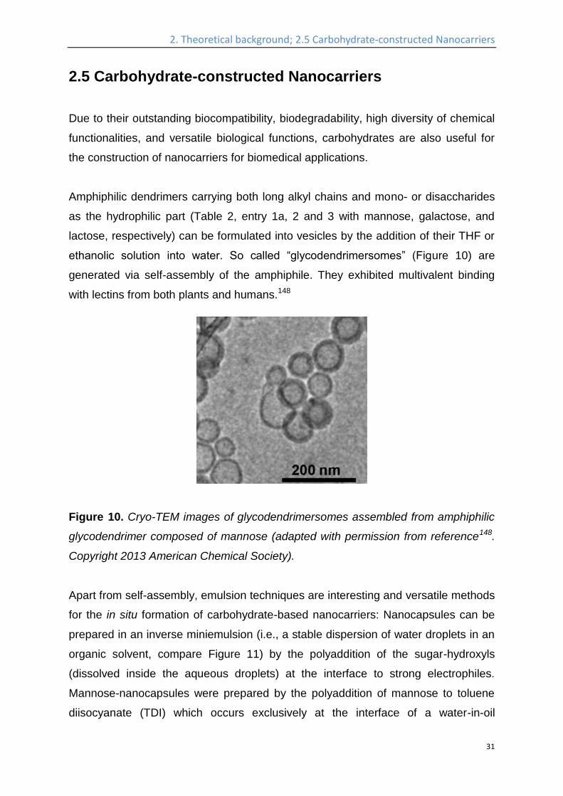
Amphiphilic dendrimers carrying both long alkyl chains and mono- or disaccharides
as the hydrophilic part (Table 2, entry 1a, 2 and 3 with mannose, galactose, and
lactose, respectively) can be formulated into vesicles by the addition of their THF or
ethanolic solution into water. So called “glycodendrimersomes” (Figure 10) are
generated via self-assembly of the amphiphile. They exhibited multivalent binding
with lectins from both plants and humans.148
Figure 10. Cryo-TEM images of glycodendrimersomes assembled from amphiphilic
glycodendrimer composed of mannose (adapted with permission from reference148.
Copyright 2013 American Chemical Society).
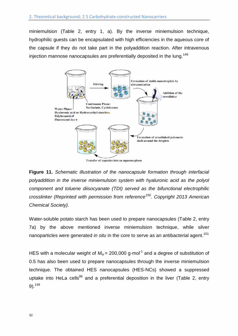
Apart from self-assembly, emulsion techniques are interesting and versatile methods
for the in situ formation of carbohydrate-based nanocarriers: Nanocapsules can be
prepared in an inverse miniemulsion (i.e., a stable dispersion of water droplets in an
organic solvent, compare Figure 11) by the polyaddition of the sugar-hydroxyls
(dissolved inside the aqueous droplets) at the interface to strong electrophiles.
Mannose-nanocapsules were prepared by the polyaddition of mannose to toluene
diisocyanate (TDI) which occurs exclusively at the interface of a water-in-oil
2. Theoretical background; 2.5 Carbohydrate-constructed Nanocarriers
32
miniemulsion (Table 2, entry 1, a). By the inverse miniemulsion technique,
hydrophilic guests can be encapsulated with high efficiencies in the aqueous core of
the capsule if they do not take part in the polyaddition reaction. After intravenous
injection mannose nanocapsules are preferentially deposited in the lung.149
Figure 11. Schematic illustration of the nanocapsule formation through interfacial
polyaddition in the inverse miniemulsion system with hyaluronic acid as the polyol
component and toluene diisocyanate (TDI) served as the bifunctional electrophilic
crosslinker (Reprinted with permission from reference150. Copyright 2013 American
Chemical Society).
Water-soluble potato starch has been used to prepare nanocapsules (Table 2, entry
7a) by the above mentioned inverse miniemulsion technique, while silver
nanoparticles were generated in situ in the core to serve as an antibacterial agent.151
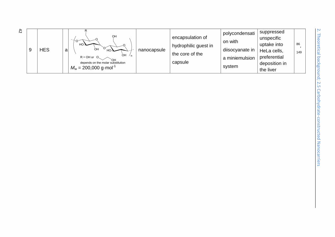
HES with a molecular weight of Mw = 200,000 g·mol-1 and a degree of substitution of
0.5 has also been used to prepare nanocapsules through the inverse miniemulsion
technique. The obtained HES nanocapsules (HES-NCs) showed a suppressed
uptake into HeLa cells86 and a preferential deposition in the liver (Table 2, entry
9).149
2. Theoretical background; 2.5 Carbohydrate-constructed Nanocarriers
33
The in vivo plasma half-life times of the HES-NCs obtained by this strategy can be
further tailored by different surface functionalization methods. PEGylation of the
capsule surface by isocyanate-terminated PEG results in increased plasma half-life
times with 20% and 5% of the nanocapsules remaining in the blood plasma after
24 h and 72 h, respectively.152
Despite the straightforward reaction setup, this strategy has limited feasibility, when
used to encapsulate and protect pharmaceutical agents, which often contain
nucleophiles like amines, thiols, or alcohols, and consequently will participate in the
polycondensation reaction with the diisocyanate electrophile. Recent work presents
strategies to use bioorthogonal reactions to generate the nanocarriers allowing the
encapsulation of more complex molecules.
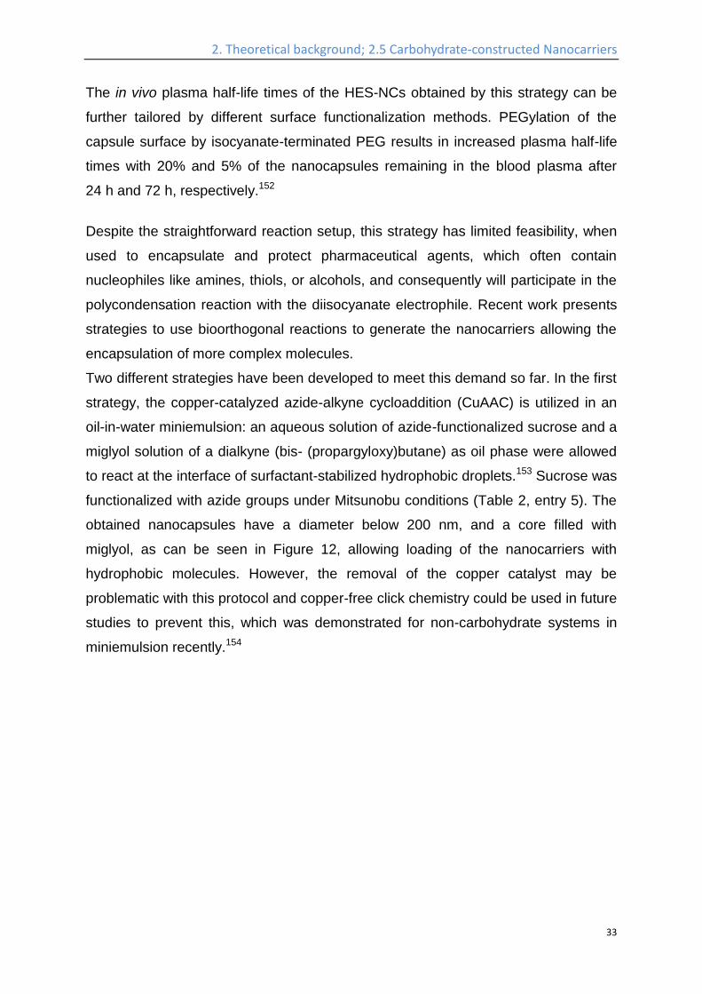
Two different strategies have been developed to meet this demand so far. In the first
strategy, the copper-catalyzed azide-alkyne cycloaddition (CuAAC) is utilized in an
oil-in-water miniemulsion: an aqueous solution of azide-functionalized sucrose and a
miglyol solution of a dialkyne (bis- (propargyloxy)butane) as oil phase were allowed
to react at the interface of surfactant-stabilized hydrophobic droplets.153 Sucrose was
functionalized with azide groups under Mitsunobu conditions (Table 2, entry 5). The
obtained nanocapsules have a diameter below 200 nm, and a core filled with
miglyol, as can be seen in Figure 12, allowing loading of the nanocarriers with
hydrophobic molecules. However, the removal of the copper catalyst may be
problematic with this protocol and copper-free click chemistry could be used in future
studies to prevent this, which was demonstrated for non-carbohydrate systems in
miniemulsion recently.154
2. Theoretical background; 2.5 Carbohydrate-constructed Nanocarriers
34
Figure 12. SEM (top) and TEM (bottom) images of the sucrose nanocapsules
generated by interfacial CuAAC polyaddition. (Reprinted with permission from
reference153. Copyright 2012 American Chemical Society).
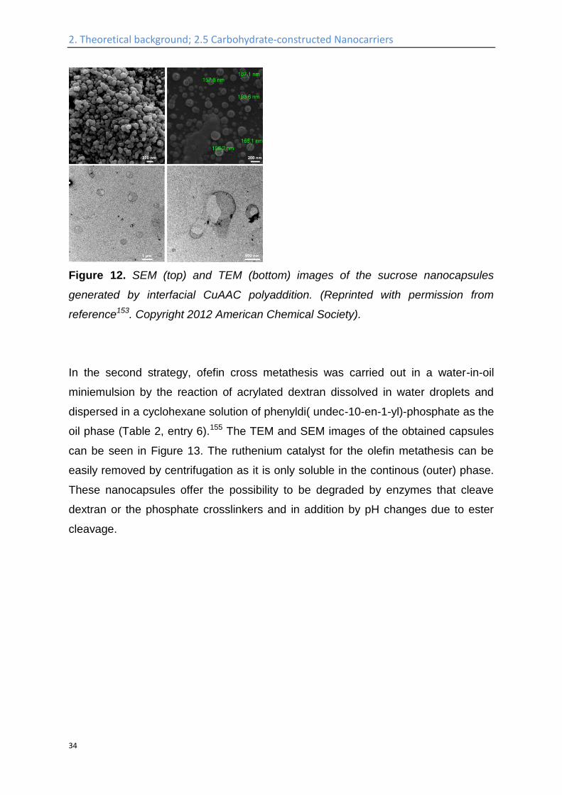
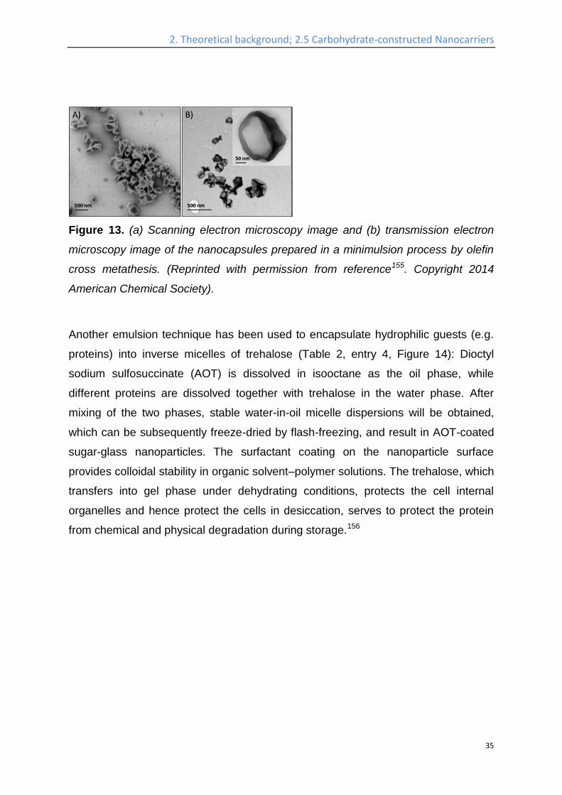
In the second strategy, ofefin cross metathesis was carried out in a water-in-oil
miniemulsion by the reaction of acrylated dextran dissolved in water droplets and
dispersed in a cyclohexane solution of phenyldi( undec-10-en-1-yl)-phosphate as the
oil phase (Table 2, entry 6).155 The TEM and SEM images of the obtained capsules
can be seen in Figure 13. The ruthenium catalyst for the olefin metathesis can be
easily removed by centrifugation as it is only soluble in the continous (outer) phase.
These nanocapsules offer the possibility to be degraded by enzymes that cleave
dextran or the phosphate crosslinkers and in addition by pH changes due to ester
cleavage.
2. Theoretical background; 2.5 Carbohydrate-constructed Nanocarriers
35
Figure 13. (a) Scanning electron microscopy image and (b) transmission electron
microscopy image of the nanocapsules prepared in a minimulsion process by olefin
cross metathesis. (Reprinted with permission from reference155. Copyright 2014
American Chemical Society).
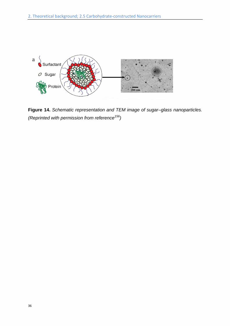
Another emulsion technique has been used to encapsulate hydrophilic guests (e.g.
proteins) into inverse micelles of trehalose (Table 2, entry 4, Figure 14): Dioctyl
sodium sulfosuccinate (AOT) is dissolved in isooctane as the oil phase, while
different proteins are dissolved together with trehalose in the water phase. After
mixing of the two phases, stable water-in-oil micelle dispersions will be obtained,
which can be subsequently freeze-dried by flash-freezing, and result in AOT-coated
sugar-glass nanoparticles. The surfactant coating on the nanoparticle surface
provides colloidal stability in organic solvent–polymer solutions. The trehalose, which
transfers into gel phase under dehydrating conditions, protects the cell internal
organelles and hence protect the cells in desiccation, serves to protect the protein
from chemical and physical degradation during storage.156
2. Theoretical background; 2.5 Carbohydrate-constructed Nanocarriers
36
Figure 14. Schematic representation and TEM image of sugar–glass nanoparticles.
(Reprinted with permission from reference156)
2. Th
eoretical b
ackgrou
nd
; 2.5 C
arbo
hyd
rate-co
nstru
cted N
ano
carriers
37
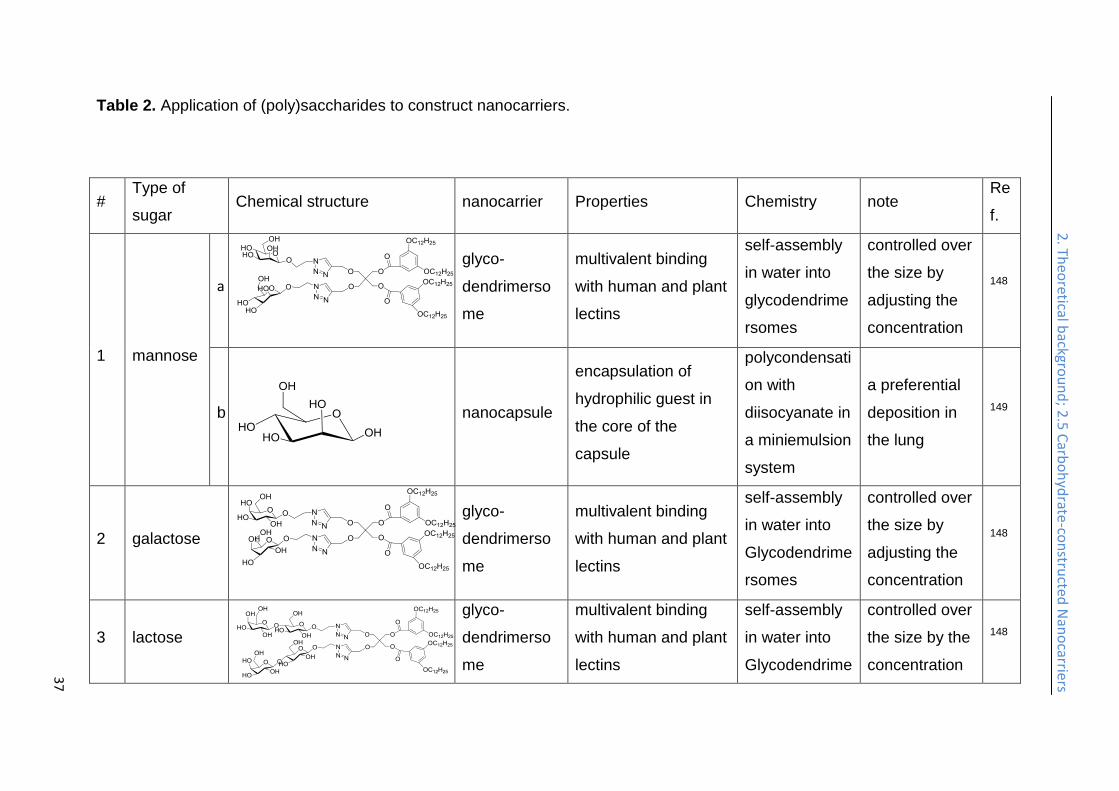
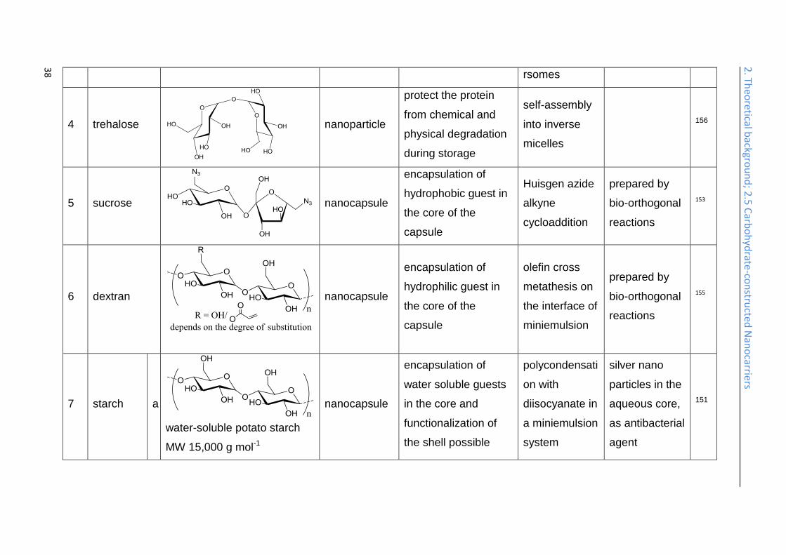
Table 2. Application of (poly)saccharides to construct nanocarriers.
# Type of
sugar Chemical structure nanocarrier Properties Chemistry note
Re
f.
1 mannose
a
glyco-
dendrimerso
me
multivalent binding
with human and plant
lectins
self-assembly
in water into
glycodendrime
rsomes
controlled over
the size by
adjusting the
concentration
148
b
nanocapsule
encapsulation of
hydrophilic guest in
the core of the
capsule
polycondensati
on with
diisocyanate in
a miniemulsion
system
a preferential
deposition in
the lung
149
2 galactose
glyco-
dendrimerso
me
multivalent binding
with human and plant
lectins
self-assembly
in water into
Glycodendrime
rsomes
controlled over
the size by
adjusting the
concentration
148
3 lactose
glyco-
dendrimerso
me
multivalent binding
with human and plant
lectins
self-assembly
in water into
Glycodendrime
controlled over
the size by the
concentration
148
2. Th
eoretical b
ackgrou
nd
; 2.5 C
arbo
hyd
rate-co
nstru
cted N
ano
carriers
38
rsomes
4 trehalose
nanoparticle
protect the protein
from chemical and
physical degradation
during storage
self-assembly
into inverse
micelles
156
5 sucrose
nanocapsule
encapsulation of
hydrophobic guest in
the core of the
capsule
Huisgen azide
alkyne
cycloaddition
prepared by
bio-orthogonal
reactions
153
6 dextran
nanocapsule
encapsulation of
hydrophilic guest in
the core of the
capsule
olefin cross
metathesis on
the interface of
miniemulsion
prepared by
bio-orthogonal
reactions
155
7 starch a
water-soluble potato starch
MW 15,000 g mol-1
nanocapsule
encapsulation of
water soluble guests
in the core and
functionalization of
the shell possible
polycondensati
on with
diisocyanate in
a miniemulsion
system
silver nano
particles in the
aqueous core,
as antibacterial
agent
151
2. Th
eoretical b
ackgrou
nd
; 2.5 C
arbo
hyd
rate-co
nstru
cted N
ano
carriers
39
b
multilayered
polysaccharid
e vesicle
the amylase
treatment of the
nanoparticles allows
the presence of a
void / hollow inner
core (resulting from
the degradation
starch molecules)
within the fabricated
particles
rehydration of
a thin film of
hyaluronate-
starch to form
vesicles
157
c
siRNA
complex
protect siRNA from
enzymatic
degradation
self-assembled
with siRNA to
form
nanocarriers
efficiently
induced P-
glycoprotein
gene silencing
in the human
ovarian
adenocarcino
macell line
158
2. Th
eoretical b
ackgrou
nd
; 2.5 C
arbo
hyd
rate-co
nstru
cted N
ano
carriers
40
8 Hyaluroni
c Acid
a
Mw =140 000 g·mol-1
nanocapsule
encapsulation of
polyhexanide in the
core of the capsule
polycondensati
on with
diisocyanate in
a miniemulsion
system
release of
polyhexanide
upon the
contact with
bacterial
150
b
nanoparticle
specific uptake by
SCC7 cancer cells,
Encapsulation of
doxorubicin and
camptothecin
amphiphilic
HA-CA is self-
assembled to
nanoparticles
in PBS
size can be
tuned between
237–424 nm ,
rapid drug
release in the
presence of
enzyme Hyal-1
159-
162
c nanoparticle
higher cellular uptake
than free paclitaxel in
HepG2 cells
hyaluronic -
paclitaxel self-
assemble in
water to
paclitaxel
could
accumulate
remarkably
134
2. Th
eoretical b
ackgrou
nd
; 2.5 C
arbo
hyd
rate-co
nstru
cted N
ano
carriers
41
form the nano-
particles
into tumor
sites after oral
administration
d
nanoparticle
binding of the particle
with CD44 over-
expressed cancer
cells
self-assembled
in water into
nanoparticles
higher
therapeutic
potential in the
presence of a
green tea
polyphenol,
epigallocatechi
n-3-gallate
163
e
multilayered
polysaccharid
e vesicle
incubation with
hyaluronidase
contributed to
accelerate the
release
rehydration of
a thin film of
hyaluronate-
starch to form
vesicles
drug release in
the presence
of
hyaluronidase
157
2. Th
eoretical b
ackgrou
nd
; 2.5 C
arbo
hyd
rate-co
nstru
cted N
ano
carriers
42
9 HES a
Mw = 200,000 g·mol-1
nanocapsule
encapsulation of
hydrophilic guest in
the core of the
capsule
polycondensati
on with
diisocyanate in
a miniemulsion
system
suppressed
unspecific
uptake into
HeLa cells,
preferential
deposition in
the liver
86,
149
2. Theoretical background; 2.5 Carbohydrate-constructed Nanocarriers
43
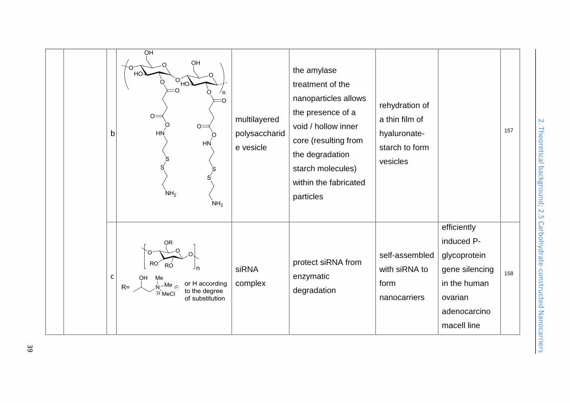
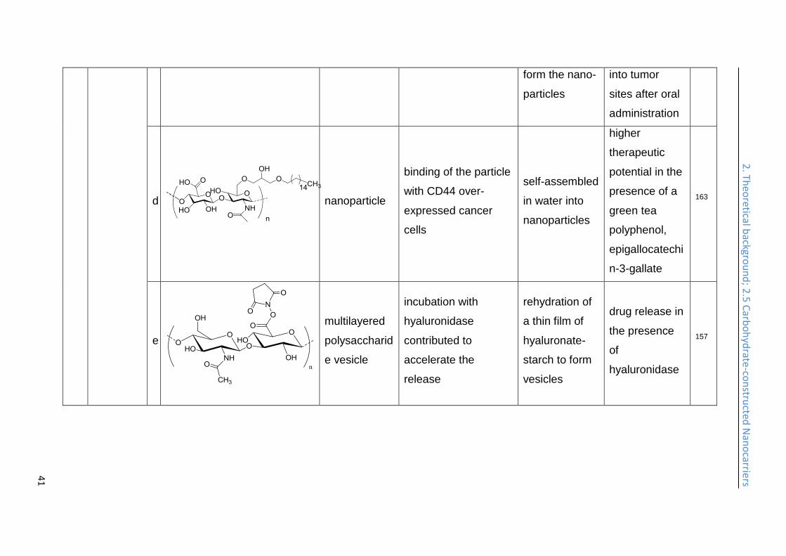
Multilayered polysaccharide vesicles are generated from starch as the core and
hyaluronic acid (HA) as the shell (Table 2, entry 7b). The hydroxyl groups of starch
were activated by succinic anhydride and then reacted with an excess of cysteamine
by Steglich esterification (i.e. N,N-dimethylamino pyridine (DMAP), dicyclohexyl
carbodiimide (DCC), and N-hydroxy succinimide (NHS) in dimethyl sulfoxide
(DMSO)) to produce amino-functionalized starch with additional disulfide bonds. The
amines were then reacted with the activated ester groups of HA. Rehydration of a
thin film of this core-shell HA-starch conjugate in PBS will result in self-assembled
nanoparticles with starch core and hyaluronic acid shell, which are subsequently
treated by amylase, and result in vesicles with a hollow inner core in the end.
Proteins/peptides can be encapsulated in these vesicles, when they are dissolved in
the PBS buffer used. In addition, the enzymatic degradation of the HA shell by
hyaluronidase (HYAL) enzyme contributed to accelerate the release of the
payload.157 In another work, starch modified with ammonium groups is complexed
with siRNA by electrostatic interaction to self-assemble into nanocarriers (Table 2,
entry 7, c), the starch can protect the siRNA from enzymatic degradation on its
delivery route. It has high cellular uptake into a human ovarian adenocarcinoma cell
line and efficiently induced P-glycoprotein (P-gp) gene silencing.158
Antibacterial nanodevices are interesting for coatings and wound dressings if the
release of antibacterial agents can be triggered by the presence of bacteria. HA-
nanocapsules (Table 2, entry 8a) containing the antimicrobial agent polyhexanide
were prepared by the interfacial polycondensation with TDI in an inverse
miniemulsion. They can be specifically cleaved in the presence of the enzyme
hyaluronidase, a factor of pathogenicity and invasion for bacteria like
Staphylococcus aureus and Escherichia coli.150
2. Theoretical background; 2.5 Carbohydrate-constructed Nanocarriers
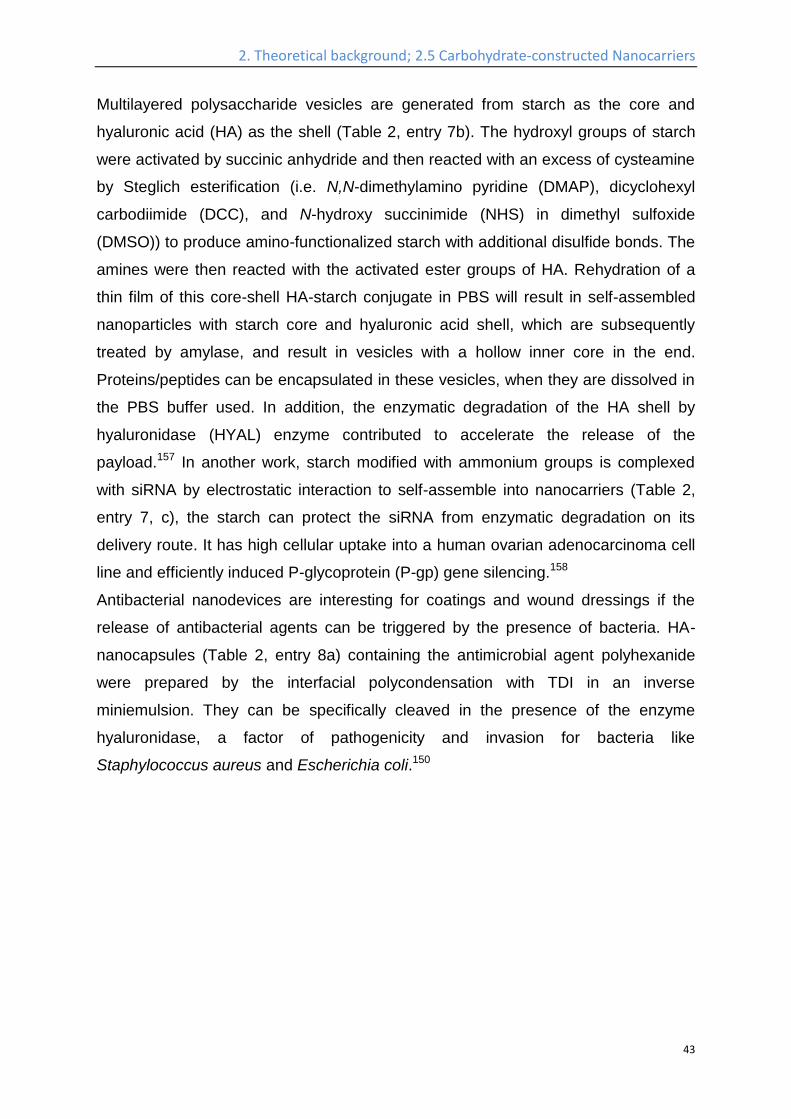
44
Figure 15. Schematic illustration of the formation of drug-loaded HA-NPs.
(Reprinted with permission from reference.160 Copyright 2011 American Chemical
Society).
Hyaluronan-cholanic acid conjugates (HA-CA conjugates) were synthesized by the
chemical conjugation of the hydrophobic bile acid (a steroic acid) to the hydrophilic
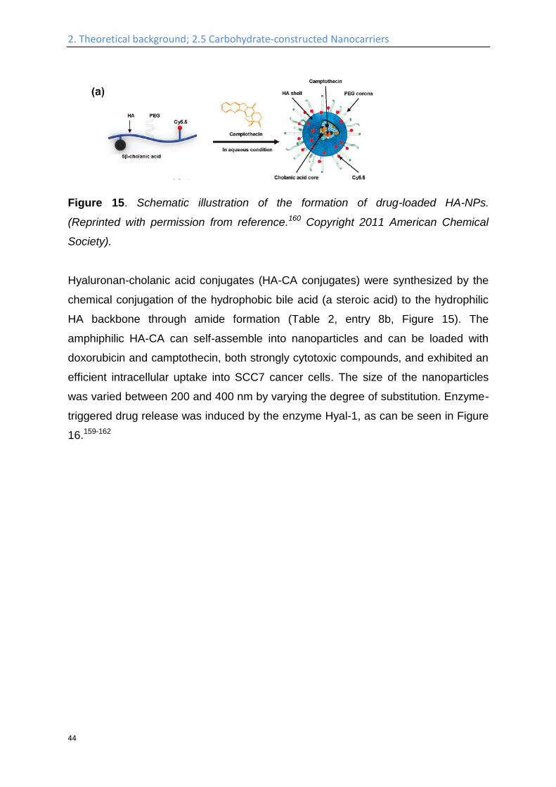
HA backbone through amide formation (Table 2, entry 8b, Figure 15). The
amphiphilic HA-CA can self-assemble into nanoparticles and can be loaded with
doxorubicin and camptothecin, both strongly cytotoxic compounds, and exhibited an
efficient intracellular uptake into SCC7 cancer cells. The size of the nanoparticles
was varied between 200 and 400 nm by varying the degree of substitution. Enzyme-
triggered drug release was induced by the enzyme Hyal-1, as can be seen in Figure
16.159-162
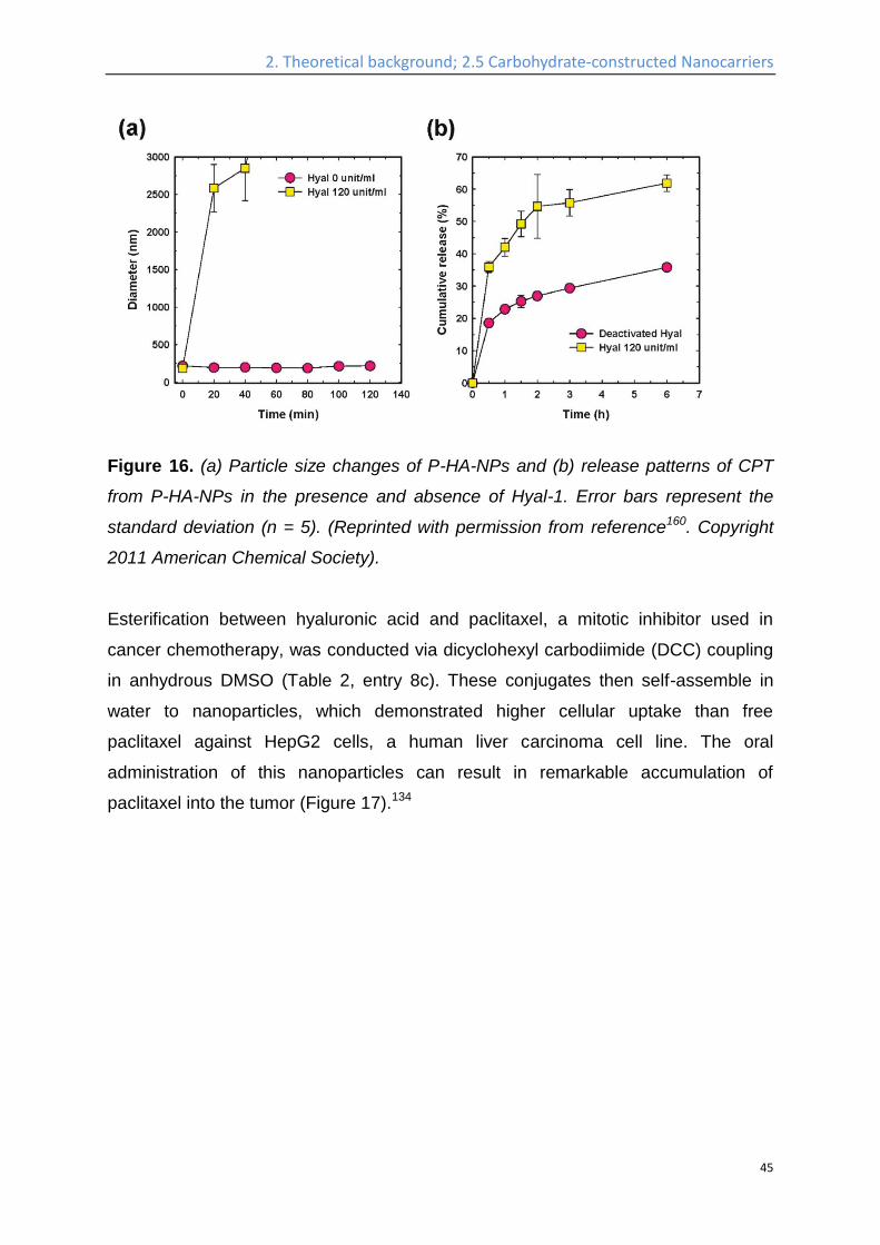
2. Theoretical background; 2.5 Carbohydrate-constructed Nanocarriers
45
Figure 16. (a) Particle size changes of P-HA-NPs and (b) release patterns of CPT
from P-HA-NPs in the presence and absence of Hyal-1. Error bars represent the
standard deviation (n = 5). (Reprinted with permission from reference160. Copyright
2011 American Chemical Society).
Esterification between hyaluronic acid and paclitaxel, a mitotic inhibitor used in
cancer chemotherapy, was conducted via dicyclohexyl carbodiimide (DCC) coupling
in anhydrous DMSO (Table 2, entry 8c). These conjugates then self-assemble in
water to nanoparticles, which demonstrated higher cellular uptake than free
paclitaxel against HepG2 cells, a human liver carcinoma cell line. The oral
administration of this nanoparticles can result in remarkable accumulation of
paclitaxel into the tumor (Figure 17).134
2. Theoretical background; 2.5 Carbohydrate-constructed Nanocarriers
46
Figure 17. Ex vivo fluorescence intensity images of the tumors and major organs after oral administration of rhodamine B labelled hyaluronic acid-paclitaxel nanoparticles (CS/RB-HA-PTX CNPs) and free Rhodamine B (RB). (Taken from reference 134 with permission).
Amphiphilic hexadecylated polysaccharides were synthesized (Table 2, entry 8d)
and self-assembled in water into nanoparticles. Doxorubicin can be encapsulated in
these nanoparticles, which showed specific binding with CD44 over-expresseing
cancer cells. Higher therapeutic potential in the presence of a green tea polyphenol,
epigallocatechin-3-gallate, was also observed for this nanoparticle system.163
Other polysaccharides like cellulose and chitin, which intrinsically are water
insoluble, have also been used to construct nanocarriers. In some cases, their water
solubility has been increased by chemical modifications, for example,
carboxymethylation. Carboxymethyl cellulose has been conjugated with docetaxel
and Poly(ethylene glycol) through acetylation, self-assembled into nanoparticles,
which was stable against dilution, and induced significantly higher toxic effects
against EMT-6 murine mammary carcinoma cells and murine Pan02 tumors cells.164
Carboxymethyl chitin nanoparticles have also been prepared by crosslinking of the
polysaccharide with CaCl2 and FeCl3.165 The obtained nanoparticle showed anti-
bacterial activity by itself, and a sustained and controlled release of the payload.
While different functions are enabled by cellulose and chitin, the intrinsic low water
solubility and poor bio-degradability in animals rendered them to be used more as
wound dressing, scaffolds for tissue engineering and medical implants.166,167

2. Theoretical background; 2.5 Carbohydrate-constructed Nanocarriers
47
It is the aim of this work to prepare sugar based nano-capsules, systematically
evaluate their encapsulation efficiency of the payload, the interaction with plasma
protein and different cell lines, the pharmaceutical behavior (in-vivo plasma
halftime), the specific targeting effect, and the possibility to prepare the capsule by
bio-orthogonal chemistry.
3. Tailoring the stealth properties of biocompatible polysaccharide nanocapsules: introduction
48
3. Tailoring the stealth properties of
biocompatible polysaccharide nanocapsules
A part of this work in this chapter has been published in Biomaterials (Kang B,
Okwieka P, Schöttler S, Seifert O, Kontermann RE, Pfizenmaier K, Musyanovych A,
Meyer R, Diken M, Sahin U, Mailänder V, Wurm FR, Landfester K, Biomaterials.
2015;49:125-34)168 and altered for this thesis with permission. I developed the
synthesis, prepared all the related samples, and conducted the characterization
concerning DLS, SEM, TEM, NMR, release, fluorescamine assay; the in-vitro, in-
vivo characterization and the protein adsorption assay were conducted by the
collaboration partners, which are acknowledged after each corresponding
contribution.
Introduction
As a rapid growing interdisciplinary research field, nanomedicine has extended the
number of possible therapeutic methods, including nanobiosensors169,
nanovaccination170,171, nanoparticle related imaging172, nanodrug carriers173,174, etc.
Among these many potential applications, nanocarriers for targeted drug delivery are
a major research field due to promising therapeutic effects, e.g. specific cell
targeting, protecting the drug simultaneously on its way to the desired site, and the
controlled release of the delivered cargo175,176. Of utmost importance for targeted
delivery and release are the functional properties of the nanocarrier itself: unspecific
cell-uptake has to be prevented, and the carrier needs to exhibit long blood
circulation half-life in order to achieve enrichment and selective, targeted activity at
the diseased tissue. High drug loading capacity is needed to maximize therapeutic
effect. In addition, the nanocontainer should be biodegradable, but only subsequent
to uptake and release of the bioactive principle at the target site. Finally, of equal
important is the precise quantification of the chemistry of nanocarrier synthesis, to
allow systematic investigation and understanding of the factors, which influence the
performance of the system. Many studies have so far addressed the question of
suitable targets and provide a large toolbox of targeting structures177-189. In contrast,
3. Tailoring the stealth properties of biocompatible polysaccharide nanocapsules: introduction
49
information on the development of new synthesis mechanisms for the nanocarrier
platform itself is sparse. The performance of the whole targeting system is often
hindered by properties of the nanocarrier itself190-193, which often possesses one or
more of the setbacks like low stability, cytotoxicity, no precise quantification (of
functional groups, drug loading, etc.), unspecific interaction with healthy cells, and
short plasma half life time.
In this study, miniemulsion was chosen as the preparation method, due to its
outstanding performance to generate nano-objects with tunable sizes, various
surface functionalization possibilities, and efficient encapsulation of a payload194.
Nanoparticles with diameters between 50 nm to 500 nm have been successfully
prepared by miniemulsion, through different reaction mechanisms, including radical
polymerization195, polycondensation196, oxidative polymerization197, and click
chemistry198.
HES is a starch derivative, whose hydroxyl groups at the C2 or C6 positions of each
glucose unit are partially functionalized with a hydroxyethyl group. It is approved for
clinical application, where in most cases it is used as a volume expander for the
treatment of a hypovolemic shock199. Due to the presence of hydroxyethyl groups,
the enzymatic degradation of HES is much slower compared to native starch, while
the biocompatibility is rather unchanged200. Furthermore, it has been shown that,
when HES is attached on the surface of micelles, i.e. “HESylation”, it shows similar
properties as membrane-grafted poly(ethylene glycol) (PEG), i.e. “PEGylation”, with
respect to prolonged blood circulation times or reduced unspecific cell uptake, often
called “stealth effect”23,24.
In the present study, we investigated the combination of both strategies: first, HES-
NCs have been prepared by interfacial polyaddition in an inverse (water-in-oil)
miniemulsion with diameters ranging from 150 to 250 nm. Then, the surface of these
polysaccharide nanocapsules was further functionalized with PEG via different
chemistries: either the coupling of PEG was conducted within the organic phase
directly after the polyaddition, or the bare HES-nanocapsules were transferred into
an aqueous dispersion and subsequently PEGylated. Both strategies were
compared with respect to toxicity, stability in long time storage, protein adsorption,
cell uptake in vitro, and plasma half life time in vivo. In addition, a detailed
3. Tailoring the stealth properties of biocompatible polysaccharide nanocapsules: introduction
50
investigation of every step in the reactions was carried out to allow a precise
quantification of functional groups and added PEG chains on the nanocontainers.
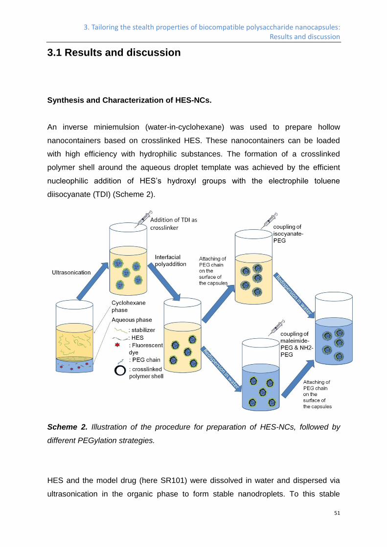
3. Tailoring the stealth properties of biocompatible polysaccharide nanocapsules: Results and discussion
51
3.1 Results and discussion
Synthesis and Characterization of HES-NCs.
An inverse miniemulsion (water-in-cyclohexane) was used to prepare hollow
nanocontainers based on crosslinked HES. These nanocontainers can be loaded
with high efficiency with hydrophilic substances. The formation of a crosslinked
polymer shell around the aqueous droplet template was achieved by the efficient
nucleophilic addition of HES’s hydroxyl groups with the electrophile toluene
diisocyanate (TDI) (Scheme 2).
Scheme 2. Illustration of the procedure for preparation of HES-NCs, followed by
different PEGylation strategies.
HES and the model drug (here SR101) were dissolved in water and dispersed via
ultrasonication in the organic phase to form stable nanodroplets. To this stable
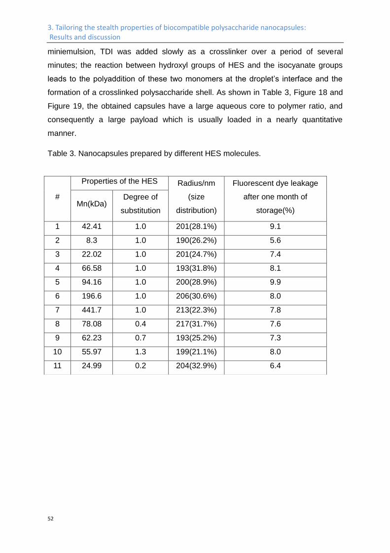
3. Tailoring the stealth properties of biocompatible polysaccharide nanocapsules: Results and discussion
52
miniemulsion, TDI was added slowly as a crosslinker over a period of several
minutes; the reaction between hydroxyl groups of HES and the isocyanate groups
leads to the polyaddition of these two monomers at the droplet’s interface and the
formation of a crosslinked polysaccharide shell. As shown in Table 3, Figure 18 and
Figure 19, the obtained capsules have a large aqueous core to polymer ratio, and
consequently a large payload which is usually loaded in a nearly quantitative
manner.
Table 3. Nanocapsules prepared by different HES molecules.
#
Properties of the HES Radius/nm
(size
distribution)
Fluorescent dye leakage
after one month of
storage(%) Mn(kDa)
Degree of
substitution
1 42.41 1.0 201(28.1%) 9.1
2 8.3 1.0 190(26.2%) 5.6
3 22.02 1.0 201(24.7%) 7.4
4 66.58 1.0 193(31.8%) 8.1
5 94.16 1.0 200(28.9%) 9.9
6 196.6 1.0 206(30.6%) 8.0
7 441.7 1.0 213(22.3%) 7.8
8 78.08 0.4 217(31.7%) 7.6
9 62.23 0.7 193(25.2%) 7.3
10 55.97 1.3 199(21.1%) 8.0
11 24.99 0.2 204(32.9%) 6.4
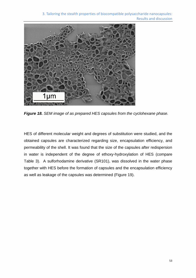
3. Tailoring the stealth properties of biocompatible polysaccharide nanocapsules: Results and discussion
53
Figure 18. SEM image of as prepared HES capsules from the cyclohexane phase.
HES of different molecular weight and degrees of substitution were studied, and the
obtained capsules are characterized regarding size, encapsulation efficiency, and
permeability of the shell. It was found that the size of the capsules after redispersion
in water is independent of the degree of ethoxy-hydroxylation of HES (compare
Table 3). A sulforhodamine derivative (SR101), was dissolved in the water phase
together with HES before the formation of capsules and the encapsulation efficiency
as well as leakage of the capsules was determined (Figure 19).
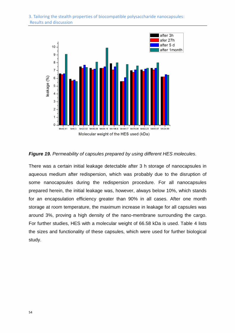
3. Tailoring the stealth properties of biocompatible polysaccharide nanocapsules: Results and discussion
54
Figure 19. Permeability of capsules prepared by using different HES molecules.
There was a certain initial leakage detectable after 3 h storage of nanocapsules in
aqueous medium after redispersion, which was probably due to the disruption of
some nanocapsules during the redispersion procedure. For all nanocapsules
prepared herein, the initial leakage was, however, always below 10%, which stands
for an encapsulation efficiency greater than 90% in all cases. After one month
storage at room temperature, the maximum increase in leakage for all capsules was
around 3%, proving a high density of the nano-membrane surrounding the cargo.
For further studies, HES with a molecular weight of 66.58 kDa is used. Table 4 lists
the sizes and functionality of these capsules, which were used for further biological
study.
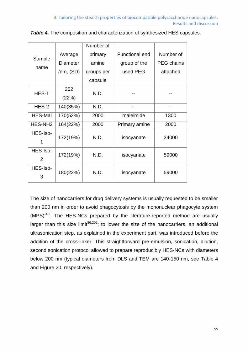
3. Tailoring the stealth properties of biocompatible polysaccharide nanocapsules: Results and discussion
55
Table 4. The composition and characterization of synthesized HES capsules.
Sample
name
Average
Diameter
/nm, (SD)
Number of
primary
amine
groups per
capsule
Functional end
group of the
used PEG
Number of
PEG chains
attached
HES-1 252
(22%) N.D. -- --
HES-2 140(35%) N.D. -- --
HES-Mal 170(52%) 2000 maleimide 1300
HES-NH2 164(22%) 2000 Primary amine 2000
HES-Iso-
1 172(19%) N.D. isocyanate 34000
HES-Iso-
2 172(19%) N.D. isocyanate 59000
HES-Iso-
3 180(22%) N.D. isocyanate 59000
The size of nanocarriers for drug delivery systems is usually requested to be smaller
than 200 nm in order to avoid phagocytosis by the mononuclear phagocyte system
(MPS)201. The HES-NCs prepared by the literature-reported method are usually
larger than this size limit86,202; to lower the size of the nanocarriers, an additional
ultrasonication step, as explained in the experiment part, was introduced before the
addition of the cross-linker. This straightforward pre-emulsion, sonication, dilution,
second sonication protocol allowed to prepare reproducibly HES-NCs with diameters
below 200 nm (typical diameters from DLS and TEM are 140-150 nm, see Table 4
and Figure 20, respectively).
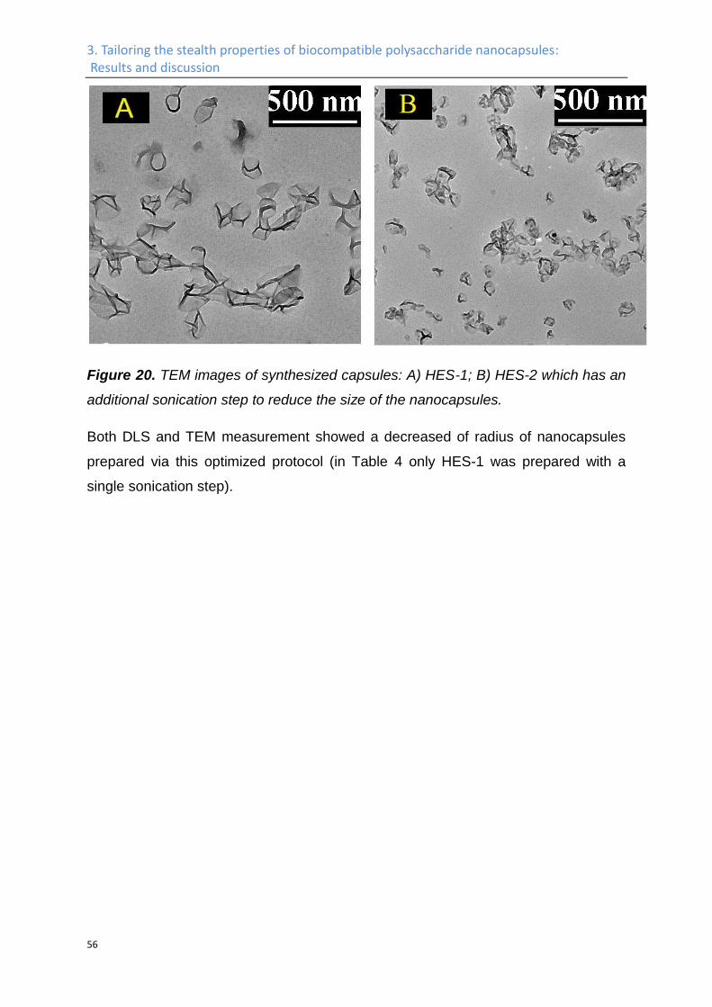
3. Tailoring the stealth properties of biocompatible polysaccharide nanocapsules: Results and discussion
56
Figure 20. TEM images of synthesized capsules: A) HES-1; B) HES-2 which has an
additional sonication step to reduce the size of the nanocapsules.
Both DLS and TEM measurement showed a decreased of radius of nanocapsules
prepared via this optimized protocol (in Table 4 only HES-1 was prepared with a
single sonication step).
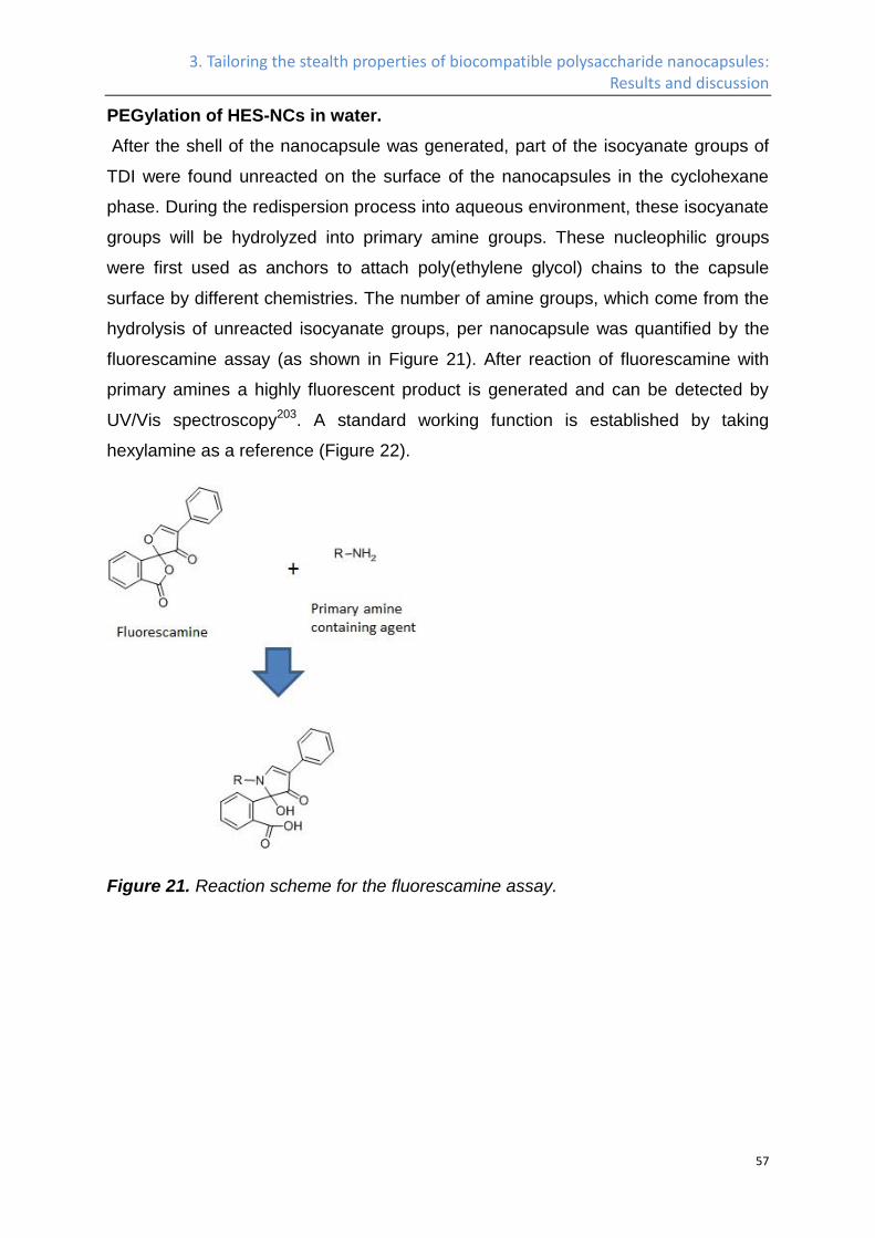
3. Tailoring the stealth properties of biocompatible polysaccharide nanocapsules: Results and discussion
57
PEGylation of HES-NCs in water.
After the shell of the nanocapsule was generated, part of the isocyanate groups of
TDI were found unreacted on the surface of the nanocapsules in the cyclohexane
phase. During the redispersion process into aqueous environment, these isocyanate
groups will be hydrolyzed into primary amine groups. These nucleophilic groups
were first used as anchors to attach poly(ethylene glycol) chains to the capsule
surface by different chemistries. The number of amine groups, which come from the
hydrolysis of unreacted isocyanate groups, per nanocapsule was quantified by the
fluorescamine assay (as shown in Figure 21). After reaction of fluorescamine with
primary amines a highly fluorescent product is generated and can be detected by
UV/Vis spectroscopy203. A standard working function is established by taking
hexylamine as a reference (Figure 22).
Figure 21. Reaction scheme for the fluorescamine assay.
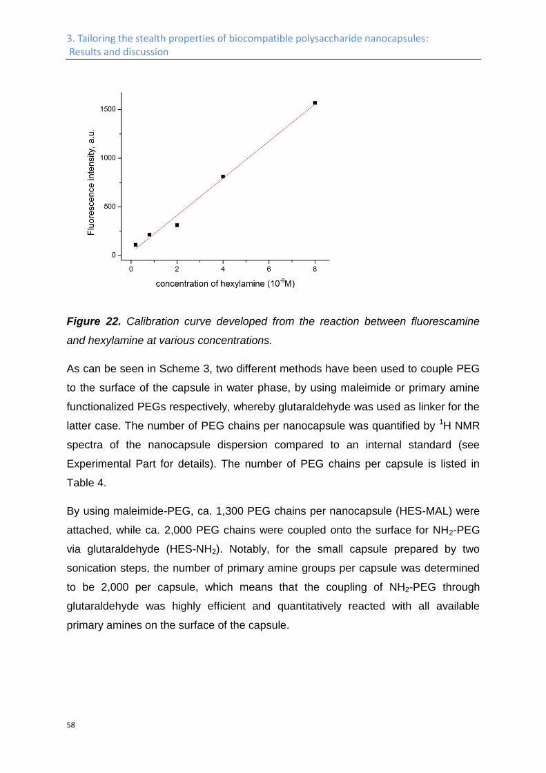
3. Tailoring the stealth properties of biocompatible polysaccharide nanocapsules: Results and discussion
58
Figure 22. Calibration curve developed from the reaction between fluorescamine
and hexylamine at various concentrations.
As can be seen in Scheme 3, two different methods have been used to couple PEG
to the surface of the capsule in water phase, by using maleimide or primary amine
functionalized PEGs respectively, whereby glutaraldehyde was used as linker for the
latter case. The number of PEG chains per nanocapsule was quantified by 1H NMR
spectra of the nanocapsule dispersion compared to an internal standard (see
Experimental Part for details). The number of PEG chains per capsule is listed in
Table 4.
By using maleimide-PEG, ca. 1,300 PEG chains per nanocapsule (HES-MAL) were
attached, while ca. 2,000 PEG chains were coupled onto the surface for NH2-PEG
via glutaraldehyde (HES-NH2). Notably, for the small capsule prepared by two
sonication steps, the number of primary amine groups per capsule was determined
to be 2,000 per capsule, which means that the coupling of NH2-PEG through
glutaraldehyde was highly efficient and quantitatively reacted with all available
primary amines on the surface of the capsule.
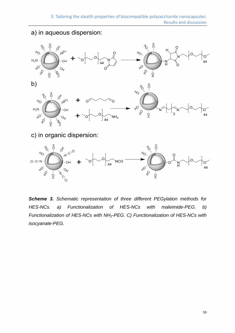
3. Tailoring the stealth properties of biocompatible polysaccharide nanocapsules: Results and discussion
59
Scheme 3. Schematic representation of three different PEGylation methods for
HES-NCs. a) Functionalization of HES-NCs with maleimide-PEG. b)
Functionalization of HES-NCs with NH2-PEG. C) Functionalization of HES-NCs with
isocyanate-PEG.
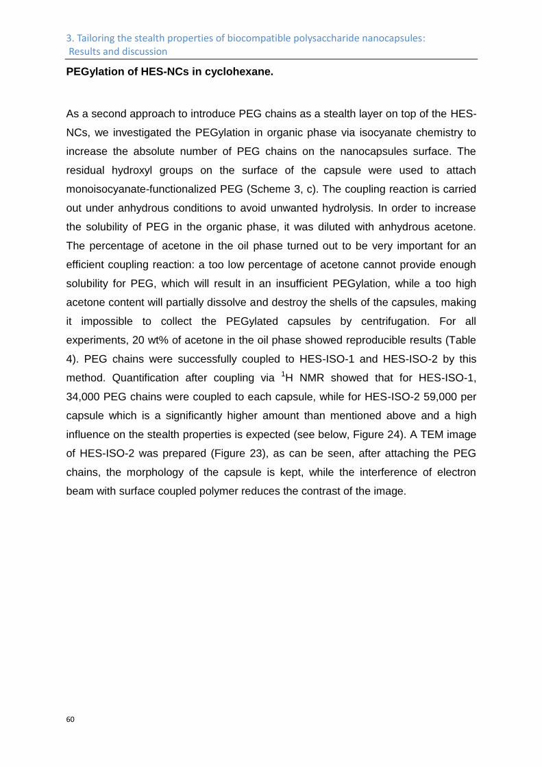
3. Tailoring the stealth properties of biocompatible polysaccharide nanocapsules: Results and discussion
60
PEGylation of HES-NCs in cyclohexane.
As a second approach to introduce PEG chains as a stealth layer on top of the HES-
NCs, we investigated the PEGylation in organic phase via isocyanate chemistry to
increase the absolute number of PEG chains on the nanocapsules surface. The
residual hydroxyl groups on the surface of the capsule were used to attach
monoisocyanate-functionalized PEG (Scheme 3, c). The coupling reaction is carried
out under anhydrous conditions to avoid unwanted hydrolysis. In order to increase
the solubility of PEG in the organic phase, it was diluted with anhydrous acetone.
The percentage of acetone in the oil phase turned out to be very important for an
efficient coupling reaction: a too low percentage of acetone cannot provide enough
solubility for PEG, which will result in an insufficient PEGylation, while a too high
acetone content will partially dissolve and destroy the shells of the capsules, making
it impossible to collect the PEGylated capsules by centrifugation. For all
experiments, 20 wt% of acetone in the oil phase showed reproducible results (Table
4). PEG chains were successfully coupled to HES-ISO-1 and HES-ISO-2 by this
method. Quantification after coupling via 1H NMR showed that for HES-ISO-1,
34,000 PEG chains were coupled to each capsule, while for HES-ISO-2 59,000 per
capsule which is a significantly higher amount than mentioned above and a high
influence on the stealth properties is expected (see below, Figure 24). A TEM image
of HES-ISO-2 was prepared (Figure 23), as can be seen, after attaching the PEG
chains, the morphology of the capsule is kept, while the interference of electron
beam with surface coupled polymer reduces the contrast of the image.
3. Tailoring the stealth properties of biocompatible polysaccharide nanocapsules: Results and discussion
61
Figure 23. TEM image of nanocapsules coupled with PEG and redispersed in water
phase. The surface coupled poly(ethylene glycol) chains interference with the
electron beam, and reduce the contrast of the image.
3. Tailoring the stealth properties of biocompatible polysaccharide nanocapsules: Results and discussion
62
In vivo plasma half-life.
Nowadays, HESylation is a common pathway to modify surfaces or micelles with
stealth properties23,24. A detailed investigation of colloidal objects based on HES,
however, is missing. Moreover, we sought to potentially improve these basic stealth
properties of HES by additional PEGylation of HES capsules. Therefore, we
compared the pharmacokinetic properties of unmodified HES-NCs with several
samples of PEGylated HES-NCs (see above) and measured their plasma half-life
time in vivo, as can be seen in Figure 24. Contrary to expectations of sufficient
stealth properties of HES on its own, the unmodified HES-NCs were rapidly cleared
from the blood plasma with less than 2% remaining after 2 h after the injection. This
rather short plasma half-life of the unmodified nanocapsules could be due to the
poor mobility of the HES chains after crosslinking or due to the presence of too
many urethane bridges in the shell. To increase the plasma half-life, we combined
the positive properties of HES such as biodegradability and easy handling
concerning the formation of loaded- nanocapsules with the known stealth properties
of PEG. As can be seen in Figure 24, compared to unmodified HES2, the circulation
time of both HES-MAL and HES-NH2 was significantly prolonged. Furthermore,
when we compared the circulation time between HES-MAL and HES-NH2, a trend of
increased circulation time as a result of increased surface PEG density became
apparent, too. In order to further increase the plasma half-life, a higher density of
surface coupled PEG chains will probably be needed. Since the measured surface
density of primary amines was also 2000 per capsule, we have consumed all the
available primary amine groups for capsule HES-NH2. Therefore, a new method is
needed in order to further increase the plasma half-time of the nanocapsules.
Subsequent plasma half-life studies showed that the circulation time of HES-ISO-1
was substantially enhanced compared to HES-MAL and HES-NH2, with 10% of the
fluorescent capsule signal staying in the blood plasma after 24 h, and 72 h after
injection, still 2% of capsules were detectable. With an increased surface PEG
density, the plasma half-life of HES-ISO-2 was further prolonged, with 20% and 5%
of capsules remaining in the blood plasma after 24 h and 72 h, respectively. To test
the reproducibility of the long circulation capsules, HES-ISO-3 was prepared under
the same condition as HES-ISO-2, and showed comparably good circulation time

3. Tailoring the stealth properties of biocompatible polysaccharide nanocapsules: Results and discussion
63
too. These results show that, when the poly(ethylene glycol) chains are attached to
the surface of the capsules in the right way and at the appropriate density, the
combination of the biocompatible HES shell and the surface attached PEG chains
can provide the system with a substantially enhanced plasma half-life, which makes
the capsule system an ideal platform for targeted drug delivery.
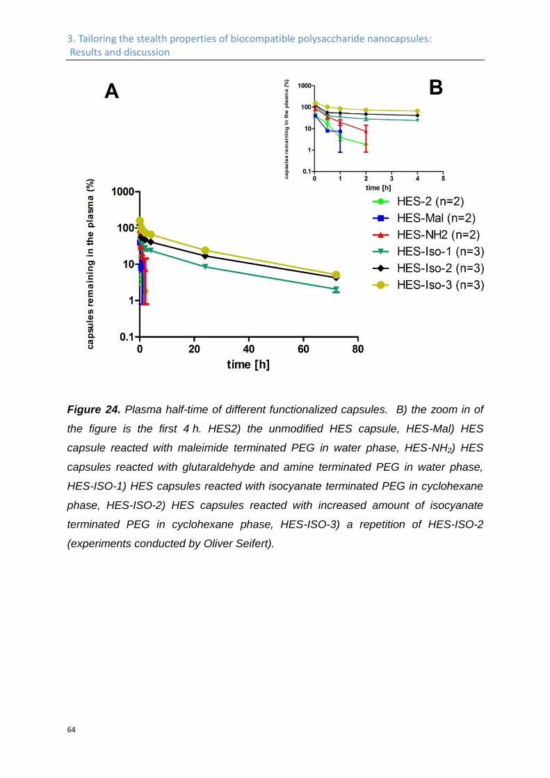
3. Tailoring the stealth properties of biocompatible polysaccharide nanocapsules: Results and discussion
64
Figure 24. Plasma half-time of different functionalized capsules. B) the zoom in of
the figure is the first 4 h. HES2) the unmodified HES capsule, HES-Mal) HES
capsule reacted with maleimide terminated PEG in water phase, HES-NH2) HES
capsules reacted with glutaraldehyde and amine terminated PEG in water phase,
HES-ISO-1) HES capsules reacted with isocyanate terminated PEG in cyclohexane
phase, HES-ISO-2) HES capsules reacted with increased amount of isocyanate
terminated PEG in cyclohexane phase, HES-ISO-3) a repetition of HES-ISO-2
(experiments conducted by Oliver Seifert).
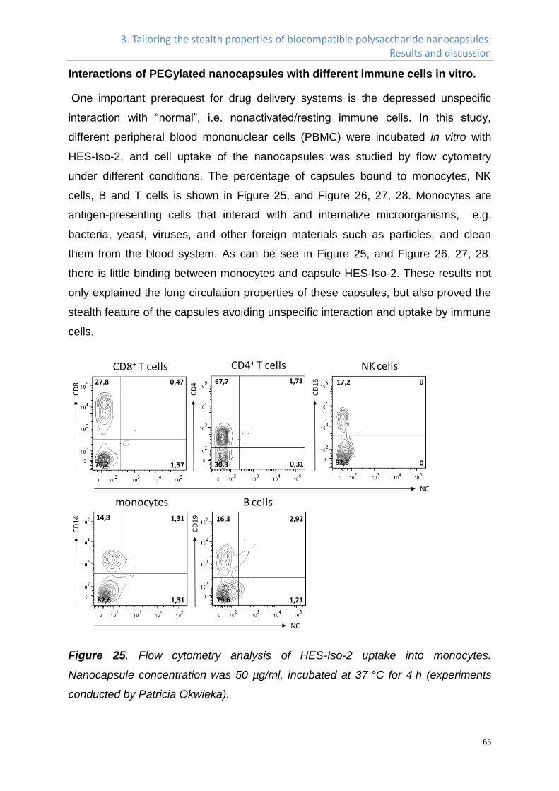
3. Tailoring the stealth properties of biocompatible polysaccharide nanocapsules: Results and discussion
65
Interactions of PEGylated nanocapsules with different immune cells in vitro.
One important prerequest for drug delivery systems is the depressed unspecific
interaction with “normal”, i.e. nonactivated/resting immune cells. In this study,
different peripheral blood mononuclear cells (PBMC) were incubated in vitro with
HES-Iso-2, and cell uptake of the nanocapsules was studied by flow cytometry
under different conditions. The percentage of capsules bound to monocytes, NK
cells, B and T cells is shown in Figure 25, and Figure 26, 27, 28. Monocytes are
antigen-presenting cells that interact with and internalize microorganisms, e.g.
bacteria, yeast, viruses, and other foreign materials such as particles, and clean
them from the blood system. As can be see in Figure 25, and Figure 26, 27, 28,
there is little binding between monocytes and capsule HES-Iso-2. These results not
only explained the long circulation properties of these capsules, but also proved the
stealth feature of the capsules avoiding unspecific interaction and uptake by immune
cells.
Figure 25. Flow cytometry analysis of HES-Iso-2 uptake into monocytes.
Nanocapsule concentration was 50 µg/ml, incubated at 37 °C for 4 h (experiments
conducted by Patricia Okwieka).
CD8+ T cells CD4+ T cells NK cells
0 17,2
0 82,8
NC
CD
8
CD
4
CD
1627,8 0,47
70,2 1,57
1,73
0,31 30,3
67,7
NC
B cells
2,92
1,21
16,3
79,6
monocytes
CD
14 1,31
1,31
14,8
82,6
CD
19
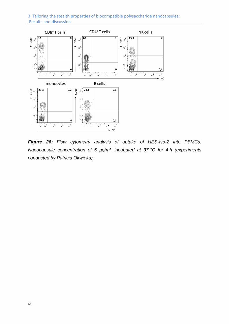
3. Tailoring the stealth properties of biocompatible polysaccharide nanocapsules: Results and discussion
66
Figure 26: Flow cytometry analysis of uptake of HES-Iso-2 into PBMCs.
Nanocapsule concentration of 5 µg/ml, incubated at 37 °C for 4 h (experiments
conducted by Patricia Okwieka).
CD8+ T cells CD4+ T cells NK cells0 15,3
0,484,3
NC
CD
8
CD
4
CD
1632 0
68 0
0
038
62
NC
B cells
0,1
0,1
24,1
75,7
monocytes
CD
14 0,2
0
23,3
76,5
CD
19
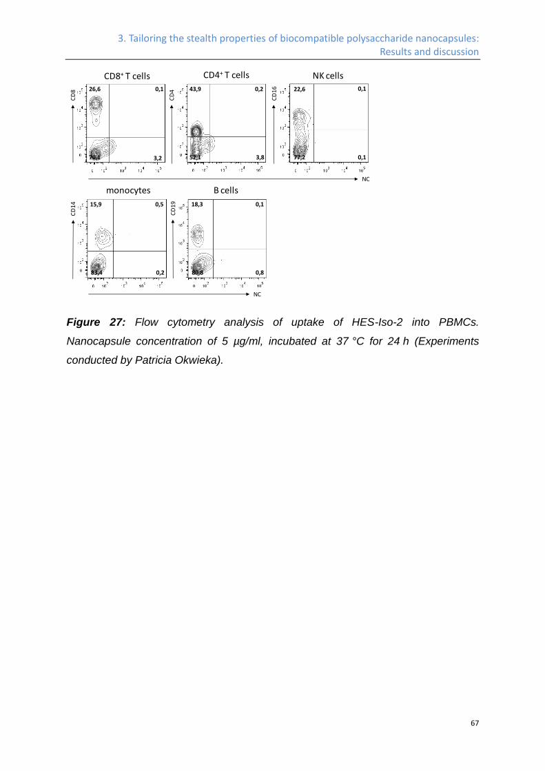
3. Tailoring the stealth properties of biocompatible polysaccharide nanocapsules: Results and discussion
67
Figure 27: Flow cytometry analysis of uptake of HES-Iso-2 into PBMCs.
Nanocapsule concentration of 5 µg/ml, incubated at 37 °C for 24 h (Experiments
conducted by Patricia Okwieka).
CD8+ T cells CD4+ T cells NK cells0,1 22,6
0,177,2
NC
CD
8
CD
4
CD
1626,6 0,1
70,1 3,2
0,2
3,852,1
43,9
NC
B cells
0,1
0,8
18,3
80,8
monocytes
CD
14 0,5
0,2
15,9
83,4
CD
19
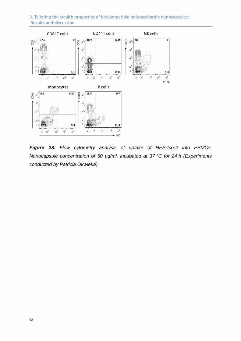
3. Tailoring the stealth properties of biocompatible polysaccharide nanocapsules: Results and discussion
68
Figure 28: Flow cytometry analysis of uptake of HES-Iso-2 into PBMCs.
Nanocapsule concentration of 50 µg/ml, incubated at 37 °C for 24 h (Experiments
conducted by Patricia Okwieka).
CD8+ T cells CD4+ T cells NK cells
4 30
5,360,7
NC
CD
8
CD
4
CD
1637,6 0
61,5 0,1
0,45
0,4550,8
48,2
NC
B cells
0,7
11,4
18,4
69,5
monocytes
CD
14 8,01
3,9
0,5
87,6
CD
19
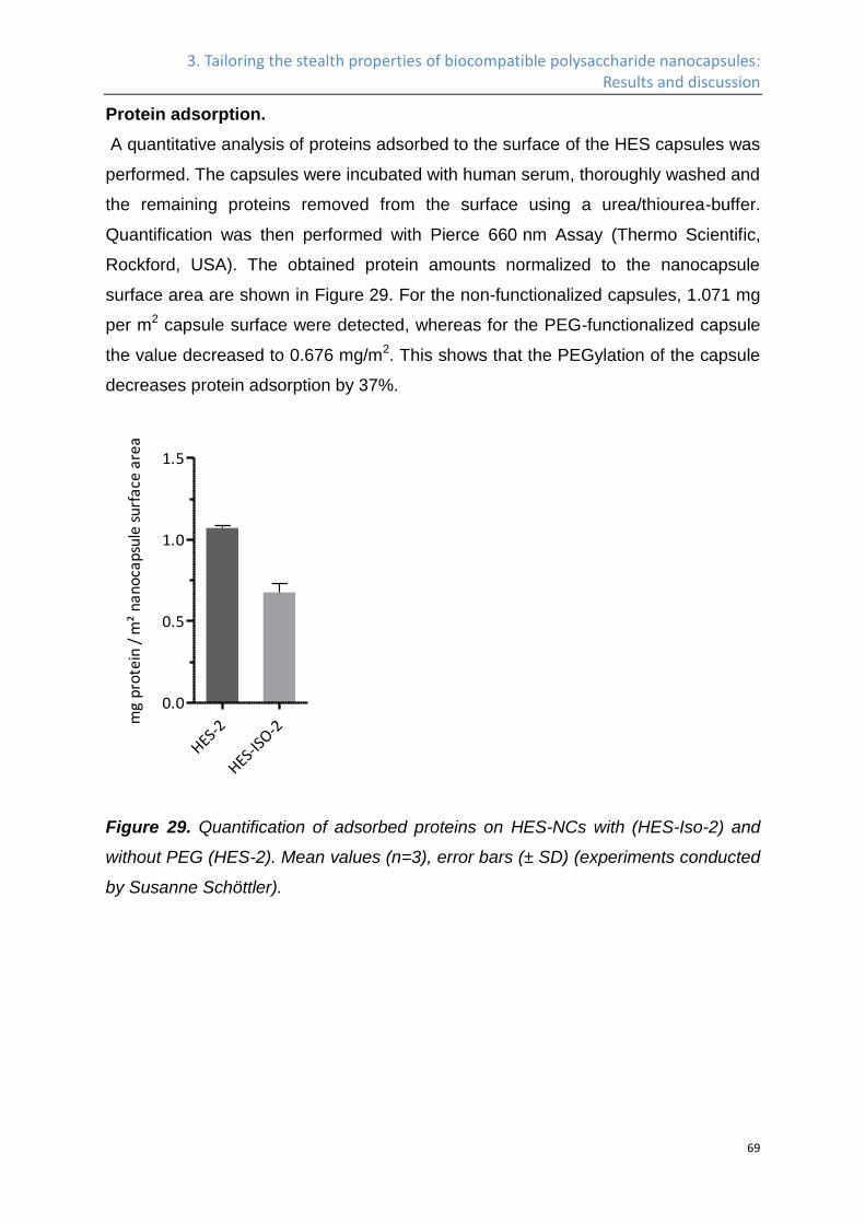
3. Tailoring the stealth properties of biocompatible polysaccharide nanocapsules: Results and discussion
69
Protein adsorption.
A quantitative analysis of proteins adsorbed to the surface of the HES capsules was
performed. The capsules were incubated with human serum, thoroughly washed and
the remaining proteins removed from the surface using a urea/thiourea-buffer.
Quantification was then performed with Pierce 660 nm Assay (Thermo Scientific,
Rockford, USA). The obtained protein amounts normalized to the nanocapsule
surface area are shown in Figure 29. For the non-functionalized capsules, 1.071 mg
per m2 capsule surface were detected, whereas for the PEG-functionalized capsule
the value decreased to 0.676 mg/m2. This shows that the PEGylation of the capsule
decreases protein adsorption by 37%.
HES-2
HES-IS
O-2
0.0
0.5
1.0
1.5
mg
pro
tein
/ m
² n
an
oca
psu
le s
urf
ace
are
a
Figure 29. Quantification of adsorbed proteins on HES-NCs with (HES-Iso-2) and
without PEG (HES-2). Mean values (n=3), error bars (± SD) (experiments conducted
by Susanne Schöttler).
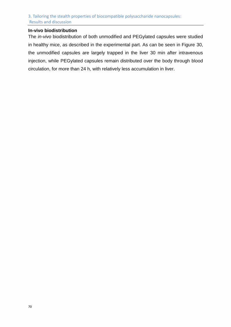
3. Tailoring the stealth properties of biocompatible polysaccharide nanocapsules: Results and discussion
70
In-vivo biodistribution
The in-vivo biodistribution of both unmodified and PEGylated capsules were studied
in healthy mice, as described in the experimental part. As can be seen in Figure 30,
the unmodified capsules are largely trapped in the liver 30 min after intravenous
injection, while PEGylated capsules remain distributed over the body through blood
circulation, for more than 24 h, with relatively less accumulation in liver.
3. Tailoring the stealth properties of biocompatible polysaccharide nanocapsules: Results and discussion
71
Figure 30. Bio-distribution of unmodified (HES2) and PEGylated (HES-Iso-2)
capsules. 1 mg of the capsules was injected intravenously and the fluorescent
intensity was recorded after 30 min, 1 h, 2 h, 4 h, 8 h, and 24 h. The mice were
sacrificed after 24 h and different organs were tested for their fluorescent intensity
(experiments conducted by Mustafa Diken).
As can be seen in Figure 30, the accumulation of capsules in liver and spleen was
much lower for PEGylated sample, compared to unmodified capsules.
3. Tailoring the stealth properties of biocompatible polysaccharide nanocapsules: Results and discussion
72
Discussion and conclusion
When attached to the surface of nanocarriers, HES has been proved, that it can
provide stealth property23,24. It is due to this reason, we decided to take it as the
building block for the nanocapsules. Previous work from our group86 did show
depressed unspecific uptake of HES capsules in HeLa cells. Stealth property of
these HES capsules is expected, when they are injected intravenously into the blood
system. However, the size of the HES capsule used in our previous publication86,
which is sample HES-1 in this work, is around 250 nm. According to the work from
other groups, the size of the nanocarrier need to be small enough to avoid
phagocytosis by the mononuclear phagocyte system (MPS)201. HES-2 is prepared
for this purpose. By straightforward pre-emulsion, sonication, dilution, second
sonication protocol, HES-NCs with diameters below 200 nm is prepared (typical
diameters from DLS and TEM are 140-150 nm, see Table 4 and Figure 20,
respectively), and this small HES capsule (HES-2) is sent for in-vivo plasma halftime
test. Contrary to expectations of sufficient stealth properties of HES on its own, the
unmodified HES-2 nanocapsules were rapidly cleared from the blood plasma with
less than 2% remaining 2 h after the injection. This rather short plasma half-life of
the unmodified nanocapsules could be due to the poor mobility of the HES chains
after crosslinking, or due to the presence of too many urethane bridges in the shell.
To increase the plasma half-life, we combined the positive properties of HES such
as biodegradability and easy handling, concerning the formation of loaded-
nanocapsules, with the known stealth properties of PEG. The residual amine and
hydroxyl groups on the surface of the capsules offer versatile functionalization
possibilities. In this work we have coupled PEG to the capsule by reacting with both
these groups. Namely, using maleimide and amine terminated PEG to couple with
the amine groups on the capsule surface (HES-Mal and HES-NH2), and using
isocyanate terminated PEG to react with hydroxyl groups (HES-Iso-1, HES-Iso-2,
and HES-Iso-3). It turned out that, by the coupling with amine groups we can attach
maximum ca. 2000 PEG chains per capsule (HES-Mal), while the maximum density
for the hydroxyl method is 59000 per capsule (HES-Iso-2). Subsequent in-vivo
plasma halftime test showed, for the HES-NCs, an increased PEG density in this
range can effectively increase the plasma halftime (see Figure 24). The HES-Iso-2
shows outstanding plasma halftime, after 24 h and 72 h, there are 20% and 5% of
3. Tailoring the stealth properties of biocompatible polysaccharide nanocapsules: Results and discussion
73
capsules remaining in the blood plasma, respectively. The high plasma halftime of
HES-Iso-2 is reproduced by HES-Iso-3. These results show that, when the
poly(ethylene glycol) chains are attached to the surface of the HES capsules in the
right way and at the appropriate density, the combination of the biocompatible HES
shell and the surface attached PEG chains can provide the system with a
substantially enhanced plasma half-life, which makes the capsule system an ideal
platform for targeted drug delivery.
On the other hand, the plasma halftime of the PEGylated HES capsules is so
outstanding, that we start to consider the possibility, whether the capsules are
internalized in some of the blood cells, therefore, the fluorescent signal can stay in
the blood plasma for such a long time. Based on these considerations, in vitro tests
of HES-Iso-2 with different blood immune cells were initiated. Different peripheral
blood mononuclear cells (PBMC) were incubated in vitro with HES-Iso-2, and the
cell uptake of the nanocapsules was studied by flow cytometry. The percentage of
capsules bound to monocytes, NK cells, B and T cells is shown in Figure 25, 26, 27,
and 28. Monocytes are antigen-presenting cells that interact with and internalize
microorganisms, e.g. bacteria, yeast, viruses, and other foreign materials such as
particles, and clean them from the blood system. As can be see in Figure 25, there
is little binding between monocytes and capsule HES-Iso-2. These results show that,
there is little unspecific binding between HES-Iso-2 and different blood immune cells,
and the origin of the long circulating properties comes from the advanced stealth
property of the capsule itself.
Due to the hydrophobic interaction between the protein and the nano-particles, and
the high surface tenstion of the nano-partcle itself, whenever nanoparticles enter
biological fluoides like blood, proteins will spontaneously absorb on their surface,
and consequently form a so called protein corona. Many researchers have proposed
that the behavior of the nanocarriers in biological liquid, for example the stealth
properties of nanocarrier in blood plasma, is often determined by the protein corona
formed on the surface of them, instead of the intrinsic functionalization of the
nanocarrier itself204-211. The protein corona formed on the nanocapsule self is
compared between HES-2 and HES-Iso-2. The results demonstrate that we can
decrease the protein absorption by 37% after PEGylation, from 1.071 mg per m2 to
0.676 mg/m2. By PEGylation we can decrease 37% of the protein absorption,
3. Tailoring the stealth properties of biocompatible polysaccharide nanocapsules: Results and discussion
74
however, the plasma halftime is increased drastically. We believe that in this case, it
is not the amount of protein absorpt, but the type of protein, which determines the
stealth properties of the HES capsules. A study of the detailed composition of the
protein corona on these capsules is under investigation with mass spectrometry, and
will be discussed in another work in our group.
The in-vivo bio-distribution of HES-2 and HES-Iso-2 is compared. As can be seen in
Figure 30, the accumulation of capsules in liver and spleen is depressed after
PEGylation. This versatile chemical platform in combination with the outstandingly
long circulation times makes the PEGylated HES nanocontainers ideal for further
studies for targeted cell uptake and subsequent drug release.
In summary, interfacial polyaddition in an inverse miniemulsion system was used to
prepare biodegradable, PEG functionalized nanocontainers with optimized
pharmacokinetic properties suited for targeted drug delivery. In the work presented
here, hydrophilic, biocompatible and biodegradable HES was reacted at the water-oil
interface with TDI in order to generate a cross-linked nanocontainer that can be
loaded with hydrophilic dyes or drugs. HES-NCs with different sizes and versatile
chemical functionality were prepared, which have good shelf-life stability over long
time storage, and are non-toxic. The surface of nanocarriers was functionalized with
PEG via different chemical protocols producing nanocapsules with a variable and
precisely determined PEG density. Hydrophilic drugs, fluorescent dyes, or other
contrast agents can be encapsulated in the nanocontainer, which will be protected
against degradation on the delivery route. The library of different PEGylated
nanocapsules was investigated in vitro with respect to biocompatibility, protein
interaction, cell uptake and in vivo plasma half-life time. Highly PEGylated
nanocapsules did show a reduced protein adsorption and a very low unspecific cell
uptake in several blood cells. As a result, these nanocontainers show a pronounced
plasma circulation time in blood which is higher than typically applied liposomal
formulations. This versatile chemical platform in combination with the outstandingly
long circulation times makes the PEGylated HES nanocontainers ideal for further
studies for targeted cell uptake, and the results will be discussed in the following
sector.
3. Tailoring the stealth properties of biocompatible polysaccharide nanocapsules: Experimental part
75
3.2 Experimental Part
Materials
The isocyanate-PEG and isocyanate-PEG-isocyanate (PEG molecular weight of
5,000 g·mol−1, 110 ethylene oxide units) were purchased from Nanocs Inc., USA.
PBS buffer (pH 7.4, without Ca2+ and Mg2+) was purchased from Gibco, Germany.
Borate buffer (1M, pH 9.5) was produced by adjusting pH of boric acid (B6768
Sigma) water solution to the desired value by sodium hydroxide solution. HES (200k,
0.5 degree of substitution) was bought from Fresenius Kabi, dialyzed in the dialyzing
tube with molecular cutting off of 14k againt MillQ water for 4 days, and
subsequently freeze-dried for further synthesis. Oil soluble surfactant poly((ethylene-
co-butylene)-b-(ethylene oxide)), P(E/B-b-EO), consisting of a poly(ethylene-co-
butylene) block (Mw = 3,700 g·mol−1) and a poly(ethylene oxide) block (Mw = 3,600
g·mol−1) was synthesized starting from ω-hydroxypoly-(ethylene-co-butylene), which
was dissolved in toluene after addition of ethylene oxide via anionic
polymerization212. The anionic surfactant sodium dodecylsulfate (SDS) was
purchased from Fluka. C-type-lectin (Galanthus nivalis snowdrop lectin fluorescein
labeled 2 mg) was bought from BIOZOL Diagnostica Vertrieb GmbH. The
fluorescent dye Cy5-oligo was purchased from BioChemica, Aldrich. DBCO-PEG4-
NHS was bought from Jana Bioscience. Dimethyl sulfoxide (<50 ppm water content)
was purchased from Acros Organics. Demineralized MilliQ water was used for the
synthesis of nanocapsules. Polystyrene nanoparticles was prepared according to
literature.195 All the other chemicals were purchased from Sigma Aldrich, and used
as received.
3. Tailoring the stealth properties of biocompatible polysaccharide nanocapsules: Experimental part
76
Characterization methods
Dynamic Light Scattering (DLS). The average size of the capsules is measured by
DLS using a PSS Nicomp Particle Sizer 380. The capsule dispersion is diluted by
MilliQ water to a solid content of 0.1%. The scattered light is detected at 90°, while
the temperature is maintained at 25 °C.
Electron Microscopy. Scanning electron microscopy (SEM) measurements are
carried out by using a LEO (Zeiss) 1530 Gemini device (Oberkochen, Germany).
The samples are diluted to a solid content of 0.01% with MilliQ water, then 15 µL of
the diluted sample is placed on the silica wafer and dried at room temperature
overnight. Transmission electron microscopy (TEM) measurements are carried out
by using Jeol 1400 device at 80 kV accelerating voltage. The samples are diluted in
the same way as for SEM sample preparation. Then 2 µL of the diluted sample is
placed on a copper grid, which is covered by a 5 nm thick carbon film.
Nuclear magnetic resonance (NMR) spectroscopy. 1H NMR spectra of
functionalized capsules are measured at 300 MHz on a Bruker Avance 300
Spectrometer.
General protocol for the synthesis of HES-NCs. HES-NCs were prepared in a
water-in-oil miniemulsion system similarly to the previously published protocols.86 An
aqueous solution of HES is emulsified with cyclohexane containing a block
copolymer, P(E/B-b-EO) as surfactant. In a typical reaction, 100 mg of the stabilizer
is dissolved in 9.5 g of cyclohexane at 60 °C. The water phase consists of 1 mg of
SR101 (dye acting as a model for a low molecular weight drug), 10 mg of NaCl, 100
mg HES and 590 mg of PBS buffer. 7.5 g of the oil phase are mixed with the water
phase and are magnetically stirred at 1000 rpm for 30 min, followed by
ultrasonication at 70% amplitude for 3 min, with a pulse of 20 s on and 10 s off
(cooling the mixture with an ice bath), using a Branson Sonifier W-450-Digital and a
½" tip. Then, 85 mg of TDI is added into 2 g of the oil phase, and added dropwise
(over a period of 2 min) into the formulated miniemulsion. The interfacial
polymerization is allowed to proceed at 25 °C for 24 h. HES molecules with different
molecular weight and degree of substitution is used, with the detailed composition
listed in (Table 4).
3. Tailoring the stealth properties of biocompatible polysaccharide nanocapsules: Experimental part
77
HES capsules with sizes lower than 200 nm. The reaction is conducted in the
same manner as described above, however, an additional amount of oil phase is
added after the sonication step, followed by an additional sonication step before the
addition of the crosslinker TDI. The oil phase is prepared by dissolving 160 mg of
P(E/B-b-EO) in 14.5 g of cyclohexane. The water phase is mixed with 7.5 g of the
oil phase, magnetically stirred at 1000 rpm for 30 min, followed by ultrasonication
under the same conditions as for the preparation of larger HES-NCs. After
ultrasonication, 5 mL additional oil phase is introduced into the system, and
magnetically stirred for the other 30 min at 1000 rpm, followed by an additional
ultrasonication step. To start the polyaddition, TDI is added into 2 g of the oil phase,
and added dropwise into the miniemulsion system. The interfacial polymerization is
allowed to proceed at 25 °C for 24 h.
Redispersion of HES-NCs in water. 1.5 g of the nanocapsule dispersion in
cyclohexane is centrifuged at 4000 rpm for 30 min, and the precipitate is redispersed
in 400 µL of fresh cyclohexane by pipetting up and down. Then, a 0.3wt% SDS
solution is prepared, and filtered (sterile, with 0.2 µm pore size). 5 mL of this SDS
solution is gently shaken in a sonication bath (Bandelin Sonorex, type RK 52H),
while the nanocapsule dispersion in cyclohexane is added slowly. Then the whole
dispersion is magnetically stirred at 1000 rpm at room temperature overnight, in an
open vial, to allow evaporation of cyclohexane. The obtained dispersion is
ultrafiltrated using Amicon Centrifugal Filter (Ultra-0.5, Ultracel-100 Membrane,
100 kDa), in order to remove the excess of SDS and used for further measurements.
Quantification of primary amine groups at the surface of nanocapsules. The
fluorescamine (FA) assay203 is used to quantify the available primary amine groups
at the surface of the nanocapsules. A stock solution of FA is prepared by mixing 2
mg of FA and 1.5 mL of anhydrous acetone. A standard working function is
established from an aqueous hexylamine solution as reference. For a typical
quantification step, 25 µL of the nanocapsule dispersion in water (1wt% solid
content) are added into 725 µL of boric buffer (pH 9.5), followed by the addition of
200 µL of the FA stock solution. The mixture is vortexed (Heidolph REAX2000 at
maximum speed) for 30 s, and the fluorescence intensity is measured by a Tecan
plate reader at 25 °C by excitation at 410 nm and detecting the emission at 470 nm.
All fluorescence measurements are repeated three times.
3. Tailoring the stealth properties of biocompatible polysaccharide nanocapsules: Experimental part
78
Functionalization of HES capsules with maleimide-PEG. 12 mg of maleamide-
PEG (MeO-PEG44-Mal, with Mn=2,000 g·mol−1) are dissolved in 1 mL of MilliQ water.
5 mL of the nanocapsule dispersion in water (1wt% solid content) are magnetically
stirred, while the maleimide-PEG solution was added in one portion. The system is
stirred at room temperature overnight, Millipore ultrafilter with a molecular cut off of
100 kDa is used to remove the free PEG chain after coupling.
Functionalization of HES capsules with NH2-PEG. To 5 mL of the nanocapsules
dispersion in water (1wt% sold content) 3µL of a glutaraldehyde stock solution (50%
water solution) are added. After 2h at room temperature, the unreacted
glutaraldehyde is removed by ultrafiltration, and 12 mg of MeO-PEG44-NH2 with
Mn=2000g·mol−1 in 1 mL of MilliQ water are added. The reaction is allowed to
proceed overnight, and the free PEG is removed by ultrafiltration.
Functionalization of the surface of HES-NCs with monoisocyanate-PEG. For a
typical coupling reaction, 2 mL of the nanocapsule dispersion is centrifuged at 4000
rpm for 30 min to remove the excess of surfactant. The upper phase is discarded,
while the precipitate is redispersed in 2.5 g of anhydrous cyclohexane by pipetting
up and down. 16 mg of monoisocyanate-PEG (MeO-PEG44-NCO, with Mn=2,000
g·mol−1) was dissolved in 0.63 g (800 µL) of anhydrous acetone and added dropwise
into the capsule dispersion, which is magnetically stirred at 500 rpm. The reaction is
allowed to proceed for 5 h, and centrifuged at 4000 rpm (Eppendorff centrifuge
5417C) afterwards to remove non-coupled PEG. The precipitate is redispersed in
400 µL of cyclohexane and used for further analyses.
Quantification of PEG chains on HES-NCs. 1H NMR spectroscopy was used to
determine the number of PEG chains coupled to HES-NCs. A glass capillary tube
containing 5% of dichloromethane (DCM) in deuterated DCM was used as a
constant internal reference for all samples, as can be seen in Figure 31. The
PEGylated capsule dispersion is freeze-dried, redispersed in D2O, and set for NMR
measurement with the internal reference inside a second glass vial, as can be seen
in Figure 31. Integration of the resonance corresponding for poly(ethylene glycol)
units (at 3.5 ppm) is compared with that of DCM (at 5.5 ppm) to quantify the number
of PEG chains in the dispersion. The number of capsules in the dispersion can be
calculated through the solid content of the dispersion, the average diameter of the
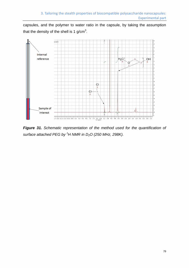
3. Tailoring the stealth properties of biocompatible polysaccharide nanocapsules: Experimental part
79
capsules, and the polymer to water ratio in the capsule, by taking the assumption
that the density of the shell is 1 g/cm3.
Figure 31. Schematic representation of the method used for the quantification of
surface attached PEG by 1H NMR in D2O (250 MHz, 298K).
3. Tailoring the stealth properties of biocompatible polysaccharide nanocapsules: Experimental part
80
In-vitro test (experiments conducted by Patricia Okwieka).
Nanocapsule incubation with human serum and protein quantification.
The nanocapsule dispersions were diluted with ultrapure water to a total particle
surface area of 0.05 m2 in 150 µL and incubated with 1 mL human serum for 1 h at
37 °C under constant agitation. The particles were separated from the supernatant
by centrifugation at 15,000 g for 1 h. The nanoparticles were washed with
phosphate-buffered saline (PBS; Gibco) in three centrifugation/redispersion steps at
15,000 g for 1 h. To elute the adsorbed proteins, the particle pellet was re-
suspended in 7 M urea, 2 M thiourea and 4% CHAPS, and the nanoparticles were
again pelleted. The supernatant was then used for protein quantification. Protein
concentrations were determined using Pierce 660 nm protein Assay (Thermo
Scientific, Rockford, USA) according to manufacturer's instructions with BSA as a
standard. Each sample was measured in triplicate.
Preparation of peripheral blood mononuclear cells (PBMC)
PBMC were isolated from healthy human donor buffy coats using standard Ficoll
density centrifugation. Donor blood was obtained according to the declaration of
Helsinki.
Incubation of nanocapsules with PBMC
For nanocapsule treatment cryopreserved PBMC were thawed in AIM-V containing
10% human serum. 5 x 10⁶ PBMC per well were seeded in a 48 well plate and
incubated with 50 µg/ mL of nanocapsules for 4 h at 37 °C/ 5% CO2. Afterwards cells
were harvested with cold PBS and stained against several expression markers.
Antibody staining of PBMC for flow cytometry analysis
Potential NC uptake by PBMC was analyzed using a LSR-Fortessa FACS analyzer
equipped with FACSDivaTM software (both Becton Dickinson). A 586/15 nm
bandpass filter with excitation by a 100 mW/561 nm laser was used to detect the
fluorescent dye SR101 of the NC. PBMC were stained against several markers:
CD3, CD4, CD8, CD14, CD16, CD19, CD45, and CD56. The following
3. Tailoring the stealth properties of biocompatible polysaccharide nanocapsules: Experimental part
81
fluorochrome-conjugated monoclonal mouse anti-human antibodies were used:
CD3-V450 (BD), CD3-FITC (Beckmann Coulter), CD4-APC-H7 (BD), CD8-APC
(Beckmann Coulter), CD14-V450 (BD), CD19-APC (BioLegend), CD16-Pacific Blue
(BioLegend), CD45-FITC (Beckmann Coulter), CD45-APC (Beckmann Coulter),
CD45-V450 (BD), CD45-APC-H7 (BD), and CD56-APC (BD). Cells were incubated
with antibodies for 15 min at 4 °C, washed with PBS/ 0.1% BSA, centrifuged (5 min,
1500 rpm) and resuspended in 500 µL PBS/ 0.1% BSA. Conjugates were excited
using a 100 mW/ 488 nm laser (filter 530/ 30 nm), a 40 mW 640 nm laser (filter 670/
30 nm and 780/ 60 nm) and a 100 mW 405 nm laser (filter 450/ 50 nm). For
analysis, cells were gated on a forward scatter/ sideward scatter dot plot to exclude
debris. The selected events were then further analyzed for the mentioned cell
markers.
3. Tailoring the stealth properties of biocompatible polysaccharide nanocapsules: Experimental part
82
In-vivo test
Pharmakokinetic studies for plasma half-life studies (experiments conducted by
Oliver Seifert).
Female SWISS (Janvier, CD1, 8 weeks old animals per construct) received an
intravenous (i.v.) injection of 300 µg of the capsule in a total volume of 100 µL (in
PBS). Blood samples (50 µL) were taken in the time intervals of 2 min, 30 min, 1 h, 2
h, 4 h, 6 h, 1 day and 3 days, incubated with heparin and stored at 4°C. Using Li-
COR, fluorescence intensity of the blood samples was measured to analyze the
concentration of the functionalized capsules. Comprising standard curve and weight
of animals, the percentage of injected doses was calculated. T1/2β and area under
the curve (AUC) were calculated with Excel.
In vivo / Ex vivo Fluorescence Imaging for the bio-distribution of the capsules
(experiments conducted by Mustafa Diken).
Six to eight-week old female BALB/c mice were obtained from Charles River. All
mice were kept and handled in accordance with federal and state policies on animal
research.
In vivo/ ex vivo fluorescence imaging of fluorescently labeled unmodified or
PEGylated HES-NCs was performed using the IVIS Spectrum Imaging system
(Perkin Elmer). Upon intravenous injection of nanocapsules (1 mg), BALB/c mice
(n = 3) were anesthetized with isofluorane at different time points (30 min, 1 h, 2 h,
4 h, 6 h, 8 h, 24 h) and placed in the light-tight chamber of the IVIS instrument.
Fluorescence emission was visualized by using the light source and filters with
excitation at 745 nm and emission at 800 nm for an exposure time of 3sec. 24 h
after injection, mice were sacrificed and fluorescence signal from isolated organs
such as brain, lungs, heart, kidneys, liver and spleen were collected ex vivo using
the same filter set and exposure time. Fluorescence images were presented as
superimposed to black & white photos of mice/organs and the fluorescence intensity
was depicted with a pseudocolor scale.

3. Tailoring the stealth properties of biocompatible polysaccharide nanocapsules: Experimental part
83
4. Surface modification of HES-NCs for cell-specific targeting: introduction
84
4. Surface modification of HES-NCs for cell-specific
targeting
The work in this chapter concerning the surface modification of HES capsules by mannose,
the study of interaction of the nanocapsules with blood plasma protein, and the influence of
the protein corona on the targeting efficient has been published in Angewandte Chemie
International Edition (Kang, B.; Okwieka, P.; Schöttler, S.; Winzen, S.; Langhanki, J.; Mohr,
K.; Opatz, T.; Mailander, V.; Landfester, K.; Wurm, F. R. Angew. Chem. Int. Ed. Engl. 2015,
54, 7436.)213 and altered for this thesis with permission. The manuscript concerning the
surface modification of HES-NCs with Microbody, scFv antibody, and Dec205 is in
preparation. I synthesized the nanocarriers and developed all the surface modification
methods, synthesized the 2-aminoethyl α-D-mannopyranoside, and conducted the
characterization concerning DLS, SEM, TEM, NMR, fluorescamine assay, anthracene azide
assay, and the c-type-lectin binding assay; the in-vitro, ITC characterization and the protein
adsorption assay was conducted by the collaboration partners, which are acknowledged
after each corresponding contribution.
Introduction
In the last decades, the application of synthetic nanocarriers for the targeted delivery
of drugs is termed as a promising therapy against cancer, due to its ability to reduce
the side effect of the traditional chemotherapy. While the specific targeting of the
tumor tissue can be achieved passively by the enhanced permeability and retention
effect (EPR),214 surface modification of the nanocarriers by targeting agent, which
can actively bind to the malignant cell line, will not only increase the targeting effect
to the tumor tissue, but also be able to target the tumor cells in metastases.
When synthetic nanocarriers enter biological fluids, it is known that, due to the high
surface energy and hydrophobic interactions, they strongly adsorb plasma
proteins.70,215 Many researchers proposed that the in vivo fate of any nanocarrier is
determined by this protein “corona” formation post injection, instead of the intrinsic
properties of the mostly polymeric nanocarrier itself.204-211,216 PEGylation is the state
of the art approach to depress the unspecific interaction with plasma proteins and
often referred to the term “stealth effect”.10,217 However, the stealth effect alone is not
4. Surface modification of HES-NCs for cell-specific targeting: introduction
85
enough: specific targeting agents have to be attached additionally in order to reach
the place of action and avoid opsonization.215 But how does this additional moiety
interact with plasma proteins? A new corona could be generated around the carriers,
consequently altering the in vivo performance by covering and deactivating the
targeting group.
While the protein corona formation for single component nanoparticles, such as
polystyrene206,210,218, zinc oxide219, silica210,219,220, gold221, and titanium dioxide219 has
been extensively studied, no study has been conducted comparing protein
interactions of a PEGylated nanocarrier before and after the attachment of an
additional targeting group. Hence, no information on the combination of stealth
properties of the PEGylated carrier and the specific targeting after “on-top
attachment” of the targeting group is known. Herein, for the first time, we compare
PEGylated stealth nanocarriers with subsequently modified analogues carrying
mannose targeting moieties with respect to protein adsorption. Isothermal titration
calorimetry (ITC), dynamic light scattering (DLS), and cell-uptake studies before and
after incubation with human plasma have been performed and the influence on the
targeting efficiency to dendritic cells is evaluated.
4. Surface modification of HES-NCs for cell-specific targeting: Results and discussion
86
4.1 Results and discussion
Surface functionalization of HES-NCs with mannose
A working strategy for specific targeting is the attachment of various small
biomolecules, such as folic acid86,222, glucose223, mannose224,225, galactose224, or
dissacharides226 to the nanocarriers. Coupling of these targeting agents to the
nanocarriers often involves several reaction steps, furthermore, quantification of the
attached groups is challenging especially when the nanocarrier itself is
biocompatible and vulnerable against chemical transformations. In this work we
present a robust and general protocol for the coupling of such small biomolecules to
the surface of nanocarriers, and a corresponding characterization method, which
can quantify the number of the coupled biomolecules very precisely.
We have been studying hydroxyethyl starch nanocapsules (HES-NCs) as
biodegradable nanocarriers intensively.86 They are prepared via an inverse
miniemulsion protocol and diameters can be adjusted precisely (Table 4). More
recently, well-defined PEGylation of HES-NCs was accomplished via different
protocols.227 Herein, we extend these protocols to generate NCs which can be
further functionalized “on-top” with specific targeting groups. Diisocyanate-PEG
(OCN-PEG110-NCO, Mn = 5000 g·mol−1) reacts both with the surface hydroxyls of the
polysaccharide and subsequently with targeting groups. After one of the isocyanate
groups has reacted with the surface, the reactivity of the other one will decrease
drastically due to the steric hindrance and loss of mobility. An excess of
diisocyanate-PEG has to be used, which will further reduce the formation of cycles,
and keep the second isocyante group available. Monoisocyanate-PEG (MeO-
PEG110-NCO, Mn = 5000 g·mol−1) has been used to prepare PEGylated capsules as
negative control for further studies. Quantification of the HES-NCs by NMR
spectroscopy after PEGylation revealed that 5*105 PEG chains have been coupled
to the surface for both PEGs (cf. Table 5), which is a first indication that the
diisocyanate-PEG did not form cycles and reacted rather selectively at one end.
Two methods have been used to couple mannose to the PEGylated HES-NCs
(Scheme 4). Route 1 is a combination of techniques with mannose carrying an azide
4. Surface modification of HES-NCs for cell-specific targeting: Results and discussion
87
group at C-1, and coupling to the capsules via metal-free click chemistry: The HES-
NCs, which were PEGylated by diisocyanate-PEG, were redispersed in water, and
the mixture is magnetically stirred in an open vessel to evaporate cyclohexane.
During this procedure, the isocyanate groups can be hydrolyzed to release primary
amine groups. The number of amine groups was determined by a fluorescence
assay (reference, support info); however, only a rather small amount of 1,000 groups
per NC was detected. The large discrepancy between the number of available NCO-
groups and the number of amines per NC can be reasoned due to the fast reaction
of already hydrolyzed amines with remaining isocyanate groups to form a urea
linkage. The reaction speed between isocyanates amines is ca. thousand times
faster than the hydrolysis of the former,228 hence most of the primary amine groups
will be consumed by the cyclization with other PEG chains. The available amines
were activated for the Huisgen-click reaction by amidation with an N-hydroxy
succinimidyl ester carrying dibenzocyclooctyne (DBCO) (Scheme 4, Route 1). The
DBCO groups are used since they react with azides in the absence of any metal
catalyst due to their ring strain, which is a great advantage for bioapplications.229,230
The number of DBCO groups per HES-NC was quantified after quantitative click
reaction with anthracene methylazide and found to be approx. 1000 (experimetal
part). 2-Azidoethyl α-D-mannopyranoside was then reacted with DBCO-modified
NCs, and after purification, the number of remaining DBCO groups was quantified as
well. No DBCO was detected after the addition of azidomannose. Therefore, both
the coupling of DBCO to the amine groups and the subsequent coupling of azide
mannose to DBCO have achieved quantitative yield by this method, however, the
total number of targeting groups remained rather low.
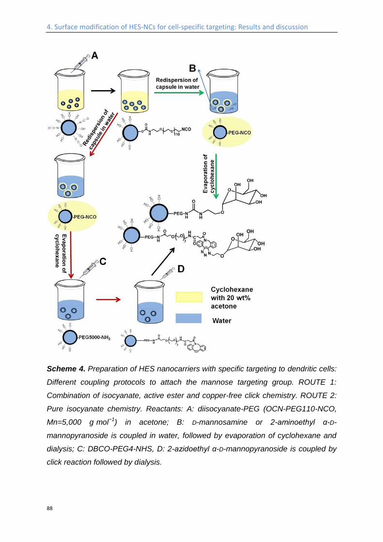
4. Surface modification of HES-NCs for cell-specific targeting: Results and discussion
88
Scheme 4. Preparation of HES nanocarriers with specific targeting to dendritic cells:
Different coupling protocols to attach the mannose targeting group. ROUTE 1:
Combination of isocyanate, active ester and copper-free click chemistry. ROUTE 2:
Pure isocyanate chemistry. Reactants: A: diisocyanate-PEG (OCN-PEG110-NCO,
Mn=5,000 g·mol−1) in acetone; B: D-mannosamine or 2-aminoethyl α-D-
mannopyranoside is coupled in water, followed by evaporation of cyclohexane and
dialysis; C: DBCO-PEG4-NHS, D: 2-azidoethyl α-D-mannopyranoside is coupled by
click reaction followed by dialysis.
4. Surface modification of HES-NCs for cell-specific targeting: Results and discussion
89
A second route utilized amine-functionalized mannose derivatives which are directly
coupled to the available NCO-groups during the redispersion procedure (Scheme 4,
Route 2). Namely, amino-derivatives of mannose are dissolved in water, before the
redispersion process. The mixture is magnetically stirred in an open vessel, to
evaporate the cyclohexane and produce a stable dispersion of PEGylated and
mannose-funcionalized HES-NCs. Due to the high reaction kinetics of isocyanates
with amines, the amino-sugars will preferably react with the nanocarriers, before
hydrolysis of the isocyanates occurs. The amount of amine-functionalized mannose
in water (before the addition of NCs) can be quantified by the fluorescamine
assay203. After the coupling reaction, the NCs are precipitated by centrifugation, and
the remaining amine-functionalized mannose in the upper phase is quantified by the
same method. By comparing both amounts, the precise number of mannose units
which are attached to the NCs is available. Two different mannose derivatives were
used, D-mannosamine and 2-aminoethyl α-D-mannopyranoside, with the amine
groups at the C-2 or the C-1 position (the functionalized HES-NCs are named HES-
PEG5000-C2-Man and HES-PEG5000-C1-Man accordingly). With this easy and fast
protocol ca. 2·105 (Table 5) mannose units can be attached to the NCs in both cases.
Compared to the first method, this method is a single step coupling reaction,
produces a 200 times higher mannose density, allows a precise quantification and
omits expensive intermediates. This general protocol can be used for any type of
small biomolecule, as long as it carries an amine group.
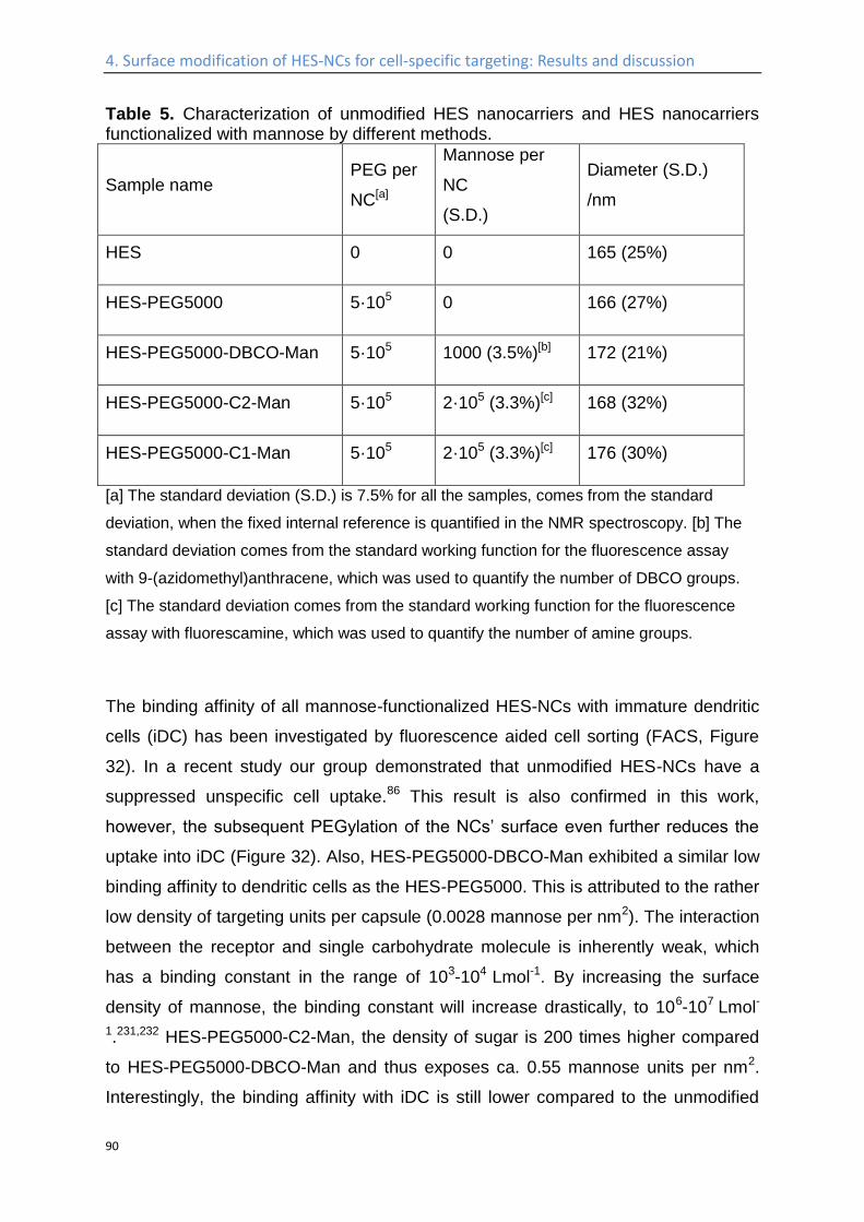
4. Surface modification of HES-NCs for cell-specific targeting: Results and discussion
90
Table 5. Characterization of unmodified HES nanocarriers and HES nanocarriers functionalized with mannose by different methods.
Sample name PEG per
NC[a]
Mannose per
NC
(S.D.)
Diameter (S.D.)
/nm
HES 0 0 165 (25%)
HES-PEG5000 5·105 0 166 (27%)
HES-PEG5000-DBCO-Man 5·105 1000 (3.5%)[b] 172 (21%)
HES-PEG5000-C2-Man 5·105 2·105 (3.3%)[c] 168 (32%)
HES-PEG5000-C1-Man 5·105 2·105 (3.3%)[c] 176 (30%)
[a] The standard deviation (S.D.) is 7.5% for all the samples, comes from the standard
deviation, when the fixed internal reference is quantified in the NMR spectroscopy. [b] The
standard deviation comes from the standard working function for the fluorescence assay
with 9-(azidomethyl)anthracene, which was used to quantify the number of DBCO groups.
[c] The standard deviation comes from the standard working function for the fluorescence
assay with fluorescamine, which was used to quantify the number of amine groups.
The binding affinity of all mannose-functionalized HES-NCs with immature dendritic
cells (iDC) has been investigated by fluorescence aided cell sorting (FACS, Figure
32). In a recent study our group demonstrated that unmodified HES-NCs have a
suppressed unspecific cell uptake.86 This result is also confirmed in this work,
however, the subsequent PEGylation of the NCs’ surface even further reduces the
uptake into iDC (Figure 32). Also, HES-PEG5000-DBCO-Man exhibited a similar low
binding affinity to dendritic cells as the HES-PEG5000. This is attributed to the rather
low density of targeting units per capsule (0.0028 mannose per nm2). The interaction
between the receptor and single carbohydrate molecule is inherently weak, which
has a binding constant in the range of 103-104 Lmol-1. By increasing the surface
density of mannose, the binding constant will increase drastically, to 106-107 Lmol-
1.231,232 HES-PEG5000-C2-Man, the density of sugar is 200 times higher compared
to HES-PEG5000-DBCO-Man and thus exposes ca. 0.55 mannose units per nm2.
Interestingly, the binding affinity with iDC is still lower compared to the unmodified
4. Surface modification of HES-NCs for cell-specific targeting: Results and discussion
91
HES-NCs (Figure 32B). The reason for this could be that the free hydroxyl group at
C-2 position of the mannose molecule is essential for interaction with protein
acceptor.233 Only, when 2-aminoethyl α-D-mannopyranoside (with the amine at the
C-1 position) was used, a much stronger binding of HES-PEG5000-C1-Man with iDC
compared to unmodified and PEGylated capsules was detected (Figure 32C). These
results clearly demonstrate that, when mannose is attached via a PEG linker to
HES-NCs, a multivalency effect is achieved, combining the stealth behavior of a
PEGylated nanocarrier with on-top mannose functionalization.
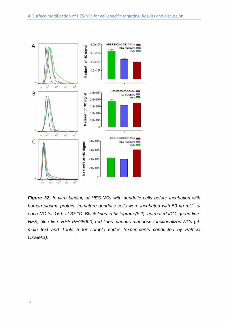
4. Surface modification of HES-NCs for cell-specific targeting: Results and discussion
92
Figure 32. In-vitro binding of HES-NCs with dendritic cells before incubation with
human plasma protein. Immature dendritic cells were incubated with 50 µg mL-1 of
each NC for 16 h at 37 °C. Black lines in histogram (left): untreated iDC; green line:
HES; blue line: HES-PEG5000; red lines: various mannose functionalized NCs (cf.
main text and Table 5 for sample codes (experiments conducted by Patricia
Okwieka).
4. Surface modification of HES-NCs for cell-specific targeting: Results and discussion
93
Surface functionalization of HES-NCs with different antibodies
Route 1 (labeled with red arrows) in Scheme 4, which involves the coupling of NHS
ester linked DBCO groups to the end of PEG and the subsequent coupling of azide
functionalized molecules to the DBCO groups, turned out to be insufficient when
used to couple mannose to the capsule for DCs targeting, since the maximum
number of mannose coupled is limited to be 1000 per capsule, which is not enough
to activate the multivalent interaction with the cells. However, this method
demonstrated many advantages at the same time, including of the ease of
functionalization of a PEGylated interface with additional targeting agent, the great
advantage for bio-applications due to the catalyst-free click reaction involved, and
the well-established quantification methods, which allows functionalizing the
nanocapsule with targeting agent in a quantitative manner. Other than mannose,
some of the targeting agent, especially antibodies, have much higher individual
binding affinity with cellular accepters, hence can be used via route 1 to surface
modify the HES-NCs for cell-specific targeting. The general strategy of surface
functionalization of HES-NCs with different antibodies is shown in Scheme 5.
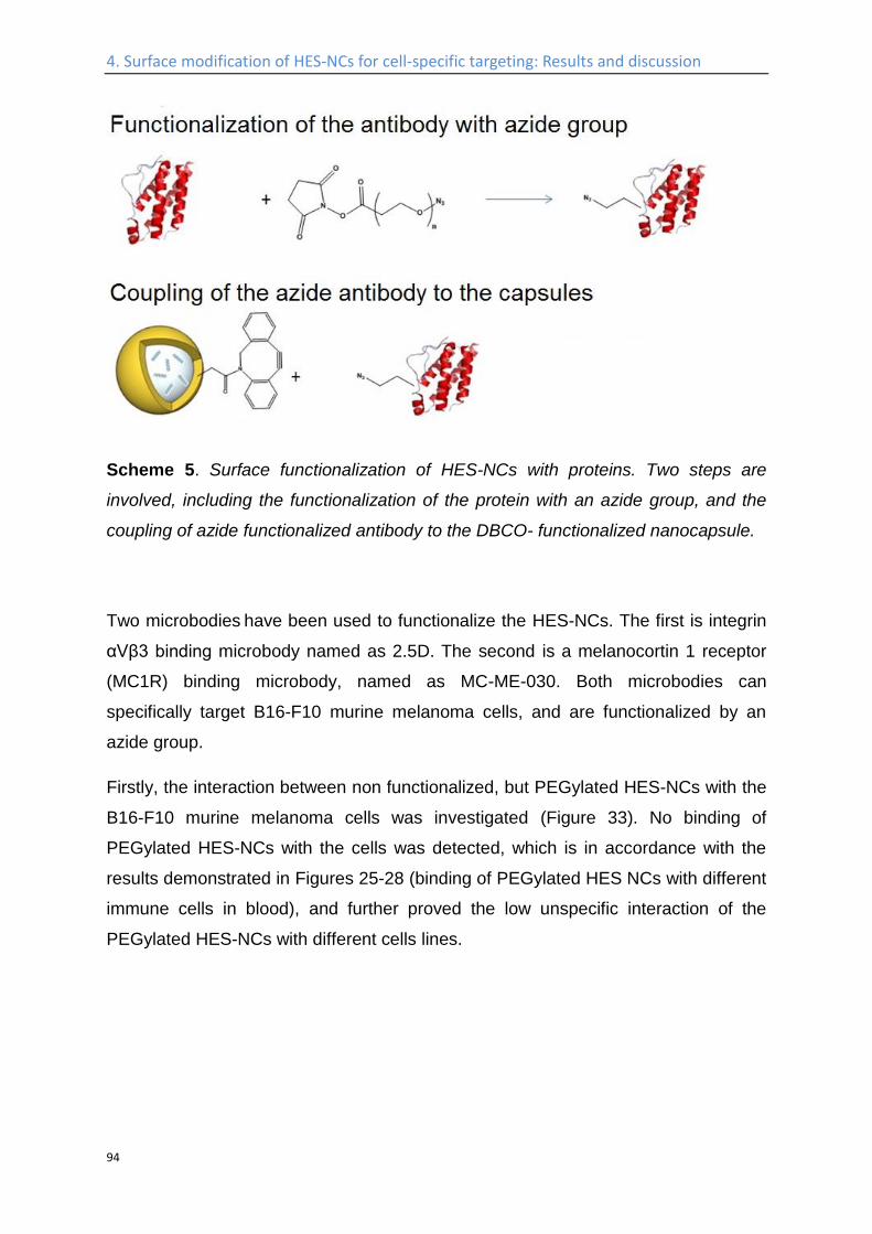
4. Surface modification of HES-NCs for cell-specific targeting: Results and discussion
94
Scheme 5. Surface functionalization of HES-NCs with proteins. Two steps are
involved, including the functionalization of the protein with an azide group, and the
coupling of azide functionalized antibody to the DBCO- functionalized nanocapsule.
Two microbodies have been used to functionalize the HES-NCs. The first is integrin
αVβ3 binding microbody named as 2.5D. The second is a melanocortin 1 receptor
(MC1R) binding microbody, named as MC-ME-030. Both microbodies can
specifically target B16-F10 murine melanoma cells, and are functionalized by an
azide group.
Firstly, the interaction between non functionalized, but PEGylated HES-NCs with the
B16-F10 murine melanoma cells was investigated (Figure 33). No binding of
PEGylated HES-NCs with the cells was detected, which is in accordance with the
results demonstrated in Figures 25-28 (binding of PEGylated HES NCs with different
immune cells in blood), and further proved the low unspecific interaction of the
PEGylated HES-NCs with different cells lines.
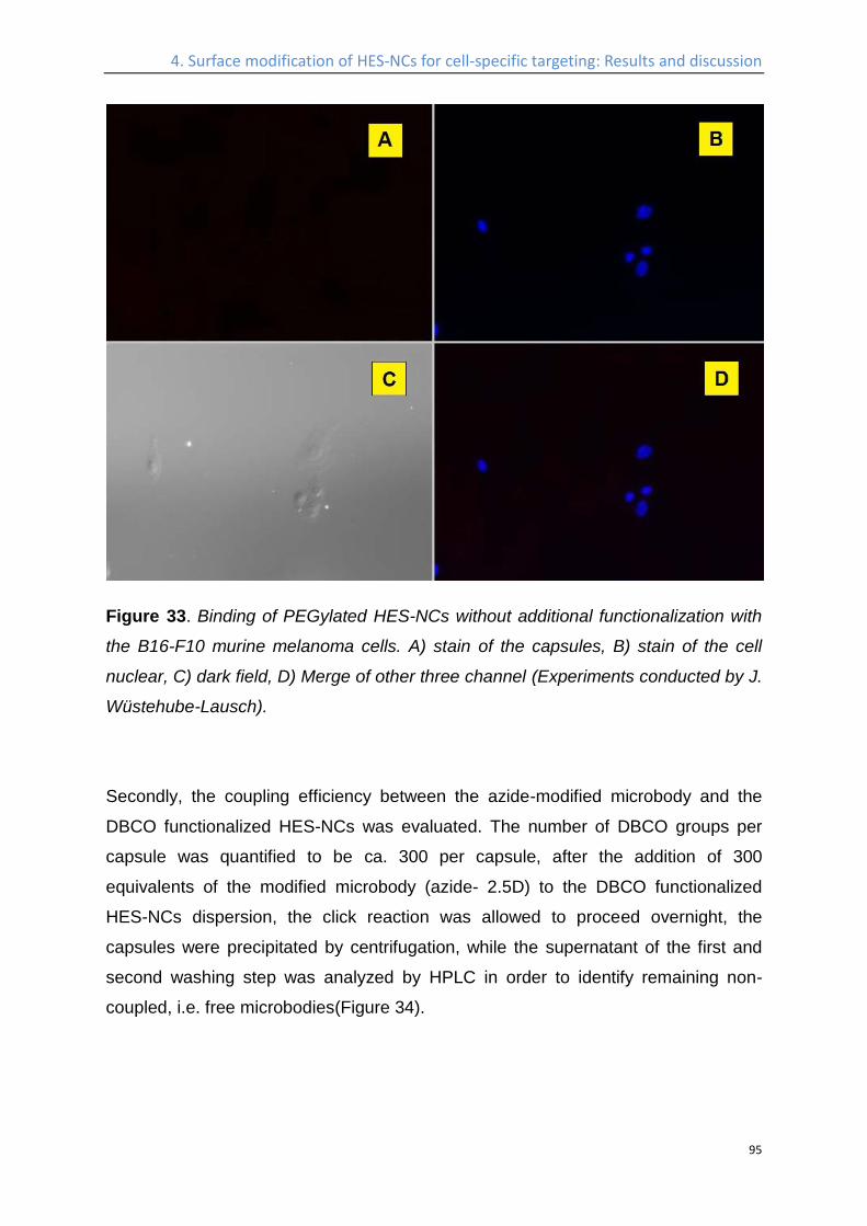
4. Surface modification of HES-NCs for cell-specific targeting: Results and discussion
95
Figure 33. Binding of PEGylated HES-NCs without additional functionalization with
the B16-F10 murine melanoma cells. A) stain of the capsules, B) stain of the cell
nuclear, C) dark field, D) Merge of other three channel (Experiments conducted by J.
Wüstehube-Lausch).
Secondly, the coupling efficiency between the azide-modified microbody and the
DBCO functionalized HES-NCs was evaluated. The number of DBCO groups per
capsule was quantified to be ca. 300 per capsule, after the addition of 300
equivalents of the modified microbody (azide- 2.5D) to the DBCO functionalized
HES-NCs dispersion, the click reaction was allowed to proceed overnight, the
capsules were precipitated by centrifugation, while the supernatant of the first and
second washing step was analyzed by HPLC in order to identify remaining non-
coupled, i.e. free microbodies(Figure 34).
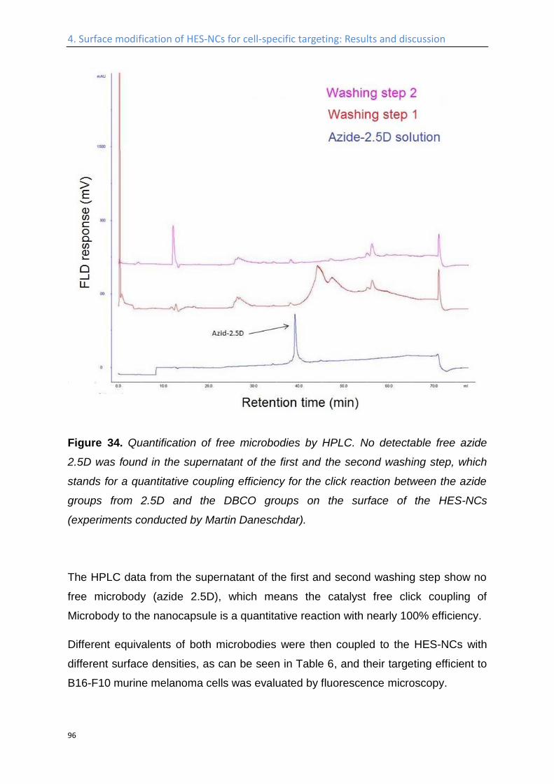
4. Surface modification of HES-NCs for cell-specific targeting: Results and discussion
96
Figure 34. Quantification of free microbodies by HPLC. No detectable free azide
2.5D was found in the supernatant of the first and the second washing step, which
stands for a quantitative coupling efficiency for the click reaction between the azide
groups from 2.5D and the DBCO groups on the surface of the HES-NCs
(experiments conducted by Martin Daneschdar).
The HPLC data from the supernatant of the first and second washing step show no
free microbody (azide 2.5D), which means the catalyst free click coupling of
Microbody to the nanocapsule is a quantitative reaction with nearly 100% efficiency.
Different equivalents of both microbodies were then coupled to the HES-NCs with
different surface densities, as can be seen in Table 6, and their targeting efficient to
B16-F10 murine melanoma cells was evaluated by fluorescence microscopy.
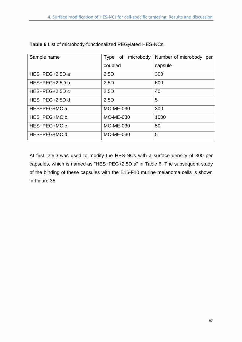
4. Surface modification of HES-NCs for cell-specific targeting: Results and discussion
97
Table 6 List of microbody-functionalized PEGylated HES-NCs.
Sample name Type of microbody
coupled
Number of microbody per
capsule
HES+PEG+2.5D a 2.5D 300
HES+PEG+2.5D b 2.5D 600
HES+PEG+2.5D c 2.5D 40
HES+PEG+2.5D d 2.5D 5
HES+PEG+MC a MC-ME-030 300
HES+PEG+MC b MC-ME-030 1000
HES+PEG+MC c MC-ME-030 50
HES+PEG+MC d MC-ME-030 5
At first, 2.5D was used to modify the HES-NCs with a surface density of 300 per
capsules, which is named as "HES+PEG+2.5D a" in Table 6. The subsequent study
of the binding of these capsules with the B16-F10 murine melanoma cells is shown
in Figure 35.
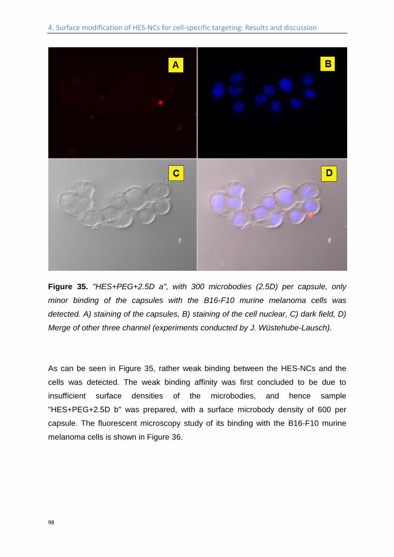
4. Surface modification of HES-NCs for cell-specific targeting: Results and discussion
98
Figure 35. "HES+PEG+2.5D a", with 300 microbodies (2.5D) per capsule, only
minor binding of the capsules with the B16-F10 murine melanoma cells was
detected. A) staining of the capsules, B) staining of the cell nuclear, C) dark field, D)
Merge of other three channel (experiments conducted by J. Wüstehube-Lausch).
As can be seen in Figure 35, rather weak binding between the HES-NCs and the
cells was detected. The weak binding affinity was first concluded to be due to
insufficient surface densities of the microbodies, and hence sample
"HES+PEG+2.5D b" was prepared, with a surface microbody density of 600 per
capsule. The fluorescent microscopy study of its binding with the B16-F10 murine
melanoma cells is shown in Figure 36.
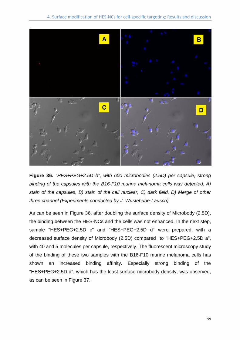
4. Surface modification of HES-NCs for cell-specific targeting: Results and discussion
99
Figure 36. "HES+PEG+2.5D b", with 600 microbodies (2.5D) per capsule, strong
binding of the capsules with the B16-F10 murine melanoma cells was detected. A)
stain of the capsules, B) stain of the cell nuclear, C) dark field, D) Merge of other
three channel (Experiments conducted by J. Wüstehube-Lausch).
As can be seen in Figure 36, after doubling the surface density of Microbody (2.5D),
the binding between the HES-NCs and the cells was not enhanced. In the next step,
sample "HES+PEG+2.5D c" and "HES+PEG+2.5D d" were prepared, with a
decreased surface density of Microbody (2.5D) compared to "HES+PEG+2.5D a",
with 40 and 5 molecules per capsule, respectively. The fluorescent microscopy study
of the binding of these two samples with the B16-F10 murine melanoma cells has
shown an increased binding affinity. Especially strong binding of the
"HES+PEG+2.5D d", which has the least surface microbody density, was observed,
as can be seen in Figure 37.
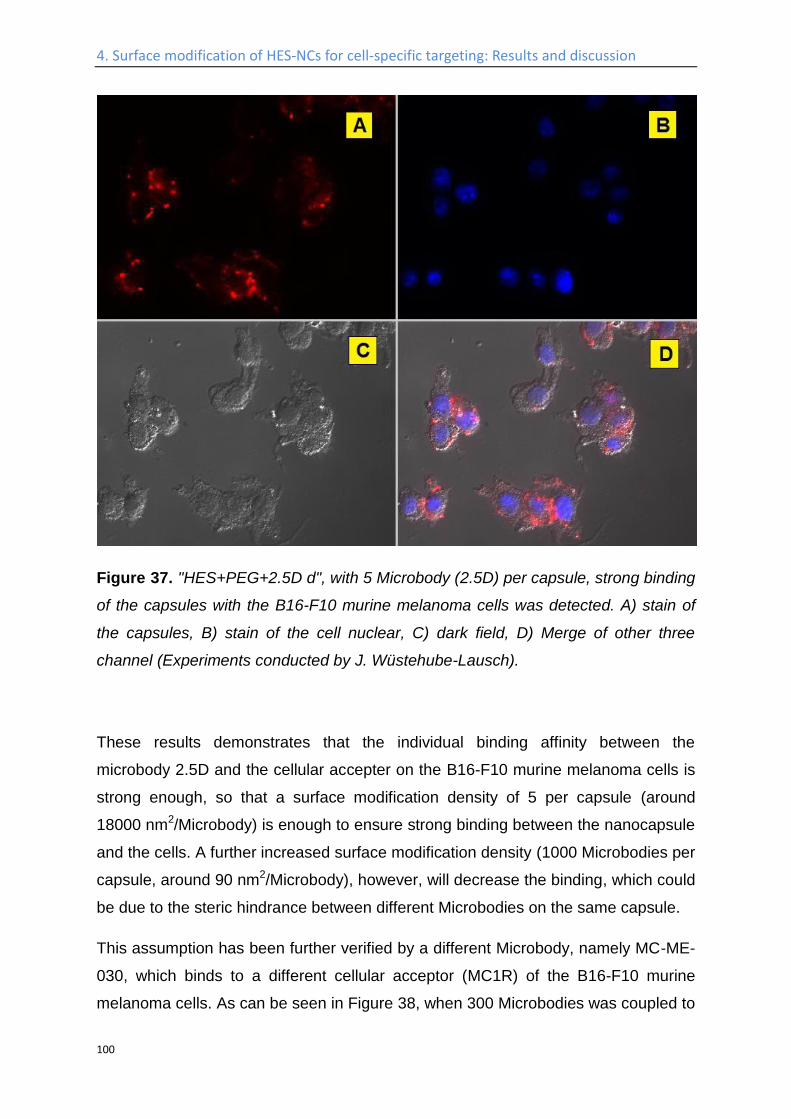
4. Surface modification of HES-NCs for cell-specific targeting: Results and discussion
100
Figure 37. "HES+PEG+2.5D d", with 5 Microbody (2.5D) per capsule, strong binding
of the capsules with the B16-F10 murine melanoma cells was detected. A) stain of
the capsules, B) stain of the cell nuclear, C) dark field, D) Merge of other three
channel (Experiments conducted by J. Wüstehube-Lausch).
These results demonstrates that the individual binding affinity between the
microbody 2.5D and the cellular accepter on the B16-F10 murine melanoma cells is
strong enough, so that a surface modification density of 5 per capsule (around
18000 nm2/Microbody) is enough to ensure strong binding between the nanocapsule
and the cells. A further increased surface modification density (1000 Microbodies per
capsule, around 90 nm2/Microbody), however, will decrease the binding, which could
be due to the steric hindrance between different Microbodies on the same capsule.
This assumption has been further verified by a different Microbody, namely MC-ME-
030, which binds to a different cellular acceptor (MC1R) of the B16-F10 murine
melanoma cells. As can be seen in Figure 38, when 300 Microbodies was coupled to
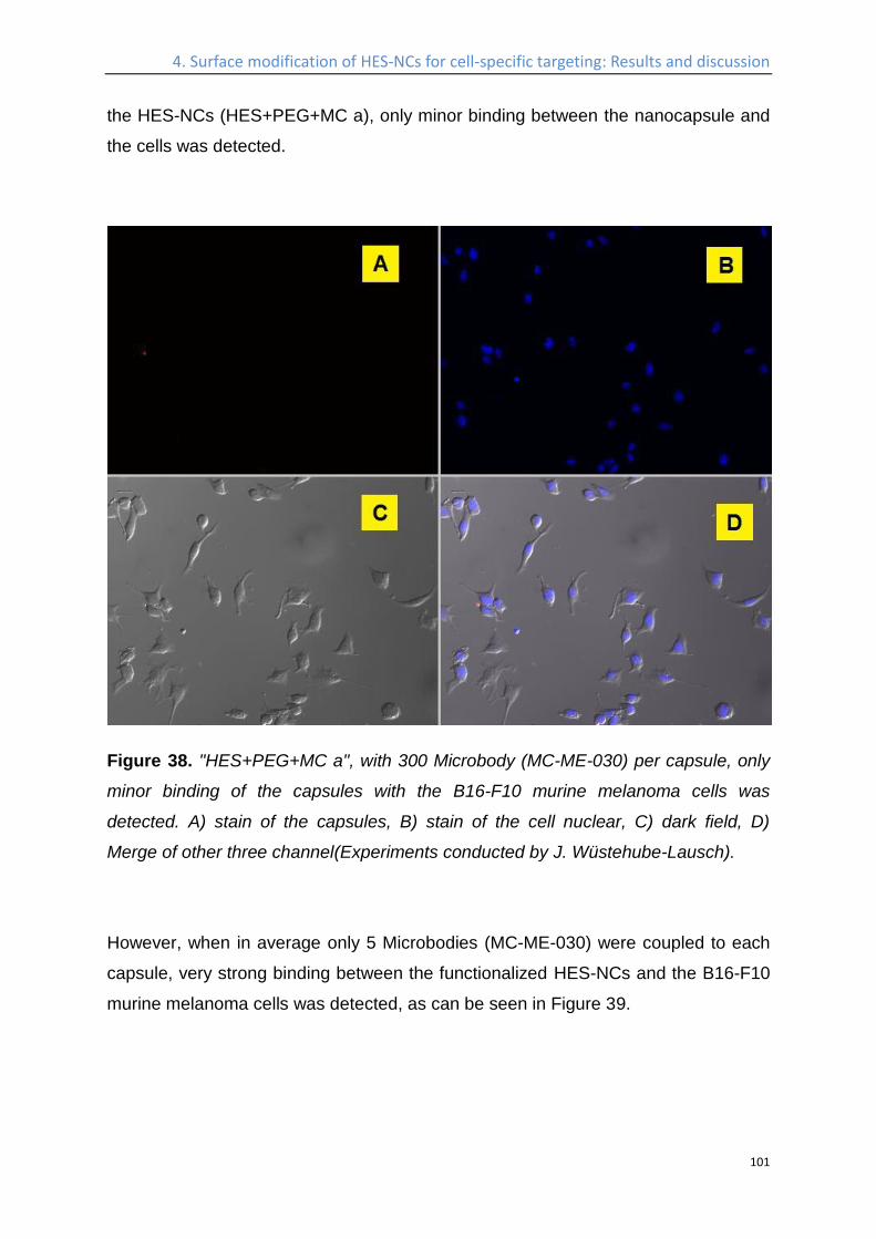
4. Surface modification of HES-NCs for cell-specific targeting: Results and discussion
101
the HES-NCs (HES+PEG+MC a), only minor binding between the nanocapsule and
the cells was detected.
Figure 38. "HES+PEG+MC a", with 300 Microbody (MC-ME-030) per capsule, only
minor binding of the capsules with the B16-F10 murine melanoma cells was
detected. A) stain of the capsules, B) stain of the cell nuclear, C) dark field, D)
Merge of other three channel(Experiments conducted by J. Wüstehube-Lausch).
However, when in average only 5 Microbodies (MC-ME-030) were coupled to each
capsule, very strong binding between the functionalized HES-NCs and the B16-F10
murine melanoma cells was detected, as can be seen in Figure 39.
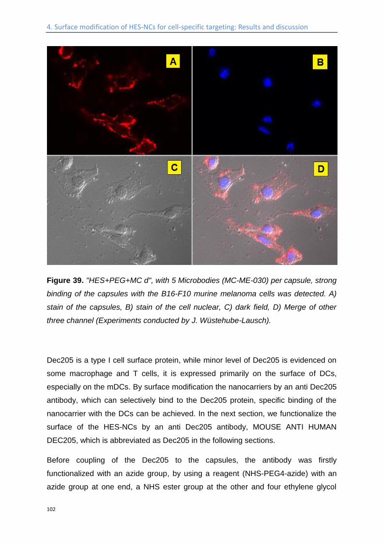
4. Surface modification of HES-NCs for cell-specific targeting: Results and discussion
102
Figure 39. "HES+PEG+MC d", with 5 Microbodies (MC-ME-030) per capsule, strong
binding of the capsules with the B16-F10 murine melanoma cells was detected. A)
stain of the capsules, B) stain of the cell nuclear, C) dark field, D) Merge of other
three channel (Experiments conducted by J. Wüstehube-Lausch).
Dec205 is a type I cell surface protein, while minor level of Dec205 is evidenced on
some macrophage and T cells, it is expressed primarily on the surface of DCs,
especially on the mDCs. By surface modification the nanocarriers by an anti Dec205
antibody, which can selectively bind to the Dec205 protein, specific binding of the
nanocarrier with the DCs can be achieved. In the next section, we functionalize the
surface of the HES-NCs by an anti Dec205 antibody, MOUSE ANTI HUMAN
DEC205, which is abbreviated as Dec205 in the following sections.
Before coupling of the Dec205 to the capsules, the antibody was firstly
functionalized with an azide group, by using a reagent (NHS-PEG4-azide) with an
azide group at one end, a NHS ester group at the other and four ethylene glycol

4. Surface modification of HES-NCs for cell-specific targeting: Results and discussion
103
repeating units in between. After introducing the azide group to the Dec205, its
biological activity was tested, and compared to those without azide functionalization
(Figure 40).
Figure 40. Characterization of the binding affinity of Dec205 with DCs before and
after azidation (Experiments conducted by Patricia Okwieka).
As can be seen in Figure 40, in comparison to unmodified Dec205, the binding
affinity of this antibody after azidation was still intact. It was then coupled to the
PEGylated HES-NCs in the same matter as for coupling of the Microbodies, the
number of Dec205 coupled to each capsule is controlled to be 8. The binding affinity
of Dec205 functionalized HES-NCs with iDCs was then evaluated by FACS (Figure
41). Strong binding between the Dec205 coupled HES-NCs and the iDC was
detected, in comparison to the unmodified HES-NCs.
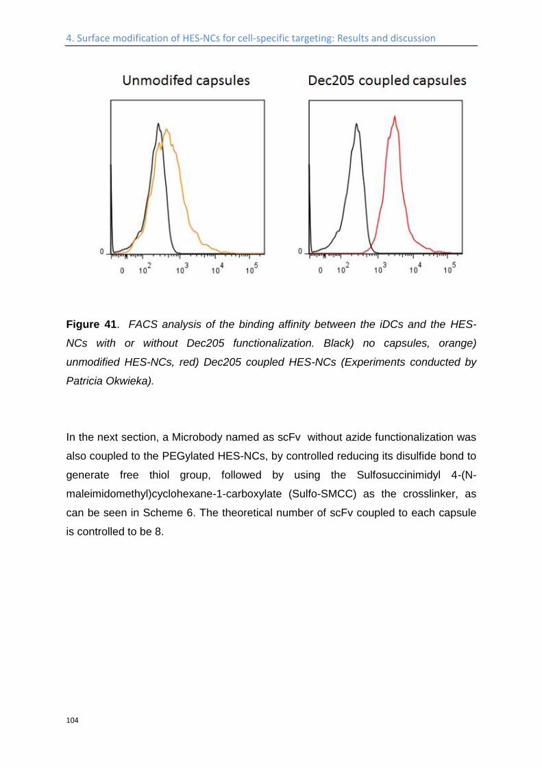
4. Surface modification of HES-NCs for cell-specific targeting: Results and discussion
104
Figure 41. FACS analysis of the binding affinity between the iDCs and the HES-
NCs with or without Dec205 functionalization. Black) no capsules, orange)
unmodified HES-NCs, red) Dec205 coupled HES-NCs (Experiments conducted by
Patricia Okwieka).
In the next section, a Microbody named as scFv without azide functionalization was
also coupled to the PEGylated HES-NCs, by controlled reducing its disulfide bond to
generate free thiol group, followed by using the Sulfosuccinimidyl 4-(N-
maleimidomethyl)cyclohexane-1-carboxylate (Sulfo-SMCC) as the crosslinker, as
can be seen in Scheme 6. The theoretical number of scFv coupled to each capsule
is controlled to be 8.
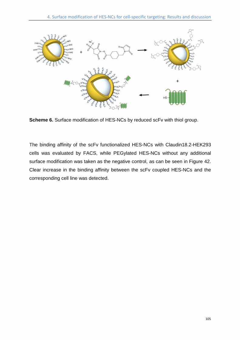
4. Surface modification of HES-NCs for cell-specific targeting: Results and discussion
105
Scheme 6. Surface modification of HES-NCs by reduced scFv with thiol group.
The binding affinity of the scFv functionalized HES-NCs with Claudin18.2-HEK293
cells was evaluated by FACS, while PEGylated HES-NCs without any additional
surface modification was taken as the negative control, as can be seen in Figure 42.
Clear increase in the binding affinity between the scFv coupled HES-NCs and the
corresponding cell line was detected.
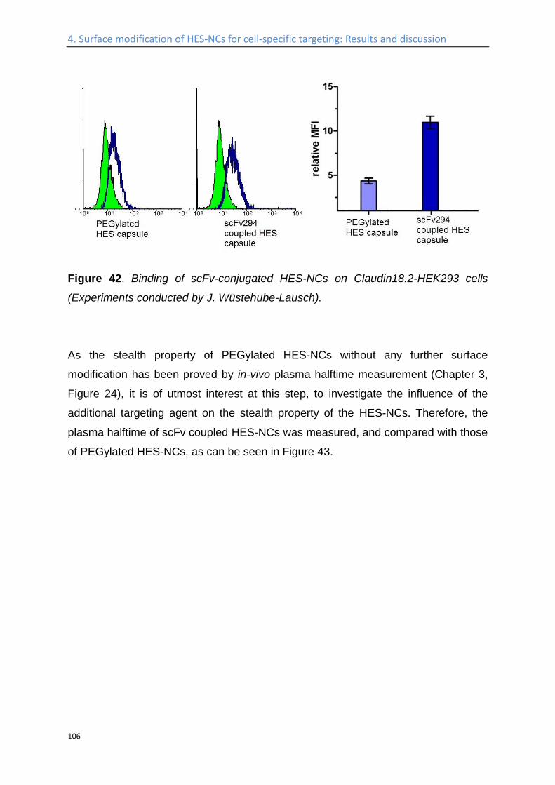
4. Surface modification of HES-NCs for cell-specific targeting: Results and discussion
106
Figure 42. Binding of scFv-conjugated HES-NCs on Claudin18.2-HEK293 cells
(Experiments conducted by J. Wüstehube-Lausch).
As the stealth property of PEGylated HES-NCs without any further surface
modification has been proved by in-vivo plasma halftime measurement (Chapter 3,
Figure 24), it is of utmost interest at this step, to investigate the influence of the
additional targeting agent on the stealth property of the HES-NCs. Therefore, the
plasma halftime of scFv coupled HES-NCs was measured, and compared with those
of PEGylated HES-NCs, as can be seen in Figure 43.
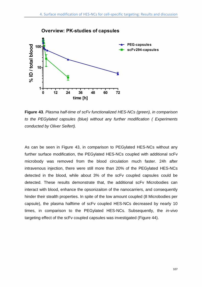
4. Surface modification of HES-NCs for cell-specific targeting: Results and discussion
107
Figure 43. Plasma half-time of scFv functionalized HES-NCs (green), in comparison
to the PEGylated capsules (blue) without any further modification ( Experiments
conducted by Oliver Seifert).
As can be seen in Figure 43, in comparison to PEGylated HES-NCs without any
further surface modification, the PEGylated HES-NCs coupled with additional scFv
microbody was removed from the blood circulation much faster. 24h after
intravenous injection, there were still more than 20% of the PEGylated HES-NCs
detected in the blood, while about 3% of the scFv coupled capsules could be
detected. These results demonstrate that, the additional scFv Microbodies can
interact with blood, enhance the opsonizaiton of the nanocarriers, and consequently
hinder their stealth properties. In spite of the low amount coupled (8 Microbodies per
capsule), the plasma halftime of scFv coupled HES-NCs decreased by nearly 10
times, in comparison to the PEGylated HES-NCs. Subsequently, the in-vivo
targeting effect of the scFv coupled capsules was investigated (Figure 44).
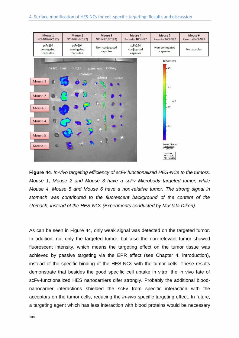
4. Surface modification of HES-NCs for cell-specific targeting: Results and discussion
108
Figure 44. In-vivo targeting efficiency of scFv functionalized HES-NCs to the tumors.
Mouse 1, Mouse 2 and Mouse 3 have a scFv Microbody targeted tumor, while
Mouse 4, Mouse 5 and Mouse 6 have a non-relative tumor. The strong signal in
stomach was contributed to the fluorescent background of the content of the
stomach, instead of the HES-NCs (Experiments conducted by Mustafa Diken).
As can be seen in Figure 44, only weak signal was detected on the targeted tumor.
In addition, not only the targeted tumor, but also the non-relevant tumor showed
fluorescent intensity, which means the targeting effect on the tumor tissue was
achieved by passive targeting via the EPR effect (see Chapter 4, introduction),
instead of the specific binding of the HES-NCs with the tumor cells. These results
demonstrate that besides the good specific cell uptake in vitro, the in vivo fate of
scFv-functionalized HES nanocarriers difer strongly. Probably the additional blood-
nanocarrier interactions shielded the scFv from specific interaction with the
acceptors on the tumor cells, reducing the in-vivo specific targeting effect. In future,
a targeting agent which has less interaction with blood proteins would be necessary
4. Surface modification of HES-NCs for cell-specific targeting: Results and discussion
109
in order to enhance the specific targeting effect in-vivo. Furthermore, additional
investigations need to be done, in order to quantify the influence of the additional
targeting agent on the protein adsorption behavior of the PEGylated HES-NCs, and
its subsequent influence on the targeting efficient.

4. Surface modification of HES-NCs for cell-specific targeting: Results and discussion
110
Study of the interaction of the nanocapsules with blood plasma proteins
In order to investigate the influence of the additional targeting agent on the protein
adsorption of the PEGylated HES-NCs, different techniques have been applied,
including ITC, DLS, SDS-PAGE, and quantitative liquid-chromatography coupled
with mass-spectrometry. Mannose-functionalized HES-NCs are studied in this case,
due to its high degree of surface functionalization (2·105 mannose/capsule), while,
due to the extremely low surface functionalization density (5-10 antibody/capsule),
the influence of antibody on the protein corona of the nanocapsules are hard to
quantify by the above mentioned characterization methods.
First, ITC was used to determine the protein interactions from human plasma with
the prepared HES-NCs. The HES-NCs were titrated with diluted human plasma to
obtain the heat released from the protein adsorption at the surfaces after each
titration step. The corresponding heats were corrected for the heat of dilution. The
same measurement was performed with standard polystyrene (PS) nanoparticles as
an reference (Figure 45), which are known to exhibit a high protein binding affinity.234
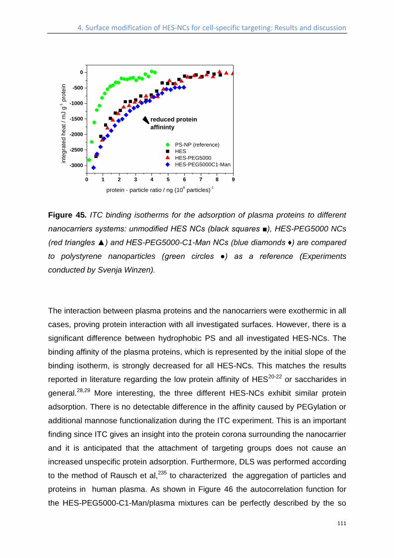
4. Surface modification of HES-NCs for cell-specific targeting: Results and discussion
111
0 1 2 3 4 5 6 7 8 9
-3000
-2500
-2000
-1500
-1000
-500
0
PS-NP (reference)
HES
HES-PEG5000
HES-PEG5000C1-Man
in
teg
rate
d h
ea
t / m
J g
-1 p
rote
in
protein - particle ratio / ng (106 particles)
-1
reduced protein
affininty
Figure 45. ITC binding isotherms for the adsorption of plasma proteins to different
nanocarriers systems: unmodified HES NCs (black squares ■), HES-PEG5000 NCs
(red triangles ▲) and HES-PEG5000-C1-Man NCs (blue diamonds ♦) are compared
to polystyrene nanoparticles (green circles ●) as a reference (Experiments
conducted by Svenja Winzen).
The interaction between plasma proteins and the nanocarriers were exothermic in all
cases, proving protein interaction with all investigated surfaces. However, there is a
significant difference between hydrophobic PS and all investigated HES-NCs. The
binding affinity of the plasma proteins, which is represented by the initial slope of the
binding isotherm, is strongly decreased for all HES-NCs. This matches the results
reported in literature regarding the low protein affinity of HES20-22 or saccharides in
general.28,29 More interesting, the three different HES-NCs exhibit similar protein
adsorption. There is no detectable difference in the affinity caused by PEGylation or
additional mannose functionalization during the ITC experiment. This is an important
finding since ITC gives an insight into the protein corona surrounding the nanocarrier
and it is anticipated that the attachment of targeting groups does not cause an
increased unspecific protein adsorption. Furthermore, DLS was performed according
to the method of Rausch et al,235 to characterized the aggregation of particles and
proteins in human plasma. As shown in Figure 46 the autocorrelation function for
the HES-PEG5000-C1-Man/plasma mixtures can be perfectly described by the so
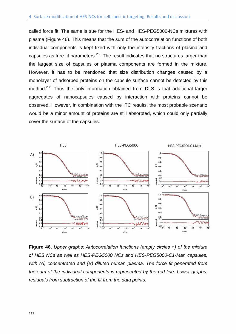
4. Surface modification of HES-NCs for cell-specific targeting: Results and discussion
112
called force fit. The same is true for the HES- and HES-PEG5000-NCs mixtures with
plasma (Figure 46). This means that the sum of the autocorrelation functions of both
individual components is kept fixed with only the intensity fractions of plasma and
capsules as free fit parameters.235 The result indicates that no structures larger than
the largest size of capsules or plasma components are formed in the mixture.
However, it has to be mentioned that size distribution changes caused by a
monolayer of adsorbed proteins on the capsule surface cannot be detected by this
method.236 Thus the only information obtained from DLS is that additional larger
aggregates of nanocapsules caused by interaction with proteins cannot be
observed. However, in combination with the ITC results, the most probable scenario
would be a minor amount of proteins are still absorpted, which could only partially
cover the surface of the capsules.
Figure 46. Upper graphs: Autocorrelation functions (empty circles ○) of the mixture
of HES NCs as well as HES-PEG5000 NCs and HES-PEG5000-C1-Man capsules,
with (A) concentrated and (B) diluted human plasma. The force fit generated from
the sum of the individual components is represented by the red line. Lower graphs:
residuals from subtraction of the fit from the data points.
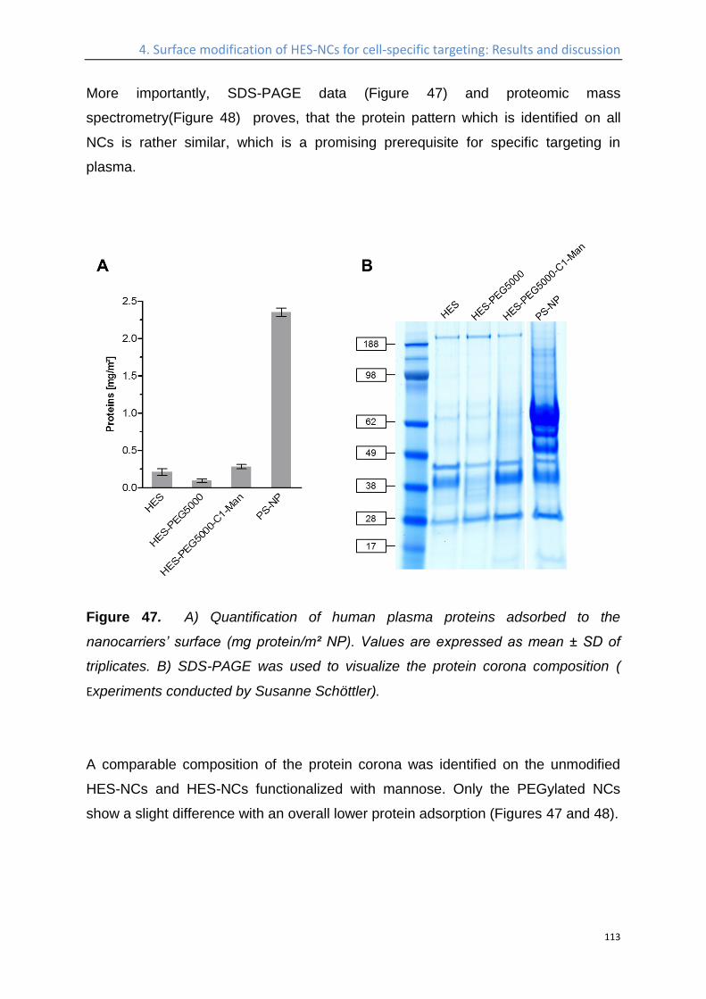
4. Surface modification of HES-NCs for cell-specific targeting: Results and discussion
113
More importantly, SDS-PAGE data (Figure 47) and proteomic mass
spectrometry(Figure 48) proves, that the protein pattern which is identified on all
NCs is rather similar, which is a promising prerequisite for specific targeting in
plasma.
Figure 47. A) Quantification of human plasma proteins adsorbed to the
nanocarriers’ surface (mg protein/m² NP). Values are expressed as mean ± SD of
triplicates. B) SDS-PAGE was used to visualize the protein corona composition (
Experiments conducted by Susanne Schöttler).
A comparable composition of the protein corona was identified on the unmodified
HES-NCs and HES-NCs functionalized with mannose. Only the PEGylated NCs
show a slight difference with an overall lower protein adsorption (Figures 47 and 48).
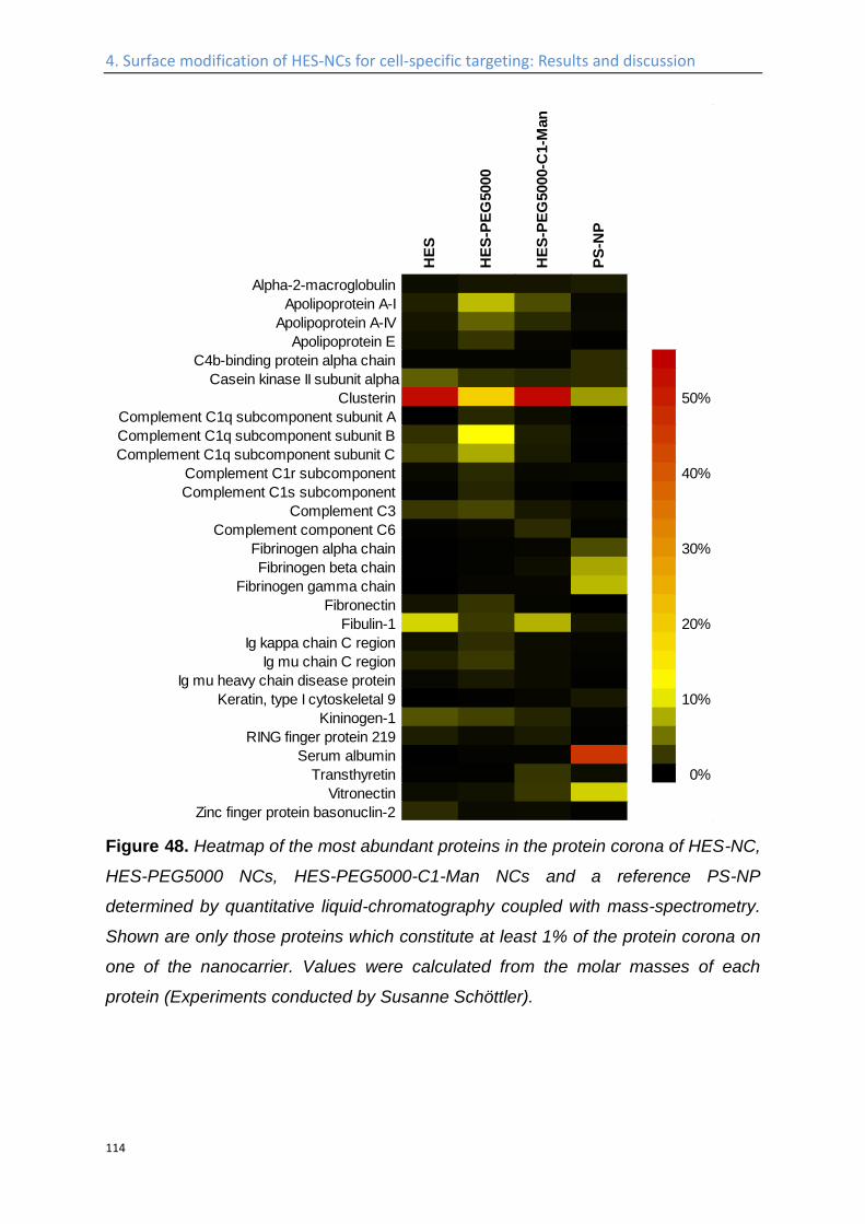
4. Surface modification of HES-NCs for cell-specific targeting: Results and discussion
114
Figure 48. Heatmap of the most abundant proteins in the protein corona of HES-NC,
HES-PEG5000 NCs, HES-PEG5000-C1-Man NCs and a reference PS-NP
determined by quantitative liquid-chromatography coupled with mass-spectrometry.
Shown are only those proteins which constitute at least 1% of the protein corona on
one of the nanocarrier. Values were calculated from the molar masses of each
protein (Experiments conducted by Susanne Schöttler).
HE
S
HE
S-P
EG
5000
HE
S-P
EG
5000-C
1-M
an
PS
-NP
Alpha-2-macroglobulin
Apolipoprotein A-I
Apolipoprotein A-IV
Apolipoprotein E
C4b-binding protein alpha chain
Casein kinase II subunit alpha
Clusterin 50%
Complement C1q subcomponent subunit A
Complement C1q subcomponent subunit B
Complement C1q subcomponent subunit C
Complement C1r subcomponent 40%
Complement C1s subcomponent
Complement C3
Complement component C6
Fibrinogen alpha chain 30%
Fibrinogen beta chain
Fibrinogen gamma chain
Fibronectin
Fibulin-1 20%
Ig kappa chain C region
Ig mu chain C region
Ig mu heavy chain disease protein
Keratin, type I cytoskeletal 9 10%
Kininogen-1
RING finger protein 219
Serum albumin
Transthyretin 0%
Vitronectin
Zinc finger protein basonuclin-2
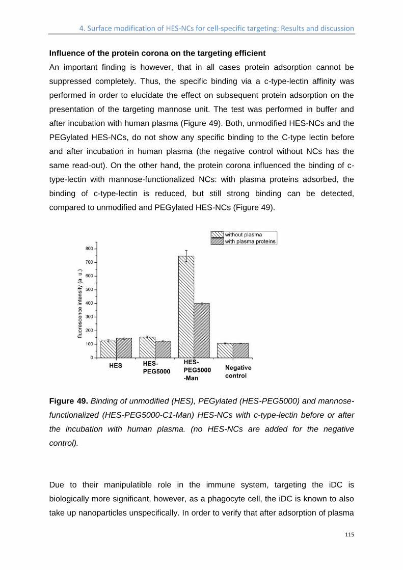
4. Surface modification of HES-NCs for cell-specific targeting: Results and discussion
115
Influence of the protein corona on the targeting efficient
An important finding is however, that in all cases protein adsorption cannot be
suppressed completely. Thus, the specific binding via a c-type-lectin affinity was
performed in order to elucidate the effect on subsequent protein adsorption on the
presentation of the targeting mannose unit. The test was performed in buffer and
after incubation with human plasma (Figure 49). Both, unmodified HES-NCs and the
PEGylated HES-NCs, do not show any specific binding to the C-type lectin before
and after incubation in human plasma (the negative control without NCs has the
same read-out). On the other hand, the protein corona influenced the binding of c-
type-lectin with mannose-functionalized NCs: with plasma proteins adsorbed, the
binding of c-type-lectin is reduced, but still strong binding can be detected,
compared to unmodified and PEGylated HES-NCs (Figure 49).
Figure 49. Binding of unmodified (HES), PEGylated (HES-PEG5000) and mannose-
functionalized (HES-PEG5000-C1-Man) HES-NCs with c-type-lectin before or after
the incubation with human plasma. (no HES-NCs are added for the negative
control).
Due to their manipulatible role in the immune system, targeting the iDC is
biologically more significant, however, as a phagocyte cell, the iDC is known to also
take up nanoparticles unspecifically. In order to verify that after adsorption of plasma
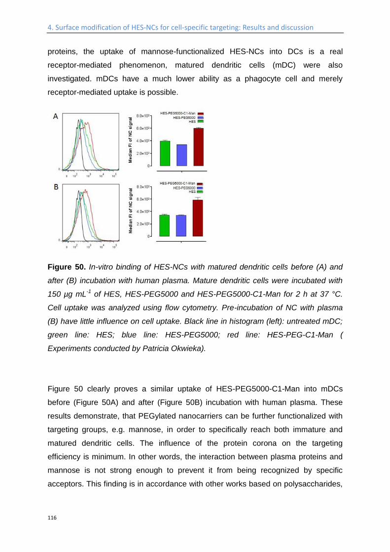
4. Surface modification of HES-NCs for cell-specific targeting: Results and discussion
116
proteins, the uptake of mannose-functionalized HES-NCs into DCs is a real
receptor-mediated phenomenon, matured dendritic cells (mDC) were also
investigated. mDCs have a much lower ability as a phagocyte cell and merely
receptor-mediated uptake is possible.
Figure 50. In-vitro binding of HES-NCs with matured dendritic cells before (A) and
after (B) incubation with human plasma. Mature dendritic cells were incubated with
150 µg mL-1 of HES, HES-PEG5000 and HES-PEG5000-C1-Man for 2 h at 37 °C.
Cell uptake was analyzed using flow cytometry. Pre-incubation of NC with plasma
(B) have little influence on cell uptake. Black line in histogram (left): untreated mDC;
green line: HES; blue line: HES-PEG5000; red line: HES-PEG-C1-Man (
Experiments conducted by Patricia Okwieka).
Figure 50 clearly proves a similar uptake of HES-PEG5000-C1-Man into mDCs
before (Figure 50A) and after (Figure 50B) incubation with human plasma. These
results demonstrate, that PEGylated nanocarriers can be further functionalized with
targeting groups, e.g. mannose, in order to specifically reach both immature and
matured dendritic cells. The influence of the protein corona on the targeting
efficiency is minimum. In other words, the interaction between plasma proteins and
mannose is not strong enough to prevent it from being recognized by specific
acceptors. This finding is in accordance with other works based on polysaccharides,
4. Surface modification of HES-NCs for cell-specific targeting: Results and discussion
117
inter alia HES23, chitosan183, hyaluronic acid138 to functionalized the surface of
nanocarriers, in order to reduce protein interactions.
Taking into consideration that, rather simple saccharide motifs, like the disaccharide
moiety in bleomycin226, have been proven to be able to selectively target cancer
cells. The use of a combination of stealth nanocarriers (i.e. PEGylated materials)
with such saccharide-based targeting moieties could solve the important dilemma,
where the stealth effect is lost after coupling of other targeting agents.
4. Surface modification of HES-NCs for cell-specific targeting: Experimental Part
118
Experimental Part
Materials
The isocyanate-PEG and isocyanate-PEG-isocyanate (PEG molecular weight of
5,000 g·mol−1, 110 ethylene oxide units) was purchased from Nanocs Inc., USA.
PBS buffer (pH 7.4, without Ca2+ and Mg2+) was purchased from Gibco, Germany.
Boric buffer (1M, pH 9.5) was produced by adjusting pH of boric acid (B6768 Sigma)
water solution to the desired value by sodium hydroxide solution. HES (200k, 0.5
degree of substitution) was bought from Fresenius Kabi. Oil soluble surfactant
poly((ethylene-co-butylene)-b-(ethylene oxide)), P(E/B-b-EO), consisting of a
poly(ethylene-co-butylene) block (Mw =3,700 g·mol−1) and a poly(ethylene oxide)
block (Mw = 3,600 g·mol−1) was synthesized starting from ω-hydroxypoly-(ethylene-
co-butylene), which was dissolved in toluene after addition of ethylene oxide under
anionic polymerization conditions212. The anionic surfactant sodium dodecylsulfate
(SDS) was purchased from Fluka. C-type-lectin (Galanthus nivalis snowdrop lectin
fluorescein labeled 2 mg) was bought from BIOZOL Diagnostica Vertrieb GmbH.
The fluorescent dye Cy5-oligo was purchased from BioChemica, Aldrich. DBCO-
PEG4-NHS was bought from Jana Bioscience. Dimethylsulfoxide (<50 ppm water
content) was purchased from Acros Organics. Demineralized MilliQ water was used
for the synthesis of nanocapsules. Mannose-azide. Polystyrene particles was
prepared according to the publication.195 The Microbody 2.5D, MC-ME-030, and
scFv was obtained from BioNTech. The MOUSE ANTI HUMAN CD205 (Dec205)
antibody was bought from Bio-Rad AbD Serotec GmbH, with the catalog number of
MCA4755. All the other chemicals were purchased from Sigma Aldrich, and used as
received.
Blood samples were taken at the Transfusion Centre of the University Medical
Center of the Johannes Gutenberg-University Mainz from 10 healthy donors after
obtaining informed consent. The study was approved by the local ethics committee.
Li-Heparin was added to prevent blood clotting. The blood was then centrifuged to
pellet red and white blood cells and the plasma supernatant was pooled. Aliquots
were stored at -80 °C. After thawing, the plasma was centrifuged at 20,000 g for 1 h

4. Surface modification of HES-NCs for cell-specific targeting: Experimental Part
119
at 4 °C to remove any residual precipitates. A protein concentration of 66 g·L-1 was
determined for the plasma.
4. Surface modification of HES-NCs for cell-specific targeting: Experimental Part
120
Methods.
NMR
1H NMR and 13C spectrums are measured at 300 MHz on a Bruker Avance 300
Spectrometer.
Fluorescence
The fluorescamine (FA) assay203 is used to quantify the available primary amine
groups at the surface of the nanocapsules, and the amine functionalized mannose in
the solution. A stock solution of FA is prepared by mixing 2 mg of FA and 1.5 mL of
anhydrous acetone. A standard working function is established from an aqueous
hexylamine solution as reference. For a typical quantification step, 25 µL of the
nanocapsule dispersion (or amine functionalized mannose solution) in water are
added into 725 µL of boric buffer (pH 9.5), followed by the addition of 200 µL of the
FA stock solution. The mixture is vortexed (Heidolph REAX2000 at maximum speed)
for 30 s, and the fluorescence intensity is measured by a Tecan plate reader at 25
°C by excitation at 410 nm and detecting the emission at 470 nm. All fluorescence
measurements are repeated three times.
Electron Microscopy.
The same diluted dispersion as for DLS is used to prepare electron microscopy
samples. Scanning electron microscopy (SEM) measurements are carried out by
using a LEO (Zeiss) 1530 Gemini device (Oberkochen, Germany). then 15 µL of the
dispersion is placed on the silica wafer and dried at room temperature overnight.
Transmission electron microscopy (TEM) measurements are carried out by using
Jeol 1400 device at 120 kV accelerating voltage. The samples are diluted in the
same way as for SEM sample preparation. Then 2 µL of the diluted sample is placed
on a copper grid, which is covered by a 5 nm thick carbon film.
4. Surface modification of HES-NCs for cell-specific targeting: Experimental Part
121
Functionalization of the surface of HES-NCs with monoisocyanate-PEG or
isocyanate-PEG-isocyanate
For a typical coupling reaction, 2 mL of the nanocapsule dispersion is centrifuged at
4000 rpm for 30 min to remove the excess of surfactant. The upper phase is
discarded, while the precipitate is redispersed in 2.5 g of anhydrous cyclohexane by
pipetting up and down. Either 16 mg of monoisocyanate-PEG (MeO-PEG110-NCO
with Mn=5000 g·mol−1) or isocyanate-PEG-isocyanate (OCN-PEG110-NCO with
Mn=5000 g·mol−1) was dissolved in 0.63 g (800 µL) of anhydrous acetone and
added dropwise into the capsule dispersion, which is magnetically stirred at 500
rpm. The reaction is allowed to proceed for 5 h, and centrifuged at 4000 rpm
(Eppendorff centrifuge 5417C) afterwards to remove non-coupled PEG. The
precipitate is redispersed in 400 µL of cyclohexane and used for further steps.
Redispersion of capsules in water
A 0.1wt% SDS solution is prepared, and filtered (sterile, with 0.2 µm pore size). 5 mL
of this SDS solution is gently shaken in a sonication bath (Bandelin Sonorex, type
RK 52H), while nanocapsule (from 2 mL of the as prepared dispersion) redispersed
in 400 µL of cyclohexane is added slowly. Then the whole dispersion is magnetically
stirred at 1000 rpm at room temperature overnight, in an open vial, to allow
evaporation of cyclohexane. The obtained dispersion is ultrafiltrated by a Amicon
Centrifugal Filter (Ultra-0.5, Ultracel-100 Membrane, 100 kDa), in order to remove
the excess of SDS and used for further steps.
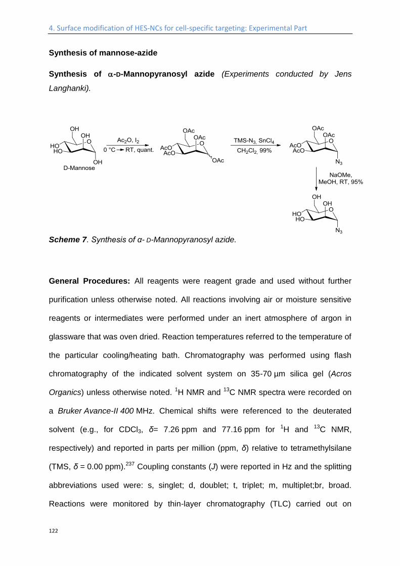
4. Surface modification of HES-NCs for cell-specific targeting: Experimental Part
122
Synthesis of mannose-azide
Synthesis of -D-Mannopyranosyl azide (Experiments conducted by Jens
Langhanki).
Scheme 7. Synthesis of α- D-Mannopyranosyl azide.
General Procedures: All reagents were reagent grade and used without further
purification unless otherwise noted. All reactions involving air or moisture sensitive
reagents or intermediates were performed under an inert atmosphere of argon in
glassware that was oven dried. Reaction temperatures referred to the temperature of
the particular cooling/heating bath. Chromatography was performed using flash
chromatography of the indicated solvent system on 35-70 µm silica gel (Acros
Organics) unless otherwise noted. 1H NMR and 13C NMR spectra were recorded on
a Bruker Avance-II 400 MHz. Chemical shifts were referenced to the deuterated
solvent (e.g., for CDCl3, δ= 7.26 ppm and 77.16 ppm for 1H and 13C NMR,
respectively) and reported in parts per million (ppm, δ) relative to tetramethylsilane
(TMS, δ = 0.00 ppm).237 Coupling constants (J) were reported in Hz and the splitting
abbreviations used were: s, singlet; d, doublet; t, triplet; m, multiplet;br, broad.
Reactions were monitored by thin-layer chromatography (TLC) carried out on
4. Surface modification of HES-NCs for cell-specific targeting: Experimental Part
123
0.25 mm E. Merck silica gel plates (60F254) using an 1M ethanolic solution of sulfuric
acid with 0.2% 3-methoxyphenol and heat as developing agents. Specific reactions
were monitored by LC-MS on a 1200 HPLC-unit from Agilent Technologies with
binary pump and integrated diode array detector coupled to a LC/MSD-Trap-mass-
spectrometer from Bruker. Ionization was achieved by an electron-spray-ionization
source (ESI) or an atmospheric-pressure-chemical-ionization source (APCI). High-
resolution masses were recorded on a Waters QTof-Ultima 3-Instrument with
Lockspray-Interface and a suitable external calibrant. Infrared spectra were recorded
as FT-IR spectra using a diamond ATR unit and are reported in terms of frequency
of absorption (, cm-1). Dichloromethane was distilled from calcium hydride.
1,2,3,4,6-Penta-O-acetyl-D-mannopyranose. Iodine (560 mg, 2.2 mmol, 0.04 eq.)
and acetic anhydride (50 mL) were mixed under Ar-atmosphere. D-Mannose (10 g,
55.5 mmol, 1 eq.) was added portion by portion at 0 °C. After stirring for 30 min at
0 °C and additionally for 18 hours at room temperature TLC
(cyclohexane/toluene/ethylacetate 3:3:1) showed complete consumption of the
starting material. The reaction mixture was diluted with CH2Cl2 (50 mL) and was
washed twice with cold saturated aqueous Na2SO3 solution (2x 80 mL), then with a
saturated aqueous solution of NaHCO3 (4x 50 mL). The separated organic layer was
dried over anhydrous Na2SO4. The solvent was removed in vacuo to afford the
desired peracetylated D-mannose (21.5 g, 55.1 mmol, 99%, mixture of both anomers
Rf 0.30 (silica gel, cyclohexane/toluene/ethyl acetate, 3:3:1);
Signals assignable to -anomer: 1H NMR, COSY (600 MHz, CDCl3) δ (ppm) = 6.09
(d, 3J = 1.9 Hz, 1H, H-1), 5.34–5.36 (m, 2H, H-3, H-4), 5.25–5.27 (m, 1H, H-2), 4.28
4. Surface modification of HES-NCs for cell-specific targeting: Experimental Part
124
(dd, 2J = 12.4 Hz, 3J = 4.9 Hz, 1H, H-6a), 4.10 (dd, 2J = 12.4 Hz, 3J = 2.5 Hz, 1H,
H-6b), 4.03-4.07 (m, 1H, H-5), 2.18, 2.17, 2.10, 2.05, 2.01 (5x s, 15H, COCH3 );
13C NMR, HSQC, HMBC (151 MHz, CDCl3) δ (ppm) = 170.8, 170.2, 169.9, 169.7,
168.2 (5x COCH3), 90.7 (C-1), 70.7 (C-5), 68.8 (C-3), 68.4 (C-2), 65.6 (C-4), 62.2
(C-6), 21.0, 20.9, 20.9, 20.8, 20.8 (5x COCH3).
2,3,4,6-Tetra-O-acetyl--D-mannopyranosyl azide. 1,2,3,4,6-Penta-O-acetyl-D-
mannopyranose (13.6 g, 34.9 mmol, 1 eq.) was dissolved in anhydrous CH2Cl2
(140 mL) under argon. Trimethylsilyl azide (16.1 g, 18.5 mL, 139 mmol, 4 eq.) and
tin(IV) chloride (2.37 g, 1.06 mL, 9.1 mmol, 0.26 eq.) was added. After stirring for
four hours at room temperature TLC (cyclohexane/toluene/ethylacetate 3:3:1)
showed complete consumption of the starting material. The reaction mixture was
washed successively with saturated aqueous NaHCO3 solution (100mL), water
(100 mL) and brine (100 mL). The organic layer was dried over anhydrous Na2SO4.
The solvent was removed in vacuo and the residue was purified by flash column
chromatography (cyclohexane/ ehtylacetate, 3:1) to afford the title compound
(12.9 g, 34.4 mmol, 99%) as a clear, colorless oil.
Rf 0.49 (silica gel, cyclohexane/toluene/ethyl acetate, 3:3:1);
1H NMR, COSY (400 MHz, CDCl3) δ (ppm) = 5.38 (d, 3J = 1.9 Hz, 1H, H-1), 5.14
(dd, 3J = 3.0 Hz, 3J = 1.9 Hz, 1H, H-2), 5.30–5.21 (m, 2H, H-3, H-4), 4.32-4.27 (m,
1H, H-6a), 4.17–4.11 (m, 2H, H-5, H-6b), 2.16, 2.10, 2.04, 1.98 (4x s, 12H, COCH3 );
13C NMR, HSQC, HMBC (100.6 MHz, CDCl3) δ (ppm) = 170.7, 170.0, 169.9, 169.7
(4x COCH3), 87.6 (C-1), 70.7 (C-5), 69.3 (C-2), 68.3 (C-3), 65.7 (C-4), 62.2 (C-6),
21.0, 20.8, 20.8, 20.7 (4x COCH3).
The spectral data are in accordance with literature. (7)
4. Surface modification of HES-NCs for cell-specific targeting: Experimental Part
125
α-D-Mannopyranosyl azide. 2,3,4,6-Tetra-O-acetyl-α-D-mannopyranosyl azide
(10 g, 26.8 mmol) was dissolved in methanol (100 mL) and sodium methoxide was
added until pH 9–10 (approx. 60 mg). The reaction mixture was stirred at room
temperature for 16 hours. Subsequently the solution was neutralized by Amberlite
120 H+ resin until pH 7. The mixture was filtered over Celite witch was washed
thoroughly with methanol. The solvent was removed in vacuo to afford the desired α-
D-Mannopyranosyl azide (5.23 g, 25.5 mmol, 95%) as a colorless syrup.
1H NMR, COSY (400 MHz, CDCl3) δ (ppm) = 5.43 (d, 3J = 1.9 Hz, 1H, H-1), 3.91–3.87 (m,
1H, H-6a), 3.85 (dd, 3J = 3.3 Hz, 3J = 1.9 Hz, 1H, H-2), 3.79–3.69 (m, 3H, H-3, H-5, H-6b),
3.65-3.59 (m, 1H, H-4); 13C NMR, HSQC, HMBC (100.6 MHz, CDCl3) δ (ppm) = 89.7 (C-1),
74.6 (C-5), 69.8, 69.7 (C-2, C-3), 66.3 (C-4), 60.7 (C-6).
Coupling of mannose-azide to isocyanate-PEG-isocyanate functionalized
capsule
To 5 mL of the capsule dispersion with a solid content of 0.1 wt% is added 500 µL of
0.8M boric acid solution. The pH of the solution is adjusted to 8.3 and 0.12 mg of
DBCO-PEG4-NHS is added, which is prepared as a stock solution of 1mg in 1mL of
anhydrous DMSO. The reaction is allowed to proceed for 4h, the uncoupled DBCO
is removed by ultrafiltration. And the coupled DBCO is quantified by a fluoregenetic
reaction with anthracene azide238. Azide mannose is added with a 1:1 mol ratio to
coupled DBCO.
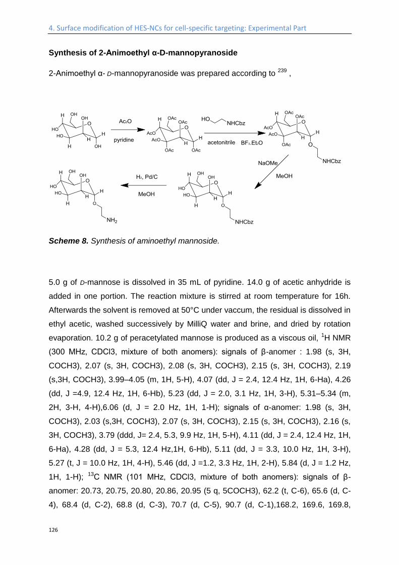
4. Surface modification of HES-NCs for cell-specific targeting: Experimental Part
126
Synthesis of 2-Animoethyl α-D-mannopyranoside
2-Animoethyl α- D-mannopyranoside was prepared according to 239 ,
Scheme 8. Synthesis of aminoethyl mannoside.
5.0 g of D-mannose is dissolved in 35 mL of pyridine. 14.0 g of acetic anhydride is
added in one portion. The reaction mixture is stirred at room temperature for 16h.
Afterwards the solvent is removed at 50°C under vaccum, the residual is dissolved in
ethyl acetic, washed successively by MilliQ water and brine, and dried by rotation
evaporation. 10.2 g of peracetylated mannose is produced as a viscous oil, 1H NMR
(300 MHz, CDCl3, mixture of both anomers): signals of β-anomer : 1.98 (s, 3H,
COCH3), 2.07 (s, 3H, COCH3), 2.08 (s, 3H, COCH3), 2.15 (s, 3H, COCH3), 2.19
(s,3H, COCH3), 3.99–4.05 (m, 1H, 5-H), 4.07 (dd, J = 2.4, 12.4 Hz, 1H, 6-Ha), 4.26
(dd, J =4.9, 12.4 Hz, 1H, 6-Hb), 5.23 (dd, J = 2.0, 3.1 Hz, 1H, 3-H), 5.31–5.34 (m,
2H, 3-H, 4-H),6.06 (d, J = 2.0 Hz, 1H, 1-H); signals of α-anomer: 1.98 (s, 3H,
COCH3), 2.03 (s,3H, COCH3), 2.07 (s, 3H, COCH3), 2.15 (s, 3H, COCH3), 2.16 (s,
3H, COCH3), 3.79 (ddd, J= 2.4, 5.3, 9.9 Hz, 1H, 5-H), 4.11 (dd, J = 2.4, 12.4 Hz, 1H,
6-Ha), 4.28 (dd, J = 5.3, 12.4 Hz,1H, 6-Hb), 5.11 (dd, J = 3.3, 10.0 Hz, 1H, 3-H),
5.27 (t, J = 10.0 Hz, 1H, 4-H), 5.46 (dd, J =1.2, 3.3 Hz, 1H, 2-H), 5.84 (d, J = 1.2 Hz,
1H, 1-H); 13C NMR (101 MHz, CDCl3, mixture of both anomers): signals of β-
anomer: 20.73, 20.75, 20.80, 20.86, 20.95 (5 q, 5COCH3), 62.2 (t, C-6), 65.6 (d, C-
4), 68.4 (d, C-2), 68.8 (d, C-3), 70.7 (d, C-5), 90.7 (d, C-1),168.2, 169.6, 169.8,
4. Surface modification of HES-NCs for cell-specific targeting: Experimental Part
127
170.1, 170.7 (5 s, 5 COCH3); signals of α-anomer: 20.63,20.76, 20.81, 20.84, 20.86
(5 q, 5 COCH3), 62.1 (t, C-6), 65.4 (d, C-4), 68.3 (d, C-2), 70.7 (d,C-3), 73.4 (d, C-
5), 90.5 (d, C-1), 168.5, 169.7, 169.9, 170.3, 170.8 (5 s, 5 COCH3).
2.52 g of Mannose pentaacetate is dissolved in 25 mL of anhydrous acetonitrile in a
50 mL schlenk tube, which is sealed by a rubber stopper and flushed with argon.
1.526 g of N-Z-ethanolamine (1.2 equi) is dissolved in 2 mL of anhydrous
acetonitrile, and added dropwise into the schlenk tube. The system is placed in ice
bath, and 4.61 g of BF3•Et2O (5 equivalent) is added dropwise. The solution is
stirred in ice bath for the other 30 min, and overnight at room temperature. 5
equivalent of TEA was added to quench the reaction. Solvent is removed by
vacuum, and the product purified by column chromatography on silica, with 1:1
mixture of ethyl acetic and petroleum ether ( low boiling point) as elute. The second
compound come out of the column is the product. 1H NMR (300 MHz, CDCl3): 2.00
(s, 3H, COCH3),2.04 (s, 3H, COCH3), 2.09 (s, 3H, COCH3), 2.16 (s, 3H, COCH3),
3.36–3.53 (m, 2H,CH2NH), 3.58 (ddd, J = 3.6, 6.8, 10.2 Hz, 1H, CHaHbCH2NH),
3.78 (ddd, J = 3.9, 6.210.2 Hz, 1H, CHaHbCH2NH), 3.97 (ddd, J = 2.3, 5.7 9.5 Hz,
1H, 5-H), 4.08 (dd, J = 2.3,12.2 H 1H, 6-Ha), 4.26 (dd, J = 5.7, 12.2 Hz, 1H, 6-Hb),
4.82 (d, J = 1.7 Hz, 1H, 1-H), 5.12(s, 2H, CH2Ph), 5.20 (bt, J = 5.8 Hz, 1H, NH),
5.25 (dd, J = 1.7, 3.2 Hz, 1H, 2-H), 5.26 (dd, JS12= 9.5, 10.1 H), 5.31 (dd, J = 3.2,
10.0 Hz, 1H, 3-H), 7.29–7.39 (m, 5H, C6H5); 13C NMR(101 MHz, CDCl3): 20.7,
20.8, 20.9 (3 q, 4 COccCH3), 40.7 (CH2NH), 62.5 (t, C-6), 66.1 (d, C-4), 66.9 (t,
CH2Ph), 67.8 (t, CH2CH2NH), 68.8 (d, C-5), 69.0 (d, C-3), 69.4 (d,C-2), 97.8 (d, C-
1), 128.2, 128.6 (2 d, o-, m-, p-C from C6H5), 136.4 (s, i-C from C6H5),156.4 (s,
NCOO), 169.8, 170.0, 170.1, 170.7 (4 s, 4 COCH3).
975 mg of the product from last step is dissolved in 30 mL MeOH, and 34 mg of
NaOMe is added (113 mg 30% solution in MeOH). The reaction is stirred overnight,
and the ions are removed by Abmerlite IR120, H+ form. The resin is removed by
filtration, and solvent removed by rotational evaporation. 1H NMR (300 MHz, MeOD):
3.27–3.39 (m, 2H,CH2NH), 3.47–3.55 (m, 2H, 5-H, CHaHbCH2NH), 3.60 (t, J = 9.5
Hz, 1H, 4-H), 3.68 (dd, J =5.8, 11.7 Hz, 1H, 6-Ha), 3.69 (dd, J = 3.4, 9.3 Hz, 1H, 3-
H), 3.74 (ddd, J = 4.9, 6.4, 10.2 Hz,1H, CHaHbCH2NH), 3.80 (dd, J = 1.7, 3.4 Hz,

4. Surface modification of HES-NCs for cell-specific targeting: Experimental Part
128
1H, 2-H), 3.81 (dd, J = 2.3, 11.7 Hz, 1H, 6-Hb), 4.75 (d, J = 1.6 Hz, 1H, 1-H), 5.06 (s,
2H, CH2Ph) , 7.24–7.36 (m, 5H, C6H5); 13C NMR(101 MHz, MeOD): 41.7 (t,
CH2CH2NH), 62.8 (t, C-6), 67.5 (t, CH2Ph,CH2CH2NH), 68.5 (d, C-4), 72.0 (d, C-2),
72.5 (d, C-3), 74.7 (d, C-5), 101.6 (d, C-1), 128.8,129.0, 129.5 (3 s, o-, m-, p-C from
C6H5), 138.3 (s, i-C from C6H5), 158.9 (s, NCOO).
The product from the third step is further hydrogenated by dissolving in MeOH on Pd
deposited active carbon. The reduction is carried out overnight. The catalyst is
removed by Celite filtration, and the solvent removed by rotational evaporation to get
the final product, 356 mg, as white powder. 1H NMR (300 MHz, MeOD): 2.82–2.86
(m, 2H,CH2NH2), 3.48 (ddd, J = 4.7, 5.9, 10.2 Hz, 1H, CHaHbCH2NH2), 3.56 (ddd,
J = 2.1, 5.8,9.7 Hz, 1H, 5-H), 3.63 (t, J = 9.4 Hz, 1H, 4-H), 3.73 (dd, J = 5.8, 11.8 Hz,
1H, 6-Ha), 3.74(dd, J = 3.4, 9.1 Hz, 1H, 3-H), 3.79 (ddd, J = 4.7, 5.9, 10.2 Hz, 1H,
CHaHbCH2NH2), 3.86(dd, J = 1.7, 3.4 Hz, 1H, 2-H), 3.86 (dd, J = 2.1, 11.8 Hz, 1H,
6-Hb), 4.80 (d, J = 1.7 Hz, 1H,1-H); 13C NMR (101 MHz, MeOD): 101.7 (d, C-1), 74.6
(d, C-5), 72.5 (d, C-4),72.0 (d, C-2), 70.0 (t, CH2CH2NH2), 68.6 (d, C-3), 62.8 (t, C-
6), 42.0 (t, CH2NH2).
Coupling of mannose-amine to isocyanate-PEG-isocyanate functionalized
capsule
The isocyanate-PEG-isocyanate functionalized HES capsule in cyclohexane phase
is redispersed in 5 g 0.1wt% SDS solution, which contains additionally 20mg of
amine functionalized mannose. Other conditions are the same as for redispersion of
PEG functionalized capsules in water.
Coupling of Microbody 2.5D and MC-ME-030 to the surface of the PEGylated
Nanocapsules
To 5 mL of the capsule dispersion with a solid content of 0.1 wt% is added 500 µL of
0.8M boric acid solution. The pH of the solution is adjusted to 8.3 and different
amounts of DBCO-PEG4-NHS is added, which is prepared as a stock solution of
1mg in 1mL of anhydrous DMSO. The reaction is allowed to proceed for 4h, the
4. Surface modification of HES-NCs for cell-specific targeting: Experimental Part
129
uncoupled DBCO is removed by ultrafiltration. And the coupled DBCO is quantified
by a fluoregenetic reaction with anthracene azide238. Azide functionalized
Microbodies are added with a 1:1 mol ratio to coupled DBCO.
Coupling of Microbody scFv to the surface of the PEGylated Nanocapsules
The scFv-Fragment is reduced by Tris(2-carboxyethyl)phosphine (TCEP) (1:62.5
(8mM); 2h;RT), to create thiol groups on the scFv-Fragment, followed by dialyzing in
Nellis-buffer (degased by flushing with N2; 100 mM Phosphate buffer (76,3 mM
NaH2PO4 & 23,7 mM Na2HPO4, 300 mM NaCl, 4 mM Ethylenediaminetetraacetic
acid (EDTA)); pH 6.7; overnight).
HES-NCs are incubated with Sulfosuccinimidyl 4-(N-maleimidomethyl)cyclohexane-
1-carboxylate (Sulfo-SMCC, 3 mM) in PBS (pH 7.4) for 1h at room temperature. The
free Sulfo-SMCC is removed by ultrafiltration (ultrafilter 100K MWCO, 5000 rpm, 20
min), followed by the addition of the reduced scFv-Fragment. The number of scFv-
Fragment added per capsule is calculated to be 8. After overnight incubation, the
dispersion is further purified by ultrafiltration, and send for biological test.
Coupling of Dec205 to the surface of the PEGylated Nanocapsules
0.4 mg of Dec205 is dissolved in 400 µL of PBS buffer, and the pH is adjusted to
7.60±0.05, the stock solution of 1.16 * 10-3 mg of NHS-PEG4-Azide in anhydrous
DMSO is added, which is 3.7 mol equivalent to the Dec205. The reaction is allowed
to proceed for 1h, while the excess of NHS-PEG4-Azide is removed by dialyzing.
The coupling of the azide Dec205 to the PEGylated HES-NCs is carried out
following the same procedure as for coupling of Microbodies to the capsules.
Incubation of capsules with human plasma
To 100 µL dispersion of each capsule dispersion with a solid content of 1.0wt%, 16.7
µL of human plasma with a protein concentration of 15 g·L-1 were added at room
4. Surface modification of HES-NCs for cell-specific targeting: Experimental Part
130
temperature. The system is incubated at room temperature over night, and purified
in the next morning by centrifugation at 6000 rpm for 20 min. The upper phase is
discarded, and the precipitate is redispersed in MilliQ water for further analysis. The
capsule to protein ratio is the same as for ITC and DLS measurement.
4. Surface modification of HES-NCs for cell-specific targeting: Experimental Part
131
Isothermal Titration Calorimetry (ITC) ( Experiments conducted by Svenja Winzen).
The isothermal titration calorimetry measurements were performed using a NanoITC
Low Volume (TA Instruments, Eschborn, Germany) with an effective cell volume of
170 µL. In an experiment 50 µL of human plasma with a protein concentration of
15 g·L-1 were titrated to 300 µL of each HES-capsule suspension (HES 1.30 g·L-1,
HES-PEG5000 1.25 g·L-1, HES-PEG5000-C1-Man 1.15 g·L-1). The same titration
was performed with a suspension of polystyrene nanoparticles at a concentration of
0.92 g/L for comparison. The experimental temperature was kept constant at 25 °C.
Additionally, the same amount of plasma was titrated into pure water to determine
the heat of dilution for reference. The number and injected volume of the titration
steps were the same for all measurements (25 x 2 µL). The spacing between
injections was set to 300 s. The integrated reference heats were then subtracted
from the integrated heats of the adsorption experiments. All titrations were measured
in triplicates to ensure proper reproducibility.
Dynamic Light Scattering (DLS) of capsule / plasma mixtures
All dynamic light scattering experiments were performed on a commercially available
instrument from ALV GmbH (Langen, Germany) consisting of a goniometer and an
ALV-5000 multiple tau full-digital correlators with 320 channels. A helium-neon laser
from JDS Uniphase (Milpitas, USA) with an operating intensity of 25 mW and a laser
wavelength of λ = 632.8 nm was used as a light source. All solutions for the light
scattering experiments were prepared in dust-free quartz cuvettes from Hellma
(Müllheim, Germany) with an inner diameter of 18 mm, which were cleaned before
with distilled acetone. For the measurements of nanocapsule-plasma mixtures 2 mL
of concentrated (60 g·L-1) or with 0.15 M NaCl solution diluted (0.075 g·L-1) human
plasma were filtered into the cuvette through a Millex-GS filter (Merck Millipore,
Darmstadt, Germany) with a pore size of 0.2 µm. To this solution 10 µL of a 7.3 g·L-1
capsule dispersion were added. The mixtures were then incubated at 37 °C for 30
min followed by the DLS measurements at the same temperature.

4. Surface modification of HES-NCs for cell-specific targeting: Experimental Part
132
C-type lectin interaction
The binding of different capsules with a mannose specific c-type-lectin, Galanthus
nivalis snowdrop lectin, was studies by incubating different capsules with a
fluorescein labeled c-type-lectin. To 500 µL of capsule dispersion (0.1 wt %) in
HEPES buffer (0.1 M, pH 7.5) was added 10 µg of c-type-lectin in MilliQ water. The
system is shaken at room temperature for 2h, and then overnight at 4 °C. The
dispersion is centrifuged at 3150 rzb for 10 min, the precipitate is isolated, and
redispersed in 500 µL of HEPES buffer (0.1 M, pH 7.5). The fluorescence of the final
dispersion is measured by a Tecan plate reader at 25 °C by excitation at 495 nm
and detecting the emission at 519 nm. All fluorescence measurements are repeated
three times.
4. Surface modification of HES-NCs for cell-specific targeting: Experimental Part
133
in-vitro test (experiments conducted by Patricia Okwieka).
Generation of immature or mature dendritic cells (iDC or mDC)
Buffy coats from healthy human donors were used to isolate peripheral blood
mononuclear cells (PBMC) by standard Ficoll density centrifugation. Donor blood
was obtained according to the declaration of Helsinki. Dendritic cells were generated
from monocytes using plastic adherence. PBMC were seeded at a density of 1,5 x
107 cells per well of a 6-well plate. Cells were allowed to adhere in a humidified
environment at 37 °C with 5% CO2 for 1 h in 3 mL of DC-medium (RMPI with 2%
heat-inactivated pooled human serum (HS), 100 U/mL streptomycin (Sigma). By
multiple washing with phosphate buffered saline (PBS, Invitrogen) non-adhering
cells were removed. Monocytes were kept in DC-medium containing 800 U/mL
granulocyte/ macrophage-colony stimulating factor (GM-CSF, Genzyme) and 500 U/
mL interleukin (IL)-4 (PromoKine) at 37 °C/ 5% CO2 for two days. Afterwards old
medium was removed and fresh medium supplemented with with 1600 U/ mL GM-
CSF and 1000 U/ mL IL-4 was added on days 3 and 5. On day 6 immature dendritic
cells (iDC) were harvested or maturation of iDC was induced to generate mature DC
(mDC). By incubation with PBS/ EDTA (2 mM) for 10 min at 37 °C and subsequent
washing with cold PBS iDC were obtained. Maturation of iDC was induced by
incubation of cells with DC medium supplemented with 500 U/ mL IL-4, 1600 U/ mL
GM-CSF , 10ng/mL IL-1β, 10 ng/ mL TNF-α, 1000 U/ mL IL-6 and 1 µg/mL PGE2 for
2 days. Mature dendritic cells were harvested using cold PBS. For NC loading of
DC, cryopreserved i or mDC were used.
Loading of DC with nanocapsules
1 x 10⁵ DC were incubated with NC at 37 °C/ 5% CO2 in DC medium supplemented
with 1600 U/ mL GM-CSF and 500 U/ mL IL-4. Two different nanocapsule
concentrations (50 or 150 μg/ mL) and incubation periods (2 or 16 h) were analyzed.
Afterwards cells were collected using multiple washing steps with cold PBS. The
nanocapsule containing supernatant was removed by centrifugation for 5 min at 470
g.
4. Surface modification of HES-NCs for cell-specific targeting: Experimental Part
134
Flow cytometry analysis of NC-loaded DC
Pelleted DC were stained with CD11c-FITC (Miltenyi), CD45-APC-Cy7 (BD) and
CD45-V450 (BD). Cells were allowed to incubate with the various antibodies for 15
min at 4 °C. After a washing step with PBS/ 0.1% BSA, the cells were centrifuged
and resuspended in 0.3 mL PBS/ 0.1% BSA. NC uptake by DC was detected using
a LSR-Fortessa or FACS Canto II analyzer, both equipped with FACSDivaTM
software (Becton Dickinson). Fluorescence of the NC dyes sulforhodamine (SR101)
or Cy5-oligo was detected using a 586/15 nm bandpass filter with excitation by a
100 mW/ 561 nm laser or a 660/20 nm bandpass filter with excitation by a 40 mW
633 nm laser, respectively. Conjugates like FITC were excited using a 100 mW 488
nm laser (filter 530/30 nm) and a 100 mW 405 nm laser (filter 450/50). Cells were
analyzed using FlowJo 7.6.5 (Tree Star Inc.).
Fluorescent Microscopy analysis of the binding of the capsules with cells
3 * 105 B16F10 cells were splitted in a well of a 6-well-plate, and incubated at 37 °C
overnight, under 5% CO2. The cells were washed by PBS buffer for once before they
were incubated with the HES-NCs, followed by the addition of 60 µL of nanocapsule
dispersion (1 wt%), and incubated on ice for 3h. The cells are fixed by using 2%
formaldehyde (15min, RT), and subsequently washed by PBS for three times. The
cells are then embedded with DAPI 1:5000 in Fluorescent Mounting Medium, and
analyzed for the binding of the nanocapsules by using Zeiss-Microscope (Axio,
Imager.M2).
In-vivo test
Pharmakokinetic studies for plasma half-life studies (experiments conducted by
Oliver Seifert).
Female SWISS (Janvier, CD1, 8 weeks old animals per construct) received an
intravenous (i.v.) injection of 300 µg of the capsule in a total volume of 100 µL (in
PBS). Blood samples (50 µL) were taken in the time intervals of 2 min, 30 min, 1 h, 2

4. Surface modification of HES-NCs for cell-specific targeting: Experimental Part
135
h, 4 h, 6 h, 1 day and 3 days, incubated with heparin and stored at 4°C. Using Li-
COR, fluorescence intensity of the blood samples was measured to analyze the
concentration of the functionalized capsules. Comprising standard curve and weight
of animals, the percentage of injected doses was calculated. T1/2β and area under
the curve (AUC) were calculated with Excel.
In vivo / Ex vivo Fluorescence Imaging for the bio-distribution of the capsules
(experiments conducted by Mustafa Diken).
Six to eight-week old female BALB/c mice were obtained from Charles River. All
mice were kept and handled in accordance with federal and state policies on animal
research.
In vivo/ ex vivo fluorescence imaging of fluorescently labeled unmodified or
PEGylated HES-NCs was performed using the IVIS Spectrum Imaging system
(Perkin Elmer). Upon intravenous injection of nanocapsules (1mg), BALB/c mice
(n = 3) were anesthetized with isofluorane at different time points (30min, 1h, 2h, 4h,
6 h, 8 h, 24 h) and placed in the light-tight chamber of IVIS Spectrum. Fluorescence
emission was visualized by using the light source and filters with excitation at 745
nm and emission at 800 nm for an exposure time of 3 s. 24 h after injection, mice
were sacrificed and fluorescence signal from isolated organs such as brain, lungs,
heart, kidneys, liver and spleen were collected ex vivo using the same filter set and
exposure time. Fluorescence images were presented as superimposed to black &
white photos of mice/organs and the fluorescence intensity was depicted with a
pseudocolor scale.
4. Surface modification of HES-NCs for cell-specific targeting: Experimental Part
136
Protein corona analysis ( experiments conducted by Susanne Schöttler).
SDS polyacrylamide gel electrophoresis (SDS-PAGE).
For SDS PAGE 16.25 µL of the protein sample was mixed with 6.25 µL NuPAGE
LDS Sample Buffer and 2.5 µL NuPAGE Sample Reducing Agent and applied onto a
NuPAGE 10% Bis-Tris Protein Gel (all Novex, Thermo Fisher Scientific). The
electrophoresis was carried out in NuPAGE MES SDS Running Buffer at 150 V for
1.5 h with SeeBlue Plus2 Pre-Stained Standard (Invitrogen) as a molecular marker.
The gel was stained using SimplyBlue SafeStain (Novex, Thermo Fisher Scientific).
Protein in-solution digestion.
Proteins were digested following the protocol of Tenzer et al.220 with following
modifications: 25 µg of each protein sample was precipitated and trypsin was used
with a 1:50 ratio (enzyme:protein). For LC-MS analysis the samples were diluted 10-
fold with aqueous 0.1% formic acid and spiked with 50 fmol/µL Hi3 EColi Standard
(Waters Corporation) for absolute quantification.
Liquid-chromatography mass-spectrometry (LC-MS) analysis.
Quantitative analysis of protein samples was performed using a nanoACQUITY
UPLC system coupled with a Synapt G2-Si mass spectrometer (Waters
Corporation). Tryptic peptides were separated on the nanoACQUITY system
equipped with a C18 analytical reversed-phase column (1.7 μm, 75 μm x 150 mm,
Waters Corporation) and a C18 nanoACQUITY Trap Column (5 µm, 180 µm x 20
mm, Waters Corporation). Samples were processed with mobile phase A consisting
of 0.1% (v/v) formic acid in water and mobile phase B consisting of acetonitrile with
0.1% (v/v) formic acid. The separation was performed at a sample flow rate of 0.3
µL/min, using a gradient of 2-37% mobile phase B over 70 min. As a reference
compound 150 fmol/µL Glu-Fibrinopeptide was infused at a flow rate of 0.5 µL/min.
Data-independent acquisition (MSE) experiments were performed on the Synapt G2-
Si operated in resolution mode. Electrospray Ionization was performed in positive ion
4. Surface modification of HES-NCs for cell-specific targeting: Experimental Part
137
mode using a NanoLockSpray source. Data was acquired over a range of m/z 50-
2000 Da with a scan time of 1 s, ramped trap collision energy from 20 to 40 V with a
total acquisition time of 90 min. All samples were analyzed in two technical
replicates. Data acquisition and processing was carried out using MassLynx 4.1 and
TansOmics Informatics software was used to process data and identify peptides.
Data were post acquisition lock mass corrected. Noise reduction thresholds for low
energy, high energy and peptide intensity were fixed at 120, 25, and 750 counts,
respectively. During database searches, the protein false discovery rate was set at
4%. The generated peptide masses were searched against a reviewed human
protein sequence database downloaded from Uniprot. The following criteria were
used for the search: one missed cleavage, maximum protein mass 600 kDa, fixed
carbamidomethyl modification for cysteine and variable oxidation for methionine. For
identification a peptide was required to have at least three assigned fragments and a
protein was required to have at least two assigned peptides and five assigned
fragments. Quantitative data were generated based on the TOP3/Hi3 approach240,
providing the amount of each protein in fmol.
5. Bioorthogonal synthesis of HES-NCs by catalyst free click chemistry: Introduction
138
5. Bioorthogonal synthesis of HES-NCs by catalyst free
click chemistry
Kang B. et al. Manuscript in preparation.
Introduction
Nanomedicine is a promising platform for the treatment of cancer, and has drawn
intensive researching intensity in the past decades, due to its outstanding properties
including specific binding with targeted cell lines and reducing the systematic toxicity
of the traditional chemotherapy. While the targeting effect of different ligands
coupled with a variety of different nanocarriers have been investigated thoroughly
and numerous in vitro and some in vivo successes have been achieved, most of
these nanocarriers are based on inorganic nanoparticles or artificial polymers, which
are not biodegradable, hence makes the safe removal of the nanocarrier after
injection difficult. Furthermore, it is often the case, that for each therapeutic agent,
an individualized nanocarrier is designed. An once well-established nanocarrier,
which can serve as can drug delivery platform for a large variety of different
drug/potential therapeutic agent, is still sparse. The HES-NCs previously prepared in
our group via polyaddition between TDI and HES also have limited feasibility,
despite the long plasma half-life times168, depressed unspecific binding with different
cells86 and specific targeting due to the attachment of ligands213, when used to
encapsulate and protect pharmaceutical agents, which often contain nucleophiles
like amines, thiols, or alcohols, and consequently will participate in the
polycondensation reaction with the diisocyanate electrophile. New strategies have to
be developed, which use bio-orthogonal reactions to generate the nanocarriers
allowing the encapsulation of more complex pharmaceutical agents.
Among the different click reactions,241 the azide-alkyne Huisgen cycloaddition is
probably the most promising, due to the stability of the resulting 1,2,3-triazoles, and
the tolerance of this reaction to a vast variety of reaction conditions and functional
groups, i.e. orthogonality against other reactions.242 Recently we were able to
prepare nanocapsules in a water-in-oil miniemulsion via click reaction.243 In this
work, an electron- deficient propiolic acid diester, namely hexanediol dipropiolate
(HDDP), was used as a crosslinker for HES. For the generation of nanocarriers by
5. Bioorthogonal synthesis of HES-NCs by catalyst free click chemistry: Introduction
139
click chemistry, HES was modified with multiple azide groups, allowing metal-free
click polyaddition. In a previous publication, our group studied the kinetics of a
heterogeneous click polyaddition by NMR spectroscopy154. Herein we describe the
synthesis of an azido-derivative that can be used to modify polysaccharides, like
HES, with a tailorable amount of azide groups by isocyanate chemistry. This
chemistry further allows us to tailor the hydrophilicity and the molecular interactions
by hydrogen bonding between the polysaccharides and the subsequent shell of the
nanocontainer. These HES nanocontainers shall be biocompatible and
biodegradable generated by bioorthogonal and copper-free click chemistry to enable
the encapsulation of basically any cargo.
In this chapter, different methods for the functionalization of HES with azide groups
are designed and their feasibilities are investigated. In the end, a novel general
azidation agent, i.e. 1-(2-(2-(2-(2-azidoethoxy)ethoxy)ethoxy)ethyl)-3-((2-
(isocyanatomethoxy)ethoxy)methyl)urea was developed, which has the following
advantages:
i) It functionalizes the HES at room temperature without any catalyst, which
helps to keep the backbone of polysaccharide intact and minimize the
cytotoxicity.
ii) The modified HES has a tunable degree of substitution with full range
water solubility.
iii) By using this general azidation agent, not only HES, but also other sugars
and peptides, can be functionalized with azide groups. In addition, it could
also be used to functionalize the end group of other polymers which is
sometimes too unstable to be functionalized otherwise, e.g. hydroxyl
group ended polyester.
iv) During coupling of the azide groups to the HES, this general azidation
agent can create additional urea and/or urethane groups in the
functionalized molecule, which will introduce hydrogen bond interaction in
the polymeric shells of the nanocapsules, and may further enhance the
stability and suppress the permeability of the nano-scaled shell.
5. Bioorthogonal synthesis of HES-NCs by catalyst free click chemistry: Introduction
140
The azide functionalized HES prepared is then used to synthesize nanocapsules in
a water-in-oil miniemulsion system, by using HDDP as catalyst free click crosslinking
agent.

5. Bioorthogonal synthesis of HES-NCs by catalyst free click chemistry: Results and discussion
141
5.1 Results and discussion
Synthesis of azide HES
The number of azide groups coupled to the HES molecule is expressed in the term
of the degree of functionalization, which is the mean number of azide groups per
glucose unit, and ranges between 0 and 3. In summary, three different strategies
have been developed, which are named as Method 1, Method 2 and Method 3 a/b in
the following section.
Method 1
In method 1, the well reported two step strategy for the substitution of alcohols to
azide groups, i.e. tosylation followed by azidation (Scheme 9), has been adopted for
HES.
Scheme 9. Tosylation of HES and subsequent azidation of tosylated HES.
Elemental analysis after the second step revealed a degree of substitution of 0.05,
i.e. in average one azide group to every 20 glucose units was coupled. Due to the
hydrophobicity of the azide group, the solubility of this modified HES in water is
below 1 wt% and no stable nanocapsules were obtained, which is expected, when
5. Bioorthogonal synthesis of HES-NCs by catalyst free click chemistry: Results and discussion
142
we consider for the nanocapsules prepared from TDI, 10 wt% of HES in water
solution was used and a 1:1 mole ratio between the hydroxyl group and the
isocyanate group was applied, which stands for a degree of substitution of 2.5, in
order to obtain the stable capsule. The azide functionalized HES in Method 1 has
neither the desired degree of substitution nor enough water solubility.
There are several other setbacks for this method. Firstly, the sugar backbone is not
stable at elevated temperatures in DMF solution at the presence of sodium azide,
after the second step, the NMR spectrum changes significantly compared to the
original HES, indicating destroyed sugar chain. Secondly, the chemical
environments for different groups in this azide HES are close, which result in the
heavily overlapping between the peaks in NMR measurement, and hence make the
quantification of the degree of functionalization by NMR spectroscopy impossible.
The only quantification method is elemental analysis. In order to circumvent all of
these setbacks, new azidation strategy has to be developed.
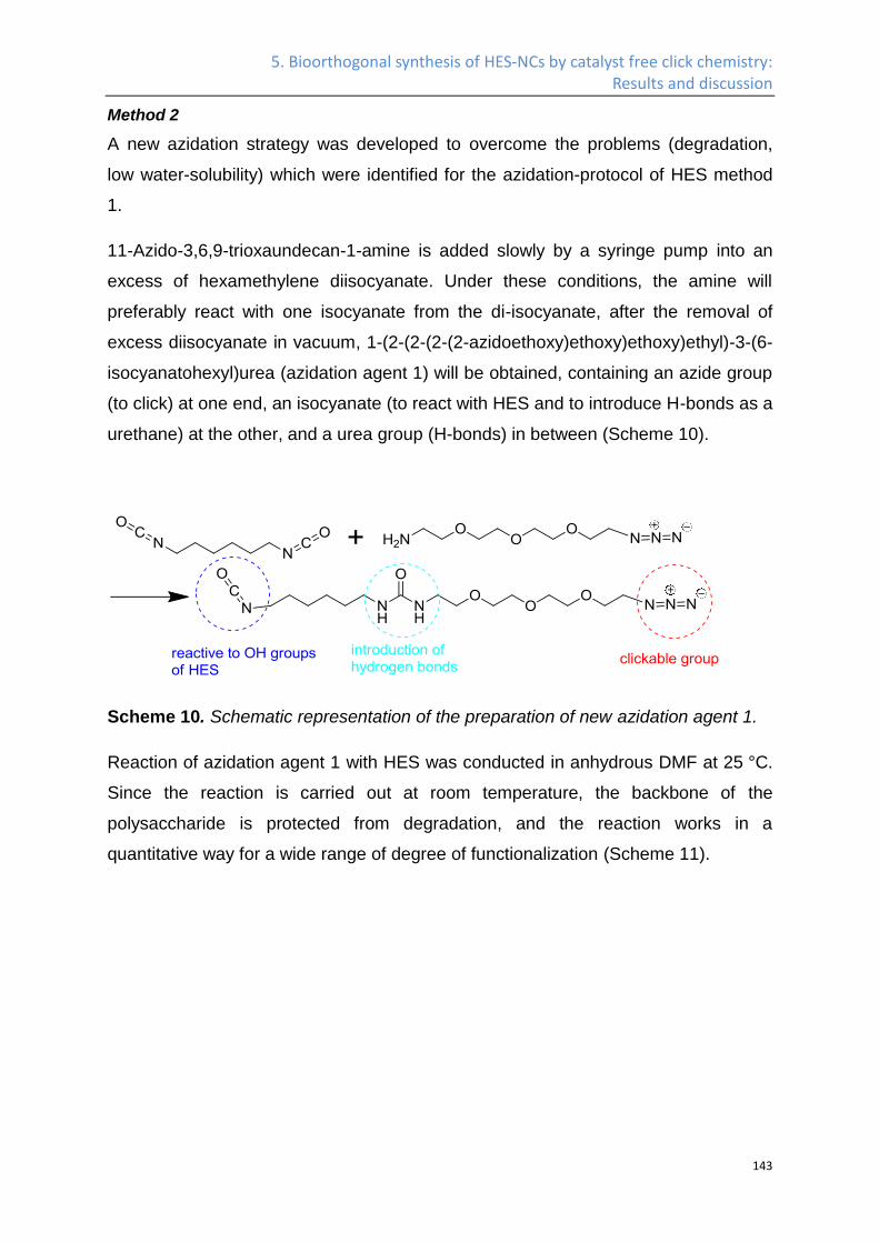
5. Bioorthogonal synthesis of HES-NCs by catalyst free click chemistry: Results and discussion
143
Method 2
A new azidation strategy was developed to overcome the problems (degradation,
low water-solubility) which were identified for the azidation-protocol of HES method
1.
11-Azido-3,6,9-trioxaundecan-1-amine is added slowly by a syringe pump into an
excess of hexamethylene diisocyanate. Under these conditions, the amine will
preferably react with one isocyanate from the di-isocyanate, after the removal of
excess diisocyanate in vacuum, 1-(2-(2-(2-(2-azidoethoxy)ethoxy)ethoxy)ethyl)-3-(6-
isocyanatohexyl)urea (azidation agent 1) will be obtained, containing an azide group
(to click) at one end, an isocyanate (to react with HES and to introduce H-bonds as a
urethane) at the other, and a urea group (H-bonds) in between (Scheme 10).
Scheme 10. Schematic representation of the preparation of new azidation agent 1.
Reaction of azidation agent 1 with HES was conducted in anhydrous DMF at 25 °C.
Since the reaction is carried out at room temperature, the backbone of the
polysaccharide is protected from degradation, and the reaction works in a
quantitative way for a wide range of degree of functionalization (Scheme 11).
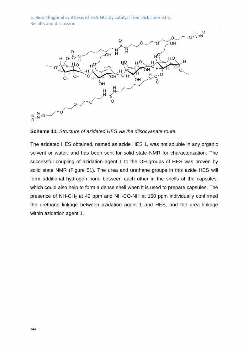
5. Bioorthogonal synthesis of HES-NCs by catalyst free click chemistry: Results and discussion
144
Scheme 11. Structure of azidated HES via the diisocyanate route.
The azidated HES obtained, named as azide HES 1, was not soluble in any organic
solvent or water, and has been sent for solid state NMR for characterization. The
successful coupling of azidation agent 1 to the OH-groups of HES was proven by
solid state NMR (Figure 51). The urea and urethane groups in this azide HES will
form additional hydrogen bond between each other in the shells of the capsules,
which could also help to form a dense shell when it is used to prepare capsules. The
presence of NH-CH2 at 42 ppm and NH-CO-NH at 160 ppm individually confirmed
the urethane linkage between azidation agent 1 and HES, and the urea linkage
within azidation agent 1.
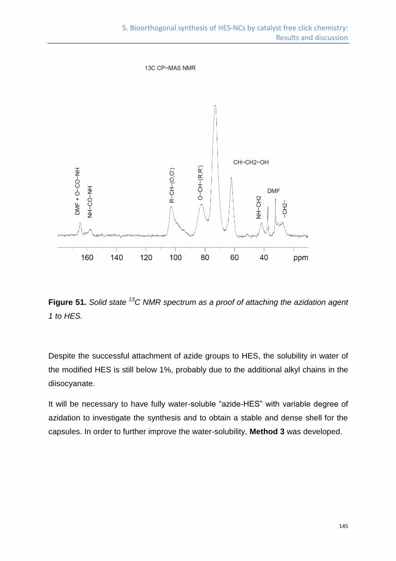
5. Bioorthogonal synthesis of HES-NCs by catalyst free click chemistry: Results and discussion
145
Figure 51. Solid state 13C NMR spectrum as a proof of attaching the azidation agent
1 to HES.
Despite the successful attachment of azide groups to HES, the solubility in water of
the modified HES is still below 1%, probably due to the additional alkyl chains in the
diisocyanate.
It will be necessary to have fully water-soluble “azide-HES” with variable degree of
azidation to investigate the synthesis and to obtain a stable and dense shell for the
capsules. In order to further improve the water-solubility, Method 3 was developed.
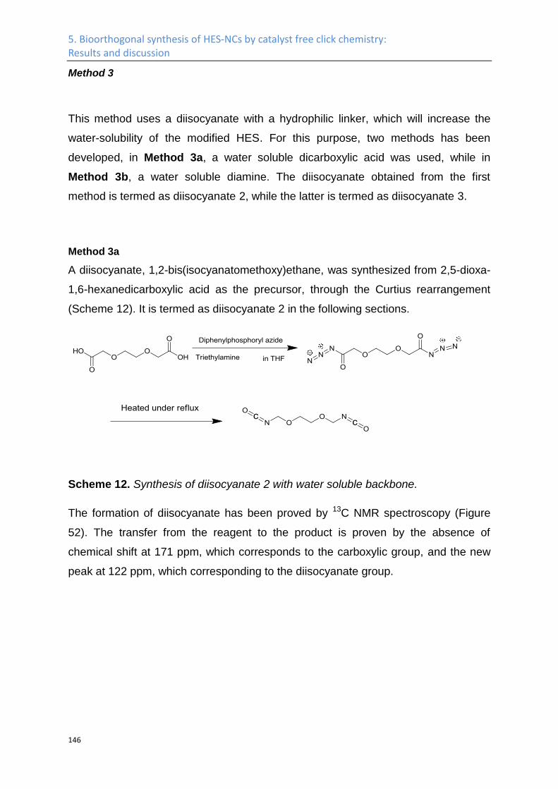
5. Bioorthogonal synthesis of HES-NCs by catalyst free click chemistry: Results and discussion
146
Method 3
This method uses a diisocyanate with a hydrophilic linker, which will increase the
water-solubility of the modified HES. For this purpose, two methods has been
developed, in Method 3a, a water soluble dicarboxylic acid was used, while in
Method 3b, a water soluble diamine. The diisocyanate obtained from the first
method is termed as diisocyanate 2, while the latter is termed as diisocyanate 3.
Method 3a
A diisocyanate, 1,2-bis(isocyanatomethoxy)ethane, was synthesized from 2,5-dioxa-
1,6-hexanedicarboxylic acid as the precursor, through the Curtius rearrangement
(Scheme 12). It is termed as diisocyanate 2 in the following sections.
Scheme 12. Synthesis of diisocyanate 2 with water soluble backbone.
The formation of diisocyanate has been proved by 13C NMR spectroscopy (Figure
52). The transfer from the reagent to the product is proven by the absence of
chemical shift at 171 ppm, which corresponds to the carboxylic group, and the new
peak at 122 ppm, which corresponding to the diisocyanate group.
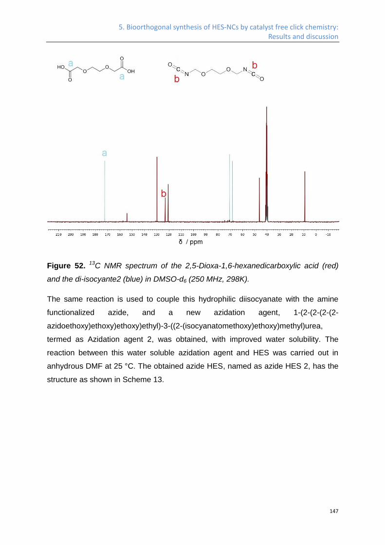
5. Bioorthogonal synthesis of HES-NCs by catalyst free click chemistry: Results and discussion
147
Figure 52. 13C NMR spectrum of the 2,5-Dioxa-1,6-hexanedicarboxylic acid (red)
and the di-isocyante2 (blue) in DMSO-d6 (250 MHz, 298K).
The same reaction is used to couple this hydrophilic diisocyanate with the amine
functionalized azide, and a new azidation agent, 1-(2-(2-(2-(2-
azidoethoxy)ethoxy)ethoxy)ethyl)-3-((2-(isocyanatomethoxy)ethoxy)methyl)urea,
termed as Azidation agent 2, was obtained, with improved water solubility. The
reaction between this water soluble azidation agent and HES was carried out in
anhydrous DMF at 25 °C. The obtained azide HES, named as azide HES 2, has the
structure as shown in Scheme 13.
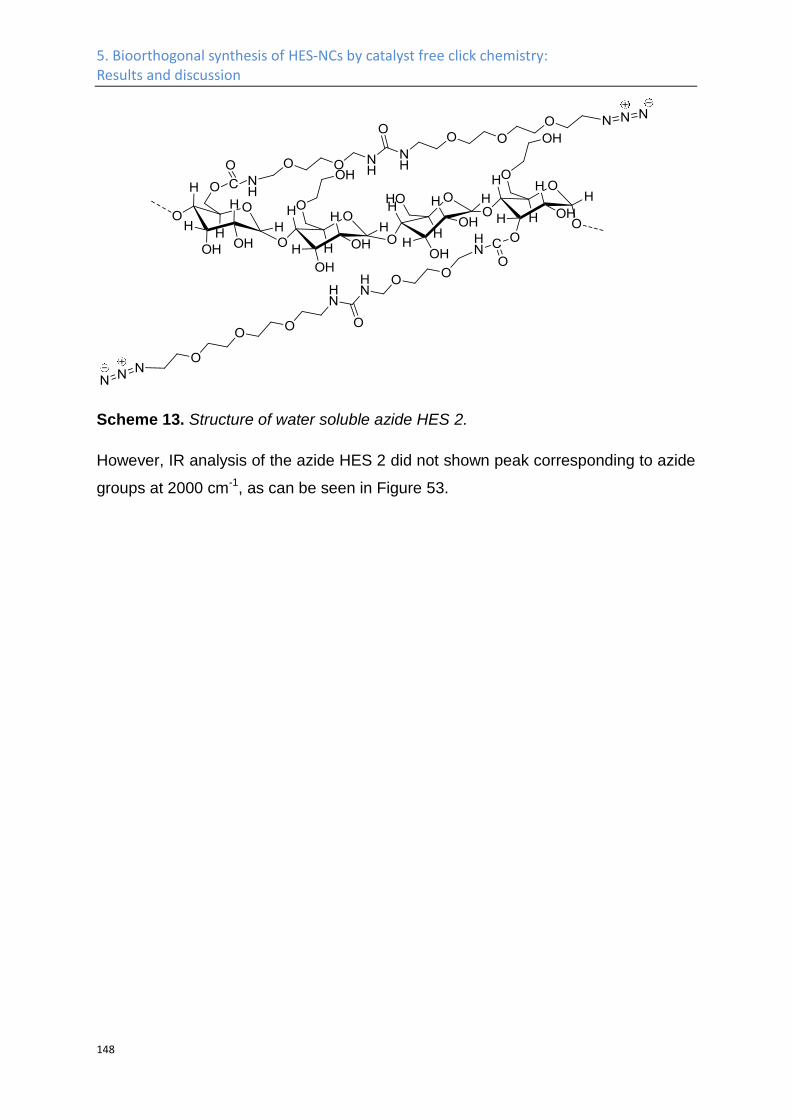
5. Bioorthogonal synthesis of HES-NCs by catalyst free click chemistry: Results and discussion
148
Scheme 13. Structure of water soluble azide HES 2.
However, IR analysis of the azide HES 2 did not shown peak corresponding to azide
groups at 2000 cm-1, as can be seen in Figure 53.
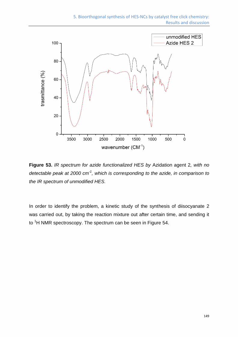
5. Bioorthogonal synthesis of HES-NCs by catalyst free click chemistry: Results and discussion
149
Figure 53. IR spectrum for azide functionalized HES by Azidation agent 2, with no
detectable peak at 2000 cm-1, which is corresponding to the azide, in comparison to
the IR spectrum of unmodified HES.
In order to identify the problem, a kinetic study of the synthesis of diisocyanate 2
was carried out, by taking the reaction mixture out after certain time, and sending it
to 1H NMR spectroscopy. The spectrum can be seen in Figure 54.
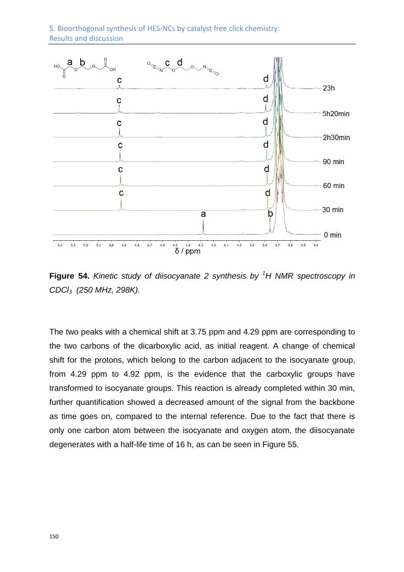
5. Bioorthogonal synthesis of HES-NCs by catalyst free click chemistry: Results and discussion
150
Figure 54. Kinetic study of diisocyanate 2 synthesis by 1H NMR spectroscopy in
CDCl3 (250 MHz, 298K).
The two peaks with a chemical shift at 3.75 ppm and 4.29 ppm are corresponding to
the two carbons of the dicarboxylic acid, as initial reagent. A change of chemical
shift for the protons, which belong to the carbon adjacent to the isocyanate group,
from 4.29 ppm to 4.92 ppm, is the evidence that the carboxylic groups have
transformed to isocyanate groups. This reaction is already completed within 30 min,
further quantification showed a decreased amount of the signal from the backbone
as time goes on, compared to the internal reference. Due to the fact that there is
only one carbon atom between the isocyanate and oxygen atom, the diisocyanate
degenerates with a half-life time of 16 h, as can be seen in Figure 55.
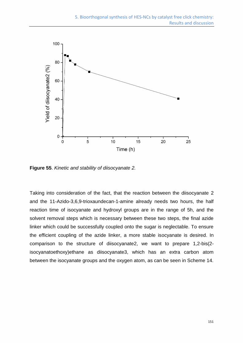
5. Bioorthogonal synthesis of HES-NCs by catalyst free click chemistry: Results and discussion
151
Figure 55. Kinetic and stability of diisocyanate 2.
Taking into consideration of the fact, that the reaction between the diisocyanate 2
and the 11-Azido-3,6,9-trioxaundecan-1-amine already needs two hours, the half
reaction time of isocyanate and hydroxyl groups are in the range of 5h, and the
solvent removal steps which is necessary between these two steps, the final azide
linker which could be successfully coupled onto the sugar is neglectable. To ensure
the efficient coupling of the azide linker, a more stable isocyanate is desired. In
comparison to the structure of diisocyanate2, we want to prepare 1,2-bis(2-
isocyanatoethoxy)ethane as diisocyanate3, which has an extra carbon atom
between the isocyanate groups and the oxygen atom, as can be seen in Scheme 14.
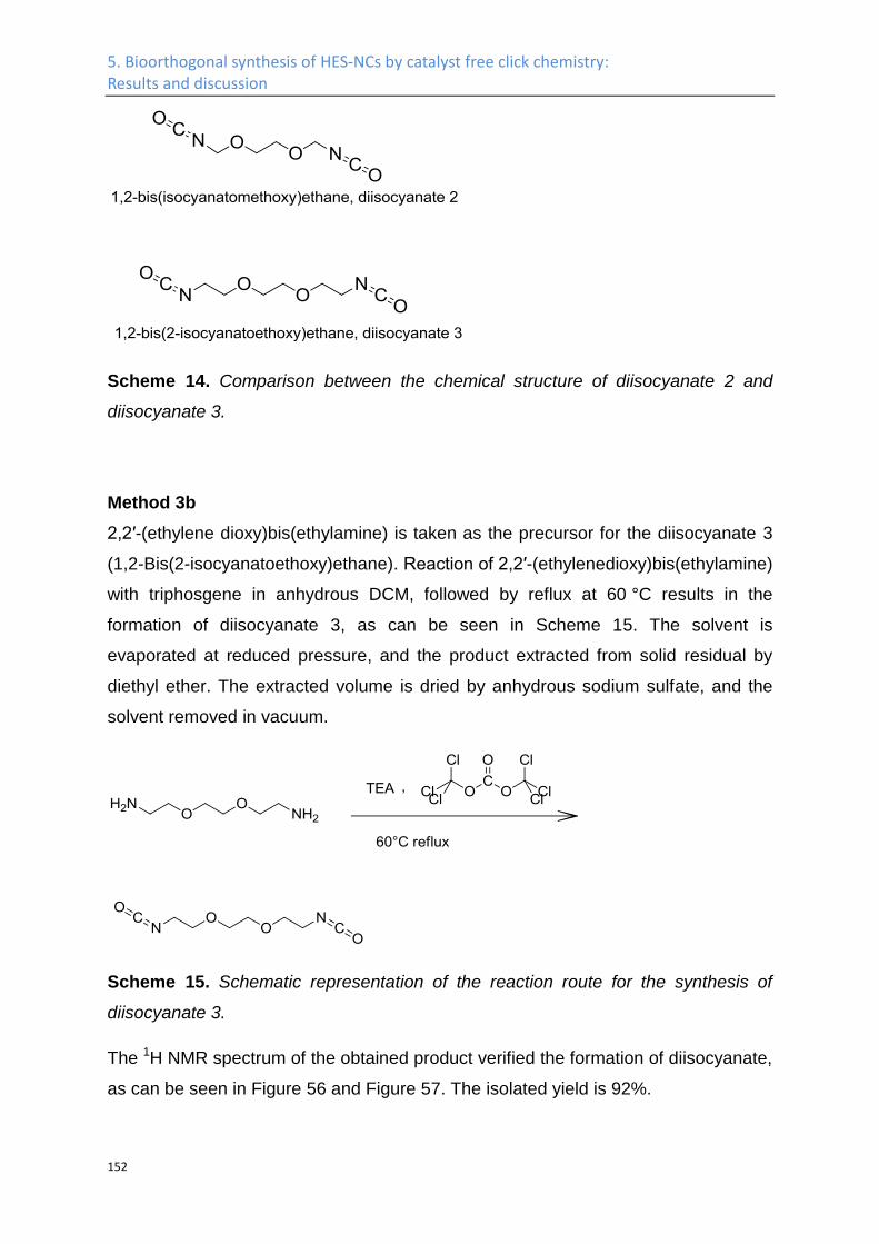
5. Bioorthogonal synthesis of HES-NCs by catalyst free click chemistry: Results and discussion
152
Scheme 14. Comparison between the chemical structure of diisocyanate 2 and
diisocyanate 3.
Method 3b
2,2′-(ethylene dioxy)bis(ethylamine) is taken as the precursor for the diisocyanate 3
(1,2-Bis(2-isocyanatoethoxy)ethane). Reaction of 2,2′-(ethylenedioxy)bis(ethylamine)
with triphosgene in anhydrous DCM, followed by reflux at 60 °C results in the
formation of diisocyanate 3, as can be seen in Scheme 15. The solvent is
evaporated at reduced pressure, and the product extracted from solid residual by
diethyl ether. The extracted volume is dried by anhydrous sodium sulfate, and the
solvent removed in vacuum.
Scheme 15. Schematic representation of the reaction route for the synthesis of
diisocyanate 3.
The 1H NMR spectrum of the obtained product verified the formation of diisocyanate,
as can be seen in Figure 56 and Figure 57. The isolated yield is 92%.
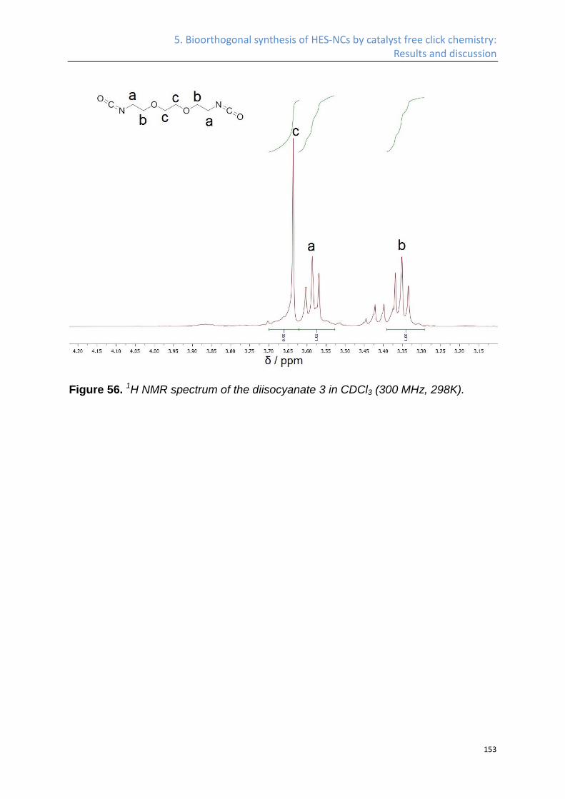
5. Bioorthogonal synthesis of HES-NCs by catalyst free click chemistry: Results and discussion
153
Figure 56. 1H NMR spectrum of the diisocyanate 3 in CDCl3 (300 MHz, 298K).
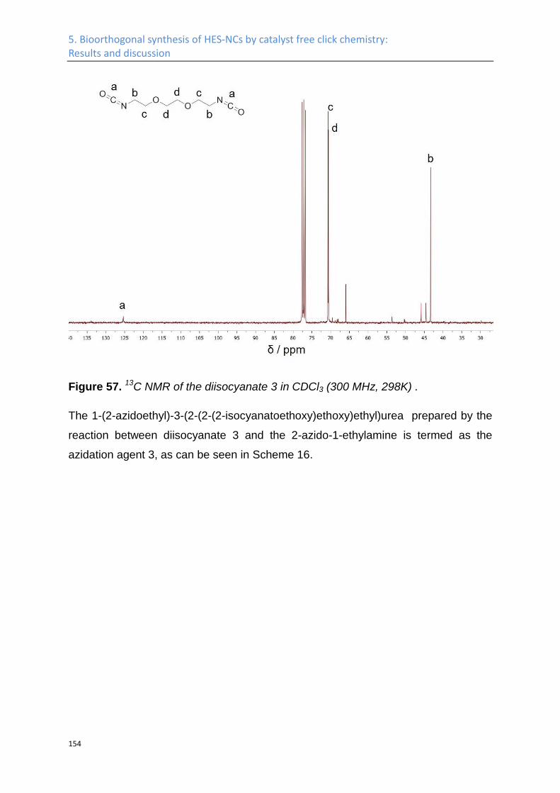
5. Bioorthogonal synthesis of HES-NCs by catalyst free click chemistry: Results and discussion
154
Figure 57. 13C NMR of the diisocyanate 3 in CDCl3 (300 MHz, 298K) .
The 1-(2-azidoethyl)-3-(2-(2-(2-isocyanatoethoxy)ethoxy)ethyl)urea prepared by the
reaction between diisocyanate 3 and the 2-azido-1-ethylamine is termed as the
azidation agent 3, as can be seen in Scheme 16.
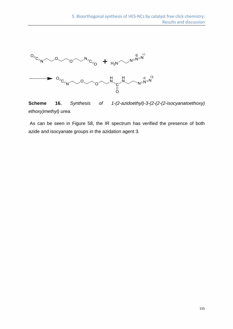
5. Bioorthogonal synthesis of HES-NCs by catalyst free click chemistry: Results and discussion
155
Scheme 16. Synthesis of 1-(2-azidoethyl)-3-(2-(2-(2-isocyanatoethoxy)
ethoxy)methyl) urea.
As can be seen in Figure 58, the IR spectrum has verified the presence of both
azide and isocyanate groups in the azidation agent 3.
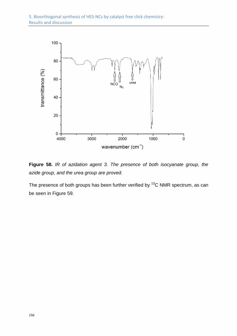
5. Bioorthogonal synthesis of HES-NCs by catalyst free click chemistry: Results and discussion
156
Figure 58. IR of azidation agent 3. The presence of both isocyanate group, the
azide group, and the urea group are proved.
The presence of both groups has been further verified by 13C NMR spectrum, as can
be seen in Figure 59.
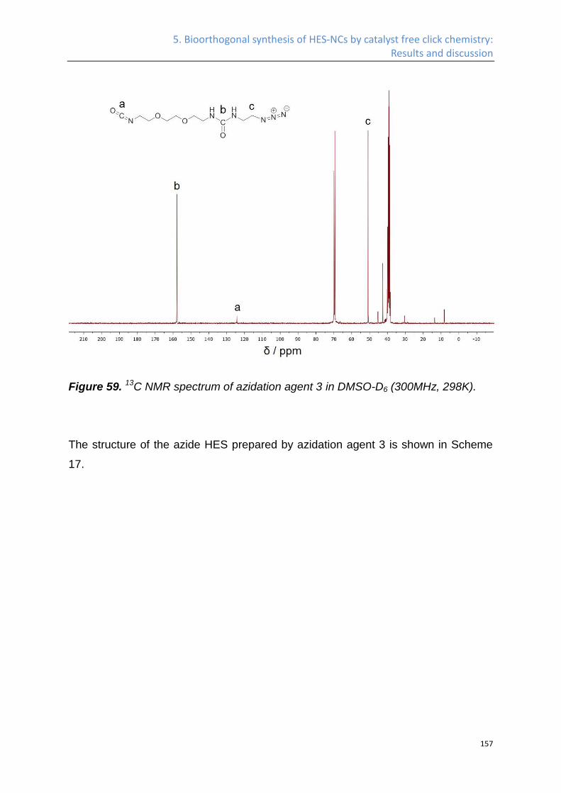
5. Bioorthogonal synthesis of HES-NCs by catalyst free click chemistry: Results and discussion
157
Figure 59. 13C NMR spectrum of azidation agent 3 in DMSO-D6 (300MHz, 298K).
The structure of the azide HES prepared by azidation agent 3 is shown in Scheme
17.
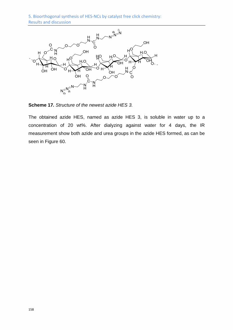
5. Bioorthogonal synthesis of HES-NCs by catalyst free click chemistry: Results and discussion
158
Scheme 17. Structure of the newest azide HES 3.
The obtained azide HES, named as azide HES 3, is soluble in water up to a
concentration of 20 wt%. After dialyzing against water for 4 days, the IR
measurement show both azide and urea groups in the azide HES formed, as can be
seen in Figure 60.
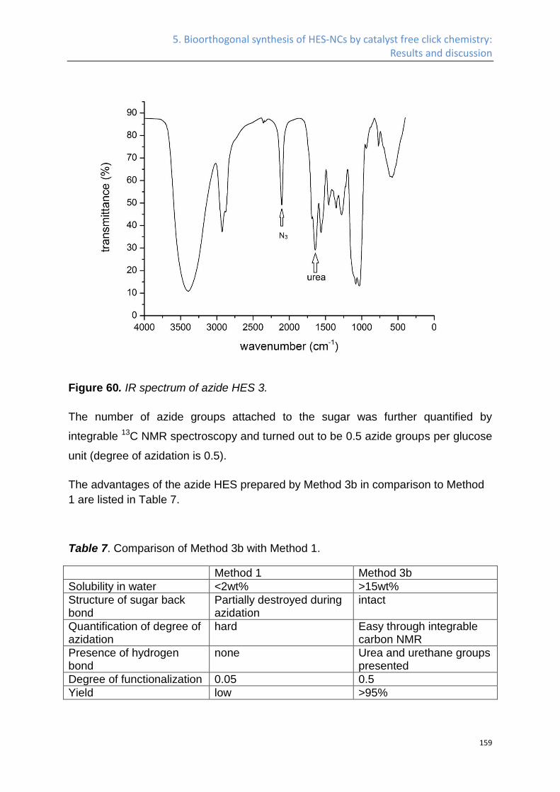
5. Bioorthogonal synthesis of HES-NCs by catalyst free click chemistry: Results and discussion
159
Figure 60. IR spectrum of azide HES 3.
The number of azide groups attached to the sugar was further quantified by
integrable 13C NMR spectroscopy and turned out to be 0.5 azide groups per glucose
unit (degree of azidation is 0.5).
The advantages of the azide HES prepared by Method 3b in comparison to Method
1 are listed in Table 7.
Table 7. Comparison of Method 3b with Method 1.
Method 1 Method 3b
Solubility in water <2wt% >15wt%
Structure of sugar back bond
Partially destroyed during azidation
intact
Quantification of degree of azidation
hard Easy through integrable carbon NMR
Presence of hydrogen bond
none Urea and urethane groups presented
Degree of functionalization 0.05 0.5
Yield low >95%
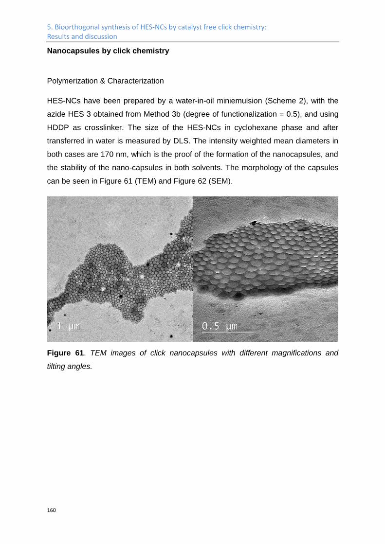
5. Bioorthogonal synthesis of HES-NCs by catalyst free click chemistry: Results and discussion
160
Nanocapsules by click chemistry
Polymerization & Characterization
HES-NCs have been prepared by a water-in-oil miniemulsion (Scheme 2), with the
azide HES 3 obtained from Method 3b (degree of functionalization = 0.5), and using
HDDP as crosslinker. The size of the HES-NCs in cyclohexane phase and after
transferred in water is measured by DLS. The intensity weighted mean diameters in
both cases are 170 nm, which is the proof of the formation of the nanocapsules, and
the stability of the nano-capsules in both solvents. The morphology of the capsules
can be seen in Figure 61 (TEM) and Figure 62 (SEM).
Figure 61. TEM images of click nanocapsules with different magnifications and
tilting angles.
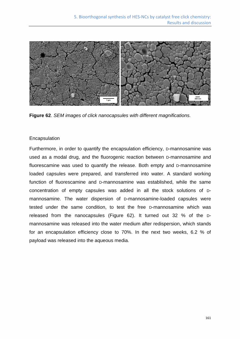
5. Bioorthogonal synthesis of HES-NCs by catalyst free click chemistry: Results and discussion
161
Figure 62. SEM images of click nanocapsules with different magnifications.
Encapsulation
Furthermore, in order to quantify the encapsulation efficiency, D-mannosamine was
used as a modal drug, and the fluorogenic reaction between D-mannosamine and
fluorescamine was used to quantify the release. Both empty and D-mannosamine
loaded capsules were prepared, and transferred into water. A standard working
function of fluorescamine and D-mannosamine was established, while the same
concentration of empty capsules was added in all the stock solutions of D-
mannosamine. The water dispersion of D-mannosamine-loaded capsules were
tested under the same condition, to test the free D-mannosamine which was
released from the nanocapsules (Figure 62). It turned out 32 % of the D-
mannosamine was released into the water medium after redispersion, which stands
for an encapsulation efficiency close to 70%. In the next two weeks, 6.2 % of
payload was released into the aqueous media.
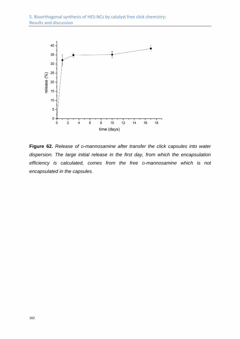
5. Bioorthogonal synthesis of HES-NCs by catalyst free click chemistry: Results and discussion
162
Figure 62. Release of D-mannosamine after transfer the click capsules into water
dispersion. The large initial release in the first day, from which the encapsulation
efficiency is calculated, comes from the free D-mannosamine which is not
encapsulated in the capsules.

5. Bioorthogonal synthesis of HES-NCs by catalyst free click chemistry: Results and discussion
163
Conclusion
These results demonstrate that HES can be further functionalized with azide groups,
while letting its sugar backbone and good water solubility intact. The azidation agent
1-(2-azidoethyl)-3-(2-(2-(2-isocyanatoethoxy)ethoxy)ethyl)urea developed in this
work was used to functionalize the HES at room temperature in the absence of any
catalyst. In addition, all the sugars, peptide, or any other molecule containing either
primary amine or hydroxyl groups can be functionalized with azide group by applying
this azidation agent. Due to the mild reaction condition of this method, it can be very
useful to functionalize polymers which are too unstable to be functionalized
otherwise, e.g. polyesters.
The azide-functionalized HES has been used to prepare nanocapsules via copper
free click reaction, in a water-in-oil miniemulsion system by using HDDP as
crosslinker. The capsule morphology has been proven by TEM, while the
encapsulation efficiency and release profile of a payload, D-mannosamine in this
case, has been studied. It has encapsulation efficiency close to 70%, and released
6.2% of the encapsulated payload in the next two weeks. Taking into consideration
that D-mannosamine is a rather small molecule in comparison to most of the
therapeutic agent, the click capsule is considered to be dense enough to use as the
carrier for drugs.
5. Bioorthogonal synthesis of HES-NCs by catalyst free click chemistry: Experimental Part
164
5.2 Experimental Part
Materials
O-(2-Aminoethyl)-O'-(2-azidoethyl)pentaethylene glycol and 2,2'-
(Ethylenedioxy)bis(ethylamine) was bought from Sigma Aldrich, and storage over
molecular sieves for 4 days before use. 2,2'-(ethylenebis(oxy))bisacetic acid was
ordered from Merck. All the other chemicals were bought from Sigma Aldrich without
further purifications.
Method 1 (by tosylation, azidation)
1.36 g of starch was dissolved in 60 mL of DMA, followed by the addition of 3.14 g of
TEA in 10 mL of DMA, the solution was then cooled to 0 °C and 15.0 g of
tosylchloride was added into the solution dropwise. The reaction was allowed to
proceed for 48 h, afterwards, the product was precipitated into cold water, filtrated,
and redissolved in DMA. The procedure was repeated for three times, and 2.2 g of
tosylated starch was dissolved in 15 mL DMF, 2.5 g of sodium azide was added, and
the reaction was magnetically stirred at 80 °C for 24 h. The product was dialyzed k
against MilliQ water for 3 days (MWCO 14 kDa). The product was freeze dried to
obtain 1,17 g of product.
Method 2 (hydrophobic diisocyanate)
1.44 g (4 times excesses) of hexamethylene diisocyanate was dissolved in 5 mL of
anhydrous DCM, while 520 mg of O-(2-Aminoethyl)-O'-(2-azidoethyl)pentaethylene
glycol in 4 mL of anhydrous DCM was added at a speed of 2 ml/h by a syringe
pump. The solvent and the excess of hexamethylene diisocyanate and the solvent
was removed by vaccum, and the obtained azidation agent is redissolved in 2 mL
anhydrous DMF, which was subsequently added dropwise into the solution of 1.36 g
of HES in 10 mL anhydrous DMF. The reaction was allowed to proceed for 18 h, and
the product was purified by dialysis. The solution and the precipitate form during
dialysis is freeze dried together to get 1.5 g of product.
5. Bioorthogonal synthesis of HES-NCs by catalyst free click chemistry: Experimental Part
165
Method 3a
Synthesis of 1,2-bis(isocyanatomethoxy)ethane (diisocyanate 2)
820 mg of 2,2'-(ethylenebis(oxy))bisacetic acid was dissolved in 12 mL anhydrous
THF. The solution was cooled to 0 °C, followed the dropwise addition of 1.01 g of
TEA in 2 mL of anhydrous THF. In the end, 2.95 g of diphenylphosphoryl azide in 3
mL of anhydrous THF was added dropwise. The system was magnetically stirred at
0 °C for 2 h, at room temperature for 1 h, and refluxed at 66 °C for 1 h, the product
was analyzed by 1H NMR and 13C NMR spectroscopy, and used without further
purification in the next step.
Synthesis of the 1-(2-(2-(2-(2-azidoethoxy)ethoxy)ethoxy)ethyl)-3-((2-
(isocyanatomethoxy)ethoxy)methyl)urea (azidation agent 2) and the azide HES 2
The THF solution of 1,2-bis(isocyanatomethoxy)ethane obtained from last step was
cooled to -56 °C, by immersing the reaction container in the mixture of n-octane and
dry ice. 420 mg of O-(2-Aminoethyl)-O'-(2-azidoethyl)pentaethylene glycol was
dissolved in 2 mL of anhydrous THF, and added at a speed of 2 mL/h by a syringe
pump into the diisocyanate 1 solution. After the addition, the precipitate at the
bottom of the bottle was isolated, redissolved into anhydrous DMF, subsequently
added dropwise into the solution of 500 mg of azide HES in 6 mL of anhydrous
DMF. The reaction was allowed to proceed for 48 h, and the product was purified by
dialysis against MilliQ water in a dialyzing tube with MWCO of 14kDa, while the
solution after dialyzing was freeze dried to obtain 520 mg of Azide HES 2 (IR in
Figure 53).
Method 3b
Synthesis of 2-azido-1-ethylamine
As a shorter substituent for O-(2-Aminoethyl)-O'-(2-azidoethyl)pentaethylene glycol,
2-Azido-1-ethylamine was prepared, by reaction of 2-Chloroethylamine
hydrochloride (3g) together with sodium azide (5.12) g in MilliQ water at 80 °C for 18
h. Afterwards 1.05 g of sodium hydroxide was added, and the product is extracted
by diethyl ether, dried over sodium sulfide, and solvent partially removed by rotary
evaporation. The diethyl ether solution of 2-Azido-1-ethylamine with a concentration
of 36.5% (quantified by 1H NMR spectroscopy), containing 1.8 g (yield 80%) of 2-
5. Bioorthogonal synthesis of HES-NCs by catalyst free click chemistry: Experimental Part
166
azido-1-ethylamine was obtained. 1H-NMR Spectroscopy (CDCl3, 300 MHz) δ(ppm)
= 3.35-3.26 (2H, t,J= 5.6 Hz, CH2), 2.88-2.75 (2H, t,J= 5.6 Hz, CH2). FT-IR (ATR)ν =
3366 (br), 2104 (s).
Synthesis of 1,2-Bis(2-isocyanatoethoxy)ethane (diisocyanate 3)
4.5 g of 2,2'-(Ethylenedioxy)bis(ethylamine) was dissolved together with 5.2 g of
TEA in 8 mL of anhydrous DCM, and added dropwise into the solution of 3 g of
triphosgene in 30 mL of anhydrous DCM, while the latter is cooled to 4 °C. After
finishing the addition, the system was kept at 4 °C for 20 min, then at room
temperature for 1h, followed by refluxing at 50 °C for 2 h. The solvent is then
removed in vaccuo, and the product extracted by anhydrous diethyl ether. The
product was checked by NMR and used without further purification in the next step.
1H-NMR Spectroscopy (CDCl3, 300 MHz) δ(ppm) = 3.65 (s, 4 H);3.60 (t, 4 H); 3.36
(t, 4 H).13C NMR (CDCl3, 300 MHz), δ (ppm) 69.7,69.6, 42.5.
Synthesis of 1-(2-azidoethyl)-3-(2-(2-(2-isocyanatoethoxy)ethoxy)ethyl)urea
(azidation agent 3)
1500 mg of 2-azido-1-ethylamine was dissolved in 2 mL of anhydrous diethyl ether,
and added at a speed of 2 mL/h by a syringe pump into the diisocyanate 3 solution.
After the addition, the solvent was removed at reduced pressure and the product
was checked by IR, redissolved in anhydrous DMF without further purification,
which was subsequently added dropwise into the solution of 500 mg of HES (with a
theoretical degree of substitution of 3) in 6 mL of anhydrous DMF. The reaction was
allowed to proceed for 48 h, and the product was purified by dialysis against MilliQ
water in a dialyzing tube with MWCO of 14kDa, while the solution after dialyzing was
freeze dried to obtain 520 mg of product (azide HES 3). FT-IR (ATR) ν= 2000 (N3),
1700 (urea). 13C NMR (DMSO-D6, 300 MHz), δ (ppm) = 50.46 (CH2-N3), 60.96 (CH2-
OH), 158.34 (urea).
Synthesis of HES nanocontainers
5. Bioorthogonal synthesis of HES-NCs by catalyst free click chemistry: Experimental Part
167
50 mg of azide HES 3 was dissolved in 460 µL of MilliQ water as the aqueous
phase, while 70 mg of P(E/B)-b-PEO was dissolved in the mixture of 5g cyclohexane
and 2 g of toluene. The water phase was shaken gently at 40 °C for 3 h, to ensure
the complete solvation of azide HES, after which it was mixed with oil phase, stirred
magnetically at 200 rpm for 30 min, followed by ultrasonication at 70% amplitude for
3 min, with a pulse of 20 s on and 10 s off at 0 °C, using a Branson Sonifier W-450-
Digital and a ½" tip. 80 mg of HDDP was dissolved together with 20 mg of P(E/B)-b-
PEO in 800 µL of toluene and added into the system after sonication. The whole
system was magnetically stirred at 500 rpm for 5 days, while the temperature was
kept at 50 °C.

Summary and Outlook
168
Summary and Outlook
In this work, hydroxyethyl starch (HES) based nanocapsules were prepared from
different chemical reactions, surface functionalized by different methods in order to
elongate its plasma halftime (improve its stealth property), and enable specific
targeting effect. In addition, the interaction between the plasma protein with the
unmodified HES-NCs, PEGlyated HES-NCs and mannose coupled HES-NCs was
studied and compared by different methods in detail.
In chapter 3, nanocapsules based on polysaccharides were prepared via the
reaction between HES and toluene diisocyanate (TDI). The surface of the
nanocarriers was functionalized with poly(ethylene glycol) (PEG) via different
methods in order to ensure a long circulation time in the blood plasma. The library of
different PEGylated nanocapsules was investigated in vitro with respect to
biocompatibility, protein interaction, cell uptake, and in vivo plasma half-life time.
Highly PEGylated nanocapsules did show a reduced protein adsorption and a very
low unspecific cell uptake in several blood cells. As a result, these nanocontainers
show a pronounced plasma circulation time in blood which is higher than typically
applied liposomal formulations. This versatile chemical platform in combination with
the outstandingly long circulation times makes the PEGylated HES nanocontainers
ideal for further studies for targeted cell uptake and subsequent drug release.
In chapter 4 it was proven that the PEGylated HES nanocarriers can be efficiently
functionalized with mannose derivatives. Different chemistries have been
investigated and a convenient two step protocol allowed a high PEG density on the
nanocarriers and a high degree of “on top” functionalization with mannose. The
influence of mannose on the PEGylated NCs on the pattern of the protein corona
after plasma incubation was proven to be minimal by ITC, DLS, SDS-PAGE, and
mass spectrometry. Furthermore, cellular uptake of the NCs by dendritic cells and a
binding assay with c-type lectin proved that the targeting moieties were active and
accessible to the biological receptors after incubation with human plasma. Taking
into consideration that rather simple carbohydrates like the disaccharide in
bleomycin,226 have been proven to be able to selectively target cancer cells, the use
of a combination of stealth nanocarriers (i.e. PEGylated materials) with such
Summary and Outlook
169
saccharide-based targeting moieties could solve the important dilemma, where the
stealth effect is lost after coupling of other targeting agents.
Furthermore, in chapter 5, in order to minimize the immobilization of the
encapsulated therapeutical agent into the shell of the nanocapsule through the
reaction between the amine, thiol, hydroxyl groups of the potential payload with
toluene diisocyanate, hydroxyethyl starch nanocapsules were also prepared by
copper free click reaction in the chapter 5. An azidation agent was developed, which
can covalently attach azide groups to any polysaccharide at room temperature, with
tunable degree of functionalization, and full range water solubility. Azide
hydroxyethyl starch prepared by this azidation agent was used to prepare
nanocapsules by copper free click reaction.
As nanocapsules with long plasma circulation time, specific cell targeting effect and
formed by bio-orthogonal chemistry were prepared in this work, the efficiency of
these nanocapsules as targeted delivery systems for different (potential) therapeutic
agents could be tested both in vitro and in vivo. In addition, further efforts could be
done, to substitute the poly(ethylene glycol) used in this work with polysaccharide, in
order to prepare completely sugar based nano-carriers, which is fully biodegradable.
Based on the work in this thesis and many other studies which are published in
recent years, when mono/oligo saccharides are used to surface functionalize nano-
carriers, they are proven to have following advantages:
(i) specific targeting to different cell types;37,39-41,128,131
(ii) diagnostics and differentiation of healthy and malignant population of the same
cell type;18,128
(iii) protecting the payload131 and prolonged in vivo plasma half-life time (due to
reduced protein adsorption);88
(iv) controlled release of a payload by specific protein interaction;38
(v) competing with and inhibiting the binding of other saccharide containing bacteria,
virus, or pathogen with the corresponding cells.36
Summary and Outlook
170
For nanocarriers that are surface modified by polysaccharides, similar properties are
obtained, including:
(i) protecting the payload134 and increasing the plasma half-life times;23,77,138
(ii) specific targeting;133,137,138,183
(iii) enzyme-induced released or activation of a therapeutic agent;23,25,26,77,78
(v) decreased toxicity of the payload;132,135,137,183
(vi) kinetics of metabolization are tunable by chemical functionalization to balance
biocompatibility and stability.23,77
As carbohydrates are omnipresent as functional surface coatings in nature, their use
in biomedical applications is obvious. They have been used to construct
nanocarriers, which enable the encapsulation and protection156,158 of different
(mainly) water-soluble guests,86,149-151,159-163 while keeping the biological properties
such as specific targeting,134,148,159-163,244 suppressed unspecific cell uptake,86 and
enzyme-triggered release.150,157,159-162 In addition, the inherent high chemical
functionality of different polysaccharides (mainly: hydroxyls, carboxylic acids,
amines) allows straightforward crosslinking or on top functionalization of these
molecules which is often a major factor of their application compared to synthetic
macromolecules. However, it always must be considered, that the molecular weight
distributions of these biopolymers are typically rather broad, making fractionation
necessary in some cases. Nevertheless, due to the biodegradability of
polysaccharides, their non-uniform molecular weight might not be too problematic for
nanoparticular drug delivery devices, however, the toxicity after any chemical
modification and also that of the degradation products after chemical modification
need to be considered.
A major feature of all nanocarriers, either modified or constructed of carbohydrates
is their low protein interaction: the high level of hydrophilicity induces for many of
them a “stealth” behavior, and the unspecific cell uptake due to opsonization is low.
Furthermore, targeting is achieved by carbohydrates in multiple cases. This behavior
plus the inherent biodegradability renders carbohydrate-based nanocarriers as a
high potential platform for developing the “magic bullet” that was coined by Paul
Summary and Outlook
171
Ehrlich more than 100 years ago and makes research in this direction promising for
many scientists. It is certain that several new developments in treating diseases or
enabling sophisticated diagnostics will rely on carbohydrates in future.
Abbreviations
172
Abbreviations ASGP-R Asialoglycoprotein receptors
BSA Bovine serum albumin
cLSM Confocal laser scanning microscopy
CuAAC Copper-catalyzed azide-alkyne cycloaddition
DBCO Dibenzocyclooctyl
DCC Dicyclohexyl carbodiimide
DCM Dichloromethane
DCs Dendritic cells
DC-SIGN Dendritic cell-specific intercellular adhesion molecule-3-grabbing
non-integrin
Dec205 MOUSE ANTI HUMAN CD205
DLS Dynamic light scattering
DMA N,N-Dimethylformamide dimethyl acetal
DMF Dimethylformamid
DMSO Dimethyl sulfoxide
DNA Deoxyribonucleic acid
DPPA Diphenylphosphoryl azide
DSC Differential scanning calorimetry
EDTA Ethylenediaminetetraacetic acid
EPR Enhanced permeability and retention
Et3N Triethylamine
Abbreviations
173
FA Fluorescamine
FACS Fluorescence activated cell sorting
FTIR Fourier transform infrared spectroscopy
GNP Gold nanoparticle
GPC Gel permeation chromatography
HA Hyaluronic acid
HES Hydroxyethyl starch
HES-NCs Hydroxyethyl starch nanocapsules
HDDP Hexanediol dipropiolate
HPLC High-performance liquid chromatography
iDC Immature dendritic cell
ITC Isothermal Titration Calorimetry
KBr Potassium bromide
kDa Kilodaltons
KLE poly((ethylene-co-butylene)-b-(ethylene oxide))
MC1R Melanocortin 1 receptor
MCF-7 Michigan Cancer Foundation - 7
mDC Mature dendritic cell
Mn Number average molecular weight
MPS Mononuclear phagocyte system
MWCO Molecular weight cutoff
Mw Weight average molecular weight
n. a. not available
Abbreviations
174
n. d. not determined
NaCl Sodium chloride
NHS N-hydroxy succinimide
NMR Nuclear magnetic resonance
nm nanometer
NP Nanoparticle
PBMC Peripheral blood mononuclear cells
PBS Phosphate-buffered saline
PDI Polydispersity index
PEG Poly(ethylene glycol)
ppm Parts per million
PS Polystyrene
SEM Scanning electron microscopy
SD Standard deviation
SDS Sodium dodecyl sulfate
SDS-PAGE Sodium dodecyl sulfate polyacrylamide gel electrophoresis
siRNA Small interfering RNA
Sulfo-SMCC Sulfosuccinimidyl 4-(N-maleimidomethyl)cyclohexane-1-carboxylate
SR101 Sulforhodamine 101
TCEP Tris(2-carboxyethyl)phosphine
TDI Toluene Diisocyanate
TEA triethylamine
TEM Transition electron microscopy
Abbreviations
175
TGA Thermogravimetric analysis
THF Tetrahydrofuran
Erklärung
176
Erklärung
Hiermit versichern ich gemäß § 10 Abs. 3d der Promotionsordung vom 24.07.2007,
dass ich die als Dissertation vorgelegte Arbeit selbst angefertigt und alle benutzen
Hilfsmittel (Literatur, Apparaturen, Material) in der Arbeit angegeben habe.
Mainz, den 19. Oktober 2015
Biao Kang

Curriculum Vitae
177
Curriculum Vitae
Biao Kang
Date of Birth: 10 September 1986
Place of Birth: Shanxi, China
Education and experiences:
12. 2011- 09. 2015 Ph.D candidate, Polymer chemistry, in the group of Prof. Dr.
Katharina Landfester, Max-Planck-Institut für Polymerforschung, Mainz,
Germany.
10. 2009- 11. 2011 Master, Advanced Materials, Ulm University, Ulm, Germany.
09. 2005- 06. 2009 Double Bachelor, Chemical Engineering and Business
Administration, Shandong University, Jinan, China
Publications during Ph.D.:
Kang B, Till Opatz, Landfester K, Wurm F R. Sugars in biomedical applications:
surface functionalization and construction of nanocarriers. Chem Soc Rev, DOI:
10.1039/C5CS00092K
Kang B, Okwieka P, Schottler S, Winzen S, Langhanki J, Mohr K, et al.
Carbohydrate-Based Nanocarriers Exhibiting Specific Cell Targeting with Minimum
Influence from the Protein Corona. Angew Chem Int Ed Engl. 2015;54:7436-40.
Kang B, Okwieka P, Schottler S, Seifert O, Kontermann RE, Pfizenmaier K, et al.
Tailoring the stealth properties of biocompatible polysaccharide nanocontainers.
Biomaterials. 2015;49:125-34.
Weilbacher M, Prestel A, Kaps L, Bamberger D, Kang B, Tomcin S, et al.
Nanoparticular targeting of tolerance inducing macrophages, DC and myeloid
suppressor cells - new strategies for therapeutic approaches in immune-mediated
diseases. Experimental Dermatology. 2014;23:E27-E.
Kang B, Landfester K, Wurm F R. Study of the influence of hydrogen bond on the
permeability of nanobarriers in a Hydroxyethyl starch nanocapsules prepared by click
chemistry, to be submitted
At least four other papers are in preparation, with me as one of the coauthers.
References
178
References
(1) Strebhardt, K.; Ullrich, A. Nature Reviews Cancer 2008, 8, 473. (2) Liu, Z.; Cai, W.; He, L.; Nakayama, N.; Chen, K.; Sun, X.; Chen, X.; Dai, H. Nature Nanotechnology 2007, 2, 47. (3) Nasongkla, N.; Bey, E.; Ren, J.; Ai, H.; Khemtong, C.; Guthi, J. S.; Chin, S.-F.; Sherry, A. D.; Boothman, D. A.; Gao, J. Nano Lett. 2006, 6, 2427. (4) Cheng, J.; Teply, B. A.; Sherifi, I.; Sung, J.; Luther, G.; Gu, F. X.; Levy-Nissenbaum, E.; Radovic-Moreno, A. F.; Langer, R.; Farokhzad, O. C. Biomaterials 2007, 28, 869. (5) Wu, W.; Wieckowski, S.; Pastorin, G.; Benincasa, M.; Klumpp, C.; Briand, J. P.; Gennaro, R.; Prato, M.; Bianco, A. Angewandte Chemie-International Edition 2005, 44, 6358. (6) Qian, X.; Peng, X.-H.; Ansari, D. O.; Yin-Goen, Q.; Chen, G. Z.; Shin, D. M.; Yang, L.; Young, A. N.; Wang, M. D.; Nie, S. Nat. Biotechnol. 2008, 26, 83. (7) Gao, X. H.; Cui, Y. Y.; Levenson, R. M.; Chung, L. W. K.; Nie, S. M. Nat. Biotechnol. 2004, 22, 969. (8) Liong, M.; Lu, J.; Kovochich, M.; Xia, T.; Ruehm, S. G.; Nel, A. E.; Tamanoi, F.; Zink, J. I. Acs Nano 2008, 2, 889. (9) Tkachenko, A. G.; Xie, H.; Coleman, D.; Glomm, W.; Ryan, J.; Anderson, M. F.; Franzen, S.; Feldheim, D. L. J. Am. Chem. Soc. 2003, 125, 4700. (10) Dai, Q.; Walkey, C.; Chan, W. C. W. Angewandte Chemie-International Edition 2014, 53, 5093. (11) Laine, R. A. In Glycosciences; Wiley-VCH Verlag GmbH: 2008, p 1. (12) Danguy, A.; Camby, I.; Kiss, R. Biochim. Biophys. Acta 2002, 1572, 285. (13) Crowe, J. H.; Crowe, L. M.; Chapman, D. Science 1984, 223, 701. (14) Kwong, P. D.; Wyatt, R.; Robinson, J.; Sweet, R. W.; Sodroski, J.; Hendrickson, W. A. Nature 1998, 393, 648. (15) Xie, Y.-L.; Wang, M.-J.; Yao, S.-J. Langmuir 2009, 25, 8999. (16) Sharon, N.; Lis, H. Sci. Am. 1993, 268, 82. (17) Ye, S. Q.; Wang, C. Y.; Liu, X. X.; Tong, Z. J Biomat Sci-Polym E 2005, 16, 909. (18) El-Boubbou, K.; Zhu, D. C.; Vasileiou, C.; Borhan, B.; Prosperi, D.; Li, W.; Huang, X. J. Am. Chem. Soc. 2010, 132, 4490.
References
179
(19) Knop, K.; Hoogenboom, R.; Fischer, D.; Schubert, U. S. Angewandte Chemie-International Edition 2010, 49, 6288. (20) Besheer, A.; Vogel, J.; Glanz, D.; Kressler, J.; Groth, T.; Maeder, K. Mol. Pharm. 2009, 6, 407. (21) Lemarchand, C.; Gref, R.; Couvreur, P. Eur. J. Pharm. Biopharm. 2004, 58, 327. (22) Orlando, M. Ph.D. Thesis, University of Giessen 2003. (23) Noga, M.; Edinger, D.; Klaeger, R.; Wegner, S. V.; Spatz, J. P.; Wagner, E.; Winter, G.; Besheer, A. Biomaterials 2013, 34, 2530. (24) Noga, M.; Edinger, D.; Roedl, W.; Wagner, E.; Winter, G.; Besheer, A. J. Controlled Release 2012, 159, 92. (25) Ferguson, E. L.; Duncan, R. Biomacromolecules 2009, 10, 1358. (26) Duncan, R.; Gilbert, H. R. P.; Carbajo, R. J.; Vicent, M. J. Biomacromolecules 2008, 9, 1146. (27) Hardwicke, J.; Ferguson, E. L.; Moseley, R.; Stephens, P.; Thomas, D. W.; Duncan, R. Journal of Controlled Release 2008, 130, 275. (28) Marchant, R. E.; Yuan, S.; Szakalasgratzl, G. Journal of Biomaterials Science-Polymer Edition 1994, 6, 549. (29) Osterberg, E.; Bergstrom, K.; Holmberg, K.; Schuman, T. P.; Riggs, J. A.; Burns, N. L.; Vanalstine, J. M.; Harris, J. M. Journal of Biomedical Materials Research 1995, 29, 741. (30) Kang, B.; Opatz, T.; Landfester, K.; Wurm, F. R. Chem. Soc. Rev. 2015. (31) Brewer, C. F.; Miceli, M. C.; Baum, L. G. Curr. Opin. Struct. Biol. 2002, 12, 616. (32) Gabius, H. J. Biochem. Soc. Trans. 2008, 36, 1491. (33) Gabius, H. J.; Andre, S.; Kaltner, H.; Siebert, H. C. Biochim. Biophys. Acta 2002, 1572, 165. (34) Yamazaki, N.; Kojima, S.; Bovin, N. V.; Andre, S.; Gabius, S.; Gabius, H. J. Adv Drug Deliv Rev 2000, 43, 225. (35) Geijtenbeek, T. B. H.; Torensma, R.; van Vliet, S. J.; van Duijnhoven, G. C. F.; Adema, G. J.; van Kooyk, Y.; Figdor, C. G. Cell 2000, 100, 575. (36) Martinez-Avila, O.; Hijazi, K.; Marradi, M.; Clavel, C.; Campion, C.; Kelly, C.; Penades, S. Chemistry 2009, 15, 9874. (37) Chavez-Santoscoy, A. V.; Roychoudhury, R.; Pohl, N. L. B.; Wannemuehler, M. J.; Narasimhan, B.; Ramer-Tait, A. E. Biomaterials 2012, 33, 4762. (38) Wu, S.; Huang, X.; Du, X. Angew. Chem. Int. Ed. Engl. 2013, 52, 5580. (39) Yu, G.; Ma, Y.; Han, C.; Yao, Y.; Tang, G.; Mao, Z.; Gao, C.; Huang, F. J. Am. Chem. Soc. 2013, 135, 10310.
References
180
(40) Chen, W.; Zou, Y.; Meng, F.; Cheng, R.; Deng, C.; Feijen, J.; Zhong, Z. Biomacromolecules 2014, 15, 900. (41) Lartigue, L.; Oumzil, K.; Guari, Y.; Larionova, J.; Guerin, C.; Montero, J. L.; Barragan-Montero, V.; Sangregorio, C.; Caneschi, A.; Innocenti, C.; Kalaivani, T.; Arosio, P.; Lascialfari, A. Org. Lett. 2009, 11, 2992. (42) Zimmer, A. K.; Chetoni, P.; Saettone, M. F.; Zerbe, H.; Kreuter, J. J. Controlled Release 1995, 33, 31. (43) Durrani, A. M.; Farr, S. J.; Kellaway, I. W. Int. J. Pharm. 1995, 118, 243. (44) Lehr, C. M.; Bouwstra, J. A.; Schacht, E. H.; Junginger, H. E. Int. J. Pharm. 1992, 78, 43. (45) Letourneur, D.; Parisel, C.; Prigent-Richard, S.; Cansell, M. J Control Release 2000, 65, 83. (46) Cansell, M.; Parisel, C.; Jozefonvicz, J.; Letourneur, D. J. Biomed. Mater. Res. 1999, 44, 140. (47) Demetriou, M.; Granovsky, M.; Quaggin, S.; Dennis, J. W. Nature 2001, 409, 733. (48) Sacchettini, J. C.; Baum, L. G.; Brewer, C. F. Biochemistry 2001, 40, 3009. (49) Lowe, J. B. Cell 2001, 104, 809. (50) Rabinovich, G. A.; Baum, L. G.; Tinari, N.; Paganelli, R.; Natoli, C.; Liu, F. T.; Iacobelli, S. Trends Immunol. 2002, 23, 313. (51) Hakomori, S. Proc Natl Acad Sci U S A 2002, 99, 10231. (52) Danishefsky, S. J.; Allen, J. R. Angew. Chem. Int. Ed. Engl. 2000, 39, 836. (53) Lis, H.; Sharon, N. Chem. Rev. 1998, 98, 637. (54) Liu, F. T.; Rabinovich, G. A. Nat. Rev. Cancer 2005, 5, 29. (55) Barondes, S. H.; Castronovo, V.; Cooper, D. N.; Cummings, R. D.; Drickamer, K.; Feizi, T.; Gitt, M. A.; Hirabayashi, J.; Hughes, C.; Kasai, K.; et al. Cell 1994, 76, 597. (56) Lotan, R.; Ito, H.; Yasui, W.; Yokozaki, H.; Lotan, D.; Tahara, E. Int. J. Cancer 1994, 56, 474. (57) Hsu, D. K.; Dowling, C. A.; Jeng, K. C. G.; Chen, J. T.; Yang, R. Y.; Liu, F. T. Int. J. Cancer 1999, 81, 519. (58) Irimura, T.; Matsushita, Y.; Sutton, R. C.; Carralero, D.; Ohannesian, D. W.; Cleary, K. R.; Ota, D. M.; Nicolson, G. L.; Lotan, R. Cancer Res. 1991, 51, 387. (59) Schoeppner, H. L.; Raz, A.; Ho, S. B.; Bresalier, R. S. Cancer 1995, 75, 2818.
References
181
(60) Sanjuan, X.; Fernandez, P. L.; Castells, A.; Castronovo, V.; van den Brule, F.; Liu, F. T.; Cardesa, A.; Campo, E. Gastroenterology 1997, 113, 1906. (61) Xu, X. C.; el-Naggar, A. K.; Lotan, R. Am J Pathol 1995, 147, 815. (62) Fernandez, P. L.; Merino, M. J.; Gomez, M.; Campo, E.; Medina, T.; Castronovo, V.; Sanjuan, X.; Cardesa, A.; Liu, F. T.; Sobel, M. E. J. Pathol. 1997, 181, 80. (63) VandenBrule, F. A.; Buicu, C.; Berchuck, A.; Bast, R. C.; Deprez, M.; Liu, F. T.; Cooper, D. N. W.; Pieters, C.; Sobel, M. E.; Castronovo, V. Hum Pathol 1996, 27, 1185. (64) Auzenne, E.; Ghosh, S. C.; Khodadadian, M.; Rivera, B.; Farquhar, D.; Price, R. E.; Ravoori, M.; Kundra, V.; Freedman, R. S.; Klostergaard, J. Neoplasia 2007, 9, 479. (65) Peer, D.; Margalit, R. Neoplasia 2004, 6, 343. (66) Zoller, M. J Mol Med-Jmm 1995, 73, 425. (67) Smadja-Joffe, F.; Legras, S.; Girard, N.; Li, Y.; Delpech, B.; Bloget, F.; Morimoto, K.; Le Bousse-Kerdiles, C.; Clay, D.; Jasmin, C.; Levesque, J. P. Leuk. Lymphoma 1996, 21, 407. (68) Jin, S.; Cheng, Y.; Reid, S.; Li, M.; Wang, B. Med Res Rev 2010, 30, 171. (69) Salvati, A.; Pitek, A. S.; Monopoli, M. P.; Prapainop, K.; Bombelli, F. B.; Hristov, D. R.; Kelly, P. M.; Aberg, C.; Mahon, E.; Dawson, K. A. Nat Nanotechnol 2013, 8, 137. (70) Pablo del Pino, B. P., Qian Zhang, Pauline Maffre, G. Ulrich Nienhaus and Wolfgang J. Parak Materials Horizons 2014, 1, 301. (71) Knop, K.; Hoogenboom, R.; Fischer, D.; Schubert, U. S. Angew. Chem. Int. Ed. Engl. 2010, 49, 6288. (72) Alconcel, S. N. S.; Baas, A. S.; Maynard, H. D. Polym. Chem. 2011, 2, 1442. (73) Bendele, A.; Seely, J.; Richey, C.; Sennello, G.; Shopp, G. Toxicol. Sci. 1998, 42, 152. (74) Conover, C. D.; Gilbert, C. W.; Shum, K. L.; Shorr, R. G. Artif. Organs 1997, 21, 907. (75) Treib, J.; Baron, J. F.; Grauer, M. T.; Strauss, R. G. Intensive Care Med 1999, 25, 258. (76) Pelegri-O'Day, E. M.; Lin, E. W.; Maynard, H. D. J. Am. Chem. Soc. 2014, 136, 14323. (77) Noga, M.; Edinger, D.; Rodl, W.; Wagner, E.; Winter, G.; Besheer, A. J Control Release 2012, 159, 92. (78) Hardwicke, J.; Ferguson, E. L.; Moseley, R.; Stephens, P.; Thomas, D. W.; Duncan, R. J Control Release 2008, 130, 275.
References
182
(79) Besheer, A.; Vogel, J.; Glanz, D.; Kressler, J.; Groth, T.; Mader, K. Mol. Pharm. 2009, 6, 407. (80) Lemarchand, C.; Gref, R.; Couvreur, P. Eur J Pharm Biopharm 2004, 58, 327. (81) Besheer, A.; Hause, G.; Kressler, J.; Maeder, K. Biomacromolecules 2007, 8, 359. (82) Marchant, R. E.; Yuan, S.; Szakalas-Gratzl, G. J Biomater Sci Polym Ed 1994, 6, 549. (83) Osterberg, E.; Bergstrom, K.; Holmberg, K.; Schuman, T. P.; Riggs, J. A.; Burns, N. L.; Van Alstine, J. M.; Harris, J. M. J. Biomed. Mater. Res. 1995, 29, 741. (84) Woodle, M. C.; Lasic, D. D. Biochim. Biophys. Acta 1992, 1113, 171. (85) Allen, T. M. Adv Drug Deliv Rev 1994, 13, 285. (86) Baier, G.; Baumann, D.; Siebert, J. M.; Musyanovych, A.; Mailaender, V.; Landfester, K. Biomacromolecules 2012, 13, 2704. (87) Jancik, J. M.; Schauer, R.; Andres, K. H.; Vonduring, M. Cell Tissue Res 1978, 186, 209. (88) Ohyanagi, T.; Nagahori, N.; Shimawaki, K.; Hinou, H.; Yamashita, T.; Sasaki, A.; Jin, T.; Iwanaga, T.; Kinjo, M.; Nishimura, S. J. Am. Chem. Soc. 2011, 133, 12507. (89) Pierce, J. G.; Parsons, T. F. Annu. Rev. Biochem 1981, 50, 465. (90) Kruse, J.; Mailhammer, R.; Wernecke, H.; Faissner, A.; Sommer, I.; Goridis, C.; Schachner, M. Nature 1984, 311, 153. (91) Yeh, Y.; Feeney, R. E. Chem. Rev. 1996, 96, 601. (92) Paulson, J. C. Trends Biochem. Sci. 1989, 14, 272. (93) Ebbinghaus, S.; Meister, K.; Born, B.; DeVries, A. L.; Gruebele, M.; Havenith, M. J. Am. Chem. Soc. 2010, 132, 12210. (94) Morell, A. G.; Gregoria.G; Scheinbe.Ih; Hickman, J.; Ashwell, G. J. Biol. Chem. 1971, 246, 1461. (95) Hase, K.; Kawano, K.; Nochi, T.; Pontes, G. S.; Fukuda, S.; Ebisawa, M.; Kadokura, K.; Tobe, T.; Fujimura, Y.; Kawano, S.; Yabashi, A.; Waguri, S.; Nakato, G.; Kimura, S.; Murakami, T.; Iimura, M.; Hamura, K.; Fukuoka, S.-I.; Lowe, A. W.; Itoh, K.; Kiyono, H.; Ohno, H. Nature 2009, 462, 226. (96) Lichtenthaler, S. F.; Dominguez, D.; Westmeyer, G. G.; Reiss, K.; Haass, C.; Saftig, P.; De Strooper, B.; Seed, B. J. Biol. Chem. 2003, 278, 48713. (97) Nesbitt, W. S.; Kulkarni, S.; Giuliano, S.; Goncalves, I.; Dopheide, S. M.; Yap, C. L.; Harper, I. S.; Salem, H. H.; Jackson, S. P. J. Biol. Chem. 2002, 277, 2965. (98) Veronese, F. M.; Pasut, G. Drug Discov Today 2005, 10, 1451.
References
183
(99) Mero, A.; Pasqualin, M.; Campisi, M.; Renier, D.; Pasut, G. Carbohydr. Polym. 2013, 92, 2163. (100) Liebner, R.; Mathaes, R.; Meyer, M.; Hey, T.; Winter, G.; Besheer, A. Eur. J. Pharm. Biopharm. 2014, 87, 378. (101) Ferguson, E. L.; Alshame, A. M. J.; Thomas, D. W. Int. J. Pharm. 2010, 402, 95. (102) Davis, J. A.; Freeze, H. H. Biochim. Biophys. Acta 2001, 1528, 116. (103) Weis, W. I.; Drickamer, K.; Hendrickson, W. A. Nature 1992, 360, 127. (104) Ahire, J. H.; Chambrier, I.; Mueller, A.; Bao, Y.; Chao, Y. ACS Appl Mater Interfaces 2013, 5, 7384. (105) Kouyoumdjian, H.; Zhu, D. C.; El-Dakdouki, M. H.; Lorenz, K.; Chen, J.; Li, W.; Huang, X. ACS Chem Neurosci 2013, 4, 575. (106) Frigell, J.; Garcia, I.; Gomez-Vallejo, V.; Llop, J.; Penades, S. J. Am. Chem. Soc. 2014, 136, 449. (107) Cedervall, T.; Lynch, I.; Lindman, S.; Berggard, T.; Thulin, E.; Nilsson, H.; Dawson, K. A.; Linse, S. Proc Natl Acad Sci U S A 2007, 104, 2050. (108) Aggarwal, P.; Hall, J. B.; McLeland, C. B.; Dobrovolskaia, M. A.; McNeil, S. E. Adv Drug Deliv Rev 2009, 61, 428. (109) Walczyk, D.; Bombelli, F. B.; Monopoli, M. P.; Lynch, I.; Dawson, K. A. J. Am. Chem. Soc. 2010, 132, 5761. (110) Rocker, C.; Potzl, M.; Zhang, F.; Parak, W. J.; Nienhaus, G. U. Nat Nanotechnol 2009, 4, 577. (111) Deng, Z. J.; Liang, M.; Monteiro, M.; Toth, I.; Minchin, R. F. Nat Nanotechnol 2011, 6, 39. (112) Walkey, C. D.; Chan, W. C. Chem. Soc. Rev. 2012, 41, 2780. (113) Monopoli, M. P.; Walczyk, D.; Campbell, A.; Elia, G.; Lynch, I.; Bombelli, F. B.; Dawson, K. A. J. Am. Chem. Soc. 2011, 133, 2525. (114) Albanese, A.; Walkey, C. D.; Olsen, J. B.; Guo, H.; Emili, A.; Chan, W. C. ACS Nano 2014, 8, 5515. (115) Kang, B.; Okwieka, P.; Schottler, S.; Winzen, S.; Langhanki, J.; Mohr, K.; Opatz, T.; Mailander, V.; Landfester, K.; Wurm, F. R. Angew. Chem. Int. Ed. Engl. 2015, 54, 7436. (116) Watkins, W. M. Science 1966, 152, 172. (117) Adokoh, C. K.; Quan, S.; Hitt, M.; Darkwa, J.; Kumar, P.; Narain, R. Biomacromolecules 2014, 15, 3802. (118) Angata, T.; Varki, A. Chem. Rev. 2002, 102, 439. (119) Schauer, R. Trends Biochem. Sci 1985, 10, 357. (120) Kelm, S.; Gerlach, J.; Brossmer, R.; Danzer, C. P.; Nitschke, L. J Exp Med 2002, 195, 1207.
References
184
(121) Jin, L.; McLean, P. A.; Neel, B. G.; Wortis, H. H. J. Exp. Med. 2002, 195, 1199. (122) Farr, T. D.; Lai, C. H.; Grunstein, D.; Orts-Gil, G.; Wang, C. C.; Boehm-Sturm, P.; Seeberger, P. H.; Harms, C. Nano Lett. 2014, 14, 2130. (123) Selim, K. M. K.; Ha, Y.-S.; Kim, S.-J.; Chang, Y.; Kim, T.-J.; Lee, G. H.; Kang, I.-K. Biomaterials 2007, 28, 710. (124) Giraud, M. F.; Naismith, J. H. Curr. Opin. Struct. Biol. 2000, 10, 687. (125) Levi, J. A.; Raghavan, D.; Harvey, V.; Thompson, D.; Sandeman, T.; Gill, G.; Stuart-Harris, R.; Snyder, R.; Byrne, M.; Kerestes, Z.; et al. J Clin Oncol 1993, 11, 1300. (126) deWit, R.; Stoter, G.; Kaye, S. B.; Sleijfer, D. T.; Jones, W. G.; Huinink, W. W. T.; Rea, L. A.; Collette, L.; Sylvester, R. J Clin Oncol 1997, 15, 1837. (127) Goodwin, D. A.; Meares, C. F.; DeRiemer, L. H.; Diamanti, C. I.; Goode, R. L.; Baumert, J. E., Jr.; Sartoris, D. J.; Lantieri, R. L.; Fawcett, H. D. J Nucl Med 1981, 22, 787. (128) Yu, Z.; Schmaltz, R. M.; Bozeman, T. C.; Paul, R.; Rishel, M. J.; Tsosie, K. S.; Hecht, S. M. J. Am. Chem. Soc. 2013, 135, 2883. (129) Willmann, J. K.; Paulmurugan, R.; Chen, K.; Gheysens, O.; Rodriguez-Porcel, M.; Lutz, A. M.; Chen, I. Y.; Chen, X.; Gambhir, S. S. Radiology 2008, 246, 508. (130) Bhattacharya, C.; Yu, Z.; Rishel, M. J.; Hecht, S. M. Biochemistry 2014, 53, 3264. (131) Sizovs, A.; Xue, L.; Tolstyka, Z. P.; Ingle, N. P.; Wu, Y.; Cortez, M.; Reineke, T. M. J. Am. Chem. Soc. 2013, 135, 15417. (132) Valodkar, M.; Rathore, P. S.; Jadeja, R. N.; Thounaojam, M.; Devkar, R. V.; Thakore, S. J. Hazard. Mater. 2012, 201-202, 244. (133) Asthana, S.; Gupta, P. K.; Konwar, R.; Chourasia, M. K. J. Nanopart. Res. 2013, 15. (134) Li, J.; Huang, P. S.; Chang, L. L.; Long, X. W.; Dong, A. J.; Liu, J. J.; Chu, L. P.; Hu, F. Q.; Liu, J. F.; Deng, L. D. Macromol Res 2013, 21, 1331. (135) Boca, S. C.; Potara, M.; Gabudean, A. M.; Juhem, A.; Baldeck, P. L.; Astilean, S. Cancer Lett. 2011, 311, 131. (136) Sun, I. C.; Na, J. H.; Jeong, S. Y.; Kim, D. E.; Kwon, I. C.; Choi, K.; Ahn, C. H.; Kim, K. Pharm. Res. 2014, 31, 1418. (137) Needham, C. J.; Williams, A. K.; Chew, S. A.; Kasper, F. K.; Mikos, A. G. Biomacromolecules 2012, 13, 1429. (138) Liu, L.; He, H.; Zhang, M.; Zhang, S.; Zhang, W.; Liu, J. Biomaterials 2014, 35, 8002. (139) Wang, Z.; Chen, Z.; Liu, Z.; Shi, P.; Dong, K.; Ju, E.; Ren, J.; Qu, X. Biomaterials 2014, 35, 9678.
References
185
(140) Stark, D. M.; Timmerman, K. P.; Barry, G. F.; Preiss, J.; Kishore, G. M. Science 1992, 258, 287. (141) Hey, T.; Knoller, H.; Vorstheim, P. In Therapeutic Proteins; Wiley-VCH Verlag GmbH & Co. KGaA: 2012, p 117. (142) Kumar, M. N.; Muzzarelli, R. A.; Muzzarelli, C.; Sashiwa, H.; Domb, A. J. Chem. Rev. 2004, 104, 6017. (143) Al-Assaf, S.; Sakata, M.; McKenna, C.; Aoki, H.; Phillips, G. O. Struct. Chem. 2009, 20, 325. (144) Dias, A. M.; Hussain, A.; Marcos, A. S.; Roque, A. C. Biotechnol. Adv. 2011, 29, 142. (145) Devi, P. R.; Kumar, C. S.; Selvamani, P.; Subramanian, N.; Ruckmani, K. Mater. Lett. 2015, 139, 241. (146) Gamal-Eldeen, A. M.; Moustafa, D.; El-Daly, S. M.; Katti, K. V. Eur. J. Cancer 2014, 50, e46. (147) Fraser, J. R.; Laurent, T. C.; Laurent, U. B. J Intern Med 1997, 242, 27. (148) Percec, V.; Leowanawat, P.; Sun, H. J.; Kulikov, O.; Nusbaum, C. D.; Tran, T. M.; Bertin, A.; Wilson, D. A.; Peterca, M.; Zhang, S.; Kamat, N. P.; Vargo, K.; Moock, D.; Johnston, E. D.; Hammer, D. A.; Pochan, D. J.; Chen, Y.; Chabre, Y. M.; Shiao, T. C.; Bergeron-Brlek, M.; Andre, S.; Roy, R.; Gabius, H. J.; Heiney, P. A. J. Am. Chem. Soc. 2013, 135, 9055. (149) Anette, P.-N.; Fichter, M.; Dedters, M.; Pretsch, L.; Gregory, S. H.; Meyer, C.; Doganci, A.; Diken, M.; Landfester, K.; Baier, G.; Gehring, S. Biomacromolecules 2014, 15, 2378. (150) Baier, G.; Cavallaro, A.; Vasilev, K.; Mailaender, V.; Musyanovych, A.; Landfester, K. Biomacromolecules 2013, 14, 1103. (151) Taheri, S.; Baier, G.; Majewski, P.; Barton, M.; Forch, R.; Landfester, K.; Vasilev, K. J Mater Chem B 2014, 2, 1838. (152) Kang, B.; Okwieka, P.; Schottler, S.; Seifert, O.; Kontermann, R. E.; Pfizenmaier, K.; Musyanovych, A.; Meyer, R.; Diken, M.; Sahin, U.; Mailander, V.; Wurm, F. R.; Landfester, K. Biomaterials 2015, 49, 125. (153) Roux, R.; Sallet, L.; Alcouffe, P.; Chambert, S.; Sintes-Zydowicz, N.; Fleury, E.; Bernard, J. Acs Macro Lett 2012, 1, 1074. (154) Alexandrino, E. M.; Buchold, P.; Wagner, M.; Fuchs, A.; Kreyes, A.; Weiss, C. K.; Landfester, K.; Wurm, F. R. Chem Commun (Camb) 2014, 50, 10495. (155) Malzahn, K.; Marsico, F.; Koynov, K.; Landfester, K.; Weiss, C. K.; Wurm, F. R. Acs Macro Letters 2014, 3, 40. (156) Giri, J.; Li, W. J.; Tuan, R. S.; Cicerone, M. T. Adv. Mater. 2011, 23, 4861. (157) Kwag, D. S.; Oh, K. T.; Lee, E. S. J Control Release 2014, 187, 83.
References
186
(158) Amar-Lewis, E.; Azagury, A.; Chintakunta, R.; Goldbart, R.; Traitel, T.; Prestwood, J.; Landesman-Milo, D.; Peer, D.; Kost, J. J Control Release 2014, 185, 109. (159) Choi, K. Y.; Chung, H.; Min, K. H.; Yoon, H. Y.; Kim, K.; Park, J. H.; Kwon, I. C.; Jeong, S. Y. Biomaterials 2010, 31, 106. (160) Choi, K. Y.; Yoon, H. Y.; Kim, J.-H.; Bae, S. M.; Park, R.-W.; Kang, Y. M.; Kim, I.-S.; Kwon, I. C.; Choi, K.; Jeong, S. Y.; Kim, K.; Park, J. H. Acs Nano 2011, 5, 8591. (161) Choi, K. Y.; Min, K. H.; Na, J. H.; Choi, K.; Kim, K.; Park, J. H.; Kwon, I. C.; Jeong, S. Y. J. Mater. Chem. 2009, 19, 4102. (162) Choi, K. Y.; Min, K. H.; Yoon, H. Y.; Kim, K.; Park, J. H.; Kwon, I. C.; Choi, K.; Jeong, S. Y. Biomaterials 2011, 32, 1880. (163) Ray, L.; Kumar, P.; Gupta, K. C. Biomaterials 2013, 34, 3064. (164) Ernsting, M. J.; Tang, W.-L.; MacCallum, N.; Li, S.-D. Bioconjugate Chem. 2011, 22, 2474. (165) Dev, A.; Mohan, J. C.; Sreeja, V.; Tamura, H.; Patzke, G. R.; Hussain, F.; Weyeneth, S.; Nair, S. V.; Jayakumar, R. Carbohyd Polym 2010, 79, 1073. (166) Jorfi, M.; Foster, E. J. J. Appl. Polym. Sci. 2015, 132, n/a. (167) Jayakumar, R.; Menon, D.; Manzoor, K.; Nair, S. V.; Tamura, H. Carbohydr. Polym. 2010, 82, 227. (168) Kang, B.; Okwieka, P.; Schoettler, S.; Seifert, O.; Kontermann, R. E.; Pfizenmaier, K.; Musyanovych, A.; Meyer, R.; Diken, M.; Sahin, U.; Mailaender, V.; Wurm, F. R.; Landfester, K. Biomaterials 2015, 49, 125. (169) Maxwell, D. J.; Taylor, J. R.; Nie, S. M. J. Am. Chem. Soc. 2002, 124, 9606. (170) Vallhov, H.; Gabrielsson, S.; Stromme, M.; Scheynius, A.; Garcia-Bennett, A. E. Nano Lett. 2007, 7, 3576. (171) Koppolu, B.; Zaharoff, D. A. Biomaterials 2013, 34, 2359. (172) Qazilbash, M. M.; Brehm, M.; Chae, B.-G.; Ho, P. C.; Andreev, G. O.; Kim, B.-J.; Yun, S. J.; Balatsky, A. V.; Maple, M. B.; Keilmann, F.; Kim, H.-T.; Basov, D. N. Science 2007, 318, 1750. (173) Giljohann, D. A.; Seferos, D. S.; Daniel, W. L.; Massich, M. D.; Patel, P. C.; Mirkin, C. A. Angewandte Chemie-International Edition 2010, 49, 3280. (174) Horcajada, P.; Chalati, T.; Serre, C.; Gillet, B.; Sebrie, C.; Baati, T.; Eubank, J. F.; Heurtaux, D.; Clayette, P.; Kreuz, C.; Chang, J.-S.; Hwang, Y. K.; Marsaud, V.; Bories, P.-N.; Cynober, L.; Gil, S.; Ferey, G.; Couvreur, P.; Gref, R. Nature Materials 2010, 9, 172. (175) Min, K. H.; Park, K.; Kim, Y.-S.; Bae, S. M.; Lee, S.; Jo, H. G.; Park, R.-W.; Kim, I.-S.; Jeong, S. Y.; Kim, K.; Kwon, I. C. J. Controlled Release 2008, 127, 208.
References
187
(176) Vila, A.; Gill, H.; McCallion, O.; Alonso, M. J. J. Controlled Release 2004, 98, 231. (177) Laouini, A.; Andrieu, V.; Vecellio, L.; Fessi, H.; Charcosset, C. Int. J. Pharm. 2014, 471, 385. (178) Du, D.; Chang, N.; Sun, S.; Li, M.; Yu, H.; Liu, M.; Liu, X.; Wang, G.; Li, H.; Liu, X.; Geng, S.; Wang, Q.; Peng, H. J. Controlled Release 2014, 182, 99. (179) Yamada, Y.; Tabata, M.; Yasuzaki, Y.; Nomura, M.; Shibata, A.; Ibayashi, Y.; Taniguchi, Y.; Sasaki, S.; Harashima, H. Biomaterials 2014, 35, 6430. (180) Ju, R.-J.; Li, X.-T.; Shi, J.-F.; Li, X.-Y.; Sun, M.-G.; Zeng, F.; Zhou, J.; Liu, L.; Zhang, C.-X.; Zhao, W.-Y.; Lu, W.-L. Biomaterials 2014, 35, 7610. (181) Ara, M. N.; Matsuda, T.; Hyodo, M.; Sakurai, Y.; Hatakeyama, H.; Ohga, N.; Hida, K.; Harashima, H. Biomaterials 2014, 35, 7110. (182) Guo, Z.; He, B.; Jin, H.; Zhang, H.; Dai, W.; Zhang, L.; Zhang, H.; Wang, X.; Wang, J.; Zhang, X.; Zhang, Q. Biomaterials 2014, 35, 6106. (183) Zhang, X.; Zhang, Q.; Peng, Q.; Zhou, J.; Liao, L.; Sun, X.; Zhang, L.; Gong, T. Biomaterials 2014, 35, 6130. (184) Qin, C.; He, B.; Dai, W.; Lin, Z.; Zhang, H.; Wang, X.; Wang, J.; Zhang, X.; Wang, G.; Yin, L.; Zhang, Q. Biomaterials 2014, 35, 5908. (185) Kuo, Y.-C.; Wang, C.-T. Biomaterials 2014, 35, 5954. (186) Miyabe, H.; Hyodo, M.; Nakamura, T.; Sato, Y.; Hayakawa, Y.; Harashima, H. J. Controlled Release 2014, 184, 20. (187) Liu, Z.; Xiong, M.; Gong, J.; Zhang, Y.; Bai, N.; Luo, Y.; Li, L.; Wei, Y.; Liu, Y.; Tan, X.; Xiang, R. Nature Communications 2014, 5. (188) Qhattal, H. S. S.; Hye, T.; Alali, A.; Liu, X. Acs Nano 2014, 8, 5423. (189) She, Z.; Zhang, T.; Wang, X.; Li, X.; Song, Y.; Cheng, X.; Huang, Z.; Deng, Y. Biomaterials 2014, 35, 5216. (190) Cheng, Z.; Al Zaki, A.; Hui, J. Z.; Muzykantov, V. R.; Tsourkas, A. Science 2012, 338, 903. (191) Davis, M. E.; Chen, Z.; Shin, D. M. Nature Reviews Drug Discovery 2008, 7, 771. (192) Yan, Y.; Such, G. K.; Johnston, A. P. R.; Best, J. P.; Caruso, F. Acs Nano 2012, 6, 3663. (193) Brannon-Peppas, L.; Blanchette, J. O. Adv. Drug Del. Rev. 2004, 56, 1649. (194) Landfester, K. Angewandte Chemie-International Edition 2009, 48, 4488. (195) Landfester, K.; Bechthold, N.; Tiarks, F.; Antonietti, M. Macromolecules 1999, 32, 5222. (196) Baier, G.; Musyanovych, A.; Dass, M.; Theisinger, S.; Landfester, K. Biomacromolecules 2010, 11, 960.
References
188
(197) Lv, L.-P.; Zhao, Y.; Vilbrandt, N.; Gallei, M.; Vimalanandan, A.; Rohwerder, M.; Landfester, K.; Crespy, D. J. Am. Chem. Soc. 2013, 135, 14198. (198) Siebert, J. M.; Baier, G.; Musyanovych, A.; Landfester, K. Chem. Commun. 2012, 48, 5470. (199) Zarychanski, R.; Abou-Setta, A. M.; Turgeon, A. F.; Houston, B. L.; McIntyre, L.; Marshall, J. C.; Fergusson, D. A. Jama-Journal of the American Medical Association 2013, 309, 678. (200) Yoshida, M.; Kishikawa, T. Starke 1984, 36, 167. (201) Stolnik, S.; Illum, L.; Davis, S. S. Adv. Drug Del. Rev. 1995, 16, 195. (202) Baier, G.; Cavallaro, A.; Vasilev, K.; Mailander, V.; Musyanovych, A.; Landfester, K. Biomacromolecules 2013, 14, 1103. (203) Bohlen, P.; Stein, S.; Dairman, W.; Udenfrie.S Arch. Biochem. Biophys. 1973, 155, 213. (204) Cedervall, T.; Lynch, I.; Lindman, S.; Berggard, T.; Thulin, E.; Nilsson, H.; Dawson, K. A.; Linse, S. Proc. Natl. Acad. Sci. U. S. A. 2007, 104, 2050. (205) Aggarwal, P.; Hall, J. B.; McLeland, C. B.; Dobrovolskaia, M. A.; McNeil, S. E. Adv. Drug Del. Rev. 2009, 61, 428. (206) Walczyk, D.; Bombelli, F. B.; Monopoli, M. P.; Lynch, I.; Dawson, K. A. J. Am. Chem. Soc. 2010, 132, 5761. (207) Roecker, C.; Poetzl, M.; Zhang, F.; Parak, W. J.; Nienhaus, G. U. Nature Nanotechnology 2009, 4, 577. (208) Deng, Z. J.; Liang, M.; Monteiro, M.; Toth, I.; Minchin, R. F. Nature Nanotechnology 2011, 6, 39. (209) Walkey, C. D.; Chan, W. C. W. Chem. Soc. Rev. 2012, 41, 2780. (210) Monopoli, M. P.; Walczyk, D.; Campbell, A.; Elia, G.; Lynch, I.; Bombelli, F. B.; Dawson, K. A. J. Am. Chem. Soc. 2011, 133, 2525. (211) Albanese, A.; Walkey, C. D.; Olsen, J. B.; Guo, H.; Emili, A.; Chan, W. C. W. Acs Nano 2014, 8, 5515. (212) Schlaad, H.; Kukula, H.; Rudloff, J.; Below, I. Macromolecules 2001, 34, 4302. (213) Kang, B.; Okwieka, P.; Schoettler, S.; Winzen, S.; Langhanki, J.; Mohr, K.; Opatz, T.; Mailaender, V.; Landfester, K.; Wurm, F. R. Angewandte Chemie-International Edition 2015, 54, 7436. (214) Meng, H.; Xue, M.; Xia, T.; Ji, Z.; Tarn, D. Y.; Zink, J. I.; Nel, A. E. ACS Nano 2011, 5, 4131. (215) Salvati, A.; Pitek, A. S.; Monopoli, M. P.; Prapainop, K.; Bombelli, F. B.; Hristov, D. R.; Kelly, P. M.; Aberg, C.; Mahon, E.; Dawson, K. A. Nature Nanotechnology 2013, 8, 137.
References
189
(216) K. Mohr, M. S., G. Baier, S. Schöttler, P. Okwieka, S. Tenzer, K. Landfester, V. Mailänder, M. Schmidt, R.G. Meyer J. Nanomed Nanotechnol. 2014, 5(2), 1000193. (217) Walkey, C. D.; Olsen, J. B.; Guo, H.; Emili, A.; Chan, W. C. W. J. Am. Chem. Soc. 2012, 134, 2139. (218) Lundqvist, M.; Stigler, J.; Elia, G.; Lynch, I.; Cedervall, T.; Dawson, K. A. Proc. Natl. Acad. Sci. U. S. A. 2008, 105, 14265. (219) Deng, Z. J.; Mortimer, G.; Schiller, T.; Musumeci, A.; Martin, D.; Minchin, R. F. Nanotechnology 2009, 20. (220) Tenzer, S.; Docter, D.; Rosfa, S.; Wlodarski, A.; Kuharev, J.; Rekik, A.; Knauer, S. K.; Bantz, C.; Nawroth, T.; Bier, C.; Sirirattanapan, J.; Mann, W.; Treuel, L.; Zellner, R.; Maskos, M.; Schild, H.; Stauber, R. H. Acs Nano 2011, 5, 7155. (221) Casals, E.; Pfaller, T.; Duschl, A.; Oostingh, G. J.; Puntes, V. Acs Nano 2010, 4, 3623. (222) Sudimack, J.; Lee, R. J. Adv. Drug Del. Rev. 2000, 41, 147. (223) Wu, S.; Huang, X.; Du, X. Angewandte Chemie-International Edition 2013, 52, 5580. (224) Kikkeri, R.; Lepenies, B.; Adibekian, A.; Laurino, P.; Seeberger, P. H. J. Am. Chem. Soc. 2009, 131, 2110. (225) Freichels, H.; Wagner, M.; Okwieka, P.; Meyer, R. G.; Mailaender, V.; Landfester, K.; Musyanovych, A. Journal of Materials Chemistry B 2013, 1, 4338. (226) Yu, Z.; Schmaltz, R. M.; Bozeman, T. C.; Paul, R.; Rishel, M. J.; Tsosie, K. S.; Hecht, S. M. J. Am. Chem. Soc. 2013, 135, 2883. (227) B. Kang, P. O., S.Schöttler, O. Seifert, R. E. Kontermann, K. Pfizenmaier, A. Musyanovych, R. Meyer, M. Diken, U. Sahin, V. Mailänder, F. R. Wurm, K. Landfester submitted. (228) K, H. R. H. The Dow Chem Co (1998). (229) Becer, C. R.; Hoogenboom, R.; Schubert, U. S. Angewandte Chemie-International Edition 2009, 48, 4900. (230) Ning, X.; Guo, J.; Wolfert, M. A.; Boons, G.-J. Angewandte Chemie-International Edition 2008, 47, 2253. (231) Liang, P.-H.; Wang, S.-K.; Wong, C.-H. J. Am. Chem. Soc. 2007, 129, 11177. (232) Smith, E. A.; Thomas, W. D.; Kiessling, L. L.; Corn, R. M. J. Am. Chem. Soc. 2003, 125, 6140. (233) Shibuya, N.; Goldstein, I. J.; Vandamme, E. J. M.; Peumans, W. J. J. Biol. Chem. 1988, 263, 728. (234) Baier, G.; Costa, C.; Zeller, A.; Baumann, D.; Sayer, C.; Araujo, P. H. H.; Mailaender, V.; Musyanovych, A.; Landfester, K. Macromol. Biosci. 2011, 11, 628.
References
190
(235) Rausch, K.; Reuter, A.; Fischer, K.; Schmidt, M. Biomacromolecules 2010, 11, 2836. (236) Hemmelmann, M.; Mohr, K.; Fischer, K.; Zentel, R.; Schmidt, M. Mol. Pharm. 2013, 10, 3769. (237) Fulmer, G. R.; Miller, A. J. M.; Sherden, N. H.; Gottlieb, H. E.; Nudelman, A.; Stoltz, B. M.; Bercaw, J. E.; Goldberg, K. I. Organometallics 2010, 29, 2176. (238) Baier, G.; Siebert, J. M.; Landfester, K.; Musyanovych, A. Macromolecules 2012, 45, 3419. (239) Sardzik, R.; Noble, G. T.; Weissenborn, M. J.; Martin, A.; Webb, S. J.; Flitsch, S. L. Beilstein J. Org. Chem. 2010, 6, 699. (240) Silva, J. C.; Gorenstein, M. V.; Li, G. Z.; Vissers, J. P.; Geromanos, S. J. Mol. Cell. Proteomics 2006, 5, 144. (241) Kolb, H. C.; Finn, M. G.; Sharpless, K. B. Angew. Chem. Int. Ed. Engl. 2001, 40, 2004. (242) Moses, J. E.; Moorhouse, A. D. Chem. Soc. Rev. 2007, 36, 1249. (243) Siebert, J. M.; Baier, G.; Musyanovych, A.; Landfester, K. Chem Commun (Camb) 2012, 48, 5470. (244) Fichter, M.; Baier, G.; Dedters, M.; Pretsch, L.; Pietrzak-Nguyen, A.; Landfester, K.; Gehring, S. Nanomedicine 2013, 9, 1223.


![Functional Inorganic Nanohybrids for Biomedical …cdn.intechopen.com/pdfs/41791/InTech-Functional_inorganic... · properties for biomedical applications.[5, 8-10] ... carbohydrate),](https://static.fdocuments.net/doc/165x107/5b8b595709d3f24a638b464a/functional-inorganic-nanohybrids-for-biomedical-cdn-properties-for-biomedical.jpg)

