IVMS Intro to Biochemistry Lecture 17 -Carbohydrate Metabolism
Upload
benedict-maxwellCategory
view
242download
4
CARBOHYDRATE CARBOHYDRATE METABOLISMMETABOLISM
MGV - CLINICAL BIOCHEMISTRY MGV - CLINICAL BIOCHEMISTRY
BLOOD GLUCOSE BLOOD GLUCOSE HOMEOSTASISHOMEOSTASIS
• Sources of glucose in the bloodSources of glucose in the blood– DietDiet– Glycogenolysis (breakdown of glycogen)Glycogenolysis (breakdown of glycogen)– Gluconeogenesis (synthesis of glucose Gluconeogenesis (synthesis of glucose
from noncarbohydrate substances)from noncarbohydrate substances)
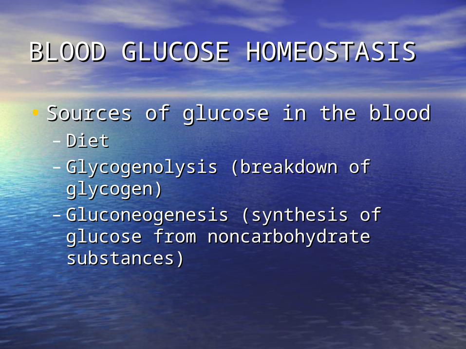
1. DIET1. DIET
Ingested carbohydrates:Ingested carbohydrates:• DigestibleDigestible - starch or disaccharides - starch or disaccharides
which after digestion are which after digestion are transformed in glucose, galactose transformed in glucose, galactose and fructose, that are absorbed, and fructose, that are absorbed, transported by the portal vein to the transported by the portal vein to the liver, where galactose and fructose liver, where galactose and fructose are conversed in glucoseare conversed in glucose
• NondigestibleNondigestible – dietary fibers – dietary fibers
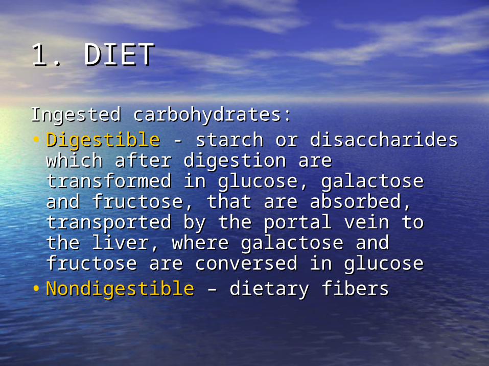
2. THE LIVER2. THE LIVER• Its importance in glucose homeostasis consists in: Its importance in glucose homeostasis consists in:
storage of the glucose as glycogen after food intake storage of the glucose as glycogen after food intake and maintaining the blood level by glycogenolysis and and maintaining the blood level by glycogenolysis and gluconeogenesis in the fasted state.gluconeogenesis in the fasted state.
• The hepatic uptake or output of glucose is controlled The hepatic uptake or output of glucose is controlled by the concentration of key intermediates and activity by the concentration of key intermediates and activity of enzymes:of enzymes:– G enters the hepatocytes relatively freely G enters the hepatocytes relatively freely
compared with extrahepatic tissuescompared with extrahepatic tissues– G phosphorilation is promoted by G-kinase with a G phosphorilation is promoted by G-kinase with a
lower affinity than hexokinase in extrahepatic lower affinity than hexokinase in extrahepatic tissues; that is why little G is taken up by the liver tissues; that is why little G is taken up by the liver at normal blood concentration compared to the at normal blood concentration compared to the more effective extraction by other tissues (brain); more effective extraction by other tissues (brain); the activity of G-kinase is increased by the activity of G-kinase is increased by hyperglycemia and the liver removes the G from hyperglycemia and the liver removes the G from the portal veinthe portal vein
– Excess G is stored in the liver as glycogen Excess G is stored in the liver as glycogen
THE LIVERTHE LIVER
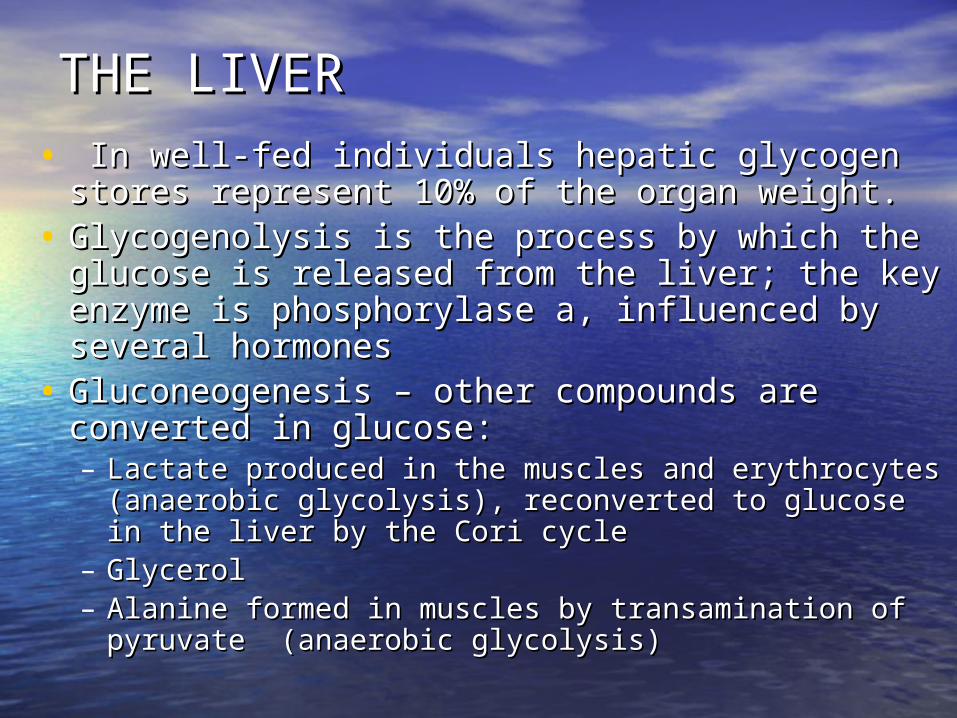
• In well-fed individuals hepatic glycogen stores In well-fed individuals hepatic glycogen stores represent 10% of the organ weight. represent 10% of the organ weight.
• Glycogenolysis is the process by which the Glycogenolysis is the process by which the glucose is released from the liver; the key glucose is released from the liver; the key enzyme is phosphorylase a, influenced by enzyme is phosphorylase a, influenced by several hormonesseveral hormones
• Gluconeogenesis – other compounds are Gluconeogenesis – other compounds are converted in glucose:converted in glucose:– Lactate produced in the muscles and erythrocytes Lactate produced in the muscles and erythrocytes
(anaerobic glycolysis), reconverted to glucose in (anaerobic glycolysis), reconverted to glucose in the liver by the Cori cyclethe liver by the Cori cycle
– GlycerolGlycerol– Alanine formed in muscles by transamination of Alanine formed in muscles by transamination of
pyruvate (anaerobic glycolysis) pyruvate (anaerobic glycolysis)
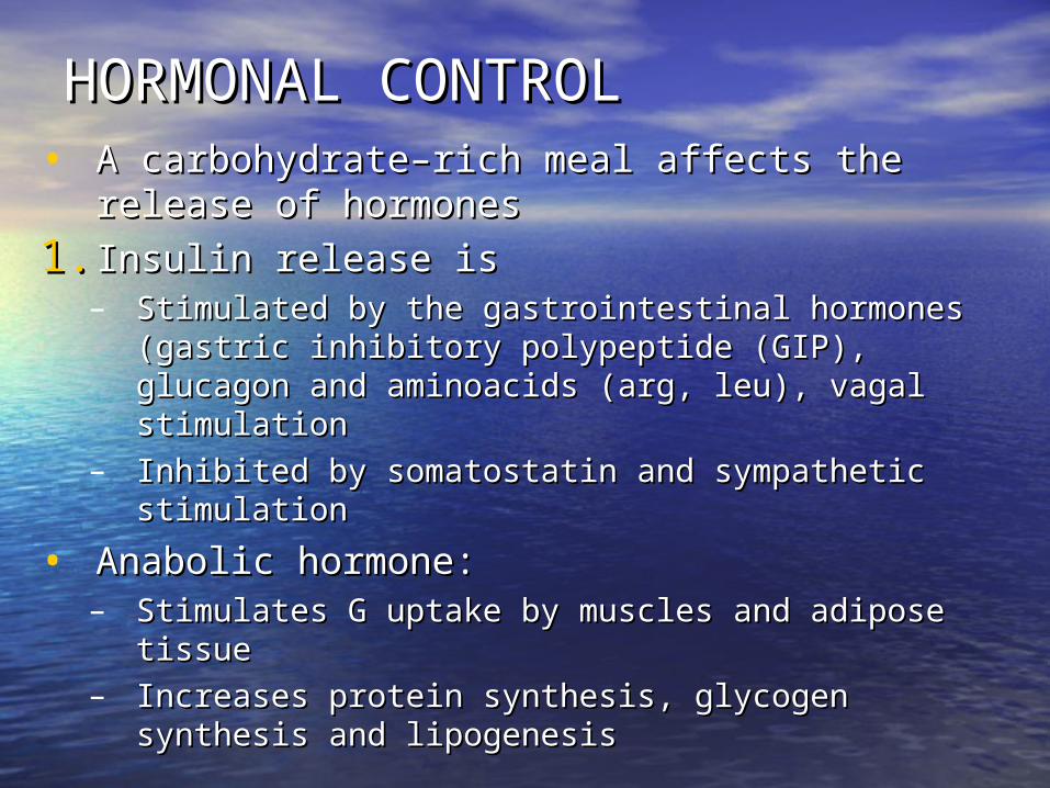
HORMONAL CONTROLHORMONAL CONTROL• A carbohydrate–rich meal affects the A carbohydrate–rich meal affects the
release of hormonesrelease of hormones
1.1. Insulin release is Insulin release is – Stimulated by the gastrointestinal hormones Stimulated by the gastrointestinal hormones
(gastric inhibitory polypeptide (GIP), glucagon (gastric inhibitory polypeptide (GIP), glucagon and aminoacids (arg, leu), vagal stimulationand aminoacids (arg, leu), vagal stimulation
– Inhibited by somatostatin and sympathetic Inhibited by somatostatin and sympathetic stimulationstimulation
• Anabolic hormone:Anabolic hormone:– Stimulates G uptake by muscles and adipose Stimulates G uptake by muscles and adipose
tissuetissue– Increases protein synthesis, glycogen synthesis Increases protein synthesis, glycogen synthesis
and lipogenesisand lipogenesis
HORMONAL CONTROLHORMONAL CONTROL
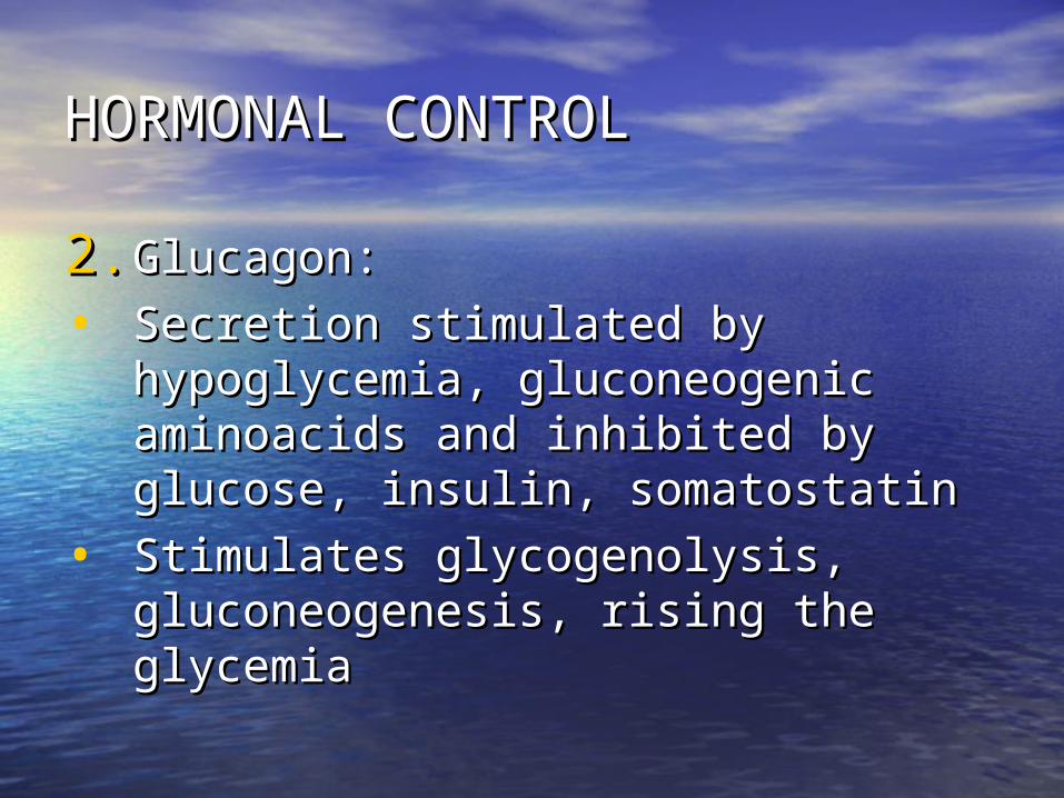
2.2. Glucagon:Glucagon:
• Secretion stimulated by Secretion stimulated by hypoglycemia, gluconeogenic hypoglycemia, gluconeogenic aminoacids and inhibited by aminoacids and inhibited by glucose, insulin, somatostatinglucose, insulin, somatostatin
• Stimulates glycogenolysis, Stimulates glycogenolysis, gluconeogenesis, rising the gluconeogenesis, rising the glycemiaglycemia
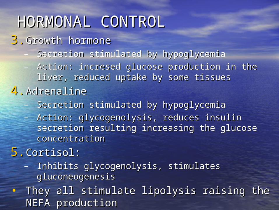
HORMONAL CONTROLHORMONAL CONTROL3.3. Growth hormone Growth hormone
– Secretion stimulated by hypoglycemia Secretion stimulated by hypoglycemia – Action: incresed glucose production in the liver, Action: incresed glucose production in the liver,
reduced uptake by some tissuesreduced uptake by some tissues
4.4. AdrenalineAdrenaline– Secretion stimulated by hypoglycemiaSecretion stimulated by hypoglycemia– Action: glycogenolysis, reduces insulin secretion Action: glycogenolysis, reduces insulin secretion
resulting increasing the glucose concentrationresulting increasing the glucose concentration
5.5. Cortisol:Cortisol:– Inhibits glycogenolysis, stimulates Inhibits glycogenolysis, stimulates
gluconeogenesisgluconeogenesis
• They all stimulate lipolysis raising the NEFA They all stimulate lipolysis raising the NEFA productionproduction
INTERRELATION OF GLUCOSE, NEFA AND INTERRELATION OF GLUCOSE, NEFA AND KETONE BODY METABOLISMKETONE BODY METABOLISM
• During prolonged fasting and starvation During prolonged fasting and starvation the muscle, brain and other tissues oxidize the muscle, brain and other tissues oxidize alternative fuels as blood concentrations alternative fuels as blood concentrations of these rise, reducing glucose utilization.of these rise, reducing glucose utilization.
• The supply of fatty acids is determined by The supply of fatty acids is determined by the rate of release of NEFA from adipose the rate of release of NEFA from adipose tissue, this being controlled by the activity tissue, this being controlled by the activity of hormone-sensitive lipase. of hormone-sensitive lipase. – Insulin inhibits this enzyme (antilipolytic); Insulin inhibits this enzyme (antilipolytic); – adrenaline, growth hormone, glucagon, cortisol adrenaline, growth hormone, glucagon, cortisol
are lipolyticare lipolytic
• When carbohydrate supply is adequate small When carbohydrate supply is adequate small amounts of NEFA are released from the adipose amounts of NEFA are released from the adipose tissuetissue
• When the carbohydrate supply is limited, greater When the carbohydrate supply is limited, greater amount of NEFA is released. They are transported amount of NEFA is released. They are transported bound with albumins in the blood, 30% ar extracted bound with albumins in the blood, 30% ar extracted by the liver:by the liver:– Re-esterified to form TGRe-esterified to form TG– Metabolized by B-oxidation in mitochondria to form acetyl-Metabolized by B-oxidation in mitochondria to form acetyl-
CoA; this can enter in Krebs cycle or form ketone bodiesCoA; this can enter in Krebs cycle or form ketone bodies
• Insulin inhibits and glucagon stimulates the Insulin inhibits and glucagon stimulates the mitochondrial carnitine-palmitoyl transferase I; it mitochondrial carnitine-palmitoyl transferase I; it enhances the transfer of FA into mitochondria, enhances the transfer of FA into mitochondria,
DIABETES MELLITUSDIABETES MELLITUS• Heterogeneous group of disorders Heterogeneous group of disorders
characterized by hyperglycemia, glycosuria, characterized by hyperglycemia, glycosuria, abnormalities of lipid and protein metabolismabnormalities of lipid and protein metabolism
• Clinical classification:Clinical classification:– Insulin-dependent diabetes mellitus (IDDM)Insulin-dependent diabetes mellitus (IDDM)– Non-insulin –dependent diabetes mellitus (NIDDM)Non-insulin –dependent diabetes mellitus (NIDDM)– Malnutrition-related DMMalnutrition-related DM– Diabetes associated with other disorders:Diabetes associated with other disorders:
• Pancreatic diseasesPancreatic diseases• Endocrine diseasesEndocrine diseases• Congenital disordersCongenital disorders
– Gestational DMGestational DM– Impared glucose toleranceImpared glucose tolerance
GLUCOSE IN THE BLOOD (GLYCEMIA)GLUCOSE IN THE BLOOD (GLYCEMIA)
• Dosing the blood glucose depends on the Dosing the blood glucose depends on the reducing propertiesreducing properties of this of this aldohexose. It is oxidized by hot alkaline copper solution, potassium aldohexose. It is oxidized by hot alkaline copper solution, potassium ferricyanide solution. These methods give 10-20 mg higher values ferricyanide solution. These methods give 10-20 mg higher values because in the blood there are other reducing substances because in the blood there are other reducing substances (gluthathion, ascorbic acid). Colorimetric methods are rapid and (gluthathion, ascorbic acid). Colorimetric methods are rapid and based on the reaction between the glucose and a chromogen (o-based on the reaction between the glucose and a chromogen (o-toluidine, anthrone). toluidine, anthrone).
• Enzymatic methodsEnzymatic methods are the most popular procedures because of are the most popular procedures because of their high specificity, rapidity of assay, use of small sample quantities their high specificity, rapidity of assay, use of small sample quantities (10 (10 l) and easy of automation. The two enzymatic systems in most l) and easy of automation. The two enzymatic systems in most general use are those with hexokinase or glucose-oxidase as the first general use are those with hexokinase or glucose-oxidase as the first enzyme in a coupled reaction; glucose dehydrogenase is used much enzyme in a coupled reaction; glucose dehydrogenase is used much less frequently.less frequently.
• No matter which method is used one must take precautions in No matter which method is used one must take precautions in sample collection to prevent glucose utilization by leukocytes, the sample collection to prevent glucose utilization by leukocytes, the glucose loss on standing in a warm room may be as high as 10 mg/dl glucose loss on standing in a warm room may be as high as 10 mg/dl per hour. The decrease in serum glucose concentration is negligible if per hour. The decrease in serum glucose concentration is negligible if the blood sample is kept cool and the serum separated from the clot the blood sample is kept cool and the serum separated from the clot within 30 minutes of drawing. Otherwise, addition to the collection within 30 minutes of drawing. Otherwise, addition to the collection tube of 2 mg sodium fluoride per ml of blood to be collected prevents tube of 2 mg sodium fluoride per ml of blood to be collected prevents glycolysis for 24 hours without interfering with the glucose glycolysis for 24 hours without interfering with the glucose determination.determination.
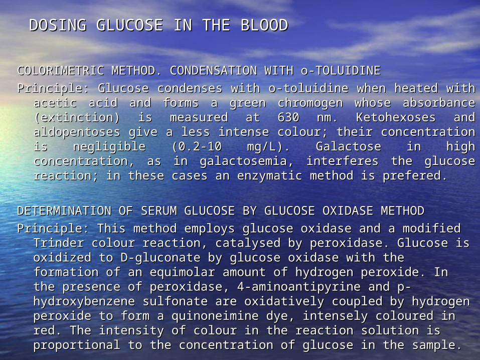
DOSING GLUCOSE IN THE BLOOD DOSING GLUCOSE IN THE BLOOD
COLORIMETRIC METHOD. CONDENSATION WITH o-TOLUIDINE COLORIMETRIC METHOD. CONDENSATION WITH o-TOLUIDINE
Principle: Glucose condenses with o-toluidine when heated with acetic Principle: Glucose condenses with o-toluidine when heated with acetic acid and forms a green chromogen whose absorbance (extinction) is acid and forms a green chromogen whose absorbance (extinction) is measured at 630 nm. Ketohexoses and aldopentoses give a less measured at 630 nm. Ketohexoses and aldopentoses give a less intense colour; their concentration is negligible (0.2-10 mg/L). intense colour; their concentration is negligible (0.2-10 mg/L). Galactose in high concentration, as in galactosemia, interferes the Galactose in high concentration, as in galactosemia, interferes the glucose reaction; in these cases an enzymatic method is prefered.glucose reaction; in these cases an enzymatic method is prefered.
DETERMINATION OF SERUM GLUCOSE BY GLUCOSE OXIDASE METHODDETERMINATION OF SERUM GLUCOSE BY GLUCOSE OXIDASE METHOD
Principle: This method employs glucose oxidase and a modified Trinder Principle: This method employs glucose oxidase and a modified Trinder colour reaction, catalysed by peroxidase. Glucose is oxidized to D-colour reaction, catalysed by peroxidase. Glucose is oxidized to D-gluconate by glucose oxidase with the formation of an equimolar gluconate by glucose oxidase with the formation of an equimolar amount of hydrogen peroxide. In the presence of peroxidase, 4-amount of hydrogen peroxide. In the presence of peroxidase, 4-aminoantipyrine and p-hydroxybenzene sulfonate are oxidatively aminoantipyrine and p-hydroxybenzene sulfonate are oxidatively coupled by hydrogen peroxide to form a quinoneimine dye, intensely coupled by hydrogen peroxide to form a quinoneimine dye, intensely coloured in red. The intensity of colour in the reaction solution is coloured in red. The intensity of colour in the reaction solution is proportional to the concentration of glucose in the sample.proportional to the concentration of glucose in the sample.
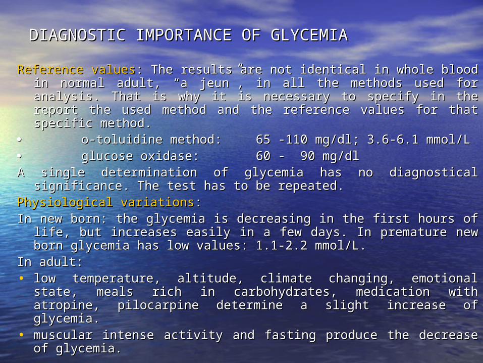
DIAGNOSTIC IMPORTANCEDIAGNOSTIC IMPORTANCE OF GLYCEMIA OF GLYCEMIA
Reference valuesReference values: The results are not identical in whole blood in : The results are not identical in whole blood in normal adult, “a jeun”, in all the methods used for analysis. That normal adult, “a jeun”, in all the methods used for analysis. That is why it is necessary to specify in the report the used method is why it is necessary to specify in the report the used method and the reference values for that specific method.and the reference values for that specific method.
o-toluidine method:o-toluidine method: 65 -110 mg/dl; 3.6-6.1 mmol/L65 -110 mg/dl; 3.6-6.1 mmol/L glucose oxidase: glucose oxidase: 60 - 90 mg/dl60 - 90 mg/dlA single determination of glycemia has no diagnostical significance. A single determination of glycemia has no diagnostical significance.
The test has to be repeated.The test has to be repeated.Physiological variationsPhysiological variations::In new born: the glycemia is decreasing in the first hours of life, but In new born: the glycemia is decreasing in the first hours of life, but
increases easily in a few days. In premature new born glycemia increases easily in a few days. In premature new born glycemia has low values: 1.1-2.2 mmol/L.has low values: 1.1-2.2 mmol/L.
In adult:In adult:• low temperature, altitude, climate changing, emotional state, low temperature, altitude, climate changing, emotional state,
meals rich in carbohydrates, medication with atropine, meals rich in carbohydrates, medication with atropine, pilocarpine determine a slight increase of glycemia. pilocarpine determine a slight increase of glycemia.
• muscular intense activity and fasting produce the decrease of muscular intense activity and fasting produce the decrease of glycemia.glycemia.
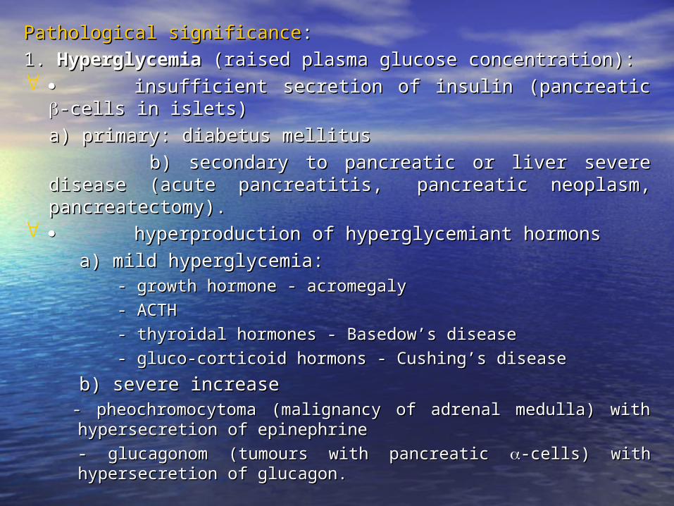
Pathological significancePathological significance::
1. 1. HyperglycemiaHyperglycemia (raised plasma glucose concentration): (raised plasma glucose concentration): insufficient secretion of insulin (pancreatic insufficient secretion of insulin (pancreatic -cells in islets)-cells in islets)
a) primary: diabetus mellitusa) primary: diabetus mellitus
b) secondary to pancreatic or liver severe b) secondary to pancreatic or liver severe disease (acute pancreatitis, disease (acute pancreatitis, pancreatic neoplasm, pancreatic neoplasm, pancreatectomy).pancreatectomy).
hyperproduction of hyperglycemiant hormonshyperproduction of hyperglycemiant hormons
a) mild hyperglycemia:a) mild hyperglycemia: - growth hormone - acromegaly- growth hormone - acromegaly
- ACTH- ACTH
- thyroidal hormones - Basedow’s disease- thyroidal hormones - Basedow’s disease
- gluco-corticoid hormons - Cushing’s disease- gluco-corticoid hormons - Cushing’s disease
b) severe increaseb) severe increase - pheochromocytoma (malignancy of adrenal medulla) with - pheochromocytoma (malignancy of adrenal medulla) with
hypersecretion of epinephrinehypersecretion of epinephrine
- glucagonom (tumours with pancreatic - glucagonom (tumours with pancreatic -cells) with hypersecretion -cells) with hypersecretion of glucagon.of glucagon.
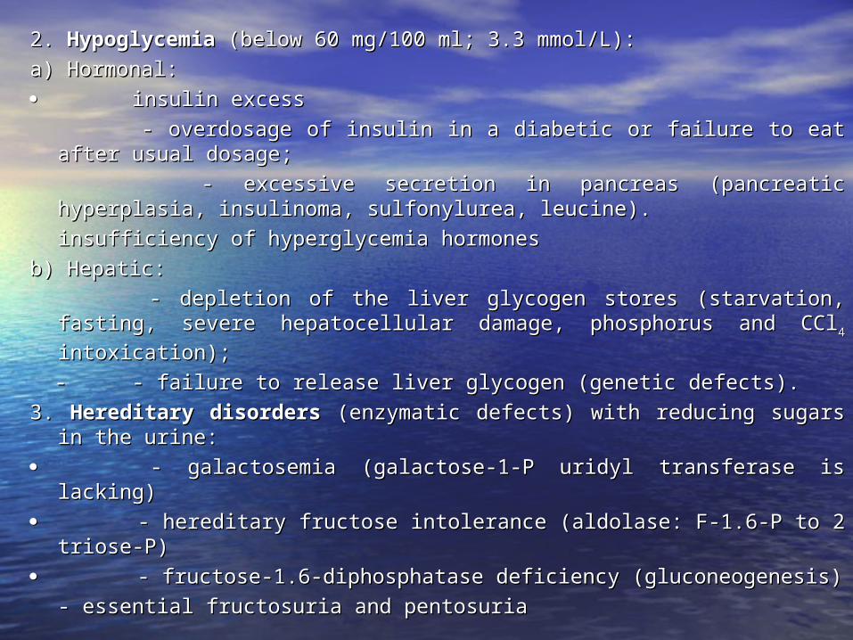
2. 2. HypoglycemiaHypoglycemia (below 60 mg/100 ml; 3.3 mmol/L): (below 60 mg/100 ml; 3.3 mmol/L):
a) Hormonal:a) Hormonal:
insulin excessinsulin excess
- overdosage of insulin in a diabetic or failure to eat after usual - overdosage of insulin in a diabetic or failure to eat after usual dosage; dosage;
- excessive secretion in pancreas (pancreatic hyperplasia, - excessive secretion in pancreas (pancreatic hyperplasia, insulinoma, sulfonylurea, leucine).insulinoma, sulfonylurea, leucine).
insufficiency of hyperglycemia hormonesinsufficiency of hyperglycemia hormones
b) Hepatic:b) Hepatic:
- depletion of the liver glycogen stores (starvation, fasting, severe - depletion of the liver glycogen stores (starvation, fasting, severe hepatocellular damage, phosphorus and CClhepatocellular damage, phosphorus and CCl44 intoxication); intoxication);
- - - failure to release liver glycogen (genetic defects).- failure to release liver glycogen (genetic defects).
3. 3. Hereditary disordersHereditary disorders (enzymatic defects) with reducing sugars (enzymatic defects) with reducing sugars in the urine:in the urine:
- - galactosemia (galactose-1-P uridyl transferase is lacking)galactosemia (galactose-1-P uridyl transferase is lacking)
- - hereditary fructose intolerance (aldolase: F-1.6-P to 2 triose-P)hereditary fructose intolerance (aldolase: F-1.6-P to 2 triose-P)
- - fructose-1.6-diphosphatase deficiency (gluconeogenesis)fructose-1.6-diphosphatase deficiency (gluconeogenesis)
- essential fructosuria and pentosuria - essential fructosuria and pentosuria
GLUCOSE IN URINE (GLYCOSURIA)GLUCOSE IN URINE (GLYCOSURIA)
Glucose is filtered through the glomerular membrane Glucose is filtered through the glomerular membrane and totally reabsorbed in proximal tubule by an active and totally reabsorbed in proximal tubule by an active transport.transport.
Normally, the urine contains very small amount of Normally, the urine contains very small amount of glucose, less than 60 mg/L (100 mg/day).glucose, less than 60 mg/L (100 mg/day).
When the glycemia is higher than 160-180 mg/dl, the When the glycemia is higher than 160-180 mg/dl, the ability of the tubular cells to transport the glucose is ability of the tubular cells to transport the glucose is overwhelmed and the glucose is eliminated in urine overwhelmed and the glucose is eliminated in urine (glycosuria or glucosuria).(glycosuria or glucosuria).
In certain pathological conditions, other saccharides can In certain pathological conditions, other saccharides can exist in urine: galactose, fructose, lactose, maltose, exist in urine: galactose, fructose, lactose, maltose, pentoses.pentoses.
The The identificationidentification of different urine saccharides is based of different urine saccharides is based on their reducing properties (except saccharose) of on their reducing properties (except saccharose) of metal salts (Fehling, Benedict tests). The methods are metal salts (Fehling, Benedict tests). The methods are less specific. Positive false results are given by less specific. Positive false results are given by increased concentrations of creatinine, uric acid, increased concentrations of creatinine, uric acid, ascorbic acid, streptomycine, phenol compoundsascorbic acid, streptomycine, phenol compounds
When the presence of glucose in urine is noticed, the When the presence of glucose in urine is noticed, the quantitative determination is necessaryquantitative determination is necessary
Qualitative and semiquantitativeQualitative and semiquantitative methods use Clinitest methods use Clinitest tablets (Ames) or glucoseoxidase impregnated strips.tablets (Ames) or glucoseoxidase impregnated strips.
Quantitative testsQuantitative tests use ortho-toluidine, hexokinase, use ortho-toluidine, hexokinase, glucose oxidase.glucose oxidase.
Reference values:Reference values: less than 60 mg/L (100 mg/day). less than 60 mg/L (100 mg/day).PhysiologicalPhysiological glycosuria appears after high glucose glycosuria appears after high glucose
intake, physical effort.intake, physical effort.PathologicalPathological significance: significance:Glycosuria + hyperglycemia:Glycosuria + hyperglycemia:• -- in diabetes mellitus (expressed in g/24 hours);in diabetes mellitus (expressed in g/24 hours);• -- increased secretion of growth hormon, thyroidal increased secretion of growth hormon, thyroidal
hormones, glucocorticoids.hormones, glucocorticoids.• -- hepatic severe damage.hepatic severe damage.Glycosuria + normal glycemia:Glycosuria + normal glycemia:• -- renal diabetes (the tubular reabsorption is renal diabetes (the tubular reabsorption is
affected);affected);• -- infectious diseases, nervous system affections;infectious diseases, nervous system affections;• -- intoxication with morphine, atropine, lead.intoxication with morphine, atropine, lead.
DIAGNOSTIC SIGNIFICANCE OF GLYCOSURIADIAGNOSTIC SIGNIFICANCE OF GLYCOSURIA
GLUCOSE IN THE URINE GLUCOSE IN THE URINE ..
When other saccharides are present, they need to be identified.When other saccharides are present, they need to be identified.
1.1. Lactose: exists physiologically in late pregnancy and lactation.Lactose: exists physiologically in late pregnancy and lactation.
2.2. Galactose: in infants during lactation;galactosemia (associated Galactose: in infants during lactation;galactosemia (associated with hypoglycemia);with hypoglycemia);
3.3. Fructose: after fruit ingestion, pregnancy, lactation; fructose Fructose: after fruit ingestion, pregnancy, lactation; fructose intolerance, essential fructosuria.intolerance, essential fructosuria.
4.4. Pentose: chronic pentosuria (deficiency of the metabolism of Pentose: chronic pentosuria (deficiency of the metabolism of glucogenetic amino acids).glucogenetic amino acids).
KETONE BODIES IN URINE (KETONURIA)KETONE BODIES IN URINE (KETONURIA)
Acetoacetic acid, Acetoacetic acid, -hydroxybutyric acid and acetone are classified as -hydroxybutyric acid and acetone are classified as ketone bodies. ketone bodies. Acetoacetic acid is the principal ketone body, Acetoacetic acid is the principal ketone body, synthesized by the liver mitochondria.synthesized by the liver mitochondria.
When there is insufficient oxalylacetic acid to derive the Krebs cycle When there is insufficient oxalylacetic acid to derive the Krebs cycle for the formation of citrate and is used to synthesize the glucose, for the formation of citrate and is used to synthesize the glucose, the acetate from the acetate from acetyl-CoAacetyl-CoA is dimerized to yield is dimerized to yield aceto-acetyl-aceto-acetyl-CoACoA. .
-hydroxybutyrate dehydrogenase reduces much of acetoacetic acid -hydroxybutyrate dehydrogenase reduces much of acetoacetic acid to to -hydroxybutyric acid. -hydroxybutyric acid.
Decarboxylase converts some of acetoacetate to Decarboxylase converts some of acetoacetate to acetoneacetone which is which is metabolized very slowly. Because it’s volatility, most evaporates metabolized very slowly. Because it’s volatility, most evaporates through the lung alveoli.through the lung alveoli.
Liver produces ketone bodies when the rate of acetyl-CoA formation Liver produces ketone bodies when the rate of acetyl-CoA formation exceeds of acetyl-CoA utilization by citric acid cycle. exceeds of acetyl-CoA utilization by citric acid cycle.

KETONE BODIES IN URINEKETONE BODIES IN URINE
Extrahepatic tissues (skeletal muscles, heart, renal cortex) utilize the Extrahepatic tissues (skeletal muscles, heart, renal cortex) utilize the ketone bodies (other than acetone) as a fuel. They oxidize ketone bodies (other than acetone) as a fuel. They oxidize --hydroxybutyrate to acetoacetate, then add CoA-SH by either of 2 hydroxybutyrate to acetoacetate, then add CoA-SH by either of 2 routes to create acetoacetyl-CoA which is cleaved into 2 acetyl-routes to create acetoacetyl-CoA which is cleaved into 2 acetyl-CoA able to enter Krebs cycle. CoA able to enter Krebs cycle.
Food and Nutrition Board of U.S. recommends that the adult diet Food and Nutrition Board of U.S. recommends that the adult diet should contain al least 100 g or 400 cal. carbohydrates daily to should contain al least 100 g or 400 cal. carbohydrates daily to generate enough oxalylacetic acid to maintain TCA cycle and generate enough oxalylacetic acid to maintain TCA cycle and prevent ketosis. Carbohydrate defficiency causes protein prevent ketosis. Carbohydrate defficiency causes protein waisting (much of dietary amino acids are converted via waisting (much of dietary amino acids are converted via deamination and gluconeogenesis to glucose). The brain deamination and gluconeogenesis to glucose). The brain acquires a limited capacity for oxidizing ketone bodies after acquires a limited capacity for oxidizing ketone bodies after about 3 weeks of fasting, to protect against muscle waisting about 3 weeks of fasting, to protect against muscle waisting (gluconeogenesis from muscular proteins).(gluconeogenesis from muscular proteins).

IDENTIFICATION OF KETONE BODIES IN URINE IDENTIFICATION OF KETONE BODIES IN URINE BY LEGAL-IMBERT REACTIONBY LEGAL-IMBERT REACTION
Principle: The most common method makes use of a reaction of sodium nitroprusside Principle: The most common method makes use of a reaction of sodium nitroprusside (Na(Na22[Fe(CN)[Fe(CN)55NO].2 HNO].2 H22O) and acetoacetate or acetone, under alkaline conditions; a O) and acetoacetate or acetone, under alkaline conditions; a lavender colour is produced; lavender colour is produced; -hydroxybutyric acid does not react.-hydroxybutyric acid does not react.
Impregnated strips or sticks with reagent are introduced in urine for few seconds. By Impregnated strips or sticks with reagent are introduced in urine for few seconds. By comparison with a colour chart, the concentration of acetoacetic acid and acetone comparison with a colour chart, the concentration of acetoacetic acid and acetone is expressed as: is expressed as:
-- negativenegative-- small small 10 mg/dl10 mg/dl-- moderate moderate 30 mg/dl30 mg/dl-- large large 80 mg/dl80 mg/dl
DIAGNOSTIC SIGNIFICANCE OF KETONE BODIESDIAGNOSTIC SIGNIFICANCE OF KETONE BODIES
• Normally, the ketone bodies are not present in the urine of healthy individuals Normally, the ketone bodies are not present in the urine of healthy individuals eating a mixed diet. (the reaction is negative)eating a mixed diet. (the reaction is negative)
• Physiological values: The ketone bodies may be present in children’s urine.Physiological values: The ketone bodies may be present in children’s urine.
PATHOLOGICAL VARIATIONS:PATHOLOGICAL VARIATIONS:When there is high serum concentration of acetoacetate and When there is high serum concentration of acetoacetate and --
hydroxybutyric acid, the state is named ketonemia. It can overwhelme hydroxybutyric acid, the state is named ketonemia. It can overwhelme the blood buffers causing metabolic acidosis.the blood buffers causing metabolic acidosis.
Ketonuria measures the acetone and acetoacetate detected by common Ketonuria measures the acetone and acetoacetate detected by common hospital tests (may fail to detect ketonuria of hospital tests (may fail to detect ketonuria of -hydroxybutyric acid -hydroxybutyric acid predominaters).predominaters).
The ketosis (ketonemia associated with ketonuria) appears whenever The ketosis (ketonemia associated with ketonuria) appears whenever • the rate of hepatic ketone body production exceeds the rate of principal the rate of hepatic ketone body production exceeds the rate of principal
utilization, utilization, • excessive amounts of fatty acids are catabolyzed andexcessive amounts of fatty acids are catabolyzed and• the availability of glucose limited.the availability of glucose limited.Hepatic overproduction is present in severe carbohydrate defficiency Hepatic overproduction is present in severe carbohydrate defficiency
(diabetic ketoacidosis, alcoholic ketoacidosis, starvation ketosis); in this (diabetic ketoacidosis, alcoholic ketoacidosis, starvation ketosis); in this situation TCA cycle intermediates are depleted and this slows the situation TCA cycle intermediates are depleted and this slows the entrance of acetyl-CoA into Krebs cycle. The acetyl-CoA carboxylase entrance of acetyl-CoA into Krebs cycle. The acetyl-CoA carboxylase (the rate controlling enzyme of fatty acid synthesis) is inhibited by the (the rate controlling enzyme of fatty acid synthesis) is inhibited by the absence of citrate, blocking another route of acetyl-CoA metabolism. absence of citrate, blocking another route of acetyl-CoA metabolism. Thus, acetyl-CoA accumulates in the liver and is excessively converted Thus, acetyl-CoA accumulates in the liver and is excessively converted to ketone bodies.to ketone bodies.
The same conditions appear when the diet is poor in glucose but rich in lipids The same conditions appear when the diet is poor in glucose but rich in lipids and proteins; in gastrointestinal troubles (acute dyspepsia, toxicosis, and proteins; in gastrointestinal troubles (acute dyspepsia, toxicosis, vomiting during pregnancy, intense muscular effort). vomiting during pregnancy, intense muscular effort).
GLYCOSYLATED HEMOGLOBINGLYCOSYLATED HEMOGLOBIN
Used to monitor the diabetes therapy.Used to monitor the diabetes therapy.Three minor hemolobins are measured:Three minor hemolobins are measured: HbA1a, HbA1b, HbA1c, HbA1a, HbA1b, HbA1c,
variants of HbA formed by glycosylation, an almost irreversible variants of HbA formed by glycosylation, an almost irreversible process in which glucose is incorporated in HbA. This reaction process in which glucose is incorporated in HbA. This reaction occurs with a constant rate during the 120 days life span of an occurs with a constant rate during the 120 days life span of an erythrocyte. erythrocyte.
Thus, the glycosylated Hb reflects the average blood glucose level Thus, the glycosylated Hb reflects the average blood glucose level during the preceding 4-6 weeks and offers information referring during the preceding 4-6 weeks and offers information referring to long-term effectiveness of diabetes therapy.to long-term effectiveness of diabetes therapy.
Levels of glucose in the erythrocytes are more stable than plasma Levels of glucose in the erythrocytes are more stable than plasma glucose.glucose.
Reference intervalReference intervalHbA1a HbA1a 1.6% of total Hb1.6% of total HbHbA1b HbA1b 0.8%0.8%HbA1c HbA1c 5%5%
Total glycosylated Hb Total glycosylated Hb 5.5-9% of total Hb5.5-9% of total HbPathologic resultsPathologic resultsDiabetes HbA1a and HbA1b 2.5-3.9%; HbA1c 8-11.9%, total 10.9-Diabetes HbA1a and HbA1b 2.5-3.9%; HbA1c 8-11.9%, total 10.9-
15.5%15.5%