Carbohydrate Binding Modules - Microbiology and Molecular
13
MICROBIOLOGY AND MOLECULAR BIOLOGY REVIEWS, June 2006, p. 283–295 Vol. 70, No. 2 1092-2172/06/$08.000 doi:10.1128/MMBR.00028-05 Copyright © 2006, American Society for Microbiology. All Rights Reserved. Carbohydrate Binding Modules: Biochemical Properties and Novel Applications Oded Shoseyov, 1 * Ziv Shani, 2 and Ilan Levy 3 The Institute of Plant Science and Genetics in Agriculture and The Otto Warburg Center for Agricultural Biotechnology, Faculty of Agricultural, Food and Environmental Quality Sciences, The Hebrew University of Jerusalem, P.O. Box 12, 1 CBD-Technologies Ltd., Tamar Science Park, P.O. Box 199, 2 and Department of Structural Biology, Faculty of Chemistry, The Weizmann Institute of Science, P.O. Box 26, 3 Rehovot 76100, Israel INTRODUCTION .......................................................................................................................................................283 CARBOHYDRATE BINDING, MODULATION OF POLYSACCHARIDE STRUCTURE, AND SOLVENT INTERFACE ........................................................................................................................................................284 Enzyme Concentration on the Surface of the Substrate—the Phase Transfer..............................................284 Substrate Binding and Selectivity ........................................................................................................................284 Nonhydrolytic Substrate Disruption ....................................................................................................................284 Surface/Interfacial Modifications .........................................................................................................................285 UTILIZATION OF CBMs .........................................................................................................................................285 Bioprocessing ...........................................................................................................................................................285 Targeting ..................................................................................................................................................................286 Cell Immobilization ................................................................................................................................................286 CBM Engineering for Different Applications .....................................................................................................286 CBMs as Analytical Tools in Research and Diagnostics ..................................................................................287 Bioremediation ........................................................................................................................................................287 Modification of Fiber..............................................................................................................................................287 MODULATION OF PLANT CELL WALLS BY CBMs ........................................................................................288 Expansins: the Main Family of Plant CBMs ......................................................................................................288 Effect of CBMs on Cellulose Biosynthesis...........................................................................................................289 FUTURE ASPECTS....................................................................................................................................................289 ACKNOWLEDGMENT..............................................................................................................................................291 REFERENCES ............................................................................................................................................................291 INTRODUCTION In the late 1940s, it was proposed that the initial stage in the enzymatic degradation of crystalline cellulose involves the ac- tion of an unknown nonhydrolytic component, termed C 1 . This component was thought to be responsible for destabilization (nonhydrolytic disruption) of the cellulose structure, making the substrate accessible to the enzyme, the C x component (161). The proteolytic susceptibility of the connecting linker between the carbohydrate binding module (CBM) moiety and the enzyme facilitated isolation of the individual domain, lead- ing to the first CBM isolation of the fungus Trichoderma reesei and the bacterium Cellulomonas fimi (69, 194, 201). While this model is still controversial, the first C 1 component was cloned from Clostridium cellulovorans and Cellulomonas fimi (49, 74, 172, 173). CBMs were initially classified as cellulose binding domains (CBDs), based on the initial discovery of several modules that bind cellulose (69, 194, 201). However, more and more mod- ules in carbohydrate-active enzymes that bind carbohydrates other than cellulose are being found. These findings prompted the need to reclassify these polypeptides with more-compre- hensive terminology. A CBM is defined as a contiguous amino acid sequence within a carbohydrate-active enzyme with a dis- crete fold having carbohydrate binding activity (22, 23, 43). To date, more than 300 putative sequences in more than 50 dif- ferent species have been identified, and the binding domains have been classified into 43 different families based on amino acid sequence, binding specificity, and structure (for reviews, see references 26, 45, 70, 88, 117, 165, 197, and 202). Extensive data and classification can be found in the Carbohydrate-Bind- ing Module Family Server (http://afmb.cnrs-mrs.fr/pedro /CAZY/cbm.html). The CBMs contain from 30 to about 200 amino acids and exist as a single, double, or triple domain in one protein. Their location within the parental protein can be both C- or N- terminal and is occasionally centrally positioned within the polypeptide chain. The three-dimensional (3D) structures of representative members of 23 CBM families have been deci- phered so far, several in complex with their ligands. These data provide insight into the underlying mechanism of CBM-ligand recognition and interaction (for reviews, see references 26 and 81). Data from these structures indicate that CBMs from dif- ferent families are structurally similar and that their carbohy- drate binding capacity can be attributed, at least in part, to several aromatic amino acids that constitute the hydrophobic surface (for extended reviews on CBMs, see references 11, 12, 26, 70, 85, 196, and 197). CBMs have been found in both hydrolytic and nonhydrolytic * Corresponding author. Mailing address: Faculty of Agricultural, Food and Environmental Quality Sciences, The Hebrew University of Jerusalem, P.O. Box 12, Rehovot 76100, Israel. Phone: 972-8-9481084. Fax: 972-8-9462283. E-mail: [email protected]. 283 on December 25, 2018 by guest http://mmbr.asm.org/ Downloaded from
Transcript of Carbohydrate Binding Modules - Microbiology and Molecular
MICROBIOLOGY AND MOLECULAR BIOLOGY REVIEWS, June 2006, p. 283–295 Vol. 70, No. 21092-2172/06/$08.00�0 doi:10.1128/MMBR.00028-05Copyright © 2006, American Society for Microbiology. All Rights Reserved.
Carbohydrate Binding Modules: Biochemical Properties andNovel Applications
Oded Shoseyov,1* Ziv Shani,2 and Ilan Levy3
The Institute of Plant Science and Genetics in Agriculture and The Otto Warburg Center for Agricultural Biotechnology,Faculty of Agricultural, Food and Environmental Quality Sciences, The Hebrew University of Jerusalem, P.O. Box 12,1
CBD-Technologies Ltd., Tamar Science Park, P.O. Box 199,2 and Department of Structural Biology, Faculty ofChemistry, The Weizmann Institute of Science, P.O. Box 26,3 Rehovot 76100, Israel
INTRODUCTION .......................................................................................................................................................283CARBOHYDRATE BINDING, MODULATION OF POLYSACCHARIDE STRUCTURE, AND SOLVENT
INTERFACE........................................................................................................................................................284Enzyme Concentration on the Surface of the Substrate—the Phase Transfer..............................................284Substrate Binding and Selectivity ........................................................................................................................284Nonhydrolytic Substrate Disruption ....................................................................................................................284Surface/Interfacial Modifications .........................................................................................................................285
UTILIZATION OF CBMs .........................................................................................................................................285Bioprocessing...........................................................................................................................................................285Targeting ..................................................................................................................................................................286Cell Immobilization ................................................................................................................................................286CBM Engineering for Different Applications .....................................................................................................286CBMs as Analytical Tools in Research and Diagnostics ..................................................................................287Bioremediation ........................................................................................................................................................287Modification of Fiber..............................................................................................................................................287
MODULATION OF PLANT CELL WALLS BY CBMs........................................................................................288Expansins: the Main Family of Plant CBMs......................................................................................................288Effect of CBMs on Cellulose Biosynthesis...........................................................................................................289
FUTURE ASPECTS....................................................................................................................................................289ACKNOWLEDGMENT..............................................................................................................................................291REFERENCES ............................................................................................................................................................291
INTRODUCTION
In the late 1940s, it was proposed that the initial stage in theenzymatic degradation of crystalline cellulose involves the ac-tion of an unknown nonhydrolytic component, termed C1. Thiscomponent was thought to be responsible for destabilization(nonhydrolytic disruption) of the cellulose structure, makingthe substrate accessible to the enzyme, the Cx component(161). The proteolytic susceptibility of the connecting linkerbetween the carbohydrate binding module (CBM) moiety andthe enzyme facilitated isolation of the individual domain, lead-ing to the first CBM isolation of the fungus Trichoderma reeseiand the bacterium Cellulomonas fimi (69, 194, 201). While thismodel is still controversial, the first C1 component was clonedfrom Clostridium cellulovorans and Cellulomonas fimi (49, 74,172, 173).
CBMs were initially classified as cellulose binding domains(CBDs), based on the initial discovery of several modules thatbind cellulose (69, 194, 201). However, more and more mod-ules in carbohydrate-active enzymes that bind carbohydratesother than cellulose are being found. These findings promptedthe need to reclassify these polypeptides with more-compre-
hensive terminology. A CBM is defined as a contiguous aminoacid sequence within a carbohydrate-active enzyme with a dis-crete fold having carbohydrate binding activity (22, 23, 43). Todate, more than 300 putative sequences in more than 50 dif-ferent species have been identified, and the binding domainshave been classified into 43 different families based on aminoacid sequence, binding specificity, and structure (for reviews,see references 26, 45, 70, 88, 117, 165, 197, and 202). Extensivedata and classification can be found in the Carbohydrate-Bind-ing Module Family Server (http://afmb.cnrs-mrs.fr/�pedro/CAZY/cbm.html).
The CBMs contain from 30 to about 200 amino acids andexist as a single, double, or triple domain in one protein. Theirlocation within the parental protein can be both C- or N-terminal and is occasionally centrally positioned within thepolypeptide chain. The three-dimensional (3D) structures ofrepresentative members of 23 CBM families have been deci-phered so far, several in complex with their ligands. These dataprovide insight into the underlying mechanism of CBM-ligandrecognition and interaction (for reviews, see references 26 and81). Data from these structures indicate that CBMs from dif-ferent families are structurally similar and that their carbohy-drate binding capacity can be attributed, at least in part, toseveral aromatic amino acids that constitute the hydrophobicsurface (for extended reviews on CBMs, see references 11, 12,26, 70, 85, 196, and 197).
CBMs have been found in both hydrolytic and nonhydrolytic
* Corresponding author. Mailing address: Faculty of Agricultural,Food and Environmental Quality Sciences, The Hebrew University ofJerusalem, P.O. Box 12, Rehovot 76100, Israel. Phone: 972-8-9481084.Fax: 972-8-9462283. E-mail: [email protected].
283
on Decem
ber 25, 2018 by guesthttp://m
mbr.asm
.org/D
ownloaded from

proteins. Proteins that possess hydrolytic activity (e.g., cellu-lases and xylanases) encompass a complex molecular architec-ture comprising discrete modules (typically, a catalytic moduleand one or more CBMs), which are normally joined by rela-tively unstructured linker sequences. The CBMs, by bringingthe biocatalyst into intimate and prolonged association with itsrecalcitrant substrate, increase the rate of catalysis (70, 124,192, 195–197). The CBMs present in proteins that do notpossess hydrolytic activity comprise part of a scaffolding sub-unit that organizes the catalytic subunits into a cohesive mul-tienzyme complex known as a cellulosome (11–13, 15, 47, 50,53, 54, 123, 173, 212). The enzymatic complex was found tofunction more efficiently in substrate degradation, and remov-ing the CBM from the enzyme or from the scaffolding incellulosomes dramatically decreased its enzymatic activity (29,42, 74, 84, 194, 201).
CBMs have also been found in several polysaccharide-de-grading enzymes other than cellulases and xylanases. In T.reesei, CBMs have been identified in hemicellulase, endoman-nanase, and acetylxylanesterase (128, 188). CBMs have beenrecognized in esterase from Penicillium funiculosum (108), iso-maltodextranase from Arthrobacter globiformis (82), arabino-furanosidases from Aspergillus kawachii and Cellvibrio japoni-cus (21, 136), pectate lyase from Pseudomonas cellulose (27),�-agarase from the marine bacterium JAMB-A94 (145), �-glu-cosidase from Phanerochaete chrysosporium (127), and dextran-ase from Paenibacillus sp. (59). An interesting observation wasrecently reported when a CBM was found in cytochrome (217).The presence of putative CBMs in plant endoglucanases hasalso been reported (30, 152, 199). Expansins, which are be-lieved to play a role in nonhydrolytic cell wall expansion, arehomologues to CBMs and possess cellulose binding capabili-ties in vitro (40). Most recently, a small olive pollen protein,Ole e 10 (10 kDa), was identified. Ole e 10 binds specifically to1,3-�-glucans. The protein was described as an independentCBM and represents the first member of the new CBM family43 (9).
In the phylum Nematodea, CBMs have been found in secre-tions of the root knot nematode Meloidogyne incognita (51) andin the soybean cyst nematode Heterodera glycines (65), andrecently it was demonstrated that nematodes express expansinduring plant feeding (110, 159).
CARBOHYDRATE BINDING, MODULATION OFPOLYSACCHARIDE STRUCTURE, AND
SOLVENT INTERFACE
Enzyme Concentration on the Surface of the Substrate—thePhase Transfer
In earlier studies of CBM-cellulose interactions, the pres-ence of CBMs was shown to increase the effective concentra-tion of the hydrolytic unit (or units, in the case of cellulosomes)on the surface of the carbohydrate substrate (14, 196). In fact,removing the CBM from the cellulase or from the scaffoldin incellulosomes dramatically decreases enzymatic activity (2, 4,20, 25, 29, 42, 74, 79, 194, 201, 218). However, a frequentobservation in these studies is that the reduction in catalyticactivity takes place only on insoluble substrates. Alternatively,CBMs were employed to improve the carbohydrate degrading
activity. Interestingly, the addition of a CBM, derived fromcellobiohydrolase II of T. reesei, to the T. harzianum chitinaseresulted in increased hydrolytic activity of insoluble substrates(122). Moreover, replacing the CBM of endo-1,4-�-glucanasefrom Bacillus subtilis (Ben) with the CBM of exoglucanase I(Texl) from T. viride resulted in higher binding affinity andenhanced hydrolytic activity on the surface of the microcrys-talline cellulose (105). Although the interaction of CBMs withcellulose is occasionally irreversible (29, 158), their contactwith the cellulose surface is a dynamic process. Jervis et al.(96), using fluorescence recovery techniques, confirmed thatCBMCex is mobile on the surface of crystalline cellulose. Themobility of CBMs may explain the function of CBMs otherthan to concentrate the catalytic activity on the substrate.
Substrate Binding and Selectivity
In recent years, the 3D structures of representative membersfrom 22 CBM families have been resolved. Data from thesestructures indicate that CBMs from different families are struc-turally similar and that their binding to cellulose can be attrib-uted, at least in part, to their hydrophobic surface, which iscomposed of several aromatic amino acids. CBMs are classi-fied into seven “fold families,” based on their 3D structuresand functional similarities, into three types: (i) “surface bind-ing” (type A), (ii) “glycan chain binding” (type B), and (iii)“small sugar binding” (type C). The structure-function rela-tionship is discussed extensively in a review by Boraston et al.(26) and is not repeated here.
Nonhydrolytic Substrate Disruption
It has been proposed that some CBMs possess additionalfunctions, such as disruption of substrate. The initial evidencesupporting this notion was from CBMCenA, from C. fimi en-doglucanase A. More specifically, the CBM was able to disruptthe structure of cellulose fibers, resulting in the release of smallparticles without any detectable hydrolytic activity (49). Inaddition, CBMCenA was able to prevent the flocculation ofmicrocrystalline bacterial cellulose (72). Similar phenomenawere also reported for other CBMs (7, 22, 66, 71, 109, 118,148, 213). In addition, it was reported that structural dis-ruption occurred when the starch binding domains (family20 CBM) were bound to starch (68, 186). Another interest-ing discovery was reported when a CBM was applied todental plaque polysaccharides (mainly fructan and glucan),which resulted in its dispersion, thereby removing and pre-venting plaque formation (64).
Lee et al. (113) provided the first physical evidence for theinvolvement of CBMs in altering of the fiber surface followingcellulase treatment. In their study, two cellulases from T. reesei,exoglucanase (EGase) CBH I and EGase EG II, were appliedseparately and in combination to cotton fibers. Treatment withCBH I resulted in the appearance of distinct tracks along thelongitudinal axis of the fiber, as visualized by atomic forcemicroscopy, whereas EG II treatment appeared to cause peel-ing and smoothing of the fiber surface. When cellulase fromThermotoga maritime, which lacks the CBM, was used, no effecton the surface of the cotton fibers was discerned.
284 SHOSEYOV ET AL. MICROBIOL. MOL. BIOL. REV.
on Decem
ber 25, 2018 by guesthttp://m
mbr.asm
.org/D
ownloaded from

Surface/Interfacial Modifications
Recent studies have indicated that treatment of cellulosefibers with CBMs alters the interfacial properties of the fibers.This phenomenon was first reported by Cavaco-Paulo et al.(33), who demonstrated that treatment of cotton fibers with aCBM alters their affinity to dye. Suurnakki et al. (191) dem-onstrated that treatment of bleached chemical pulp with en-doglucanases, cellobiohydrolases, and the catalytic domainsfrom T. reesei could also change the interfacial properties.According to this study, the presence of the CBM in the intactenzyme had a beneficial effect on pulp properties such asviscosity and strength after refinement. Later, Pala et al. (149)demonstrated that treatment of fibers recycled from old pa-perboard containers composed of CBMs may improve both thetensile and burst indexes, as well as increase the pulp drainagerate. These observations inspired an in-depth research studyinto CBM-cellulose fiber interactions. It was shown that CBMtreatment of cellulosic fibers results in an increased surfacearea as a consequence of cellulosic aggregate disruption, re-duction in fiber acidity, and reduction of surface polarity (158).In general, the interaction between cellulose surfaces is dom-inated by double-layer repulsive forces attributed to the neg-ative charge of cellulose surfaces. Nigmatullin et al. (140) havedemonstrated, using atomic force microscopy, that despite anincrease in surface charge following CBM binding, interfacialforce profiles are less repulsive. This phenomenon may assistother molecules, such as xyloglucan and pectin, in interactingwith cellulose surfaces.
UTILIZATION OF CBMs
In recent years the practical use of CBMs has been estab-lished in different fields of biotechnology, and the number ofpublished articles and patents is constantly on the rise. Threebasic properties have contributed to CBMs being perfect can-didates for many applications: (i) CBMs are usually indepen-dently folding units and therefore can function autonomouslyin chimeric proteins; (ii) the attachment matrices are abundantand inexpensive and have excellent chemical and physicalproperties; and (iii) the binding specificities can be controlled,and therefore the right solution can be adapted to an existingproblem. Utilization of CBMs has been extensively reportedand reviewed in the literature (10, 75, 117, 146, 197, 202), andtheir use has been described in several patents (103, 104, 133,134, 174–176). Therefore, this section summarizes only thebasic principles of CBM application, along with recent devel-opments.
Bioprocessing
Bioprocessing is the major application for CBMs, given thatlarge-scale recovery and purification of biologically active mol-ecules continue to be challenges for many biotechnologicalproducts. Biospecific affinity purification (affinity chromatog-raphy) has become one of the most rapidly developing divi-sions of immobilized affinity ligand technology. To date, sev-eral affinity tags, which vary in size from several amino acids toa complete protein, have been developed. Each individual af-finity-based purification system embodies specific advantages
(for reviews, see references 57, 80, 126, 167, 193, and 211).Many protein entities have been expressed when fused toCBMs, establishing CBMs as high-capacity purification tags forthe isolation of biologically active target peptides at relativelylow cost (24, 52, 102, 160, 164, 180, 183).
Production of recombinant proteins in plants has been re-cently recognized as one of the most cost-effective productionsystems. However, a major drawback of this system is thatplants contain high levels of polysaccharides and phenolic com-ponents, which interfere with the purification process (55, 87,90). The utilization of CBMs in the production of CBM fusionproteins in plants permits efficient production, taking advan-tage of the fact that the plant cell wall is composed of cellulose.In this system the plant manufactures both the target proteinand its purification matrix (171).
Two-phase liquid separation systems for protein purificationhave been proposed in order to reduce the downstream pro-cessing of biological molecules (141, 205). Haynes et al. (83)proposed a novel two-phase separation system to purify pro-teins from aqueous solutions by utilizing family 4 CBMs, whichbind to water-soluble cellulosic materials such as hydroxyethyl-cellulose. The system was composed of a phase-forming poly-saccharide polymer to which a CBM can bind and a phase-inducing agent such as a polyethylene glycol. The solutioncontaining a CBM-fused peptide or protein was mixed with thephase-forming oligosaccharide, followed by the addition of thephase-inducing agent. The two phases were then separated,and the target protein was purified. Shortly thereafter, Lam etal. (111) designed an advanced system based on a two-phaseaqueous micellar system utilizing family 9 CBMs. Interestingly,the detergent n-decyl-�-D-glucopyranoside operates simulta-neously as a phase former and as an affinity ligand. Thesesystems may be useful for protein separation in large-scale,industrial fermentation plants.
Numerous reports have affirmed the feasibility of employinga CBM as an affinity tag for enzyme immobilization and pro-cessing. In these studies carbohydrates were used as an affinitysupport for enzyme immobilization, with high capacity, whileretaining enzymatic activity; in some instances, increased en-zymatic activity was reported (19, 77, 93, 98, 101, 102, 119, 130,155, 162, 166). Recent studies have shown that a CBM servingas a fusion partner may have additional values. In the expres-sion of CBM-lipase fusion protein in yeast, for example, it wasshown that CBM also enhanced secretion (1).
Another area of increasing interest is bioethanol productionfrom cellulosic material. Lignocellulose is the most abundantrenewable natural resource for conversion to fuels. CBMs arethe pivotal proteins able to target the catalytic modules ofpolysaccharidases that are needed for the breakdown of thecellulosic biomass to sugars, which can then be converted toliquid fuel (for extended reviews, see references 47, 54, and139).
Matrix-assisted refolding of recombinant proteins is one ofthe approaches taken in order to prevent the aggregation ofprotein during the course of renaturation. At present, onlyhistidine and arginine tags have been found to be suitable forthis process, because they maintain matrix binding ability un-der denaturing conditions (73, 189). Recently, Berdichevsky etal. (17) demonstrated that a CBM (C. thermocellum) can beused as the attachment support for matrix-assisted refolding
VOL. 70, 2006 CBMs: BIOCHEMICAL PROPERTIES AND NOVEL APPLICATIONS 285
on Decem
ber 25, 2018 by guesthttp://m
mbr.asm
.org/D
ownloaded from

of a single-chain antibody expressed in Escherichia coli. ThisCBM can bind cellulose in the presence of 6 M urea, and thismethod was shown to provide a threefold increase in proteinyield compared with standard refolding procedures.
Phage display technology is a well-established tool for iso-lating biologically active molecules (38, 61, 67, 99, 163). One ofthe limitations preventing extensive implementation of thistechnology is the relatively high proportion of clones that lackinsertions within the library. In a recent study, a CBM from C.thermocellum was fused to a single-chain antibody (scFv) andexpressed as an scFv-CBM phage display library. The CBM tagallowed for rapid recovery of phages that displayed functionalinserts, thus increasing the efficiency of the screening processfor recombinant antibodies (16). Furthermore, a novel ap-proach for high-throughput screening of shuffled recombinantscFvs was developed, based on their immobilization on cellu-lose-based supports (5).
Targeting
Cellulose is a major component of numerous commercialproducts, several of which are capable of being recycled.Therefore, CBMs can be used for the targeting of functionalmolecules to materials containing cellulose. The commercialpotential of CBMs in this context was first realized for denimstonewashing, where cellulases were used as an alternative tothe original abrasive stones (31, 32, 100). Another textile-associated CBM application used in numerous laundry pow-ders is fabric targeting of recombinant enzymes that do notpossess a native affinity for the cellulosic fibers (e.g., amy-lases, proteases, lipases, and oxidoreductases). This can beachieved by recombinant enzyme technology, where fusionto CBMs with a desired enzyme is achieved (203, 204).Additional substances can also be targeted to cellulosic fab-rics. Fragrance-bearing particles conjugated to CBMs can beadded to laundry powder, hence reducing the amount offragrance needed in the product (18).
Cell Immobilization
Cell immobilization technology ranges from ethanol produc-tion and phenol degradation (137, 139) to mammalian cellattachment (107, 215) and whole-cell diagnostics (76, 169,187). Surface-exposed CBMs can be an efficient means ofwhole-cell immobilization. Whole-cell immobilization by cel-lulosic material was first demonstrated when an E. coli surface-anchored CBM, derived from C. fimi, was attached to cellulose(62). The cells bound tightly to cellulose at a wide range ofpHs, and the extent of immobilization was dependent on theamount of surface-exposed CBM (206). In a different study,Staphylococcus carnosus was chosen to display CBMCel6A fromT. reesei on its cell surface, and the addition of the CBMpredisposed the anchoring of bacterial cells to cotton fibers(115). Yeast was also shown to be cellulose immobilized viacell surface display of CBMs (138).
A different strategy for cell immobilization was demon-strated by the attachment of mammalian cells to a cellulosicsurface coated with recombinant protein composed of the cellattachment peptide RGD fused to CBMCenA from C. fimi. Thisapproach is based on the preservation of the functional prop-
erties of the attachment ligand following its immobilization. Inaddition, it enabled cell immobilization without the need forexpensive attachment factors (210). Furthermore, it was dem-onstrated that cellulose is an excellent inert matrix for present-ing cytokines to target cells, where it demonstrated a morestimulating effect of proliferation (52), improved cellular ad-hesion (92), and stimulated receptor polarization in the cellmembrane (97). This approach was used to improve the per-formance of vascular grafts (91) and tissue-engineered scaf-folding for cartilage regeneration (92). In a study by Nordon etal. (142), a hollow-fiber device for analysis of ligand-mediatedcell adhesion wherein cell adhesion strength can be measuredunder shear stress was established. This system permits evalu-ation of the interaction of molecular domains with cell surfacereceptors.
CBM Engineering for Different Applications
It is well established that expression of foreign proteinsfused to CBMs results, for the most part, in high expressionlevels (24, 52, 101, 105, 147, 151, 160, 162, 166, 181–183). Asa result, expression vectors (pET34 to pET38) incorporatingCBMs as fusion tags were developed (143).
Several studies have shown the potential of CBMs for mod-ifying the characteristics of several enzymes. The basic ap-proach in CBM engineering was to replace or add a CBM inorder to improve hydrolytic activity. Addition of a CBM de-rived from cellobiohydrolase II of T. reesei to T. harzianumchitinase resulted in increased hydrolytic activity of insolublesubstrates (122). Replacement of the CBM of endo-1,4-�-glu-canase from B. subtilis (Ben) with the CBM of exoglucanase I(Texl) from T. viride conferred higher binding, with enhancedhydrolytic activity on the microcrystalline cellulose. In addi-tion, the hybrid enzyme was more resistant to tryptic digestion(105). Similar results were obtained with other endoglucanases(94, 112, 153).
Other studies involved actual modification of the CBM moi-ety to match a set of defined reaction conditions. Linder et al.(125), for example, rationally modified the small CBM fromCel7A cellobiohydrolase, derived from T. reesei, to be sensitiveto changes in pH. By replacing the tyrosine residues in twodifferent positions with histidine, they obtained a definite pHdependency. As a result of this manipulation, the binding ef-ficiency of the mutant CBM, at an optimal pH value, wasinferior to that of the wild type. In another study, a combina-torial library was created by introducing restricted variations indefined positions in CBM4-2, originating from Rhodothermusmarinus. This library was then used to select CBM variants thatwould interact with different carbohydrates and with humanimmunoglobulin G, thus demonstrating that a CBM is a suit-able scaffold for creating binding modules for different sub-strates (37). A similar approach was taken by Lehtio et al.(114) when screening for �-amylase inhibition in a combina-torial library of a CBM scaffold that was displayed on phage.Interestingly, they were able to recognize variants that selec-tively inhibit �-amylase and that were capable of competingwith the binding of the amylase inhibitor acarbose. Using thesame CBM library, Wernerus et al. (209) generated a metalbinding protein that lost its original cellulose binding capacity.In another example, Smith et al. (184) utilized the flat hydro-
286 SHOSEYOV ET AL. MICROBIOL. MOL. BIOL. REV.
on Decem
ber 25, 2018 by guesthttp://m
mbr.asm
.org/D
ownloaded from
phobic face of the wedge-shaped CBM from T. reesei for in-troducing random mutations in seven side chains. The mutatedCBM was then displayed on phage, and variants with highaffinity to alkaline phosphatase were selected.
Fierobe et al. (58) employed a different strategy to designand produce active cellulosome. To construct the desired com-plex, they prepared a series of chimeric scaffolds. They ob-tained the molecular building blocks from the two Clostridiumcellulosomes, C. thermocellum and C. cellulolyticum. The de-signed chimeric cellulosomes exhibited enhanced synergisticaction on crystalline cellulose. Later, the same research groupwas able to show that active cellulosome can be assembled andsecreted in bacteria (135). A similar approach was employedfor the use of these proteins in affinity chromatography (44).
CBMs as Analytical Tools in Research and Diagnostics
The use of CBMs as analytical tools was first introducedwhen a bioassay was developed for characterizing the pulpfiber surface using cellulase (216). Unfortunately, for manyyears after this seminal work was published, there were nofurther research studies in that direction. However, in recentyears this idea has been revived. McCartney et al. (129) devel-oped novel molecular probes for detection of polysaccharidesin plant cell walls using CBMs of different types. In theirapproach, recombinant CBMs fused to polyhistidine tags andanti-polyhistidine antibodies were used to detect polysaccha-ride-CBM interactions. Jamal-Talabani et al. (95) proposedthat CBMs could be used for mapping the “glyco-architecture”of plant cells. Degani et al. (46) took a different approach whenthey fused the CBD to �-glucuronidase to determine the ex-tent of wax removal from cotton fibers.
The latency of CBMs was also demonstrated in diagnostics.In order to optimize bioprocesses such as fermentation, it isvery important to monitor the glucose levels. Phelps et al. (156)addressed this problem based on the reversible immobilizationof chemically conjugated CBM-glucose oxidase (CBMCex fromC. fimi), which can be repeatedly loaded onto a celluloseprobe. Given that the binding is reversible, the sensor can beregenerated by replacing the originally bound enzyme with afresh one (157, 200). Shoseyov et al. (177) developed a systembased on CBMs that permits rapid detection of pathogenicmicrobes in food samples. In this method, a CBM is conjugatedto a bacterium binding protein such as an epitope-specificmonoclonal antibody and is loaded onto a cellulosic matrix(e.g., cotton gauze) that acts as a bacterial cell concentrator.The structure of the cotton gauze enables the passage of rel-atively large volumes of liquids, consequently permitting theisolation of sufficient bacteria, even from dilute samples. Theeluted bacteria can then be utilized for enumeration and/orclassification.
Recently, a simple and efficient strategy for the productionof non-DNA microarrays was demonstrated, based on the af-finity of a CBM for its 3D substrate. In this study, variousmicroarray formats (conventional and single-chain antibodymicroarrays and peptide microarrays for serodiagnosis of hu-man immunodeficiency virus patients), in which the bindingdeterminant is fused to the CBM, were described. This CBM-based microarray technology overcomes many of the previousobstacles that have hindered fabrication of non-DNA microar-
rays and provides a technically simple alternative to conven-tional microarray technology (144).
Bioremediation
Wang et al. (207) genetically engineered bacteria to displaysimultaneously a CBM and an organophosphorus hydrolase.The CBM was used to immobilize the bacterial cells onto acellulose support, and the organophosphorus hydrolase wasused to hydrolyze nerve gas. The high degradation capacity andaffinity for cellulose make this immobilized cell system an at-tractive alternative for nerve agent detoxification. Future ap-plications may include protective cotton clothing against nervegas and filters that may be charged and recharged by laundrycycles.
Heavy metals are major contributors to pollution; therefore,efficient removal systems are required. Recently, Xu et al.(214) reported the cloning and expression of a recombinantprotein composed of a CBM fused to a synthetic phytochelatin.The immobilized sorbent was shown to be highly effective inremoving cadmium at the level of parts per million.
Atrazine is a commonly used pesticide that is persistent inwater, is mobile in soil, and is among the most frequentlydetected pesticides in groundwater. Therefore, its removal ordetoxification from industrial wastewater is required beforeits disposal. Recently Kauffmann et al. (101) reported a novelmethod for enzymatic removal of atrazine from water. Atrazinechlorohydrolase (AtzA) was fused to a CBM and immobilizedon cellulose. The active cellulose-AtzA resin was then used todechlorinate atrazine. Hydroxyatrazine is an unregulated com-pound and is not leached from the soil. We recently con-structed a fused protein consisting of a CBM and horserad-ish peroxidase for the oxidation of a model toxic phenol,4-bromophenol. The oxidation reaction resulted in the for-mation of dimers to pentamers of phenols that adsorbed tothe cellulosic matrix. These findings may have potentialimpact in treatment of wastewater contaminated with toxicphenols (119).
Modification of Fiber
The nonhydrolytic fiber disruption activity demonstrated byDin et al. (49) provided the first evidence that CBMs havepotential in modification of fiber. It was shown that modifica-tion of the polysaccharide structure could be achieved withisolated CBMs. In this study the surface area of cellulosicmaterials (ramie cotton fibers) was roughened after treatmentwith a CBM (CBMCenA from C. fimi). It was proposed thatthese treatments could be used to alter the dyeing character-istics of cellulose fibers (71). Cavaco-Paulo et al. (33) providedadditional evidence when they demonstrated elevated levels ofdye affinity following treatment with family II CBMs from C.fimi. This was especially notable with acid dyes.
We have used a genetic engineering approach in order toconstruct a novel reagent for cellulose cross-linking. Two cel-lulose binding modules from Clostridium cellulovorans werefused together to form a cellulose cross-linking protein (CCP).The recombinant bifunctional cellulose-binding protein wasapplied to Whatman filter paper and was found to enhance itsmechanical properties, such as tensile strength and ability to
VOL. 70, 2006 CBMs: BIOCHEMICAL PROPERTIES AND NOVEL APPLICATIONS 287
on Decem
ber 25, 2018 by guesthttp://m
mbr.asm
.org/D
ownloaded from

stretch, as well as Young’s modulus and the energy to thebreakpoint. In addition, it was shown that CCP treatmentcould transform filter paper into water-repellent material(116). Furthermore, a synergistic effect between CCP and cat-ionic starch that resulted in higher mechanical performance ofpaper made in the presence of CCP was observed (120).Kitaoka and Tanaka (106) reported the production of a novelpapermaking reagent by covalently binding activated anionicpolyacrylamide (A-PAM) to a CBM originating from T. viride1,4-�-glucan (CBM–A-PAM). In this manner they were able toproduce a molecule containing more than one CBM copy thatis capable of cellulose fiber cross-linking. Importantly, the dryand wet tensile strengths of paper prepared from CBM–A-PAM were increased. Recently, we constructed a bifunctionalcross-linking molecule composed of starch and cellulose bind-ing modules, termed CSCP (starch-cellulose cross-linking pro-tein). This molecule was able to bind soluble and insolublestarch to cellulose. Additionally, this molecule was able toimprove the mechanical properties of paper composed of cel-lulose fibers and starch (121).
Suurnakki et al. (191) tested the effects of EGases, cellobio-hydrolases, and their core proteins (from T. reesei) on bleachedchemical pulp. They reported that the presence of CBMs in theEGase had a beneficial effect on the pulp’s properties. Simi-larly, Pala et al. (150) demonstrated that application of CBMsto secondary paper fibers improved drainability and resulted inpaper with improved mechanical properties. They proposedthat CBMs affect the interfacial properties of the fibers in bothfiber-water and fiber-air interactions. A novel approach for thesynthesis of cellulose synthetic polyester composite materialwas demonstrated, wherein a CBM fused to lipase, an enzymecapable of polymerizing monomers to polymer, was made. Theproximity, enabled by the CBM, of the enzyme to the cellulosesurface, facilitates a template-like synthesis of the polymer(78). At this stage, this novel approach takes advantage of onlyone property of the CBM, namely its binding to cellulose. Inthe future, however, it is likely that both the CBM’s mobility onthe fiber surface and its nonhydrolytic fiber disruption proper-ties will enable the synthesis of interlaced composite materialswith superior physical properties.
MODULATION OF PLANT CELL WALLS BY CBMs
Plant cell walls are important structures specifically designedfor a variety of apparently opposing functions. On the onehand, cell walls are responsible for tensile strength, cell shape,and resistance to pathogen invasion. On the other hand, theymust maintain reasonable flexibility against breaking forcesand just enough permeability to allow building blocks andsignaling molecules to enter the living cells. Therefore, modi-fications of living cell walls require a sensitive, highly synchro-nized system of signals, enzymes, and building blocks (185).Among plant enzymes, several families have been shown tobind to different carbohydrate components of the plant cellwall. The first and largest families identified were the expansinfamilies (discussed extensively below). A novel E-type endo-�-1,4-glucanase with a putative CBM was isolated from ripen-ing strawberry fruits (199), and, most recently, a small olivepollen protein, Ole e 10 (10 kDa) was identified. The ability ofOle e 10 to bind soluble polysaccharides has been demon-
strated (9). Ole e 10 binds specifically to 1,3-�-glucans; inaddition, this protein shows sequence identity with the non-catalytic C-terminal domains of several plant 1,3-�-glucanases(27 to 53% identity, 44 to 69% similarity). The protein can bedescribed as an independent CBM and is the first member ofthe new CBM family 43 (9).
It is now well established that living microorganisms containcomplex systems for the management of cellulose-containingmaterials. One of the pivotal players in these systems, whichappear to be present primarily in systems devoted to cellulosedegradation rather than to cellulose synthesis, is the cellulosebinding module. This important, naturally occurring proteinentity is part of a family of many endo-1,4-�-glucanase- andother polysaccharide-degrading enzymes. It plays an essentialrole in cellulose degradation and has the potential of modify-ing cellulose-containing materials. Significant progress hasbeen made in recent years in better understanding and usingthese genes as tools for improving plants in modern agricul-tural and forest systems.
Expansins: the Main Family of Plant CBMs
A crude protein extract from the cell walls of growing cu-cumber seedlings was shown to possess the ability to induce theextension of isolated cell walls (131). A specific protein pos-sessing that expansion activity was isolated and named expan-sin. It was further established that expansins induce nonhydro-lytic activity on cell wall polymers, e.g., pectins and xyloglucans,which are tightly bound to the cellulose microfibrils (132). Thein vitro effect of expansins on plant cell walls was found to besimilar to that of bacterial and fungal CBMs, including swol-lenin, which is a family II CBM with a sequence similarity toplant expansins (168). This distinct CBM family, known todayas the expansin family, was first identified as the grass allergenof group I, the major allergen from grasses (63). Some genesfrom this family have been characterized (3, 41, 56), but theirfunction in plant pollen became clear only years later when theidentity between these proteins and a second group of ex-pansins, known as �-expansins, was established (39). Expansinsare cell wall proteins that are involved in the loosening of theplant cell wall during plant growth as well as in the fruitsoftening process (40). The relatively high level of �-expansinsin the pollen suggests its involvement in pollen germinationand pollen penetration and in growth through the pistil. Ex-pansins are composed of two segregated domains, a C-terminalCBM and an N-terminal domain that exhibits some sequencesimilarity with the family 45 endoglucanases (86). A 3D modelof the CBM of the rye grass pollen allergen Lo1 pI (�-expan-sin) was constructed through homology modeling by Barre andRouge (8), who found a groove and an extended strip ofaromatic and polar residues that remarkably resemble the 3Dstructure of bacterial family III CBMs. Furthermore, the Barreand Rouge (8) model was developed according to the 3Dstructure of Phl p2 (48), a small (10.7-kDa) protein that be-longs to group 2/3 grass allergens (expansin-like protein) andhas a striking homology to the C-terminal (CBM) half of the�-expansins. Lol p3 (Lolium perenne) and its homologue,Phl p2 (both expansin-like proteins), possess significant cellwall loosening capability (L. C. Li, M. W. Shieh, and D. J.Cosgrove, Abstr. Plant Biol. Meet. Am. Soc. Plant Biol., abstr.
288 SHOSEYOV ET AL. MICROBIOL. MOL. BIOL. REV.
on Decem
ber 25, 2018 by guesthttp://m
mbr.asm
.org/D
ownloaded from

1259, 2003). These findings indicate that the minimal structuralrequirement for their activity is the CBM. A BLAST search ofthe protein databases against Phl p2 showed that almost all ofthe hits belong to proteins from the plant allergens and areclassified as group I or group 2/3. On average, the sequenceidentity was found to be about 60%; the region of homologycovers the whole sequence of group 2/3 allergens (expansin-like proteins) and the C-terminal (CBM) sequence of group Iallergens (�-expansin) (48). A 10-kDa expansin-like allergenderived from maize pollen has already been implicated in thepast as part of the group of proteins involved in the hydrolysisof style cell walls during pollen tube penetration (190). Al-though the specific carbohydrate binding determinant of ex-pansins remains controversial, its ability to bind to cellulosewas demonstrated. Microcrystalline cellulose, which was addedto a plant extract containing �-expansin, was shown to depleteits creep activity (39).
A structural similarity search using the Dali, version 2.0,server (EMBL-EBI) (89) for CBM III (CBM of the cellulo-some subunit S1 from Clostridium thermocellum, accession no.1nbc) against the protein data bank revealed significant simi-larities with both the Phl p2 (expansin-like protein, 1who) andPhl p1 (�-expansin, 1n10) allergens from timothy grass(Phleum pretense). Cellulose binding sites typically contain ar-omatic/hydrophobic and polar amino acids. The amino acids oftwo cellulose binding sites of CBM III (Fig. 1A) were denotedaccording to Tormo et al. (198). The site shown at the bottomof Fig. 1A was proposed as a planar cleft, which binds crystal-line cellulose. The site shown at the top of Fig. 1A was pro-posed as a groove that binds the amorphous single cellulosechain. Hypothetical cellulose binding clefts of the �-expansinand expansin-like proteins are presented in Fig. 1B and C,respectively. His16 and Glu48 in Ph1 p2 (expansin-like pro-tein) and their analogs in �-expansin have already been pro-posed as part of their cellulose binding cleft (8). CBM III andPhl p2 were matched by pairwise protein structure alignmentwith the C-Alpha Matching Program (6, 60). The successfulalignment revealed a close similarity along the protein back-bone (Fig. 1D) and striking similarities of specific amino acidsgroups (Fig. 1E). The homology pattern, shown in Fig. 1D,reveals two potential parallel cellulose binding grooves in bothCBM III and Phl p2. Cleft A is that reported by Tormo et al.(198), whereas cleft B was never reported for CBM III. It waspreviously suggested that Arg67, Val77, and Asp79 of Phl p2cleft B are part of the binding site of this protein (8). Theplanar cellulose binding site of CBM III (bacterial CBD familyIII) is absent from the Phl p2 protein (Fig. 1F); this finding is inagreement with the results of McQueen-Mason and Cosgrove(132), which implicated the affinity of expansin to the para-crystalline (amorphous) part of the cellulose fibers. Further-more, the two cellulose binding grooves (Fig. 1D) are parallelto each other, enabling the protein to slide between two cel-lulose chains and facilitate the disruption of the hydrogenbonding between adjacent chains in a wedge-like action. Thesimilar 3D conformations of the two binding clefts of the twoproteins further suggest the existence of a mutual potentialsubstrate. Although this model is very attractive, more exper-imental work is necessary to validate it.
Effect of CBMs on Cellulose Biosynthesis
The recombinant bacterial family III CBM was shown tomodulate cell elongation in vitro in peach (Prunus persica L.)pollen tubes and Arabidopsis thaliana seedlings. At low con-centrations, the CBM enhanced the elongation of pollen tubesand roots, whereas at high concentrations, the CBM inhibitedroot and pollen elongation in a dose-dependent manner (181).The in vivo effect of the CBM on cellulose biosynthesis wasalso demonstrated. Recombinant CBM increased the rate ofcellulose biosynthesis in Acetobacter xylinum by up to fivefoldover the control. Electron microscopy of cellulose produced inthe presence of the CBM revealed that the newly formed fibrilsappear as splayed ribbons, instead of the uniform, thin, packedribbons of the control fibers (181). The underlying mechanismby which the CBM affects cell wall metabolism is yet to bestudied. The synthesis of cellulose can be divided into an initialpolymerization step and a second step in which the individualglucan chains associate to form crystalline cellulose (28). A physi-comechanical mechanism whereby the CBM slides between cel-lulose fibers and separates them in a wedge-like action hasbeen postulated (118). This physicomechanical interferenceuncouples the cellulose-biosynthetic polymerization step fromthe crystallization step, resulting in an increased rate of cellu-lose biosynthesis (118, 170, 181). This model was further sup-ported by additional in vitro experiments in which the appli-cation of recombinant CBM markedly reduced the wet tensilestrength of cellulose paper when tested in an Instron UniversalTesting Machine (118).
By introducing the CBM gene into plants under elongationconditions by a specific promoter and a cell wall-targetingsignal peptide, we were able to express CBM proteins withinthe cell wall of plant tissue in vivo. Expression of a bacterialCBM (family III CBM) in transgenic plants resulted in accel-erated growth, as demonstrated in tobacco (170, 178), poplar(118, 170, 179), and potato (L. Safra, Z. Shani, O. Shoseyov,and S. Wolf, Abstr. 6th Int. Cong. Plant Mol. Biol., abstr.S3-103, 2000). A similar effect was observed with a plant CBM(expansin) in transgenic A. thaliana (35), in rice (36) and intransgenic poplar plants (E. Mellerowicz, N. Nishikubo, M.Gray-Mitsumune, A. Siedlecka, and B. Sundberg, Abstr. 10thCell Wall Meet., abstr. 61, 2004). Introduction of the CBMgene under the control of the elongation-specific cel1 promoterinto transgenic poplar plants led to a marked increase in bio-mass production in selected clones compared to wild-type con-trol plants (118). Analysis of the wood properties from trans-genic poplar trees showed a marked increase in fiber celllength and in the average molecular weight of cellulose poly-mers and a significant decrease in the microfibril angle (179).These results coincided with increased burst, tear, and tensileindices of paper prepared from these transgenic wood fibers.
FUTURE ASPECTS
Numerous scientific publications describing the CBM struc-ture’s putative and well-proven modes of action and novelapplications of CBMs are available today. It seems that natureselected this fascinating group of proteins to function in dif-ferent life forms. The use of human CBMs for nonimmuno-genic drug targeting and medical devices has much potential
VOL. 70, 2006 CBMs: BIOCHEMICAL PROPERTIES AND NOVEL APPLICATIONS 289
on Decem
ber 25, 2018 by guesthttp://m
mbr.asm
.org/D
ownloaded from
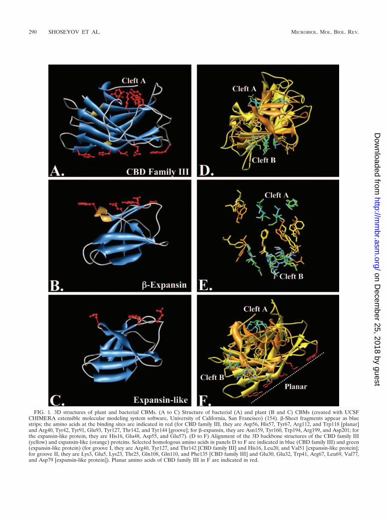
FIG. 1. 3D structures of plant and bacterial CBMs. (A to C) Structure of bacterial (A) and plant (B and C) CBMs (created with UCSFCHIMERA extensible molecular modeling system software, University of California, San Francisco) (154). �-Sheet fragments appear as bluestrips; the amino acids at the binding sites are indicated in red (for CBD family III, they are Asp56, His57, Tyr67, Arg112, and Trp118 [planar]and Arg40, Tyr42, Tyr91, Glu93, Tyr127, Thr142, and Tyr144 [groove]; for �-expansin, they are Asn159, Tyr160, Trp194, Arg199, and Asp201; forthe expansin-like protein, they are His16, Glu48, Asp55, and Glu57). (D to F) Alignment of the 3D backbone structures of the CBD family III(yellow) and expansin-like (orange) proteins. Selected homologous amino acids in panels D to F are indicated in blue (CBD family III) and green(expansin-like protein) (for groove I, they are Arg40, Tyr127, and Thr142 [CBD family III] and His16, Leu20, and Val51 [expansin-like protein];for groove II, they are Lys3, Glu5, Lys23, Thr25, Gln108, Gln110, and Phe135 [CBD family III] and Glu30, Glu32, Trp41, Arg67, Leu69, Val77,and Asp79 [expansin-like protein]). Planar amino acids of CBD family III in F are indicated in red.
290 SHOSEYOV ET AL. MICROBIOL. MOL. BIOL. REV.
on Decem
ber 25, 2018 by guesthttp://m
mbr.asm
.org/D
ownloaded from

for application in the future. CBMs may bind cytokines, growthfactors, and structural proteins to biocompatible polysaccha-ride scaffolds in order to selectively direct hard- and soft-tissueremodeling in reconstructive surgeries.
Another example of the important roles of CBMs in humanmetabolism is the unique CBM in the N-terminal region of theLaforin gene, a protein phosphatase involved in glycogen me-tabolism. A single mutation in the CBM, depleting its carbo-hydrate binding capability, is the cause of Lafora disease (34,208). This CBM may be utilized in the future as a possiblynonimmunogenic CBM for drug targeting.
The self-assembly properties of different polysaccharides,together with novel nano-fabrication techniques, may enablethe construction of 2D and 3D molecular crossroads throughwhich different CBMs may be used to carry and transfer mo-lecular cargo. These devices may be used to transfer drugs inone direction and simultaneously remove toxic molecules inthe other direction, as well as to store and remove molecularinformation in computational devices. The realization of thisconcept will require a better understanding of the molecularmechanism by which CBMs bind and move on the respectivepolysaccharides. It will take time and effort to harness the fullpotential of these molecules. However, the potential of thesemolecules for improving life in many aspects cannot be over-stated.
ACKNOWLEDGMENT
We gratefully acknowledge Deborah Weisman, from the computercenter of The Hebrew University, Faculty of Agriculture, for outstand-ing help with computer analysis of the 3D CBMs.
REFERENCES
1. Ahn, J. O., E. S. Choi, H. W. Lee, S. H. Hwang, C. S. Kim, H. W. Jang, S. J.Haam, and J. K. Jung. 2004. Enhanced secretion of Bacillus stearother-mophilus L1 lipase in Saccharomyces cerevisiae by translational fusion tocellulose-binding domain. Appl. Microbiol. Biotechnol. 64:833–839.
2. Ali, E., G. Zhao, M. Sakka, T. Kimura, K. Ohmiya, and K. Sakka. 2005.Functions of family-22 carbohydrate-binding module in Clostridium ther-mocellum Xyn10C. Biosci. Biotechnol. Biochem. 69:160–165.
3. Ansari, A. A., P. Shenbagamurthi, and D. G. Marsh. 1989. Completeprimary structure of a Lolium perenne (perennial rye grass) pollen allergen,Lol p III: comparison with Lol pI and II sequences. Biochemistry 28:8665–8667.
4. Araki, R., M. K. Ali, M. Sakka, T. Kimura, K. Sakka, and K. Ohmiya. 2004.Essential role of the family-22 carbohydrate-binding modules for beta-1,3-1,4-glucanase activity of Clostridium stercorarium Xyn10B. FEBS Lett. 561:155–158.
5. Azriel-Rosenfeld, R., M. Valensi, and I. Benhar. 2004. A human syntheticcombinatorial library of arrayable single-chain antibodies based on shufflingin vivo formed CDRs into general framework regions. J. Mol. Biol. 335:177–192.
6. Bachar, O., D. Fischer, R. Nussinov, and H. J. Wolfson. 1993. A computervision based technique for 3-D sequence independent structural compari-son of proteins. Protein Eng. 6:279–288.
7. Banka, R. R., S. Mishra, and T. K. Ghose. 1998. Fibril formation fromcellulose by a novel protein from Trichoderma reesei: a non-hydrolytic cel-lulolytic component? World J. Microb. Biotechnol. 4:551–558.
8. Barre, A., and P. Rouge. 2002. Homology modeling of the cellulose-bindingdomain of a pollen allergen from rye grass: structural basis for the celluloserecognition and associated allergenic properties. Biochem. Biophys. Res.Commun. 296:1346–1351.
9. Barrel, P., C. Suarezs, E. Batanero, C. Alfonso, J. D. Alche, M. I. Rodriguez-Garcia, M. Villalba, G. Rivas, and R. Rodriguez. 2005. An olive pollenprotein with allergenic activity, Ole e 10, defines a novel family of carbo-hydrate-binding modules and is potentially implicated in pollen germina-tion. Biochem. J. 390:77–84.
10. Bayer, E. A., E. Morag, and R. Lamed. 1994. The cellulosome—a treasure-trove for biotechnology. Trends Biotechnol. 12:379–386.
11. Bayer, E. A., H. Chanzy, R. Lamed, and Y. Shoham. 1998. Cellulose,cellulases and cellulosomes. Curr. Opin. Struct. Biol. 8:548–557.
12. Bayer, E. A., L. J. Shimon, Y. Shoham, and R. Lamed. 1998. Cellulo-somes—structure and ultrastructure. J. Struct. Biol. 124:221–234.
13. Bayer, E. A., J. P. Belaich, Y. Shoham, and R. Lamed. 2004. The cellulo-somes: multienzyme machines for degradation of plant cell wall polysac-charides. Annu. Rev. Microbiol. 58:521–554.
14. Beguin, P., and J. P. Aubert. 1994. The biological degradation of cellulose.FEMS Microbiol. Rev. 13:25–58.
15. Beguin, P., and P. M. Alzari. 1998. The cellulosome of Clostridium thermo-cellum. Biochem. Soc. Trans. 26:178–185.
16. Berdichevsky, Y., E. Ben-Zeev, R. Lamed, and I. Benhar. 1999. Phagedisplay of a cellulose binding domain from Clostridium thermocellum and itsapplication as a tool for antibody engineering. J. Immunol. Methods 228:151–162.
17. Berdichevsky, Y., R. Lamed, D. Frenkel, U. Gophna, E. A. Bayer, S. Yaron,Y. Shoham, and I. Benhar. 1999. Matrix-assisted refolding of single-chainFv� cellulose binding domain fusion proteins. Protein Expr. Purif. 17:249–259.
18. Berry, M. J., P. J. Davis, and M. J. Gidley. May 2001. Conjugated polysac-charide fabric detergent and conditioning products. U.S. patent 6,225,462.
19. Bjornvad, M., S. Pedersen, M. Schulein, and H. Bisg Rd-Fratzen. 1998.Alpha-amylase fused to cellulose binding domain, for starch degradation.WO98/16633A1, PCT.
20. Bolam, D. N., A. Ciruela, S. McQueen-Mason, P. Simpson, M. P. Williamson,J. E. Rixon, A. Boraston, G. P. Hazlewood, and H. J. Gilbert. 1998. Pseudo-monas cellulose-binding domains mediate their effects by increasing en-zyme substrate proximity. Biochem. J. 331:775–781.
21. Bolam, D. N., H. Xie, G. Pell, D. Hogg, G. Galbraith, B. Henrissat, and H. J.Gilbert. 2004. X4 modules represent a new family of carbohydrate-bindingmodules that display novel properties. J. Biol. Chem. 279:22953–22963.
22. Boraston, A., M. Bray, E. Burn, A. L. Creagh, N. Gilkes, M. Guarna, E.Jervis, P. Johnson, J. Kormos, L. McIntosh, B. McLean, L. Sandercock, P.Tomme, C. Haynes, A. Warren, and D. Kilburn. 1998. The structure andfunction of cellulose binding domains, p. 139–146. In M. Claeyssen, W.Nerinkx, and K. Piens (ed.), Carbohydrate from Trichoderma reesei andother microorganisms. The Royal Society of Chemistry, Cambridge, UnitedKingdom.
23. Boraston, A. B., B. W. McLean, J. M. Kormos, M. Alam, N. R. Gilkes, C. A.Haynes, P. Tomme, D. G. Kilburn, and R. A. J. Warren. 1999. Carbohy-drate-binding modules: diversity of structure and function, p. 202–211. InH. J. Gilbert, G. J. Davies, B. Henrissat, and B. Svensson (ed.), Recentadvances in carbohydrate bioengineering. The Royal Society of Chemistry,Cambridge, United Kingdom.
24. Boraston, A. B., B. W. McLean, M. M. Guarna, E. Amandaron-Akow, andD. G. Kilburn. 2001. A family 2a carbohydrate-binding module suitable asan affinity tag for proteins produced in Pichia pastoris. Protein Expr. Purif.21:417–423.
25. Boraston, A. B., E. Kwan, P. Chiu, R. A. Warren, and D. G. Kilburn. 2003.Recognition and hydrolysis of noncrystalline cellulose. J. Biol. Chem. 278:6120–6127.
26. Boraston, A. B., D. N. Bolam, H. J. Gilbert, and G. J. Davies. 2004.Carbohydrate-binding modules: fine-tuning polysaccharide recognition.Biochem. J. 382:769–781.
27. Brown, I. E., M. H. Mallen, S. J. Charnock, G. J. Davies, and G. W. Black.2001. Pectate lyase 10A from Pseudomonas cellulosa is a modular enzymecontaining a family 2a carbohydrate-binding module. Biochem. J. 355:155–165.
28. Brown, R. M. J., I. M. Saxena, and K. Kudlica. 1996. Cellulose biosynthesisin higher plants. Trends Plant Sci. 1:149–155.
29. Carrard, G., and M. Linder. 1999. Widely different off rates of two closelyrelated cellulose-binding domains from Trichoderma reesei. Eur. J. Bio-chem. 262:637–643.
30. Catala, C., and A. B. Bennett. 1998. Cloning and sequence analysis ofTomcel8, a new plant endo-1,4-�-D-glucanase gene, encoding a protein witha putative carbohydrate binding domain (accession no. AF098292). PlantPhysiol. 118:1535.
31. Cavaco-Paulo, A. 1998. Mechanism of cellulase action in textile processes.Carbohydr. Polymers 37:273–277.
32. Cavaco-Paulo, A. 1998. Processing textile fibers with enzymes, p. 180–189.In K. E. Eriksson and A. Cavaco-Paulo (ed.), Enzyme application in fiberprocessing. ACS symposium series 687. American Chemical Society, Wash-ington, D.C.
33. Cavaco-Paulo, A., J. Morgado, J. Andreaus, and D. G. Kilburn. 1999.Interactions of cotton with CBD peptides. Enzyme Microb. Technol. 25:639–643.
34. Chan, E. M., C. A. Ackerley, H. Lohi, L. Ianzano, M. A. Cortez, P. Shannon,S. W. Scherer, and B. A. Minassian. 2004. Laforin preferentially binds theneurotoxic starch-like polyglucosans, which form in its absence in progres-sive myoclonus epilepsy. Hum. Mol. Genet. 13:1117–1129.
35. Cho, H. T., and D. J. Cosgrove. 2000. Altered expression of expansinmodulates leaf growth and pedicel abscission in Arabidopsis thaliana. Proc.Natl. Acad. Sci. USA 97:9783–9788.
36. Choi, D., Y. Lee, H. T. Cho, and H. Kende. 2003. Regulation of expansin
VOL. 70, 2006 CBMs: BIOCHEMICAL PROPERTIES AND NOVEL APPLICATIONS 291
on Decem
ber 25, 2018 by guesthttp://m
mbr.asm
.org/D
ownloaded from

gene expression affects growth and development in transgenic rice plants.Plant Cell 15:1386–1398.
37. Cicortas Gunnarsson, L., E. Nordberg Karlsson, A. S. Albrekt, M. Andersson,O. Holst, and M. Ohlin. 2004. A carbohydrate binding module as a diver-sity-carrying scaffold. Protein Eng. Des. Sel. 17:213–221.
38. Cortese, R., P. Monaci, A. Luzzago, C. Santini, F. Bartoli, I. Cortese, P.Fortugno, G. Galfre, A. Nicosia, and F. Felici. 1996. Selection of biologicallyactive peptides by phage display of random peptide libraries. Curr. Opin.Biotechnol. 7:616–621.
39. Cosgrove, D. J., P. Bedinger, and D. M. Durachko. 1997. Group I allergensof grass pollen as cell wall-loosening agents. Proc. Natl. Acad. Sci. USA94:6559–6564.
40. Cosgrove, D. J. 2000. Loosening of plant cell walls by expansins. Nature407:321–326.
41. Cottam, G. P., D. M. Moran, and R. Standring. 1986. Physicochemical andimmunochemical characterization of allergenic proteins from rye-grass(Lolium perenne) pollen prepared by a rapid and efficient purificationmethod. Biochem. J. 234:305–310.
42. Coutinho, J. B., N. R. Gilkes, D. G. Kilburn, R. A. J. Warren, and R. C.Miller. 1993. The nature of the cellulose-binding domain affects the activ-ities of a bacterial endoglucanase on different forms of cellulose. FEMSMicrobiol. Lett. 113:211–218.
43. Coutinho, P. M., and B. Henrissat. 1999. Carbohydrate-active enzyme: anintegrated database approach, p. 3–12. In H. J. Gilbert, G. J. Davies, B.Henrissat, and B. Svensson (ed.), Recent advances in carbohydrate bioengi-neering. The Royal Society of Chemistry, Cambridge, United Kingdom.
44. Craig, S. J., F. C. Foong, and R. Nordon. 2006. Engineered proteins con-taining the cohesin and dockerin domains from Clostridium thermocellumprovides a reversible, high affinity interaction for biotechnology applica-tions. J. Biotechnol. 121:165–173.
45. Davis, G. 1998. Structural studies on cellulases. Biochem. Soc. Trans. 26:167–173.
46. Degani, O., S. Gepstein, and C. G. Dosoretz. 2004. A new method formeasuring scouring efficiency of natural fibers based on the cellulose-bind-ing domain–beta-glucuronidase fused protein. J. Biotechnol. 107:265–273.
47. Demain, A. L., M. Newcomb, and J. H. Wu. 2005. Cellulase, clostridia, andethanol. Microbiol. Mol. Biol. Rev. 69:124–154.
48. De Marino, S., M. A. Castiglione Morelli, F. Fraternali, E. Tamborini, G.Musco, S. Vrtala, C. Dolecek, P. Arossio, R. Valenta, and A. Pastore. 1999.An immunoglobulin-like fold in a major plant allergen: the solution struc-ture of Phl p 2 from timothy grass pollen. Structure 7:943–952.
49. Din, N., R. N. Gilkes, B. Tekant, R. C. Miller, Jr., R. A. J. Warren, and D. G.Kilburn. 1991. Non-hydrolytic disruption of cellulose fibers by the bindingdomain of a bacterial cellulase. Bio/Technology 9:1096–1099.
50. Ding, S. Y., R. Lamed, E. A. Bayer, and M. E. Himmel. 2003. The bacterialscaffoldin: structure, function and potential applications in the nano-sciences. Genet. Eng. 25:209–225.
51. Ding, X., J. Shields, R. Allen, and R. S. Hussey. 1998. A secretory cellulose-binding protein cDNA cloned from the root-knot nematode (Meloidogyneincognita). Mol. Plant-Microbe Interact. 11:952–959.
52. Doheny, J. G., E. J. Jervis, M. M. Guarna, R. K. Humphries, R. A. Warren,and D. G. Kilburn. 1999. Cellulose as an inert matrix for presenting cyto-kines to target cells: production and properties of a stem cell factor–cellulose-binding domain fusion protein. Biochem. J. 339:429–434.
53. Doi, R. H., M. Goldstein, S. Hashida, J. S. Park, and M. Takagi. 1994. TheClostridium cellulovorans cellulosome. Crit. Rev. Microbiol. 20:87–93.
54. Doi, R. H., and A. Kosugi. 2004. Cellulosomes: plant-cell-wall-degradingenzyme complexes. Nat. Rev. Microbiol. 2:541–551.
55. Doran, P. M. 2000. Foreign protein production in plant tissue cultures.Curr. Opin. Biotechnol. 11:199–204.
56. Esch, R. E., and D. G. Klapper. 1989. Identification and localization ofallergenic determinant on grass group-I antigens using mono-clonal anti-bodies. J. Immunol. 142:179–184.
57. Fexby, S., and L. Bulow. 2004. Hydrophobic peptide tags as tools in bio-separation. Trends Biotechnol. 22:511–516.
58. Fierobe, H. P., A. Mechaly, C. Tardif, A. Belaich, R. Lamed, Y. Shoham,J. P. Belaich, and E. A. Bayer. 2001. Design and production of activecellulosome chimeras. Selective incorporation of dockerin-containing en-zymes into defined functional complexes. J. Biol. Chem. 276:21257–21261.
59. Finnegan, P. M., S. M. Brumbley, M. G. O’Shea, H. Nevalainen, and P. L.Bergquist. 2005. Diverse dextranase genes from Paenibacillus species.Arch. Microbiol. 183:140–147.
60. Fischer, D., O. Bachar, R. Nussinov, and H. J. Wolfson. 1992. An efficientautomated computer vision based technique for detection of three dimen-sional structural motifs in proteins. J. Biomol. Struct. Dyn. 9:769–789.
61. Forrer, P., S. Jung, and A. Pluckthun. 1999. Beyond binding: using phagedisplay to select for structure, folding and enzymatic activity in proteins.Curr. Opin. Struct. Biol. 9:514–520.
62. Francisco, J. A., C. Stathopoulos, R. A. Warren, D. G. Kilburn, and G.Georgiou. 1993. Specific adhesion and hydrolysis of cellulose by intactEscherichia coli expressing surface anchored cellulase or cellulose bindingdomains. Bio/Technology 11:491–495.
63. Freidhoff, L. R., E. E. Kautzky, J. H. Grant, D. A. Meyers, and D. G. Marsh.1986. A study on the human immune response to Lilium perenne (rye)pollen and its components, Lol p I and Lol p II (rye I and rye II). I.Prevalence of reactivity to the allergens and correlations among skin test,IgE antibody and IgG antibody data. J. Allergy Clin. Immunol. 78:1190–1201.
64. Fuglsang, C. C., and R. Tsuchiya. July 2001. Cellulose binding domains(CBDs) for oral care products. U.S. patent 6,264,925.
65. Gao, B., R. Allen, E. L. Davis, T. J. Baum, and R. S. Hussey. 2004. Molec-ular characterization and developmental expression of a cellulose-bindingprotein gene in the soybean cyst nematode Heterodera glycines. Int. J.Parasitol. 34:1377–1383.
66. Gao, P. J., G. J. Chen, T. H. Wang, Y. S. Zhang, and J. Liu. 2001. Non-hydrolytic disruption of crystalline structure of cellulose by cellulose bind-ing domain and linker sequence of cellobiohydrolase I from Penicilliumjanthinellum. Acta Biochim. Biophys. Sin. 33:13–18.
67. Gaskin, D. J., K. Starck, N. A. Turner, and E. N. Vulfson. 2001. Phagedisplay combinatorial libraries of short peptides: ligand selection for pro-tein purification. Enzyme Microb. Technol. 28:766–772.
68. Giardina, T., A. P. Gunning, N. Juge, C. B. Faulds, C. S. Furniss, B.Svensson, V. J. Morris, and G. Williamson. 2001. Both binding sites of thestarch-binding domain of Aspergillus niger glucoamylase are essential forinducing a conformational change in amylose. J. Mol. Biol. 313:1149–1159.
69. Gilkes, N. R., R. A. Warren, R. J. Miller, and D. G. Kilburn. 1988. Preciseexcision of the cellulose binding domains from two Cellulomonas fimi cel-lulases by a homologous protease and the effect on catalysis. J. Biol. Chem.263:10401–10407.
70. Gilkes, N. R., B. Henrissat, D. G. Kilburn, R. J. Miller, and R. A. Warren.1991. Domains in microbial �-1,4-glycanases: sequence conservation, func-tion, and enzyme families. Microbiol. Rev. 55:303–315.
71. Gilkes, N. R., D. G. Kilburn, R. C. Miller, Jr., and A. Warren. October1998. Methods and compositions for modification of polysaccharide char-acteristics. U.S. patent 5,821,358.
72. Gilkes, N. R., D. G. Kilburn, R. C. Miller, Jr., R. A Warren, J. Sugiyama,H. Chanzy, and B. Henrissat. 1993. Visualization of the adsorption of abacterial endo-beta-1,4-glucanase and its isolated cellulose-binding domainto crystalline cellulose. Int. J. Biol. Macromol. 15:347–351.
73. Glansbeek, H. L., H. M. van Beuningen, E. L. Vitters, P. M. van der Kraan,and W. B. van den Berg. 1998. Expression of recombinant human solubletype II transforming growth factor-beta receptor in Pichia pastoris andEscherichia coli: two powerful systems to express a potent inhibitor oftransforming growth factor-beta. Protein Expr. Purif. 12:201–207.
74. Goldstein, M. A., M. Takagi, S. Hashida, O. Shoseyov, R. H. Doi, and I. H.Segel. 1993. Characterization of the cellulose-binding domain of the Clos-tridium cellulovorans cellulose-binding protein A. J. Bacteriol. 175:5762–5768.
75. Greenwood, J. M., E. Ong, N. R. Gilkes, R. A. Warren, R. C. Miller, Jr., andD. G. Kilburn. 1992. Cellulose-binding domains: potential for purificationof complex proteins. Protein Eng. 5:361–365.
76. Gunneriusson, E., P. Samuelson, M. Uhlen, P. A. Nygren, and S. Stahl.1996. Surface display of a functional single-chain Fv antibody on staphylo-cocci. J. Bacteriol. 178:1341–1346.
77. Gustavsson, M., J. Lehtio, S. Denman, T. T. Teeri, K. Hult, and M. Martinelle.2001. Stable linker peptides for a cellulose-binding domain–lipase fusionprotein expressed in Pichia pastoris. Protein Eng. 14:711–7115.
78. Gustavsson, M. T., P. V. Persson, T. Iversen, K. Hult, and M. Martinelle.2004. Polyester coating of cellulose fiber surfaces catalyzed by a cellulose-binding module–Candida antarctica lipase B fusion protein. Biomacromol-ecules 5:106–112.
79. Hall, J., G. W. Black, L. M. Ferreira, S. J. Millward-Sadler, B. R. Ali, G. P.Hazlewood, and H. J. Gilbert. 1995. The non-catalytic cellulose-bindingdomain of a novel cellulase from Pseudomonas fluorescens subsp. cellulosais important for the efficient hydrolysis of Avicel. Biochem. J. 309:749–756.
80. Harakas, N. K. 1994. Protein purification process engineering. Biospecificaffinity chromatography. Bioprocess Technol. 18:259–316.
81. Hashimoto, H., Y. Tamai, F. Okazaki, Y. Tamaru, T. Shimizu, T. Araki, andM. Sato. 2005. The first crystal structure of a family 31 carbohydrate-binding module with affinity to beta-1,3-xylan. FEBS Lett. 579:4324–4328.
82. Hatada, Y., Y. Hidaka, Y. Nogi, K. Uchimura, K. Katayama, Z. Li, M.Akita, Y. Ohta, S. Goda, H. Ito, H. Matsui, S. Ito, and K. Horikoshi. 2004.Hyper-production of an isomalto-dextranase of an Arthrobacter sp. by aprotease-deficient Bacillus subtilis: sequencing, properties, and crystalliza-tion of the recombinant enzyme. Appl. Microbiol. Biotechnol. 65:583–592.
83. Haynes, C. A., P. Tomme, and D. G. Kilburn. April 2000. Two-phasepartition affinity separation system and affinity separated cell-containingcomposition. U.S. patent 6,048,715.
84. Hefford, M. A., K. Laderoute, G. E. Willick, M. Yaguchi, and V. L. Seligy.1992. Bipartite organization of the Bacillus subtilis endo-beta-1,4-glucanaserevealed by C-terminal mutations. Protein Eng. 5:433–439.
85. Henrissat, B. 1994. Cellulases and their interaction with cellulose. Cellulose1:169–196.
86. Henrissat, B., T. T. Teeri, and R. A. Warren. 1998. A scheme for designat-
292 SHOSEYOV ET AL. MICROBIOL. MOL. BIOL. REV.
on Decem
ber 25, 2018 by guesthttp://m
mbr.asm
.org/D
ownloaded from

ing enzymes that hydrolyse the polysaccharides in the cell walls of plants.FEBS Lett. 425:352–354.
87. Herbers, K., and U. Sonnewald. 1999. Production of new/modified proteinsin transgenic plants. Curr. Opin. Biotechnol. 10:163–168.
88. Hilden, L., and G. Johansson. 2004. Recent developments on cellulases andcarbohydrate-binding modules with cellulose affinity. Biotechnol. Lett. 26:1683–1693.
89. Holm, L., and C. Sander. 1993. Protein structure comparison by alignmentof distance matrices. J. Mol. Biol. 233:123–138.
90. Hood, E. E., and J. M. Jilka. 1999. Plant-based production of xenogenicproteins. Curr. Opin. Biotechnol. 10:382–386.
91. Hsu, S. H., S. W. Whu, S. C. Hsieh, C. L. Tsai, D. C. Chen, and T. S. Tan.2004. Evaluation of chitosan-alginate-hyaluronate complexes modified byan RGD-containing protein as tissue-engineering scaffolds for cartilageregeneration. Artif. Organs 28:693–703.
92. Hsu, S. H., W. P. Chu, Y. S. Lin, Y. L. Chiang, D. C. Chen, and C. L. Tsai.2004. The effect of an RGD-containing fusion protein CBD-RGD in pro-moting cellular adhesion. J. Biotechnol. 111:143–154.
93. Hwang, S., J. Ahn, S. Lee, T. G. Lee, S. Haam, K. Lee, I. S. Ahn, and J. K.Jung. 2004. Evaluation of cellulose-binding domain fused to a lipase for thelipase immobilization. Biotechnol. Lett. 26:603–605.
94. Ito, J., Y. Fujita, M. Ueda, H. Fukuda, and A. Kondo. 2004. Improvementof cellulose-degrading ability of a yeast strain displaying Trichoderma reeseiendoglucanase II by recombination of cellulose-binding domains. Biotech-nol. Prog. 20:688–691.
95. Jamal-Talabani, S., A. B. Boraston, J. P. Turkenburg, N. Tarbouriech,V. M. Ducros, and G. J. Davies. 2004. Ab initio structure determination andfunctional characterization of CBM36, a new family of calcium-dependentcarbohydrate-binding modules. Structure 12:1177–1187.
96. Jervis, E. J., C. A. Haynes, and D. G. Kilburn. 1997. Surface diffusion ofcellulases and their isolated binding domains on cellulose. J. Biol. Chem.272:24016–24023.
97. Jervis, E. J., M. M. Guarna, J. G. Doheny, C. A. Haynes, and D. G. Kilburn.2005. Dynamic localization and persistent stimulation of factor-dependentcells by a stem cell factor/cellulose binding domain fusion protein. Biotech-nol. Bioeng. 91:314–324.
98. Jiang, M., and A. Radford. 2000. Exploitation of a cellulose-binding domainfrom Neurospora crassa. Enzyme Microb. Technol. 27:434–442.
99. Johnsson, K., and L. Ge. 1999. Phage display of combinatorial peptide andprotein libraries and their applications in biology and chemistry. Curr. Top.Microbiol. Immunol. 243:87–105.
100. Kalum, L., and B. K. Andersen. November 2000. Enzymatic treatment ofdenim. U.S. patent 6,146,428.
101. Kauffmann, C., O. Shoseyov, E. Shpigel, E. A. Bayer, R. Lamed, Y. Shoham,and R. T. Mandelbaum. 2000. A novel methodology for enzymatic removalof atrazine from water by CBD-fusion protein immobilized on cellulose.Environ. Sci. Technol. 34:1292–1296.
102. Kavoosi, M., J. Meijer, E. Kwan, A. L. Creagh, D. G. Kilburn, and C. A.Haynes. 2004. Inexpensive one-step purification of polypeptides expressedin Escherichia coli as fusions with the family 9 carbohydrate-binding moduleof xylanase 10A from T. maritima. J. Chromatogr. B 807:87–94.
103. Kilburn, D. G., R. C. Miller, N. R. Gilkes, and R. A. J. Warren. July 1996.Conjugate of non-protein chemical moiety and polypeptide having cellu-lose-binding region. U.S. patent 5,928,917.
104. Kilburn, D. G., R. C. Miller, R. A. J. Warren, and N. R. Gilkes. October1999. Polysaccharide binding fusion proteins and conjugates. U.S. patent5,962,289.
105. Kim, H., M. Goto, H. J. Jeong, K. H. Jung, I. Kwon, and K. Furukawa. 1998.Functional analysis of a hybrid endoglucanase of bacterial origin having acellulose binding domain from a fungal exoglucanase. Appl. Biochem. Bio-technol. 75:193–204.
106. Kitaoka, T., and H. Tanaka. 2001. Novel paper strength additive containingcellulose-binding domain of cellulase. J. Wood Sci. 47:322–324.
107. Kleinman, H. K., L. Luckenbill-Edds, F. W. Cannon, and G. C. Sephel.1987. Use of extracellular matrix components for cell culture. Anal. Bio-chem. 166:1–13.
108. Kroon, P. A., G. Williamson, N. M. Fish, D. B. Archer, and N. J. Belshaw.2000. A modular esterase from Penicillium funiculosum which releasesferulic acid from plant cell walls and binds crystalline cellulose contains acarbohydrate binding module. Eur. J. Biochem. 267:6740–6752.
109. Krull, L. H., F. R. Dintzis, H. L. Griffin, and F. L. Baker. 1988. A micro-fibril-generating factor from the cellulase of Trichoderma reesei. Biotechnol.Bioeng. 31:321–327.
110. Kudla, U., L. Qin, A. Milac, A. Kielak, C. Maissen, H. Overmars, H.Popeijus, E. Roze, A. Petrescu, G. Smant, J. Bakker, and J. Helder. 2005.Origin, distribution and 3D-modeling of Gr-EXPB1, an expansin from thepotato cyst nematode Globodera rostochiensis. FEBS Lett. 579:2451–2457.
111. Lam, H., M. Kavoosi, C. A. Haynes, D. I. Wang, and D. Blankschtein. 2005.Affinity-enhanced protein partitioning in decyl beta-D-glucopyranoside two-phase aqueous micellar systems. Biotechnol. Bioeng. 89:381–392.
112. Latorre-Garcia, L., A. C. Adam, P. Manzanares, and J. Polaina. 2005.
Improving the amylolytic activity of Saccharomyces cerevisiae glucoamylaseby the addition of a starch binding domain. J. Biotechnol. 118:167–176.
113. Lee, I., B. R. Evans, and J. Woodward. 2000. The mechanism of cellulaseaction on cotton fibers: evidence from atomic force microscopy. Ultrami-croscopy 82:213–221.
114. Lehtio, J., T. T. Teeri, and P. A. Nygren. 2000. Alpha-amylase inhibitorsselected from a combinatorial library of a cellulose binding domain scaffold.Proteins 41:316–322.
115. Lehtio, J., H. Wernerus, P. Samuelson, T. T. Teeri, and S. Stahl. 2001.Directed immobilization of recombinant staphylococci on cotton fibers byfunctional display of a fungal cellulose-binding domain. FEMS Microbiol.Lett. 195:197–204.
116. Levy, I., A. Nussinovitch, E. Shpigel, and O. Shoseyov. 2002. Recombinantcellulose crosslinking protein: a novel paper-modification biomaterial. Cel-lulose 9:91–98.
117. Levy, I., and O. Shoseyov. 2002. Cellulose binding domains: industrial andbiotechnological application. Biotechnol. Adv. 20:191–213.
118. Levy, I., Z. Shani, and O. Shoseyov. 2002. Modification of polysaccharidesand plant cell wall by endo-1,4-�-glucanase (EGase) and cellulose bindingdomains (CBD). Biomol. Eng. 19:17–30.
119. Levy, I., G. Ward, Y. Hadar, O. Shoseyov, and C. G. Dosoretz. 2003.Oxidation of 4-bromophenol by recombinant cellulose binding domainhorseradish peroxidase fused protein immobilized to cellulose. Biotechnol.Bioeng. 82:223–231.
120. Levy, I., T. Paldi, D. Siegel, and O. Shoseyov. 2003. Cellulose bindingdomain from Clostridium cellulovorans as a paper modification reagent.Nordic Pulp Paper Res. J. 18:421–428.
121. Levy, I., T. Paldi, and O. Shoseyov. 2004. Engineering a bifunctional proteinthat cross-bridges between starch and cellulose. Biomaterials 25:1841–1849.
122. Limon, M. C., E. Margolles-Clark, T. Benitez, and M. Penttila. 2001.Addition of substrate-binding domains increases substrate-binding capacityand specific activity of a chitinase from Trichoderma harzianum. FEMSMicrobiol. Lett. 198:57–63.
123. Limon, M. C., M. R. Chacon, R. Mejias, J. Delgado-Jarana, A. M. Rincon,A. C. Codon, and T. Benitez. 2004. Increased antifungal and chitinasespecific activities of Trichoderma harzianum CECT 2413 by addition of acellulose binding domain. Appl. Microbiol. Biotechnol. 64:675–685.
124. Linder, M., E. Margolles-Clark, T. Reinikainen, and T. T. Teeri. 1997.Trichoderma reesei cellobiohydrolase I with an endoglucanase cellulose-binding domain: action on bacterial microcrystalline cellulose. J. Biotech-nol. 57:49–57.
125. Linder, M., T. Nevanen, and T. T. Teeri. 1999. Design of a pH-dependentcellulose-binding domain. FEBS Lett. 447:13–16.
126. Lowe, C. R. 2001. Combinatorial approaches to affinity chromatography.Curr. Opin. Chem. Biol. 5:248–256.
127. Lymar, E. S., B. Li, and V. Renganathan. 1995. Purification and character-ization of a cellulose-binding �-glucosidase from cellulose-degrading cul-tures of Phanerochaete chrysosporium. Appl. Environ. Microbiol. 61:2976–2980.
128. Margolles-Clark, E., M. Tenkanen, H. Soderlund, and M. Pentilla. 1996.Acetyl xylan esterase from Trichoderma reesei contains an active site serineand a cellulose-binding domain. Eur. J. Biochem. 237:553–560.
129. McCartney, L., H. J. Gilbert, D. N. Bolam, A. B. Boraston, and J. P. Knox.2004. Glycoside hydrolase carbohydrate-binding modules as molecularprobes for the analysis of plant cell wall polymers. Anal. Biochem. 326:49–54.
130. McDonald, J. K., C. M. Taylor, and S. Rafferty. 2003. Design, preparation,and characterization of mixed dimers of inducible nitric oxide synthaseoxygenase domains. Protein Expr. Purif. 27:115–127.
131. McQueen-Mason, S. J., D. M. Durachko, and D. J. Cosgrove. 1992. Twoendogenous proteins that induce cell wall extension in plants. Plant Cell4:1425–1433.
132. McQueen-Mason, S. J., and D. J. Cosgrove. 1995. Expansin mode of actionon cell walls. Plant Physiol. 107:87–100.
133. Meade, H. 2000. Method of purifying heterologous proteins. WO/61725A1,PCT.
134. Meade, H., S. P. Fulton, and Y. Echelard. 2001. Methods of producing atarget molecule in transgenic animal and purification of the target mole-cule. WO/126455A1, PCT.
135. Mingardon, F., S. Perret, A. Belaich, C. Tardif, J. P. Belaich, and H. P.Fierobe. 2005. Heterologous production, assembly, and secretion of a mini-cellulosome by Clostridium acetobutylicum ATCC 824. Appl. Environ. Mi-crobiol. 71:1215–1222.
136. Miyanaga, A., T. Koseki, H. Matsuzawa, T. Wakagi, H. Shoun, and S.Fushinobu. 2004. Crystal structure of a family 54 alpha-L-arabinofuranosi-dase reveals a novel carbohydrate-binding module that can bind arabinose.J. Biol. Chem. 279:44907–44914.
137. Mordoccoa, A., C. Kuek, and R. Jenkinsa. 1999. Continuous degradation ofphenol at low concentration using immobilized Pseudomonas putida. En-zyme Microb. Technol. 25:530–536.
138. Nam, J., Y. Fujita, T. Arai, A. Kondo, Y. Morikaw, H. Okada, M. Ueda, and
VOL. 70, 2006 CBMs: BIOCHEMICAL PROPERTIES AND NOVEL APPLICATIONS 293
on Decem
ber 25, 2018 by guesthttp://m
mbr.asm
.org/D
ownloaded from

A. Tanaka. 2002. Construction of engineered yeast with the ability of bind-ing to cellulose. J. Mol. Catalys. 17:197–202.
139. Nigam, J. N. 2000. Continuous ethanol production from pineapple cannerywaste using immobilized yeast cells. J. Biotechnol. 80:189–193.
140. Nigmatullin, R., R. Lovitt, C. Wright, M. Linder, T. Nakari-Setala, and M.Gama. 2004. Atomic force microscopy study of cellulose surface interactioncontrolled by cellulose binding domains. Colloids Surf. B 35:125–135.
141. Nikas, Y. J., C. L. Liu, T. Srivastava, N. L. Abbott, and D. Blankschtein.1992. Protein partitioning in two-phase aqueous nonionic micellar solu-tions. Macromolecules 25:4797–4806.
142. Nordon, R. E., A. Shu, F. Camacho, and B. K. Milthorpe. 2004. Hollow-fiber assay for ligand-mediated cell adhesion. Cytometry A 57:39–44.
143. Novy, R., K. Yaeger, S. Monsma, M. McCormick, J. Berg, O. Shoseyov, E.Shpigel, D. Seigel, A. Goldlust, G. Efroni, Y. Singer, D. Kilburn, P. Tomme,and N. Gilkes. 1997. Cellulose binding domain expression vectors for therapid, low cost purification of CBD-fusion proteins. FASEB J. 11:1715.
144. Ofir, K., Y. Berdichevsky, I. Benhar, R. Azriel-Rosenfeld, R. Lamed, Y.Barak, E. A. Bayer, and E. Morag. 2005. Versatile protein microarray basedon carbohydrate-binding modules. Proteomics 5:1806–1814.
145. Ohta, Y., Y. Hatada, Y. Nogi, Z. Li, S. Ito, and K. Horikoshi. 2004. Cloning,expression, and characterization of a glycoside hydrolase family 86 beta-agarase from a deep-sea Microbulbifer-like isolate. Appl. Microbiol. Bio-technol. 66:266–275.
146. Ong, E., J. M. Greenwood, N. R. Gilkes, D. G. Kilburn, R. C. Miller, Jr.,and A. J. Warren. 1989. The cellulose-binding domain of cellulases: toolsfor biotechnology. Trends Biotechnol. 7:239–243.
147. Otomo, T., K. Teruya, K. Uegaki, T. Yamazaki, and Y. Kyogoku. 1999.Improved segmental isotope labeling of proteins and application to a largerprotein. J. Biomol. NMR 14:105–114.
148. Pages, S., L. Gal, A. Belaich, C. Gaudin, C. Tardif, and J. P. Belaich. 1997.Role of scaffolding protein CipC of Clostridium cellulolyticum in cellulosedegradation. J. Bacteriol. 179:2810–2816.
149. Pala, H., M. A. Lemos, M. Mota, and F. M. Gama. 2001. Enzymatic upgradeof old paperboard containers. Enzyme Microb. Technol. 29:274–279.
150. Pala, H., R. Pinto, M. Mota, A. P. Duarte, and F. M. Gama. 2003. Cellulose-binding domains as a tool for paper recycling, p. 105–115. In S. D. Mansfieldand J. N. Saddler (ed.), Application of enzymes to lignocellulosics. ACSsymposium series 855. American Chemical Society, Washington, D.C.
151. Paloheimo, M., A. Mantyla, J. Kallio, and P. Suominen. 2003. High-yieldproduction of a bacterial xylanase in the filamentous fungus Trichodermareesei requires a carrier polypeptide with an intact domain structure. Appl.Environ. Microbiol. 69:7073–7082.
152. Palomer, X., E. Domı́nguez-Puigjaner, M. Vendrell, and I. Llop-Tous. 2004.Study of the strawberry cell endo-beta-(1,4)-glucanase protein accumula-tion and characterization of its in vitro activity by heterologous expressionin Pichia pastoris. Plant Sci. 167:509–518.
153. Palonen, H., F. Tjerneld, G. Zacchi, and M. Tenkanen. 2004. Adsorption ofTrichoderma reesei CBH I and EG II and their catalytic domains on steampretreated softwood and isolated lignin. J. Biotechnol. 107:65–72.
154. Pettersen, E. F., T. D. Goddard, C. C. Huang, G. S. Couch, D. M. Greenblatt,E. C. Meng, and T. E. Ferrin. 2004. UCSF Chimera—a visualization systemfor exploratory research and analysis. J. Comput. Chem. 25:1605–1612.
155. Pfeifer, T. A., M. M. Guarna, E. M. Kwan, G. Lesnicki, D. A. Theilmann,T. A. Grigliatti, and D. G. Kilburn. 2001. Expression analysis of a modifiedfactor X in stably transformed insect cell lines. Protein Expr. Purif. 23:233–241.
156. Phelps, M. R., J. B. Hobbs, D. G. Kilburn, and R. F. B. Turner. 1994.Technology for regenerable biosensor probes based on enzyme cellulose-binding domain conjugates. Biotechnol. Prog. 10:433–440.
157. Phelps, M. R., J. B. Hobbs, D. G. Kilburn, and R. F. B. Turner. 1995. Anautoclavable glucose biosensor for microbial fermentation monitoring andcontrol. Biotechnol. Bioeng. 46:514–524.
158. Pinto, R., S. Moreira, M. Mota, and M. Gama. 2004. Studies on thecellulose-binding domains adsorption to cellulose. Langmuir 20:1409–1413.
159. Qin, L., U. Kudla, E. H. Roze, A. Goverse, H. Popeijus, J. Nieuwland, H.Overmars, J. T. Jones, A. Schots, G. Smant, J. Bakker, and J. Helder. 2004.Plant degradation: a nematode expansin acting on plants. Nature 427:30.
160. Rechter, M., O. Lider, L. Cahalon, E. Baharav, M. Dekel, D. Seigel, I.Vlodavsky, H. Aingorn, I. R. Cohen, and O. Shoseyov. 1999. A cellulose-binding domain-fused recombinant human T cell connective tissue-activat-ing peptide-III manifests heparanase activity. Biochem. Biophys. Res. Com-mun. 255:657–662.
161. Reese, E. T., R. G. H. Sui, and H. S. Levinson. 1950. The biologicaldegradation of soluble cellulose derivatives and its relationship to themechanism of cellulose hydrolysis. J. Bacteriol. 59:485–497.
162. Richins, R. D., A. Mulchandani, and W. Chen. 2000. Expression, immobi-lization, and enzymatic characterization of cellulose-binding domain-organ-ophosphorus hydrolase fusion enzymes. Biotechnol. Bioeng. 69:591–596.
163. Rodi, D. J., and L. Makowski. 1999. Phage-display technology—finding aneedle in a vast molecular haystack. Curr. Opin. Biotechnol. 10:87–93.
164. Rodriguez, B., M. Kavoosi, J. Koska, A. L. Creagh, D. G. Kilburn, and C. A.
Haynes. 2004. Inexpensive and generic affinity purification of recombinantproteins using a family 2a CBM fusion tag. Biotechnol. Prog. 20:1479–1489.
165. Rodriguez-Sanoja, R., N. Oviedo, and S. Sanchez. 2005. Microbial starch-binding domain. Curr. Opin. Microbiol. 8:260–267.
166. Rotticci-Mulder, J. C., M. Gustavsson, M. Holmquist, K. Hult, and M.Martinelle. 2001. Expression in Pichia pastoris of Candida antarctica lipaseB and lipase B fused to a cellulose-binding domain. Protein Expr. Purif.21:386–392.
167. Saleemuddin, M. 1999. Bioaffinity based immobilization of enzymes. Adv.Biochem. Eng. Biotechnol. 64:203–226.
168. Saloheimo, M., M. Paloheimo, S. Hakola, J. Pere, B. Swanson, E. Nyyssonen,A. Bhatia, M. Ward, and M. Penttila. 2002. Swollenin, a Trichoderma reeseiprotein with sequence similarity to the plant expansins, exhibits disruptionactivity on cellulosic materials. Eur. J. Biochem. 269:4202–4211.
169. Samuelson, P., H. Wernerus, M. Svedberg, and S. Stahl. 2000. Staphylo-coccal surface display of metal-binding polyhistidyl peptides. Appl. Envi-ron. Microbiol. 66:1243–1248.
170. Shani, Z., E. Shpigel, L. Roiz, R. Goren, B. Vinocur, T. Tzfira, A. Altman,and O. Shoseyov. 1999. Cellulose-binding domain increases cellulose syn-thase activity in Acetobacter xylinum and biomass of transgenic plants, p.213–218. In A. Altman, M. Ziv, and S. Izhar (ed.), Plant biotechnology andin vitro biology in the 21st century. Kluwer Academic Publishers, Dor-drecht, The Netherlands.
171. Shani, Z., and O. Shoseyov. December 2001. Process of expressing andisolating recombinant proteins and recombinant protein products fromplants, plant derived tissues or cultured plant cells. U.S. patent 6,331,416.
172. Shoseyov, O., and R. H. Doi. 1990. Essential 170-kDa subunit for degrada-tion of crystalline cellulose by Clostridium cellulovorans cellulase. Proc.Natl. Acad. Sci. USA 87:2192–2195.
173. Shoseyov, O., T. Hamamoto, F. Foong, and R. H. Doi. 1990. Cloning ofClostridium cellulovorans endo-1,4-beta-glucanase genes. Biochem. Bio-phys. Res. Commun. 169:667–672.
174. Shoseyov, O., I. Shpigel, M. Goldstein, and R. Doi. September 1997. Meth-ods of use of cellulose binding domain proteins. U.S. patent 5,670,623.
175. Shoseyov, O., I. Shpigel, M. Goldstein, and R. Doi. November 1998. Cel-lulose binding domain proteins. U.S. patent 5,837,814.
176. Shoseyov, O., I. Shpigel, M. Goldstein, and R. Doi. February 1998. Cellu-lose binding domain fusion proteins. U.S. patent 5,719,044.
177. Shoseyov, O., I. Shpigel, M. M. Goldstein, and R. H. Doi. January 1999.Methods of detection using a cellulose binding domain fusion product. U.S.patent 5,856,201.
178. Shoseyov, O., Z. Shani, and E. Shpigel. February 2001. Transgenic plants ofaltered morphology. U.S. patent 6,184,440.
179. Shoseyov, O., I. Levy, Z. Shani, and S. D. Mansfield. 2003. Modulation ofwood fibers and paper by cellulose-binding domains, p. 116–131. In S. D.Mansfield and J. N. Saddler (ed.), Application of enzymes to lignocellu-losics. ACS symposium series 855. American Chemical Society, Washing-ton, D.C.
180. Shpigel, E., D. Elias, I. R. Cohen, and O. Shoseyov. 1998. Production andpurification of a recombinant human hsp60 epitope using the cellulose-binding domain in Escherichia coli. Protein Expr. Purif. 14:185–191.
181. Shpigel, E., L. Roiz, R. Goren, and O. Shoseyov. 1998. Bacterial cellulose-binding domain modulates in-vitro elongation of different plant cells. PlantPhysiol. 117:1185–1194.
182. Shpigel, E., A. Goldlust, G. Efroni, A. Avraham, A. Eshel, M. Dekel, and O.Shoseyov. 1999. Immobilization of recombinant heparinase I fused to cel-lulose-binding domain. Biotechnol. Bioeng. 65:17–23.
183. Shpigel, E., A. Goldlust, A. Eshel, I. K. Ber, G. Efroni, Y. Singer, I. Levy, M.Dekel, and O. Shoseyov. 2000. Expression, purification and applications ofstaphylococcal protein A fused to cellulose-binding domain. Biotechnol.Appl. Biochem. 31:197–203.
184. Smith, G. P., S. U. Patel, J. D. Windass, J. M. Thornton, G. Winter, andA. D. Griffiths. 1998. Small binding proteins selected from a combinatorialrepertoire of knottins displayed on phage. J. Mol. Biol. 277:317–332.
185. Somerville, C., S. Bauer, G. Brininstool, M. Facette, T. Hamann, J., Milne,E. Osborne, A. Paredez, S. Persson, T. Raab, S. Vorwerk, and H. Youngs.2004. Toward a systems approach to understanding plant cell walls. Science306:2206–2211.
186. Southall, S. M. P. J. Simpson, H. J. Gilbert, G. Williamson, and M. P.Williamson. 1999. The starch-binding domain from glucoamylase disruptsthe structure of starch. FEBS Lett. 447:58–60.
187. Stahl, S., and M. Uhlen. 1997. Bacterial surface display: trends andprogress. Trends Biotechnol. 15:185–192.
188. Stalbrand, H., A. Saloheimo, J. Vehmaanpera, B. Henrissat, and M. Penttila.1995. Cloning and expression in Saccharomyces cerevisiae of a Trichodermareesei �-mannanase gene containing a cellulose-binding domain. Appl. En-viron. Microbiol. 61:1090–1097.
189. Stempfer, G., B. Holl-Neugebauer, and R. Rudolph. 1996. Improved refold-ing of an immobilized fusion protein. Nat. Biotechnol. 14:329–334.
190. Suent, D. F., S. S. H. Wu, H. C. Cang, K. S. Dhugga, and H. C. Huang. 2003.Cell wall reactive proteins in the coat and wall of maize pollen. J. Biol.Chem. 278:43672–43681.
294 SHOSEYOV ET AL. MICROBIOL. MOL. BIOL. REV.
on Decem
ber 25, 2018 by guesthttp://m
mbr.asm
.org/D
ownloaded from

191. Suurnakki, A., M. Tenkanen, M. Siika-aho, M. L. Niku-Paavola, L. Viikari,and J. Buchert. 2000. Trichoderma reesei cellulases and their core domainsin the hydrolysis and modification of chemical pulp. Cellulose 7:189–209.
192. Teeri, T. T., A. Koivula, M. Linder, G. Wohlfahrt, C. Divne, and T. A. Jones.1998. Trichoderma reesei cellobiohydrolases: why so efficient on crystallinecellulose? Biochem. Soc. Trans. 26:173–178.
193. Terpe, K. 2003. Overview of tag protein fusions: from molecular and bio-chemical fundamentals to commercial systems. Appl. Microbiol. Biotech-nol. 60:523–533.
194. Tomme, P., H. Van Tilbeurgh, G. Pettersson, J. Van Damme, J. Vande-kerckhove, J. Knowles, and M. Teeri Claeyssens. 1988. Studies of thecellulolytic system of Trichoderma reesei QM 9414. Analysis of domainfunction in two cellobiohydrolases by limited proteolysis. Eur. J. Biochem.170:575–581.
195. Tomme, P., D. P. Driver, E. A. Amandoron, R. C. Miller, Jr., R. Antony, J.Warren, and D. G. Kilburn. 1995. Comparison of a fungal (family I) andbacterial (family II) cellulose-binding domain. J. Bacteriol. 177:4356–4363.
196. Tomme, P., R. A. Warren, and N. R. Gilkes. 1995. Cellulose hydrolysis bybacteria and fungi. Adv. Microb. Physiol. 37:1–81.
197. Tomme, P., A. Boraston, B. McLean, J. Kormos, A. L. Creagh, K. Sturch,N. R. Gilkes, C. A. Haynes, R. A. Warren, and D. G. Kilburn. 1998. Charac-terization and affinity applications of cellulose-binding domains. J. Chro-matogr. B 715:283–296.
198. Tormo, J., R. Lamed, A. J. Chirino, E. Morag, E. A. Bayer, Y. Shoham, andT. A. Steitz. 1996. Crystal structure of a bacterial family-III cellulose-binding domain: a general mechanism for attachment to cellulose. EMBOJ. 15:5739–5751.
199. Trainotti, L., S. Spolaore, A. Pavanello, B. Baldan, and G. Casadoro. 1999.A novel E-type endo-1,4-�-glucanase with a putative cellulose-binding do-main is highly expressed in ripening strawberry fruits. Plant Mol. Biol.40:323–332.
200. Turner, R. F. B., D. G. Kilburn, and M. R. Phelps. April 1997. Biosensorand interface membrane. U.S. patent 5,624,537.
201. Van Tilbeurgh, H., P. Tomme, M. Claeyssens, and R. Bhikhabhai. 1986.Limited proteolysis of the cellubiohydrolase I from Trichoderma reesei.Separation of functional domains. FEBS Lett. 204:223–227.
202. Volkov, I. Y., N. A. Lunina, and G. A. Velikodvorskaya. 2004. Prospects forthe practical application of substrate-binding modules of glycosyl hydro-lases. Appl. Biochem. Microbiol. 40:427–432.
203. von der Osten, C., M. E. Bjornvad, J. Vind, and M. D. Rasmussen. January2000. Process and composition for desizing cellulosic fabric with an enzymehybrid. U.S. patent 6,017,751.
204. von der Osten, C., M. E. Bjornvad, J. Vind, and M. D. Rasmussen. January2000. Process for removal or bleaching of soiling or stains from cellulosicfabric. U.S. patent 6,015,783.
205. Walter, H., D. E. Brooks, and D. Fisher. 1985. Partitioning in aqueous
two-phase systems: theory, methods, uses, and application to biotechnology.Academic Press, Orlando, Fla.
206. Wang, A. A., A. Mulchandani, and W. Chen. 2001. Whole-cell immobiliza-tion using cell surface-exposed cellulose-binding domain. Biotechnol. Prog.17:407–411.
207. Wang, A. A., A. Mulchandani, and W. Chen. 2002. Specific adhesion tocellulose and hydrolysis of organophosphate nerve agents by a geneticallyengineered Escherichia coli strain with a surface-expressed cellulose-bind-ing domain and organophosphorus hydrolase. Appl. Environ. Microbiol.68:1684–1689.
208. Wang, J., J. A. Stuckey, M. J. Wishart, and J. E. Dixon. 2002. A uniquecarbohydrate binding domain targets the lafora disease phosphatase toglycogen. J. Biol. Chem. 277:2377–2380.
209. Wernerus, H., J. Lehtio, T. Teerim, P. A. Nygren, and S. Stahl. 2001.Generation of metal-binding staphylococci through surface display of com-binatorially engineered cellulose-binding domains. Appl. Environ. Micro-biol. 67:4678–4684.
210. Wierzba, A., U. Reichl, R. F. B. Turner, R. A. J. Warren, and D. G. Kilburn.1995. Adhesion of mammalian-cells to a recombinant attachment factor,CBD/RGD, analyzed by image-analysis. Biotechnol. Bioeng. 46:185–193.
211. Wilchek, M., and I. Chaiken. 2000. An overview of affinity chromatography.Methods Mol. Biol. 147:1–6.
212. Woodward, J., M. K. Hayes, and N. E. Lee. 1988. Hydrolysis of cellulose bysaturating and non-saturating concentrations of cellulase—implications forsynergism. Bio/Technology 6:301–304.
213. Xiao, Z. Z., P. J. Gao, Y. B. Qu, and T. H. Wang. 2001. Cellulose-bindingdomain of endoglucanase III from Trichoderma reesei disrupting the struc-ture of cellulose. Biotechnol. Lett. 23:711–715.
214. Xu, Z., W. Bae, A. Mulchandani, R. K. Mehra, and W. Chen. 2002. Heavymetal removal by novel CBD-EC20 sorbents immobilized on cellulose.Biomacromolecules 3:462–465.
215. Yamada, K. M. 1983. Cell surface interactions with extracellular materials.Annu. Rev. Biochem. 52:761–799.
216. Yang, J. I., B. Petterson, and K. E. Eriksson. 1988. Development of bio-assays for the characterization of pulp fiber surfaces. I. Characterization ofvarious mechanical pulp fiber surfaces by specific cellulolitic enzymes.Nord. Pulp Paper Res. J. 42:19–25.
217. Yoshida, M., K. Igarashi, M. Wada, S. Kaneko, N. Suzuki, H. Matsumura,N. Nakamura, H. Ohno, and M. Samejima. 2005. Characterization of car-bohydrate-binding cytochrome b562 from the white-rot fungus Phanero-chaete chrysosporium. Appl. Environ. Microbiol. 71:4548–4555.
218. Zverlov, V. V., I. Y. Volkov, G. A. Velikodvorskaya, and W. H. Schwarz.2001. The binding pattern of two carbohydrate-binding modules of lami-narinase Lam16A from Thermotoga neapolitana: differences in �-glucanbinding within family CBM4. Microbiology 147:621–629.
VOL. 70, 2006 CBMs: BIOCHEMICAL PROPERTIES AND NOVEL APPLICATIONS 295
on Decem
ber 25, 2018 by guesthttp://m
mbr.asm
.org/D
ownloaded from