SMALL INTESTINE Dr IramTassaduq. SMALL INTESTINE Dr IramTassaduq.
Protocol for the Examination of Specimens From Patients With Carcinoma of the Small IntestineVersion: SmallIntestine 4.0.0.0 Protocol Posting Date: June 2017Includes pTNM requirements from the 8th Edition, AJCC Staging Manual
For accreditation purposes, this protocol should be used for the following procedures AND tumor types:Procedure DescriptionResection Includes specimens designated segmental resection,
pancreaticoduodenectomy (Whipple resection), ileocolic resection
Tumor Type DescriptionCarcinoma Including carcinomas arising in the duodenum, jejunum, and
ileum
This protocol is NOT required for accreditation purposes for the following:ProcedureBiopsyPrimary resection specimen with no residual cancer (eg, following neoadjuvant therapy)Recurrent tumorCytologic specimens
The following tumor types should NOT be reported using this protocol:Tumor TypeCarcinoma of the ampulla (consider Ampullary Carcinoma protocol)Well-differentiated neuroendocrine tumor of the duodenum (consider the Duodenal and Ampullary NET protocol)Well-differentiated neuroendocrine tumor of the jejunum and ileum (consider the Jejunal and Ilial NET protocol)Lymphoma (consider the Hodgkin or non-Hodgkin Lymphoma protocols)Gastrointestinal stromal tumor (GIST) (consider the GIST protocol)Non-GIST sarcoma (consider the Soft Tissue protocol)
AuthorsChanjuan Shi, MD, PhD*; Jordan Berlin, MD; Philip A. Branton, MD; Patrick L. Fitzgibbons, MD; Wendy L. Frankel, MD; Wayne L. Hofstetter, MD; Sanjay Kakar, MD; David Kelsen, MD; Veronica Klepeis, MD, PhD; Jason Talmadge Lewis, MD; Laura H. Tan, MD, PhD; Mary K. Washington, MD, PhD
With guidance from the CAP Cancer and CAP Pathology Electronic Reporting Committees.* Denotes primary author. All other contributing authors are listed alphabetically.
© 2017 College of American Pathologists (CAP). All rights reserved. For Terms of Use please visit www.cap.org/cancerprotocols.

Gastrointestinal • Small IntestineSmallIntestine 4.0.0.0
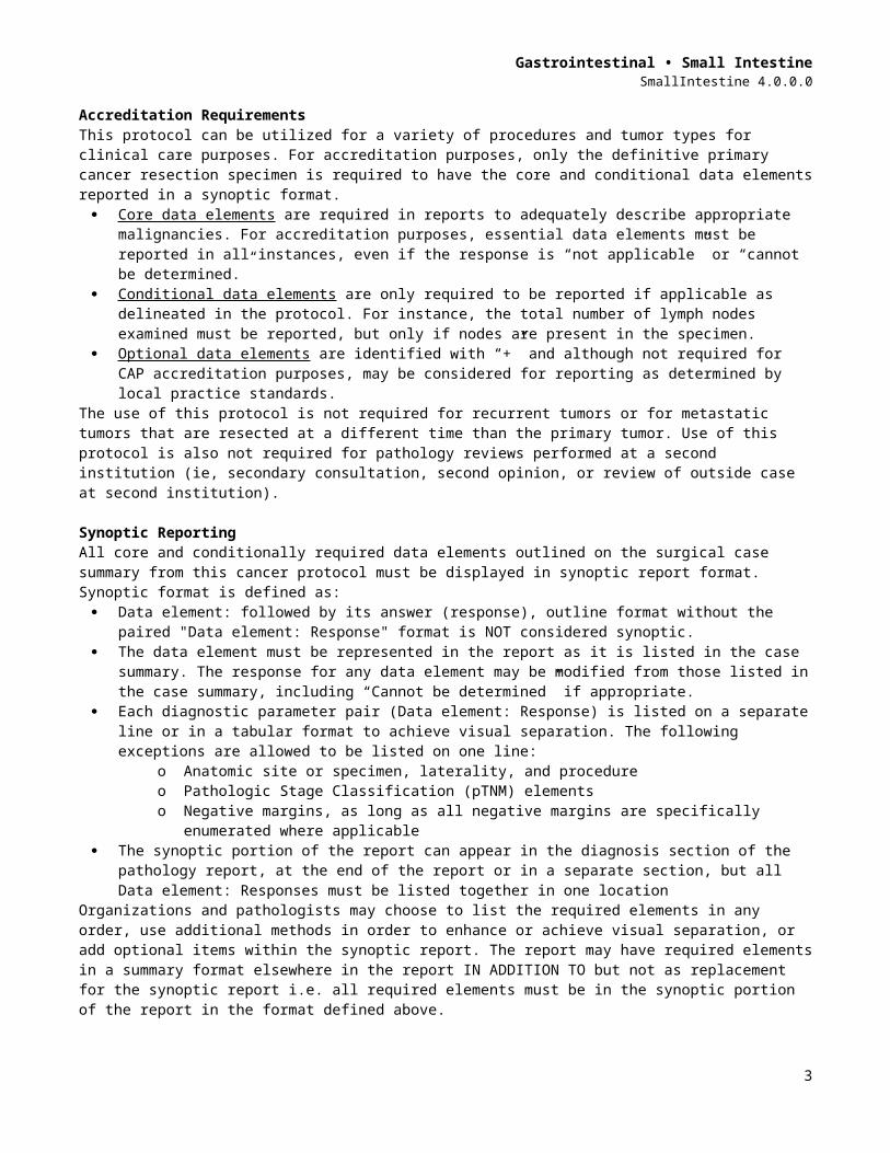
Accreditation RequirementsThis protocol can be utilized for a variety of procedures and tumor types for clinical care purposes. For accreditation purposes, only the definitive primary cancer resection specimen is required to have the core and conditional data elements reported in a synoptic format.
Core data elements are required in reports to adequately describe appropriate malignancies. For accreditation purposes, essential data elements must be reported in all instances, even if the response is “not applicable” or “cannot be determined.”
Conditional data elements are only required to be reported if applicable as delineated in the protocol. For instance, the total number of lymph nodes examined must be reported, but only if nodes are present in the specimen.
Optional data elements are identified with “+” and although not required for CAP accreditation purposes, may be considered for reporting as determined by local practice standards.
The use of this protocol is not required for recurrent tumors or for metastatic tumors that are resected at a different time than the primary tumor. Use of this protocol is also not required for pathology reviews performed at a second institution (ie, secondary consultation, second opinion, or review of outside case at second institution).
Synoptic ReportingAll core and conditionally required data elements outlined on the surgical case summary from this cancer protocol must be displayed in synoptic report format. Synoptic format is defined as:
Data element: followed by its answer (response), outline format without the paired "Data element: Response" format is NOT considered synoptic.
The data element must be represented in the report as it is listed in the case summary. The response for any data element may be modified from those listed in the case summary, including “Cannot be determined” if appropriate.
Each diagnostic parameter pair (Data element: Response) is listed on a separate line or in a tabular format to achieve visual separation. The following exceptions are allowed to be listed on one line:
o Anatomic site or specimen, laterality, and procedureo Pathologic Stage Classification (pTNM) elementso Negative margins, as long as all negative margins are specifically enumerated where applicable
The synoptic portion of the report can appear in the diagnosis section of the pathology report, at the end of the report or in a separate section, but all Data element: Responses must be listed together in one location
Organizations and pathologists may choose to list the required elements in any order, use additional methods in order to enhance or achieve visual separation, or add optional items within the synoptic report. The report may have required elements in a summary format elsewhere in the report IN ADDITION TO but not as replacement for the synoptic report i.e. all required elements must be in the synoptic portion of the report in the format defined above.
CAP Laboratory Accreditation Program Protocol Required Use Date: March 2018** Beginning January 1, 2018, the 8th edition AJCC Staging Manual should be used for reporting pTNM.
CAP Small Intestine Protocol Summary of Changes
The following data elements were modified:Pathologic Stage Classification (pTNM, AJCC 8th Edition)Histologic TypeMicroscopic Tumor ExtensionMarginsAncillary Studies
The following data element was deleted:Clinical History
2
CAP Approved Gastrointestinal • Small IntestineSmallIntestine 4.0.0.0
Surgical Pathology Cancer Case Summary
Protocol posting date: June 2017
SMALL INTESTINE:
Select a single response unless otherwise indicated.
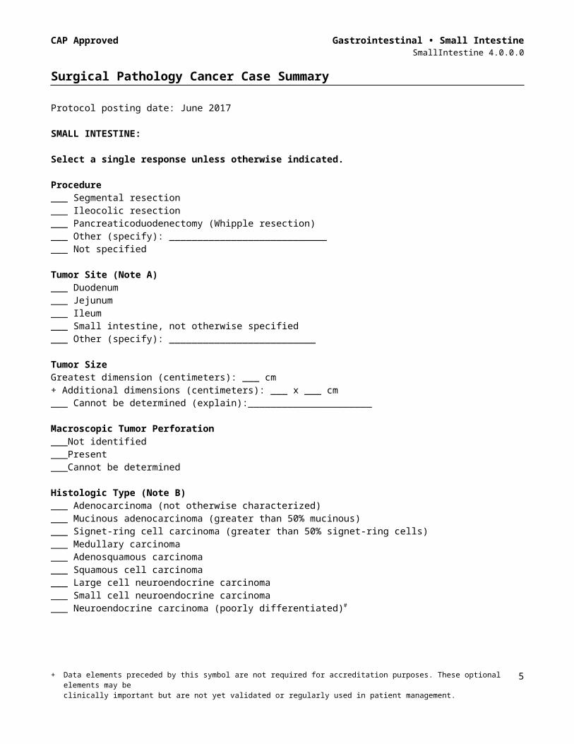
Procedure___ Segmental resection___ Ileocolic resection___ Pancreaticoduodenectomy (Whipple resection)___ Other (specify): _______________________________ Not specified
Tumor Site (Note A)___ Duodenum___ Jejunum___ Ileum___ Small intestine, not otherwise specified___ Other (specify): __________________________
Tumor SizeGreatest dimension (centimeters): ___ cm+ Additional dimensions (centimeters): ___ x ___ cm___ Cannot be determined (explain):______________________
Macroscopic Tumor Perforation ___Not identified___Present___Cannot be determined
Histologic Type (Note B)___ Adenocarcinoma (not otherwise characterized)___ Mucinous adenocarcinoma (greater than 50% mucinous)___ Signet-ring cell carcinoma (greater than 50% signet-ring cells)___ Medullary carcinoma___ Adenosquamous carcinoma___ Squamous cell carcinoma___ Large cell neuroendocrine carcinoma___ Small cell neuroendocrine carcinoma___ Neuroendocrine carcinoma (poorly differentiated)#
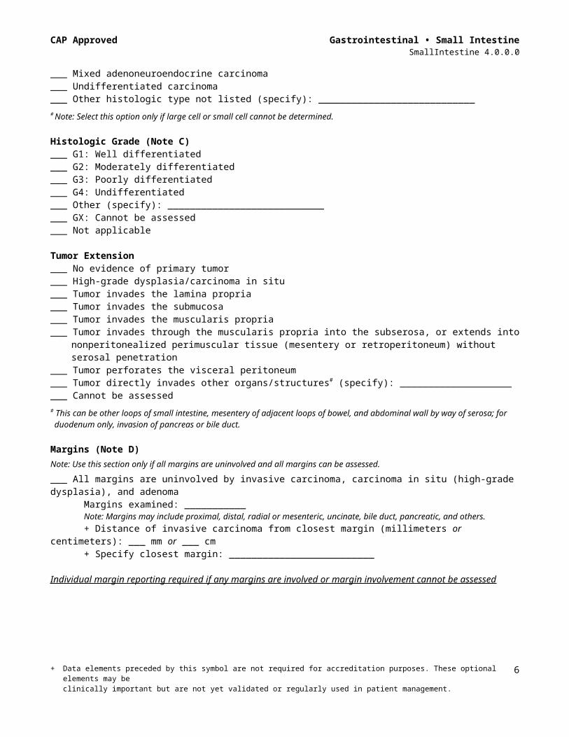
___ Mixed adenoneuroendocrine carcinoma___ Undifferentiated carcinoma___ Other histologic type not listed (specify): ____________________________# Note: Select this option only if large cell or small cell cannot be determined.
Histologic Grade (Note C)___ G1: Well differentiated ___ G2: Moderately differentiated ___ G3: Poorly differentiated___ G4: Undifferentiated___ Other (specify): _______________________________ GX: Cannot be assessed___ Not applicable
+ Data elements preceded by this symbol are not required for accreditation purposes. These optional elements may be clinically important but are not yet validated or regularly used in patient management.
3

CAP Approved Gastrointestinal • Small IntestineSmallIntestine 4.0.0.0
Tumor Extension___ No evidence of primary tumor___ High-grade dysplasia/carcinoma in situ___ Tumor invades the lamina propria___ Tumor invades the submucosa___ Tumor invades the muscularis propria___ Tumor invades through the muscularis propria into the subserosa, or extends into nonperitonealized
perimuscular tissue (mesentery or retroperitoneum) without serosal penetration ___ Tumor perforates the visceral peritoneum ___ Tumor directly invades other organs/structures# (specify): _______________________ Cannot be assessed# This can be other loops of small intestine, mesentery of adjacent loops of bowel, and abdominal wall by way of serosa; for duodenum only, invasion of pancreas or bile duct.
Margins (Note D)Note: Use this section only if all margins are uninvolved and all margins can be assessed.___ All margins are uninvolved by invasive carcinoma, carcinoma in situ (high-grade dysplasia), and adenoma
Margins examined: ___________Note: Margins may include proximal, distal, radial or mesenteric, uncinate, bile duct, pancreatic, and others.+ Distance of invasive carcinoma from closest margin (millimeters or centimeters): ___ mm or ___ cm+ Specify closest margin: __________________________
Individual margin reporting required if any margins are involved or margin involvement cannot be assessed
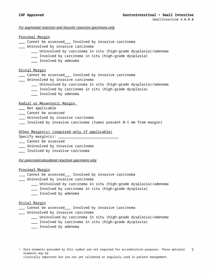
For segmental resection and ileocolic resection specimens only
Proximal Margin___ Cannot be assessed___ Involved by invasive carcinoma___ Uninvolved by invasive carcinoma
___ Uninvolved by carcinoma in situ (high-grade dysplasia)/adenoma___ Involved by carcinoma in situ (high-grade dysplasia)___ Involved by adenoma
Distal Margin___ Cannot be assessed___ Involved by invasive carcinoma___ Uninvolved by invasive carcinoma
___ Uninvolved by carcinoma in situ (high-grade dysplasia)/adenoma___ Involved by carcinoma in situ (high-grade dysplasia)___ Involved by adenoma
Radial or Mesenteric Margin ___ Not applicable___ Cannot be assessed___ Uninvolved by invasive carcinoma___ Involved by invasive carcinoma (tumor present 0-1 mm from margin)
Other Margin(s) (required only if applicable)Specify margin(s): ________________________________ Cannot be assessed___ Uninvolved by invasive carcinoma___ Involved by invasive carcinoma
+ Data elements preceded by this symbol are not required for accreditation purposes. These optional elements may be clinically important but are not yet validated or regularly used in patient management.
4
CAP Approved Gastrointestinal • Small IntestineSmallIntestine 4.0.0.0
For pancreaticoduodenal resection specimens only
Proximal Margin___ Cannot be assessed___ Involved by invasive carcinoma___ Uninvolved by invasive carcinoma
___ Uninvolved by carcinoma in situ (high-grade dysplasia)/adenoma___ Involved by carcinoma in situ (high-grade dysplasia)___ Involved by adenoma
Distal Margin___ Cannot be assessed___ Involved by invasive carcinoma___ Uninvolved by invasive carcinoma
___ Uninvolved by carcinoma in situ (high-grade dysplasia)/adenoma___ Involved by carcinoma in situ (high-grade dysplasia)___ Involved by adenoma
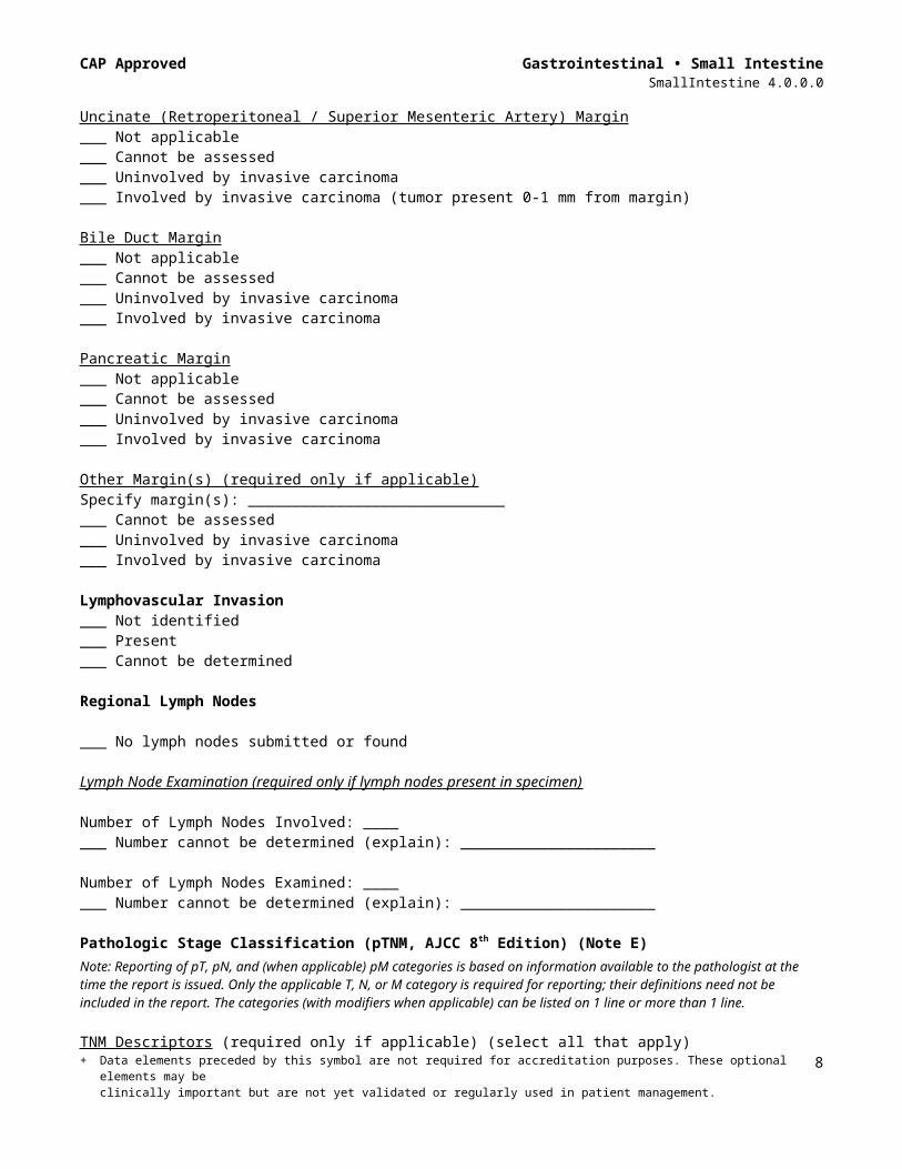
Uncinate (Retroperitoneal / Superior Mesenteric Artery) Margin___ Not applicable___ Cannot be assessed___ Uninvolved by invasive carcinoma___ Involved by invasive carcinoma (tumor present 0-1 mm from margin)
Bile Duct Margin___ Not applicable___ Cannot be assessed___ Uninvolved by invasive carcinoma___ Involved by invasive carcinoma
Pancreatic Margin___ Not applicable___ Cannot be assessed___ Uninvolved by invasive carcinoma___ Involved by invasive carcinoma
Other Margin(s) (required only if applicable)Specify margin(s): ________________________________ Cannot be assessed___ Uninvolved by invasive carcinoma___ Involved by invasive carcinoma
Lymphovascular Invasion ___ Not identified___ Present___ Cannot be determined
Regional Lymph Nodes
___ No lymph nodes submitted or found
Lymph Node Examination (required only if lymph nodes present in specimen)
Number of Lymph Nodes Involved: _______ Number cannot be determined (explain): ______________________
Number of Lymph Nodes Examined: ____
+ Data elements preceded by this symbol are not required for accreditation purposes. These optional elements may be clinically important but are not yet validated or regularly used in patient management.
5
CAP Approved Gastrointestinal • Small IntestineSmallIntestine 4.0.0.0
___ Number cannot be determined (explain): ______________________
Pathologic Stage Classification (pTNM, AJCC 8th Edition) (Note E)Note: Reporting of pT, pN, and (when applicable) pM categories is based on information available to the pathologist at the time the report is issued. Only the applicable T, N, or M category is required for reporting; their definitions need not be included in the report. The categories (with modifiers when applicable) can be listed on 1 line or more than 1 line.
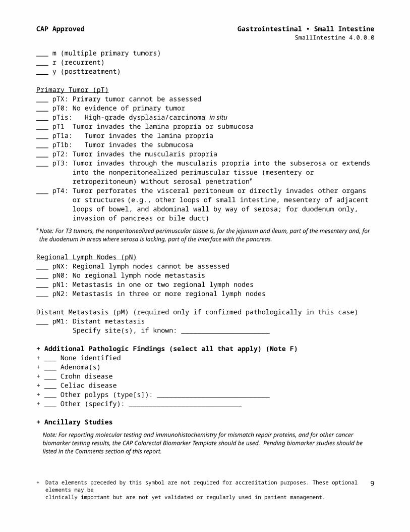
TNM Descriptors (required only if applicable) (select all that apply)___ m (multiple primary tumors)___ r (recurrent)___ y (posttreatment)
Primary Tumor (pT)___ pTX: Primary tumor cannot be assessed___ pT0: No evidence of primary tumor___ pTis: High-grade dysplasia/carcinoma in situ___ pT1 Tumor invades the lamina propria or submucosa ___ pT1a: Tumor invades the lamina propria ___ pT1b: Tumor invades the submucosa___ pT2: Tumor invades the muscularis propria___ pT3: Tumor invades through the muscularis propria into the subserosa or extends into the
nonperitonealized perimuscular tissue (mesentery or retroperitoneum) without serosal penetration#
___ pT4: Tumor perforates the visceral peritoneum or directly invades other organs or structures (e.g., other loops of small intestine, mesentery of adjacent loops of bowel, and abdominal wall by way of serosa; for duodenum only, invasion of pancreas or bile duct)
# Note: For T3 tumors, the nonperitonealized perimuscular tissue is, for the jejunum and ileum, part of the mesentery and, for the duodenum in areas where serosa is lacking, part of the interface with the pancreas.
Regional Lymph Nodes (pN)___ pNX: Regional lymph nodes cannot be assessed___ pN0: No regional lymph node metastasis___ pN1: Metastasis in one or two regional lymph nodes___ pN2: Metastasis in three or more regional lymph nodes
Distant Metastasis (pM) (required only if confirmed pathologically in this case)___ pM1: Distant metastasis
Specify site(s), if known: ______________________
+ Additional Pathologic Findings (select all that apply) (Note F)+ ___ None identified+ ___ Adenoma(s)+ ___ Crohn disease+ ___ Celiac disease+ ___ Other polyps (type[s]): ____________________________+ ___ Other (specify): ____________________________
+ Ancillary StudiesNote: For reporting molecular testing and immunohistochemistry for mismatch repair proteins, and for other cancer biomarker testing results, the CAP Colorectal Biomarker Template should be used. Pending biomarker studies should be listed in the Comments section of this report.
+ Comment(s)
+ Data elements preceded by this symbol are not required for accreditation purposes. These optional elements may be clinically important but are not yet validated or regularly used in patient management.
6
Background Documentation Gastrointestinal • Small IntestineSmallIntestine 4.0.0.0
Explanatory Notes
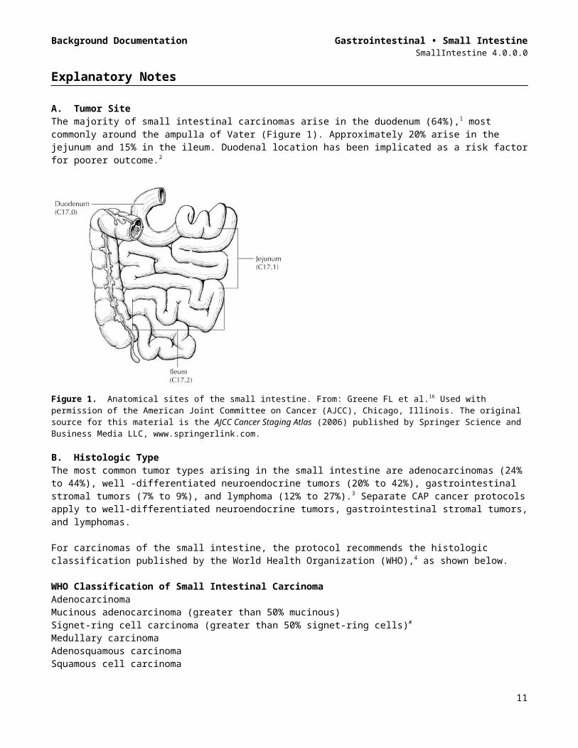
A. Tumor Site The majority of small intestinal carcinomas arise in the duodenum (64%),1 most commonly around the ampulla of Vater (Figure 1). Approximately 20% arise in the jejunum and 15% in the ileum. Duodenal location has been implicated as a risk factor for poorer outcome.2
Figure 1. Anatomical sites of the small intestine. From: Greene FL et al.16 Used with permission of the American Joint Committee on Cancer (AJCC), Chicago, Illinois. The original source for this material is the AJCC Cancer Staging Atlas (2006) published by Springer Science and Business Media LLC, www.springerlink.com.
B. Histologic TypeThe most common tumor types arising in the small intestine are adenocarcinomas (24% to 44%), well -differentiated neuroendocrine tumors (20% to 42%), gastrointestinal stromal tumors (7% to 9%), and lymphoma (12% to 27%).3 Separate CAP cancer protocols apply to well-differentiated neuroendocrine tumors, gastrointestinal stromal tumors, and lymphomas.
For carcinomas of the small intestine, the protocol recommends the histologic classification published by the World Health Organization (WHO),4 as shown below.
WHO Classification of Small Intestinal CarcinomaAdenocarcinomaMucinous adenocarcinoma (greater than 50% mucinous)Signet-ring cell carcinoma (greater than 50% signet-ring cells)#
Medullary carcinomaAdenosquamous carcinomaSquamous cell carcinomaHigh-grade neuroendocrine carcinoma
Large cell neuroendocrine carcinomaSmall cell neuroendocrine- -carcinoma##
Mixed adenoneuroendoncrine carcinomaUndifferentiated carcinoma##
Other (specify)# By convention, signet-ring cell carcinoma is always assigned grade 3 (see Note C).
7
Background Documentation Gastrointestinal • Small IntestineSmallIntestine 4.0.0.0
## By convention, small cell neuroendocrine carcinoma and undifferentiated carcinoma are assigned grade 4 (see Note C).
The term “carcinoma, NOS (not otherwise specified)” is not part of the WHO classification.
C. Histologic GradeA histologic grading system for adenocarcinomas based on the extent of glandular formation in the tumor is recommended, as shown below.
Grade X Grade cannot be assessedGrade 1 Well differentiated (more than 95% of tumor composed of glands)Grade 2 Moderately differentiated (50% to 95% of tumor composed of glands)Grade 3 Poorly differentiated (less than 50% of tumor composed of glands)
Grade 4 is reserved for small cell neuroendocrine carcinoma and undifferentiated carcinoma (WHO classification).
Most small bowel carcinomas are moderately differentiated, followed by poorly differentiated; a minority are well differentiated. Grade does not appear to be a strong predictor of outcome.1,2
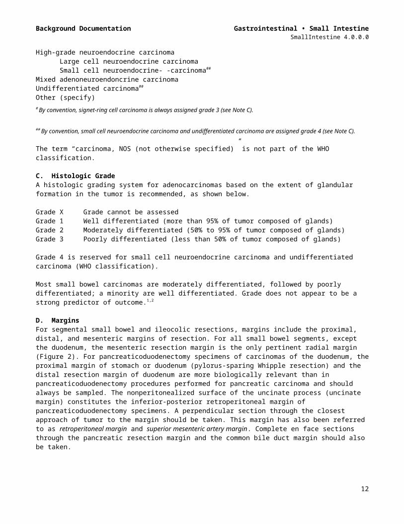
D. MarginsFor segmental small bowel and ileocolic resections, margins include the proximal, distal, and mesenteric margins of resection. For all small bowel segments, except the duodenum, the mesenteric resection margin is the only pertinent radial margin (Figure 2). For pancreaticoduodenectomy specimens of carcinomas of the duodenum, the proximal margin of stomach or duodenum (pylorus-sparing Whipple resection) and the distal resection margin of duodenum are more biologically relevant than in pancreaticoduodenectomy procedures performed for pancreatic carcinoma and should always be sampled. The nonperitonealized surface of the uncinate process (uncinate margin) constitutes the inferior-posterior retroperitoneal margin of pancreaticoduodenectomy specimens. A perpendicular section through the closest approach of tumor to the margin should be taken. This margin has also been referred to as retroperitoneal margin and superior mesenteric artery margin. Complete en face sections through the pancreatic resection margin and the common bile duct margin should also be taken.
A B C
Figure 2. A. Mesenteric margin in small intestine completely encased by peritoneum (dotted line). B. Circumferential margin (dotted line) in portion of proximal duodenum incompletely encased by peritoneum. C. Circumferential margin (dotted line) in retroperitoneal portion of duodenum completely unencased by peritoneum.
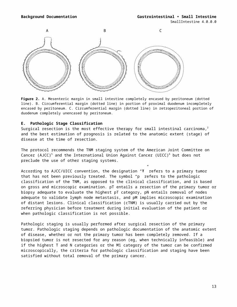
E. Pathologic Stage ClassificationSurgical resection is the most effective therapy for small intestinal carcinoma,2 and the best estimation of prognosis is related to the anatomic extent (stage) of disease at the time of resection.
The protocol recommends the TNM staging system of the American Joint Committee on Cancer (AJCC)5 and the International Union Against Cancer (UICC)6 but does not preclude the use of other staging systems.
8
Background Documentation Gastrointestinal • Small IntestineSmallIntestine 4.0.0.0
According to AJCC/UICC convention, the designation “T” refers to a primary tumor that has not been previously treated. The symbol “p” refers to the pathologic classification of the TNM, as opposed to the clinical classification, and is based on gross and microscopic examination. pT entails a resection of the primary tumor or biopsy adequate to evaluate the highest pT category, pN entails removal of nodes adequate to validate lymph node metastasis, and pM implies microscopic examination of distant lesions. Clinical classification (cTNM) is usually carried out by the referring physician before treatment during initial evaluation of the patient or when pathologic classification is not possible.
Pathologic staging is usually performed after surgical resection of the primary tumor. Pathologic staging depends on pathologic documentation of the anatomic extent of disease, whether or not the primary tumor has been completely removed. If a biopsied tumor is not resected for any reason (eg, when technically infeasible) and if the highest T and N categories or the M1 category of the tumor can be confirmed microscopically, the criteria for pathologic classification and staging have been satisfied without total removal of the primary cancer.
TNM Descriptors For identification of special cases of TNM or pTNM classifications, the “m” suffix and “y” and “r” prefixes are used. Although they do not affect the stage grouping, they indicate cases needing separate analysis.
The “m” suffix indicates the presence of multiple primary tumors in a single site and is recorded in parentheses: pT(m)NM.
The “y” prefix indicates those cases in which classification is performed during or after initial multimodality therapy (ie, neoadjuvant chemotherapy, radiation therapy, or both chemotherapy and radiation therapy). The cTNM or pTNM category is identified by a “y” prefix. The ycTNM or ypTNM categorizes the extent of tumor actually present at the time of that examination. The “y” categorization is not an estimate of tumor before multimodality therapy (ie, before initiation of neoadjuvant therapy).
The “r” prefix indicates a recurrent tumor when staged after a documented disease-free interval and is identified by the “r” prefix: rTNM.
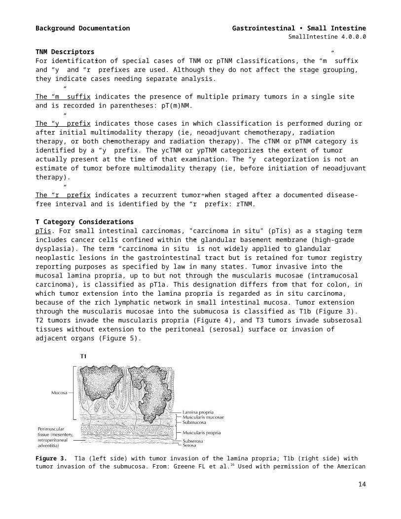
T Category Considerations pTis. For small intestinal carcinomas, "carcinoma in situ" (pTis) as a staging term includes cancer cells confined within the glandular basement membrane (high-grade dysplasia). The term “carcinoma in situ” is not widely applied to glandular neoplastic lesions in the gastrointestinal tract but is retained for tumor registry reporting purposes as specified by law in many states. Tumor invasive into the mucosal lamina propria, up to but not through the muscularis mucosae (intramucosal carcinoma), is classified as pT1a. This designation differs from that for colon, in which tumor extension into the lamina propria is regarded as in situ carcinoma, because of the rich lymphatic network in small intestinal mucosa. Tumor extension through the muscularis mucosae into the submucosa is classified as T1b (Figure 3). T2 tumors invade the muscularis propria (Figure 4), and T3 tumors invade subserosal tissues without extension to the peritoneal (serosal) surface or invasion of adjacent organs (Figure 5).
9
Background Documentation Gastrointestinal • Small IntestineSmallIntestine 4.0.0.0
Figure 3. T1a (left side) with tumor invasion of the lamina propria; T1b (right side) with tumor invasion of the submucosa. From: Greene FL et al.16 Used with permission of the American Joint Committee on Cancer (AJCC), Chicago, Illinois. The original source for this material is the AJCC Cancer Staging Atlas (2006) published by Springer Science and Business Media LLC, www.springerlink.com.
Figure 4. T2 is defined as tumor invading the muscularis propria. From: Greene FL et al.16 Used with permission of the American Joint Committee on Cancer (AJCC), Chicago, Illinois. The original source for this material is the AJCC Cancer Staging Atlas (2006) published by Springer Science and Business Media LLC, www.springerlink.com.
10
Background Documentation Gastrointestinal • Small IntestineSmallIntestine 4.0.0.0
Figure 5. T3 tumors invade through the muscularis propria into subserosal adipose tissue; T4 tumor perforates the peritoneal (serosal) surface. From: Greene FL et al.16 Used with permission of the American Joint Committee on Cancer (AJCC), Chicago, Illinois. The original source for this material is the AJCC Cancer Staging Atlas (2006) published by Springer Science and Business Media LLC, www.springerlink.com.
pT4 includes tumors perforating the visceral peritoneum (Figure 5) or directly invading other organs or structures, including invasion of other segments of small intestine, mesentery of adjacent loops of bowel, and abdominal wall by way of the serosa (Figure 6). In such a case, both an adjacent organ and the visceral peritoneum are penetrated by tumor. Intramural extension of tumor from the terminal ileum into the cecum does not affect the pT classification.4 For duodenal tumors, invasion of the pancreas and the bile duct is considered as T4 (Figure 7).
Figure 6. T4 tumor directly invades other organs or structures, including other loops of small intestine. From: Greene FL et al.16 Used with permission of the American Joint Committee on Cancer (AJCC), Chicago, Illinois. The original source for this material is the AJCC Cancer Staging Atlas (2006) published by Springer Science and Business Media LLC, www.springerlink.com.
11
Background Documentation Gastrointestinal • Small IntestineSmallIntestine 4.0.0.0
Figure 7. T4 tumor of the duodenum invades the pancreas. From: Greene FL et al.16 Used with permission of the American Joint Committee on Cancer (AJCC), Chicago, Illinois. The original source for this material is the AJCC Cancer Staging Atlas (2006) published by Springer Science and Business Media LLC, www.springerlink.com.
Tumor that is adherent to other organs or structures macroscopically is classified as cT4. However, if no tumor is found within the adhesion and no perforation of the visceral peritoneum identified microscopically, the tumor should be assigned pT3.
Tumor in veins or lymphatics does not affect the pT classification.
There are no T4a and T4b subcategories for small intestinal carcinomas in the AJCC 8th edition.
N Category Considerations The regional lymph nodes for the anatomical subsites of the small intestine are as follows:5
Duodenum: retropancreatic, hepatic artery, inferior pancreaticoduodenal, and superior mesenteric
Ileum and jejunum: cecal (terminal ileum only), ileocolic (terminal ileum only), superior mesenteric, mesenteric, NOS
Submission of lymph nodes for microscopic examination. All grossly negative or equivocal lymph nodes are to be submitted entirely. Grossly positive lymph nodes may be partially submitted for microscopic confirmation of metastasis.
The minimum number of lymph nodes that predicts regional node negativity has not been defined for small intestinal cancers. The pathology report should clearly state the total number of lymph nodes examined and the total number involved by metastases. Data are insufficient to recommend routine use of tissue levels or special/ancillary techniques to detect micrometastases or isolated tumor cells.
Nonregional lymph nodes. For microscopic examination of lymph nodes in large resection specimens, lymph nodes must be designated as regional versus nonregional, according to the anatomic location of the tumor. Metastasis to nonregional lymph nodes is classified as distant metastasis and designated as M1. Nonregional lymph nodes include celiac and para-aortic nodes.
Stage GroupingsStage 0 Tis N0 M0Stage I T1-2 N0 M0Stage IIA T3 N0 M0Stage IIB T4 N0 M0Stage IIIA Any T N1 M0
12
Background Documentation Gastrointestinal • Small IntestineSmallIntestine 4.0.0.0
Stage IIIB Any T N2 M0Stage IV Any T Any N M1
Additional DescriptorsLymphovascular InvasionLymphovascular invasion (LVI) indicates whether microscopic lymphatic and/or vascular invasion is identified in the pathology report. LVI includes lymphatic invasion, vascular invasion, or lymph-vascular invasion. By AJCC/UICC convention, LVI does not affect the T category indicating local extent of tumor unless specifically included in the definition of a T category.
F. Additional Pathologic FindingsConditions that predispose to small bowel malignancy include Crohn disease, celiac disease, inherited polyposis syndromes (including familial adenomatous polyposis and Peutz-Jeghers syndrome), and Lynch syndrome.
Small intestinal adenocarcinomas in Crohn disease arise in the setting of long-standing ileal inflammation; cumulative risk increases after 10 years of Crohn disease, although absolute risk (2.2% at 25 years) remains low.7 Signet-ring cell carcinomas appear to be more common in Crohn disease than as de novo small intestinal carcinomas.8
Small intestinal carcinomas are more frequent in polyposis syndromes, most notably in familial adenomatous polyposis, in which approximately 2.3% of patients developed a duodenal adenocarcinoma;9 most tumors in these patients develop in the periampullary region, and the duodenum may be carpeted with adenomas. Peutz-Jeghers syndrome10 is also associated with higher risk of small intestinal carcinoma.
Patients with Lynche syndrome have an approximately 4% lifetime risk of developing a small bowel carcinoma; this risk exceeds that of the normal population by 100-fold. Duodenum and jejunum are the most common primary sites, and the small bowel is the first site of cancer in approximately one-fourth of Lynche syndrome patients who develop small bowel tumors.11 Histopathologic features of Lynche syndrome-associated small intestinal carcinomas are similar to those of colorectal carcinomas arising in this setting; mucinous carcinomas are overrepresented, and tumors often show a high number of intratumoral lymphocytes and Crohn-like lymphoid reaction.12
G. Ancillary ProceduresTesting for defects in mismatch repair in small intestinal carcinomas is important for detection of Lynch syndrome. Examination of the tissue for defective DNA mismatch repair should be considered in small intestinal carcinomas regardless of the patient’s age,13 if other predisposing conditions such as familial adenomatous polyposis coli are absent. In addition, emerging data suggest that the frequency of microsatellite instability in small intestinal carcinomas is approximately equal to that of colon cancer14 and may be associated with better survival.15 However, this latter indication for testing is not clearly established and has not been accepted as standard of care.
References1. Howe JR, Karnell LH, Menck HR, Scott-Conner C. Adenocarcinoma of the small bowel: review of the
National Cancer Data Base, 1985-1995. Cancer. 1999;86:2693-2706.2. Dabaja BS, Suki D, Pro B, Bonnen M, Ajani J. Adenocarcinoma of the small bowel: presentation, prognostic
factors, and outcome of 217 patients. Cancer. 2004;101:518-526.3. Zeh HJ. Cancer of the small intestine. In: DeVita VT, Hellman S, Rosenberg SA, eds. Cancer: Principles
and Practice of Oncology. 7th ed. Philadelphia, PA: Lippincott Williams & Wilkins; 2005.4. Bosman FT, Carneiro F, Hruban RH, Theise ND, eds. WHO Classification of Tumours of the Digestive
System. Geneva, Switzerland: WHO Press; 2010.5. Amin MB, Edge SB, Greene FL, et al, eds. AJCC Cancer Staging Manual. 8th ed. New York, NY: Springer;
2017.6. Brierley JD, Gospodarowicz MK, Wittekind C, et al, eds. TNM Classification of Malignant Tumours. 8th ed.
Oxford, UK: Wiley; 2016.7. Friedman S. Cancer in Crohn's disease. [Review] [102 refs]. Gastroenterol Clin North Am. 2006;35:621-
639.
13
Background Documentation Gastrointestinal • Small IntestineSmallIntestine 4.0.0.0
8. Palascak-Juif V, Bouvier AM, Cosnes J, et al. Small bowel adenocarcinoma in patients with Crohn's disease compared with small bowel adenocarcinoma de novo. Inflamm Bowel Dis. 2005;11:828-832.
9. Jagelman DG, DeCosse JJ, Bussey HJ. Upper gastrointestinal cancer in familial adenomatous polyposis. Lancet. 1988;1:1149-1151.
10. Hearle N, Schumacher V, Menko FH, et al. Frequency and spectrum of cancers in the Peutz-Jeghers syndrome. Clin Cancer Res. 2006;12:3209-3215.
11. Rodriguez-Bigas MA, Vasen HF, Lynch HT, et al. Characteristics of small bowel carcinoma in hereditary nonpolyposis colorectal carcinoma. International Collaborative Group on HNPCC. Cancer. 1998;83:240-244.
12. Schulmann K, Brasch FE, Kunstmann E, et al. HNPCC-associated small bowel cancer: clinical and molecular characteristics. Gastroenterology. 2005;128:590-599.
13. Umar A, Boland R, Terdiman JP, et al. Revised Bethesda guidelines for hereditary nonpolyposis colorectal cancer (Lynch syndrome) and microsatellite instability. J Nat Cancer Inst. 2004;96:261-268.
14. Planck M, Ericson K, Piotrowska Z, Halvarsson B, Rambech E, Nilbert M. Microsatellite instability and expression of MLH1 and MSH2 in carcinomas of the small intestine. Cancer. 2003;97:1551-1557.
15. Brueckl WM, Heinze E, Milsmann C, et al. Prognostic significance of microsatellite instability in curatively resected adenocarcinoma of the small intestine. Cancer Letters. 2004;203:181-190.
16. Greene FL, Compton, CC, Fritz AG, et al, eds. AJCC Cancer Staging Atlas. New York: Springer; 2006.
14