
CAP Cancer Protocol Perihilar Bile Ducts · Web viewZen Y, Adsay NV, Bardadin K, et al. Biliary...
17
Protocol for the Examination of Specimens From Patients With Carcinoma of the Perihilar Bile Ducts Version: PerihilarBileDuct 4.0.0.1 Protocol Posting Date: June 2017 Includes pTNM requirements from the 8 th Edition, AJCC Staging Manual For accreditation purposes, this protocol should be used for the following procedures AND tumor types: Procedure Description Resection Includes specimens designated bile duct resection, local or segmental, hilar resection with or without hepatic resection Tumor Type Description Carcinoma Invasive carcinomas including small cell and large cell (poorly differentiated) neuroendocrine carcinoma This protocol is NOT required for accreditation purposes for the following: Procedure Biopsy Primary resection specimen with no residual cancer (eg, following neoadjuvant therapy) Cytologic specimens The following tumor types should NOT be reported using this protocol: Tumor Type Intraductal papillary neoplasm without associated invasive carcinoma Mucinous cystic neoplasm without associated invasive carcinoma Well-differentiated neuroendocrine tumors of perihilar bile ducts Lymphoma (consider the Hodgkin or non-Hodgkin Lymphoma protocols) Sarcoma (consider the Soft Tissue protocol) Authors Sanjay Kakar, MD*; Chanjuan Shi, MD, PhD*; Patrick L. Fitzgibbons, MD; Wendy L. Frankel, MD; Alyssa M. Krasinskas, MD; Mari Mino-Kenudson, MD; Timothy Pawlik, MD, MPH, PhD; Jean-Nicolas Vauthey, MD; Mary K. Washington, MD, PhD With guidance from the CAP Cancer and CAP Pathology Electronic Reporting Committees. * Denotes primary author. All other contributing authors are listed alphabetically. © 2017 College of American Pathologists (CAP). All rights reserved. For Terms of Use please visit www.cap.org/cancerprotocols .
Transcript of CAP Cancer Protocol Perihilar Bile Ducts · Web viewZen Y, Adsay NV, Bardadin K, et al. Biliary...

Protocol for the Examination of Specimens From Patients With Carcinoma of the Perihilar Bile DuctsVersion: PerihilarBileDuct 4.0.0.1 Protocol Posting Date: June 2017Includes pTNM requirements from the 8th Edition, AJCC Staging Manual
For accreditation purposes, this protocol should be used for the following procedures AND tumor types:Procedure DescriptionResection Includes specimens designated bile duct resection, local or segmental,
hilar resection with or without hepatic resectionTumor Type DescriptionCarcinoma Invasive carcinomas including small cell and large cell (poorly
differentiated) neuroendocrine carcinoma
This protocol is NOT required for accreditation purposes for the following:ProcedureBiopsyPrimary resection specimen with no residual cancer (eg, following neoadjuvant therapy)Cytologic specimens
The following tumor types should NOT be reported using this protocol:Tumor TypeIntraductal papillary neoplasm without associated invasive carcinomaMucinous cystic neoplasm without associated invasive carcinomaWell-differentiated neuroendocrine tumors of perihilar bile ductsLymphoma (consider the Hodgkin or non-Hodgkin Lymphoma protocols)Sarcoma (consider the Soft Tissue protocol)
Authors
Sanjay Kakar, MD*; Chanjuan Shi, MD, PhD*; Patrick L. Fitzgibbons, MD; Wendy L. Frankel, MD; Alyssa M. Krasinskas, MD; Mari Mino-Kenudson, MD; Timothy Pawlik, MD, MPH, PhD; Jean-Nicolas Vauthey, MD; Mary K. Washington, MD, PhD
With guidance from the CAP Cancer and CAP Pathology Electronic Reporting Committees.* Denotes primary author. All other contributing authors are listed alphabetically.
© 2017 College of American Pathologists (CAP). All rights reserved. For Terms of Use please visit www.cap.org/cancerprotocols.
Gastrointestinal • Perihilar Bile DuctsPerihilarBileDuct 4.0.0.1
Accreditation RequirementsThis protocol can be utilized for a variety of procedures and tumor types for clinical care purposes. For accreditation purposes, only the definitive primary cancer resection specimen is required to have the core and conditional data elements reported in a synoptic format.
Core data elements are required in reports to adequately describe appropriate malignancies. For accreditation purposes, essential data elements must be reported in all instances, even if the response is “not applicable” or “cannot be determined.”
Conditional data elements are only required to be reported if applicable as delineated in the protocol. For instance, the total number of lymph nodes examined must be reported, but only if nodes are present in the specimen.
Optional data elements are identified with “+” and although not required for CAP accreditation purposes, may be considered for reporting as determined by local practice standards.
The use of this protocol is not required for recurrent tumors or for metastatic tumors that are resected at a different time than the primary tumor. Use of this protocol is also not required for pathology reviews performed at a second institution (ie, secondary consultation, second opinion, or review of outside case at second institution).
Synoptic ReportingAll core and conditionally required data elements outlined on the surgical case summary from this cancer protocol must be displayed in synoptic report format. Synoptic format is defined as:
Data element: followed by its answer (response), outline format without the paired "Data element: Response" format is NOT considered synoptic.
The data element should be represented in the report as it is listed in the case summary. The response for any data element may be modified from those listed in the case summary, including “Cannot be determined” if appropriate.
Each diagnostic parameter pair (Data element: Response) is listed on a separate line or in a tabular format to achieve visual separation. The following exceptions are allowed to be listed on one line:
o Anatomic site or specimen, laterality, and procedureo Pathologic Stage Classification (pTNM) elementso Negative margins, as long as all negative margins are specifically enumerated where applicable
The synoptic portion of the report can appear in the diagnosis section of the pathology report, at the end of the report or in a separate section, but all Data element: Responses must be listed together in one location
Organizations and pathologists may choose to list the required elements in any order, use additional methods in order to enhance or achieve visual separation, or add optional items within the synoptic report. The report may have required elements in a summary format elsewhere in the report IN ADDITION TO but not as replacement for the synoptic report i.e. all required elements must be in the synoptic portion of the report in the format defined above.
CAP Laboratory Accreditation Program Protocol Required Use Date: March 2018** Beginning January 1, 2018, the 8th edition AJCC Staging Manual should be used for reporting pTNM.
CAP Perihilar Bile Duct Protocol Summary of Changes
Version 4.0.0.1Modified Tumor Extension
Version 4.0.0.0The following data elements were modified:Pathologic Stage Classification (pTNM, AJCC 8th Edition)Histologic TypeMicroscopic Tumor ExtensionMargins
The following data element was deleted:Clinical History
2

CAP Approved Gastrointestinal • Perihilar Bile DuctsPerihilarBileDuct 4.0.0.1
Surgical Pathology Cancer Case Summary
Protocol posting date: June 2017
PERIHILAR BILE DUCTS:
Select a single response unless otherwise indicated.
Procedure (Notes A and B)___ Hilar and hepatic resection___ Segmental resection of bile ducts(s)___ Choledochal cyst resection ___ Total hepatectomy___ Other (specify): _______________________________ Not specified
Tumor Site (select all that apply)
___ Right hepatic duct___ Left hepatic duct___ Junction of right and left hepatic ducts___ Cystic duct___ Common hepatic duct___ Common bile duct___ Not specified
Tumor Size
Greatest dimension (centimeters): ___ cm+ Additional dimensions (centimeters): ___ x ___ cm___ Cannot be determined (explain): ___________________________
Histologic Type (Note C)
___ Adenocarcinoma ___ Adenocarcinoma, intestinal type___ Adenocarcinoma, biliary type___ Adenocarcinoma, gastric foveolar type___ Intraductal papillary neoplasm with an associated invasive carcinoma___ Mucinous cystic neoplasm with an associated invasive carcinoma___ Mucinous adenocarcinoma___ Clear cell adenocarcinoma___ Signet-ring cell carcinoma___ Large cell neuroendocrine carcinoma___ Small cell neuroendocrine carcinoma___ Neuroendocrine carcinoma (poorly differentiated)#
___ Adenosquamous carcinoma___ Squamous cell carcinoma___ Mixed adenoneuroendocrine carcinoma___ Undifferentiated carcinoma___ Other histologic type not listed (specify): _______________________________ Carcinoma, not otherwise specified# Note: Select this option only if large cell or small cell cannot be determined.
Histologic Grade (Note D)
___ G1: Well differentiated___ G2: Moderately differentiated___ G3: Poorly differentiated
+ Data elements preceded by this symbol are not required for accreditation purposes. These optional elements may be clinically important but are not yet validated or regularly used in patient management.
3

CAP Approved Gastrointestinal • Perihilar Bile DuctsPerihilarBileDuct 4.0.0.1
___ GX: Cannot be assessed___ Not applicable
Tumor Extension (select all that apply)___ No evidence of primary tumor___ No invasion (carcinoma in situ/high-grade dysplasia)___ Tumor confined to the bile duct ___ Tumor invades beyond the wall of the bile duct into surrounding connective tissue___ Tumor invades the adjacent liver parenchyma___ Tumor invades the gallbladder___ Tumor invades the unilateral branches of the portal vein (right or left) ___ Tumor invades the unilateral branches of the hepatic artery (right or left) ___ Tumor invades main portal vein or its branches bilaterally ___ Tumor invades common hepatic artery___ Tumor invades second-order biliary radicals, unilateral___ Tumor invades second-order biliary radicals, bilateral___ Cannot be assessed
Margins (Note E)
For segmental resection specimens only
Proximal Margin___ Cannot be assessed___ Uninvolved by invasive carcinoma and high-grade intraepithelial neoplasia
+ Distance of invasive carcinoma from margin (millimeters or centimeters): ___ mm or ___ cm___ Uninvolved by invasive carcinoma
+ Distance of invasive carcinoma from margin (millimeters or centimeters): ___ mm or ___ cm___ Involved by invasive carcinoma___ Involved by high-grade intraepithelial neoplasia
Distal Margin___ Cannot be assessed___ Uninvolved by invasive carcinoma and high-grade intraepithelial neoplasia
+ Distance of invasive carcinoma from margin: ___ mm or ___ cm___ Uninvolved by invasive carcinoma
+ Distance of invasive carcinoma from margin: ___ mm or ___ cm___ Involved by invasive carcinoma___ Involved by high-grade intraepithelial neoplasia
Radial Margin___ Cannot be assessed___ Uninvolved by invasive carcinoma
+ Distance of invasive carcinoma from margin (millimeters or centimeters): ___ mm or ___ cm___ Involved by invasive carcinoma
For hilar resection and partial hepatectomy specimens only
Hepatic Parenchymal Margin___ Cannot be assessed___ Uninvolved by invasive carcinoma
+Distance of invasive carcinoma from margin (millimeters or centimeters): ___ mm or ___ cm___ Involved by invasive carcinoma
+ Data elements preceded by this symbol are not required for accreditation purposes. These optional elements may be clinically important but are not yet validated or regularly used in patient management.
4
CAP Approved Gastrointestinal • Perihilar Bile DuctsPerihilarBileDuct 4.0.0.1
Bile Duct Margin___ Cannot be assessed___ Uninvolved by invasive carcinoma and high-grade intraepithelial neoplasia
+ Distance of invasive carcinoma from margin (millimeters or centimeters): ___ mm or ___ cm___ Uninvolved by invasive carcinoma
+ Distance of invasive carcinoma from margin (millimeters or centimeters): ___ mm or ___ cm___ Involved by invasive carcinoma___ Involved by high-grade intraepithelial neoplasia
Radial Margin___ Cannot be assessed___ Uninvolved by invasive carcinoma
+ Distance of invasive carcinoma from margin (millimeters or centimeters): ___ mm or ___ cm___ Involved by invasive carcinoma
For total hepatectomy specimens only
Bile Duct Margin___ Cannot be assessed___ Uninvolved by invasive carcinoma and high-grade intraepithelial neoplasia
+ Distance of invasive carcinoma from margin (millimeters or centimeters): ___ mm or ___ cm___ Uninvolved by invasive carcinoma
+ Distance of invasive carcinoma from margin: ___ mm or ___ cm___ Involved by invasive carcinoma___ Involved by high-grade intraepithelial neoplasia
Radial Margin___ Cannot be assessed___ Uninvolved by invasive carcinoma
+ Distance of invasive carcinoma from margin (millimeters or centimeters): ___ mm or ___ cm___ Involved by invasive carcinoma
Other Margin(s) (required only if applicable)Specify margin(s): _____________________________ ___ Cannot be assessed___ Uninvolved by invasive carcinoma ___ Involved by invasive carcinoma
Lymphovascular Invasion (Note F)
___ Not identified___ Present___ Cannot be determined
Perineural Invasion (Note F)
___ Not identified___ Present___ Cannot be determined
Regional Lymph Nodes
___ No lymph nodes submitted or found
Lymph Node Examination (required only if lymph nodes are present in the specimen)
Number of Lymph Nodes Involved: _______ Number cannot be determined (explain): ______________________
+ Data elements preceded by this symbol are not required for accreditation purposes. These optional elements may be clinically important but are not yet validated or regularly used in patient management.
5

CAP Approved Gastrointestinal • Perihilar Bile DuctsPerihilarBileDuct 4.0.0.1
Number of Lymph Nodes Examined: _______ Number cannot be determined (explain): ______________________
Pathologic Stage Classification (pTNM, AJCC 8th Edition) (Note G)
Note: Reporting of pT, pN, and (when applicable) pM categories is based on information available to the pathologist at the time the report is issued. Only the applicable T, N, or M category is required for reporting; their definitions need not be included in the report. The categories (with modifiers when applicable) can be listed on 1 line or more than 1 line.
TNM Descriptors (required only if applicable) (select all that apply)___ m (multiple primary tumors)___ r (recurrent)___ y (posttreatment)
Primary Tumor (pT)___ pTX: Primary tumor cannot be assessed___ pT0: No evidence of primary tumor___ pTis: Carcinoma in situ/high-grade dysplasia___ pT1: Tumor confined to the bile duct, with extension up to the muscle layer or fibrous tissue___ pT2: Tumor invades beyond the wall of the bile duct to surrounding adipose tissue, or tumor invades
adjacent hepatic parenchyma___ pT2a: Tumor invades beyond the wall of the bile duct to surrounding adipose tissue ___ pT2b: Tumor invades adjacent hepatic parenchyma ___ pT3: Tumor invades unilateral branches of the portal vein or hepatic artery___ pT4: Tumor invades the main portal vein or its branches bilaterally, or the common hepatic artery; or
unilateral second-order biliary radicals with contralateral portal vein or hepatic artery involvement
Regional Lymph Nodes (pN)___ pNX: Regional lymph nodes cannot be assessed___ pN0: No regional lymph node metastasis___ pN1: One to three positive regional lymph nodes typically involving the hilar, cystic duct, common bile duct,
hepatic artery, posterior pancreatoduodenal, and portal vein lymph nodes ___ pN2: Four or more positive lymph nodes from the sites described for N1
Distant Metastasis (pM) (required only if confirmed pathologically in this case) ___ pM1: Distant metastasis
Specify site(s), if known: _________________________
+ Additional Pathologic Findings (select all that apply) (Note H)
+ ___ None identified+ ___ Choledochal cyst+ ___ Dysplasia+ ___ Primary sclerosing cholangitis (PSC)+ ___ Biliary stones+ ___ Other (specify): ___________________________
+ Ancillary Studies+ Specify: ___________________________________
+ Comment(s)
+ Data elements preceded by this symbol are not required for accreditation purposes. These optional elements may be clinically important but are not yet validated or regularly used in patient management.
6
Background Documentation Gastrointestinal • Perihilar Bile DuctsPerihilarBileDuct 4.0.0.1
Explanatory Notes
A. Application
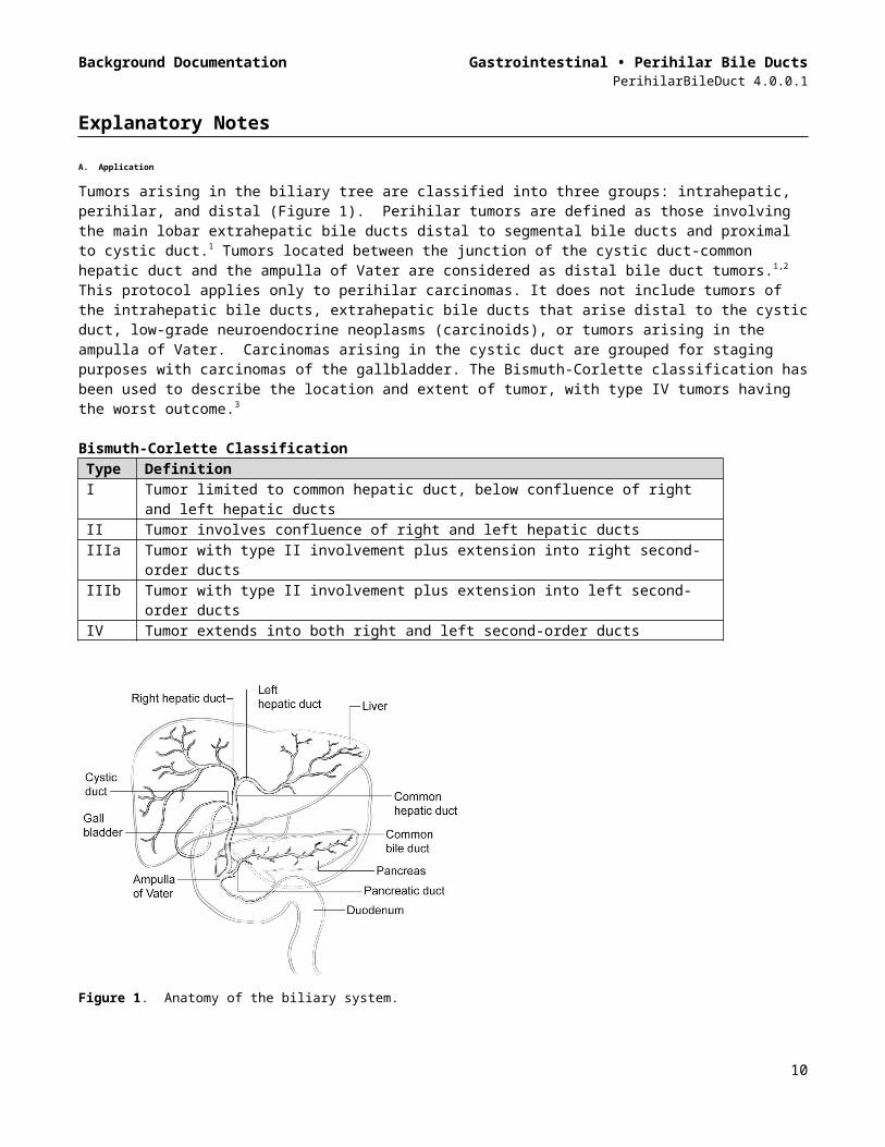
Tumors arising in the biliary tree are classified into three groups: intrahepatic, perihilar, and distal (Figure 1). Perihilar tumors are defined as those involving the main lobar extrahepatic bile ducts distal to segmental bile ducts and proximal to cystic duct.1 Tumors located between the junction of the cystic duct-common hepatic duct and the ampulla of Vater are considered as distal bile duct tumors.1,2 This protocol applies only to perihilar carcinomas. It does not include tumors of the intrahepatic bile ducts, extrahepatic bile ducts that arise distal to the cystic duct, low-grade neuroendocrine neoplasms (carcinoids), or tumors arising in the ampulla of Vater. Carcinomas arising in the cystic duct are grouped for staging purposes with carcinomas of the gallbladder. The Bismuth-Corlette classification has been used to describe the location and extent of tumor, with type IV tumors having the worst outcome.3
Bismuth-Corlette ClassificationType DefinitionI Tumor limited to common hepatic duct, below confluence of right and left hepatic ductsII Tumor involves confluence of right and left hepatic ductsIIIa Tumor with type II involvement plus extension into right second-order ductsIIIb Tumor with type II involvement plus extension into left second-order ductsIV Tumor extends into both right and left second-order ducts
Figure 1. Anatomy of the biliary system.
B. Choledochal Cyst
Carcinomas may arise in choledochal cysts (congenital cystic dilatation or duplications) of the bile duct. Histologically, they are classified in the same way as those arising in the gallbladder or bile ducts. Stones may be found in these cysts. If dysplasia or carcinoma in situ is found on initial microscopic sections, then multiple additional sections should be examined to exclude invasive cancer in other areas of the cyst.
C. Histologic Type
For consistency in reporting, the histologic classification published by the World Health Organization (WHO), shown below, is recommended.4 However, this protocol does not preclude the use of other systems of classification or histologic types. According to WHO convention, the term “cholangiocarcinoma” is reserved for carcinomas arising in the intrahepatic bile ducts (see CAP protocol for intrahepatic bile ducts).
7

Background Documentation Gastrointestinal • Perihilar Bile DuctsPerihilarBileDuct 4.0.0.1
Intraductal neoplasms have a relatively favorable prognosis,5-7 while signet-ring cell carcinoma, poorly differentiated neuroendocrine carcinomas, and undifferentiated carcinomas are associated with a poorer prognosis.
Modified WHO Classification of Carcinoma of the Extrahepatic Bile Ducts
Adenocarcinoma Adenocarcinoma, biliary type Adenocarcinoma, intestinal type Adenocarcinoma, gastric foveloar type Mucinous adenocarcinoma Clear cell adenocarcinoma Signet-ring cell carcinoma Intraductal papillary neoplasm with an associated invasive carcinomaMucinous cystic neoplasm with an associated invasive carcinoma
Adenosquamous carcinomaSquamous cell carcinomaPoorly differentiated neuroendocrine carcinoma
Large cell neuroendocrine carcinomaSmall cell neuroendocrine carcinoma
Mixed adenoneuroendocrine carcinomaUndifferentiated carcinoma
D. Histologic Grade
For adenocarcinomas, a quantitative grading system based on the proportion of gland formation within the tumor is suggested3 and shown below.
Grade X Grade cannot be assessedGrade 1 Well differentiated (greater than 95% of tumor composed of glands)Grade 2 Moderately differentiated (50% to 95% of tumor composed of glands)Grade 3 Poorly differentiated (less than 50% of tumor composed of glands)
Definitions corresponding to the above histologic grades are as follows:
Grade 1 Composed entirely of glands or has less than 5% solid or cordlike growth patternsGrade 2 Has more than 5% but less than 50% solid or cordlike growth patternsGrade 3 Has 50% to 100% solid or cordlike growth patterns
For squamous cell carcinomas, a rare tumor type in the extrahepatic bile ducts, a suggested grading system is shown below. If there are variations in the differentiation within the tumor, the highest (least favorable) grade is recorded.
Grade X Grade cannot be assessedGrade 1 Well differentiatedGrade 2 Moderately differentiatedGrade 3 Poorly differentiated
By convention, signet-ring cell carcinomas are assigned grade 3. Undifferentiated carcinomas lack morphologic or immunohistochemical evidence of glandular, squamous, or neuroendocrine differentiation. This category is not included in the AJCC grading scheme. This grading scheme is not applicable to poorly differentiated neuroendocrine carcinomas.
E. Margins
Locoregional recurrence, as opposed to distant metastases, is usually the first site of disease recurrence and occurs in up to 59% of patients with perihilar bile duct carcinomas.8 Tumor recurrence is often related to residual tumor located in the proximal or distal surgical margins of the bile duct or from tumor located along the dissected
8

Background Documentation Gastrointestinal • Perihilar Bile DuctsPerihilarBileDuct 4.0.0.1
soft tissue margin in the portal area. Local recurrence (usually at the surgical margins) can be attributed in many cases to tumor spread longitudinally along the duct wall and to perineural and lymphovascular invasion.9
Complete surgical resection with microscopically negative surgical margins is an important predictor of outcome in multivariate analysis for both perihilar and distal bile duct carcinomas, with overall 5-year survival for perihilar tumor improved from 10% for all patients to 30% for those with negative resection margins.1
Malignant tumors of the extrahepatic bile ducts are often multifocal. Therefore, microscopic foci of carcinoma or intraepithelial neoplasia may be found at the margin(s) even though the main tumor mass has been resected. In some cases it may be difficult to evaluate margins on frozen section preparations because of inflammation and reactive change of the surface epithelium or within the intramural mucous glands. If surgical margins are free of carcinoma, the distance between the closest margin and the tumor edge should be measured.
The gallbladder specimen should be examined as bile duct carcinoma can be associated with synchronous carcinomas of the gallbladder.
F. Perineural and Vascular/Lymphatic Invasion
Perineural and lymphatic invasion are common in extrahepatic bile duct carcinomas, although they are found less often in early-stage cancers (11%).10 They should be specifically evaluated because they are associated with adverse outcome on univariate analysis.11 Although perineural invasion is sometimes useful for distinguishing carcinoma from nonneoplastic glands, caution should be used in interpretation of this finding in ducts affected by primary sclerosing cholangitis, as benign hyperplastic intramural glands adjacent to nerves has been reported in this setting.12
G. TNM and Anatomic Stage/Prognostic Groupings
Surgical resection is the most effective therapy for extrahepatic biliary tract carcinomas, and the best estimation of prognosis is related to the anatomic extent (stage) of disease at the time of resection. In particular, lymph node metastases are predictors of poorer outcome.1,13
For malignant tumors of the perihilar bile ducts, the TNM staging system of the American Joint Committee on Cancer (AJCC) and the International Union Against Cancer (UICC) is recommended.2 The staging system also applies to tumors arising in choledochal cysts.
According to AJCC/UICC convention, the designation “T” refers to a primary tumor that has not been previously treated. The symbol “p” refers to the pathologic classification of the TNM, as opposed to the clinical classification, and is based on gross and microscopic examination. pT entails a resection of the primary tumor or biopsy adequate to evaluate the highest pT category, pN entails removal of nodes adequate to validate lymph node metastasis, and pM implies microscopic examination of distant lesions. Clinical classification (cTNM) is usually carried out by the referring physician before treatment during initial evaluation of the patient or when pathologic classification is not possible.
Pathologic staging is usually performed after surgical resection of the primary tumor. Pathologic staging depends on pathologic documentation of the anatomic extent of disease, whether or not the primary tumor has been completely removed. If a biopsied tumor is not resected for any reason (eg, when technically infeasible) and if the highest T and N categories or the M1 category of the tumor can be confirmed microscopically, the criteria for pathologic classification and staging have been satisfied without total removal of the primary cancer.
TNM Descriptors
For identification of special cases of TNM or pTNM classifications, the “m” suffix and “y,” “r,” and “a” prefixes are used. Although they do not affect the stage grouping, they indicate cases needing separate analysis.
The “m” suffix indicates the presence of multiple primary tumors in a single site and is recorded in parentheses: pT(m)NM.
9
Background Documentation Gastrointestinal • Perihilar Bile DuctsPerihilarBileDuct 4.0.0.1
The “y” prefix indicates those cases in which classification is performed during or after initial multimodality therapy (ie, neoadjuvant chemotherapy, radiation therapy, or both chemotherapy and radiation therapy). The cTNM or pTNM category is identified by a “y” prefix. The ycTNM or ypTNM categorizes the extent of tumor actually present at the time of that examination. The “y” categorization is not an estimate of tumor before multimodality therapy (ie, before initiation of neoadjuvant therapy).
The “r” prefix indicates a recurrent tumor when staged after a documented disease-free interval and is identified by the “r” prefix: rTNM.
The “a” prefix designates the stage determined at autopsy: aTNM.
T Category Considerations (Figures 2 and 3)Tis includes high-grade biliary intraepithelial neoplasia (BilIn-3), intraductal papillary neoplasm with high-grade dysplasia, and mucinous cystic neoplasm with high-grade dysplasia. For intraepithelial lesions, a 3-tier biliary intraepithelial neoplasia classification has been proposed.14 The term carcinoma in situ is not widely applied to glandular neoplastic lesions but is retained for tumor registry reporting purposes as specified by law in many states. A synoptic report is not required for intraductal papillary neoplasms and mucinous cystic neoplasms in the absence of an invasive component. For invasive carcinoma associated with intraductal papillary neoplasms and mucinous cystic neoplasms, only the size of the invasive component should be used to determine the T category. The invasive portion in these cases can be multifocal. The size of the largest focus as well as cumulative size of all invasive carcinoma foci should be included in the report. Till further data becomes available, the T category should be determined based on size of the largest invasive focus.
The histology of the extrahepatic biliary tree varies along its length, with little smooth muscle in the wall of the proximal ducts compared with the distal bile duct. This can make it difficult to assess the depth of tumor invasion. In addition to the problem caused by lack of discrete tissue boundaries, inflammatory changes in the bile ducts and desmoplastic stromal response to tumor may cause distortion. To overcome these difficulties, it has been proposed that the pathologist should measure the depth of invasion of tumor from the basal lamina of normal epithelium to the point of deepest tumor invasion.15 However, this system has not yet been adopted for staging purposes for perihilar location.
T3 is defined by tumor involvement of unilateral branches of portal vein or hepatic artery, while T4 is defined by tumor involvement of main portal vein or its branches bilaterally or common hepatic artery. In most instances, this determination is based on imaging except in rare instances (eg, portion of portal vein or its branch is resected and identified by the surgeon). Involvement of second order biliary radicles is also one of the features in the definition of T4 tumors; this determination is generally based on imaging. Invasion of lymphatics or smaller venous channels does not affect the T category.
10
Background Documentation Gastrointestinal • Perihilar Bile DuctsPerihilarBileDuct 4.0.0.1
Figure 2. T1 tumors are confined to the bile duct histologically. From Greene et al.16 Used with permission of the American Joint Committee on Cancer (AJCC), Chicago, Illinois. The original source for this material is the AJCC Cancer Staging Atlas (2006) published by Springer Science and Business Media LLC, www.springerlink.com.
Figure 3. T2 tumors invade beyond the wall of the bile duct. From Greene et al.16 Used with permission of the American Joint Committee on Cancer (AJCC), Chicago, Illinois. The original source for this material is the AJCC Cancer Staging Atlas (2006) published by Springer Science and Business Media LLC, www.springerlink.com.
N Category Considerations
The regional nodes for perihilar bile duct carcinomas are hilar, nodes along the cystic duct, common bile duct, hepatic artery, and portal vein and posterior pancreaticoduodenal lymph nodes. N1 and N2 categories are now defined by number of involved lymph nodes and not by location of involved lymph nodes.
Tumor involvement of other nodal groups distal to hepatoduodenal ligament is considered distant metastasis. Anatomic division of regional lymph nodes is not necessary, but separately submitted lymph nodes should be reported as submitted.
Routine assessment of regional lymph nodes is limited to conventional pathologic techniques (gross assessment and histologic examination), and data are currently insufficient to recommend special measures to detect micrometastasis or isolated tumor cells. Thus, neither multiple levels of paraffin blocks nor the use of special/ancillary techniques, such as immunohistochemistry, are recommended for routine examination of regional lymph nodes.
11

Background Documentation Gastrointestinal • Perihilar Bile DuctsPerihilarBileDuct 4.0.0.1
H. Additional Findings
Chronic inflammatory conditions affecting the bile ducts are associated with higher risk for biliary tract carcinomas. The most common risk factor for adenocarcinoma of the extrahepatic bile ducts in Western countries is primary sclerosing cholangitis, characterized by multifocal strictures and inflammation of the extrahepatic and intrahepatic biliary tree. Patients with PSC are at risk for multifocal biliary carcinomas. In Japan and Southeast Asia, hepatolithiasis due to recurrent pyogenic cholangitis with biliary stones is a more common risk factor for biliary malignancy. Biliary parasites such as Clonorchis sinensis and Opisthorchis viverrini, prevalent in parts of Asia, are also associated with carcinomas of the extrahepatic bile ducts.
References1. DeOliveira ML, Cunningham SC, Cameron JL, et al. Cholangiocarcinoma: thirty-one-year experience with
564 patients at a single institution. Ann Surg. 2007;245(5):755-762.2. Amin MB, Edge SB, Greene FL, et al, eds. AJCC Cancer Staging Manual. 8th ed. New York, NY: Springer;
2017. 3. Ebata T, Kosuge T, Hirano S, et al. Proposal to modify the International Union Against Cancer staging
system for perihilar cholangiocarcinomas. Br J Surg. 2014;101(2):79-88.4. Bosman FT, Carneiro F, Hruban RH, Theise ND, eds. WHO Classification of Tumours of the Digestive
System. Geneva, Switzerland: WHO Press; 2010.5. Albores-Saavedra J, Murakata L, Krueger JE, Henson DE. Noninvasive and minimally invasive papillary
carcinomas of the extrahepatic bile ducts. Cancer. 2000;89(3):508-515.6. Jarnagin WR, Bowne W, Klimstra DS, et al. Papillary phenotype confers improved survival after resection of
hilar cholangiocarcinoma. Ann Surg. 2005;241(5):703-712.7. Matsuo K, Rocha FG, Ito K, et al. The Blumgart preoperative staging system for hilar cholangiocarcinoma:
analysis of resectability and outcomes in 380 patients. J Am Coll Surg. 2012;215(3):343-355.8. Jarnagin WR, Ruo L, Little SA, et al. Patterns of initial disease recurrence after resection of gallbladder
carcinoma and hilar cholangiocarcinoma: implications for adjuvant therapeutic strategies. Cancer. 2003;98(8):1689-1700.
9. Jarnagin WR. Cholangiocarcinoma of the extrahepatic bile ducts. Semin Surg Oncol. 2000;19(2):156-176.10. Cha JM, Kim MH, Lee SK, et al. Clinicopathological review of 61 patients with early bile duct cancer. Clin
Oncol. 2006;18(9):669-677.11. Murakami Y, Uemura K, Hayashidani Y, Sudo T, Ohge H, Sueda T. Pancreatoduodenectomy for distal
cholangiocarcinoma: prognostic impact of lymph node metastasis. World J Surg. 2007;31(2):337-342; discussion 343-344.
12. Katabi N, Albores-Saavedra J. The extrahepatic bile duct lesions in end-stage primary sclerosing cholangitis. Am J Surg Pathol. 2003;27(3):349-355.
13. Hong SM, Cho H, Lee OJ, Ro JY. The number of metastatic lymph nodes in extrahepatic bile duct carcinoma as a prognostic factor. Am J Surg Pathol. 2005;29(9):1177-1183. [Erratum in Am J Surg Pathol. 2005;29(11):1548.]
14. Zen Y, Adsay NV, Bardadin K, et al. Biliary intraepithelial neoplasia: an international interobserver agreement study and proposal for diagnostic criteria. Mod Pathol. 2007;20(6):701-709.
15. Hong SM, Cho H, Moskaluk CA, Yu E. Measurement of the invasion depth of extrahepatic bile duct carcinoma: an alternative method overcoming the current T classification problems of the AJCC staging system. Am J Surg Pathol. 2007;31(2):199-206.
16. Greene FL, Compton, CC, Fritz AG, et al, eds. AJCC Cancer Staging Atlas. New York, NY: Springer; 2006.
12


![Lodds was a better Predictor for Lymph Node Status …...perihilar Cholangiocarcinoma as well as other cancers [21,31-35]. However, the LNR showed some limitations in patients with](https://static.fdocuments.net/doc/165x107/5e26a84aa3cdb4153406c3bc/lodds-was-a-better-predictor-for-lymph-node-status-perihilar-cholangiocarcinoma.jpg)


