
CANDIDOSIS - ECMMecmm.eu/files/1_segal.pdf · candidosis, focusing on the present knowledge in the...
45
Esther Segal Dep. of Clinical Microbiology & Immunology Sackler School of Medicine, Tel-Aviv University CANDIDOSIS ECMM Workshop 2010 Advances in the Understanding of the Pathogenesis of Opportunistic Fungal Diseases:
Transcript of CANDIDOSIS - ECMMecmm.eu/files/1_segal.pdf · candidosis, focusing on the present knowledge in the...

Esther SegalDep. of Clinical Microbiology & Immunology
Sackler School of Medicine, Tel-Aviv University
CANDIDOSIS
ECMM Workshop 2010Advances in the Understanding of the Pathogenesis
of Opportunistic Fungal Diseases:
Review article:Candida, still number one – what do we know and where are we going from there?
Candida, immer noch Nummer Eins: Was wissen wir, und wie geht es weiter?
Esther SegalDepartment of Human Microbiology, Sackler School of Medicine, Tel-Aviv University, Tel-Aviv, Israel
Mycoses, 48, (Suppl. 1) 3–11, 2005
So, what is new since then?

This presentation will overview the state of art of the pathogenesis of candidosis, focusing on the present knowledge in the areas of: (1) specific virulence factors of the fungus, such as biofilm formation, tissue
invasion, role of yeast-hyphal transition and site specific adaptation(2) novel models for studying pathogenesis: the Drosophila system-what can we
learn from it (3) epidemiologic, demographic changes in Candida species distribution: Candida
albicans vs non-Candida albicans species; are there explanations for thechanges?
(4) intra-species genotypic and possible correlations with the various clinicalentities: are there differences in the virulence of strains and could it be possibleto detect such differences and identify specific strains with defined virulenceattributes?
(5) interaction between the fungus and the host: events in the microbe and in thehost following the interaction - transcriptional changes in the microbe andeffects in the host, such as cytoskeletal rearrangements affecting cellularfunctions and host immune response
ADVANCES IN THE UNDERSTANDING OF THE PATHOGENESIS OF OPPORTUNISTIC FUNGAL DISEASES : CANDIDOSIS
Esther SegalTel-Aviv University, Tel-Aviv, Israel

MAJOR CANDIDA VIRULENCEATTRIBUTES
Host recognitionAdhesion to host tissue (Biofilm formation)Transition yeast hyphaeTissue invasion: hydrolytic enzymes (e.g.
SAPs, phospholipases) “Switching” (in C.albicans) Immunomodulation : overcomes host
defense

WHAT IS A BIOFILM ?SEM Of Candida Biofilm
From: Douglas,Trends in Microbiol. 2003
• A biofilm is an aggregate of microorganismsin which cells adhere to each other &/or to a surface.
These cells are frequently embedded within a self-produced matrix of extracellular polymeric substance (EPS).• Biofilm EPS, is a polymeric conglomeration composed of extracellular DNA, proteins & polysaccharides .• Biofilms may form on living or non-living surfacesand represent a prevalent mode of microbial lifein natural, industrial and hospital settings.
• The microbial cells growing in a biofilm arephysiologically distinct from planktonic cells.
• Microbes form a biofilm in response to many factors, which may include cellular recognition of specificor non-specific attachment sites, nutrients orexposure of planktonic cells to sub-inhibitoryconcentrations of antibiotics
Biofilm Formation on Catheter

From: Ramage et al.: Crit Rev Microbiol, 2009
CURRENT UNDERSTANDING OF FUNGAL BIOFILMS

WHAT IS THE CLINICAL SIGNIFANCE OF BIOFILMS?– BIOFILM FORMATION ON MEDICAL DEVICES
Candida has the capacity to adhere to inert surfaces of various polymeric materials of medical devices forming biofilms, e.g:
*contact lenses*artificial dentures*urinary catheters * Intravenous
catheters
Mortalityrate (%)
% Candida infection out of total (most common)
Infection rate (%)
Annual use in USA
Device
26-3810C.albicansC.glabrata
3-85x106Vascularcatheters
25-50<<1C.albicans
C.parapsilosis
1-42.4x10⁶Dialysis
332-10C.albicansC.glabrata
C.parapsilosis
2.98.5x104CardiacDevices(valves)
19.8-3921C.albicansC.glabrata
10-303x107Urinary catheters
Modified from Kojic & Darouiche, Clin. Microbiol. Rev.
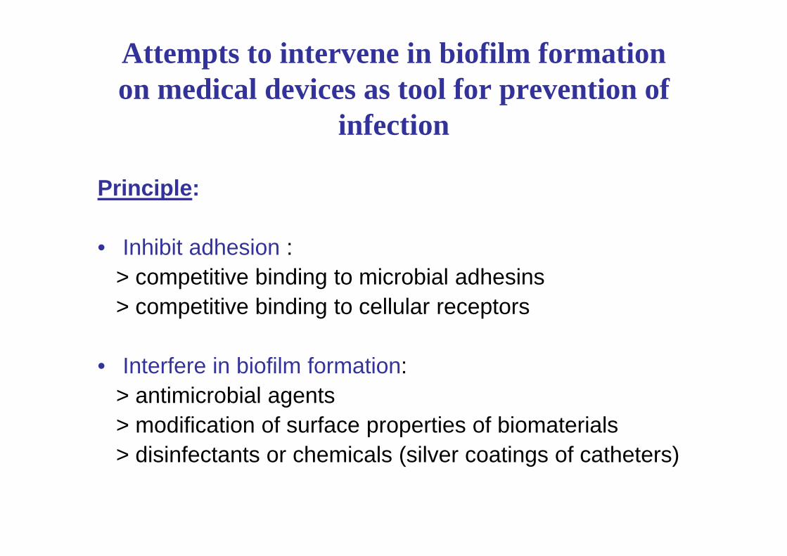
Attempts to intervene in biofilm formationon medical devices as tool for prevention of
infection
Principle:
• Inhibit adhesion :> competitive binding to microbial adhesins> competitive binding to cellular receptors
• Interfere in biofilm formation:> antimicrobial agents> modification of surface properties of biomaterials> disinfectants or chemicals (silver coatings of catheters)
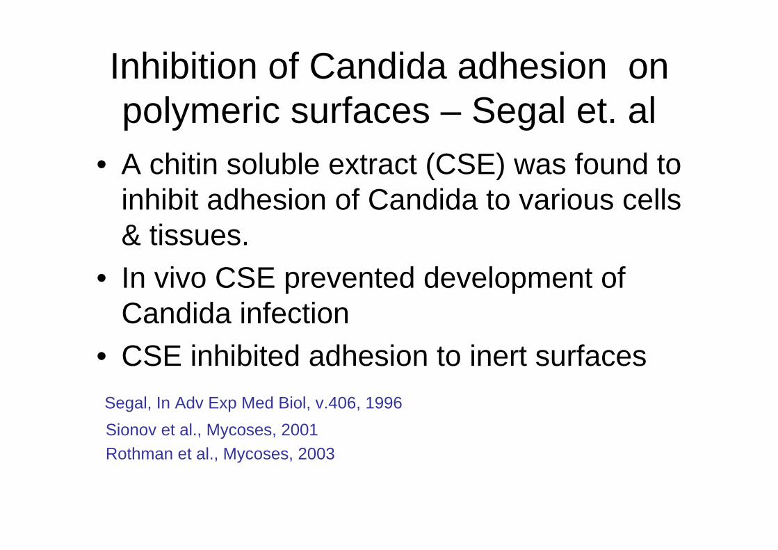
Inhibition of Candida adhesion on polymeric surfaces – Segal et. al
• A chitin soluble extract (CSE) was found to inhibit adhesion of Candida to various cells & tissues.
• In vivo CSE prevented development of Candida infection
• CSE inhibited adhesion to inert surfacesSegal, In Adv Exp Med Biol, v.406, 1996Sionov et al., Mycoses, 2001Rothman et al., Mycoses, 2003

Coating of polyurethane (polymer of IV catheters) with CSE
From: Zilberman, Navon, Sandovsky-Losica, Segal; 2007, J.Biomed.MaterialsResearch
coated non-coated
A schematic representation showing CSEchemical binding to polyurethane via spacer
(WSC) molecule.
Polyurethane Coated With CSE To Prevent Candida Adherence
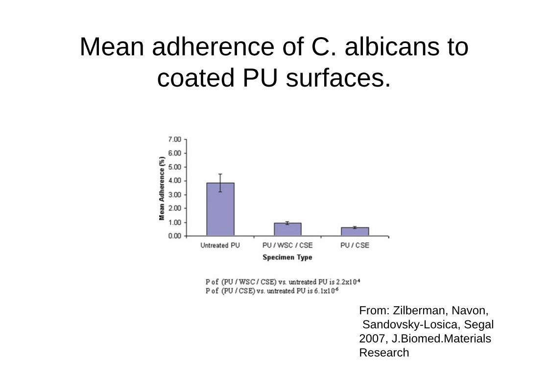
Mean adherence of C. albicans to coated PU surfaces.
From: Zilberman, Navon,Sandovsky-Losica, Segal2007, J.Biomed.MaterialsResearch

Examples of In Vivo Studies to prevent biofilm formation
• Demonstration of Antibiofilm and Antifungal Efficacy of Chitosan against Candidal Biofilms, Using an In Vivo Central Venous Catheter Model.Martinez,et al. 2010A C. albicans CVC biofilm model was used for in vivo experiments. Chitosan-treated CVCs were generated by incubating polyethylene catheters in chitosan. Chitosan-treated catheters were inserted in Sprague-Dawley rats. Chitosan inhibits candidal biofilm formation in vivo.
• Treatment and prevention of Candida albicans biofilms with caspofungin in a novel central venous catheter murine model of candidiasis. Lazell..Lopez-Ribot; 2009
We developed murine model of CVC-associated candidiasis for in vivobiofilm formation. Following model development, caspofungin was instilled in the catheter to treat preformed biofilms. The results indicated that caspofungin treatment significantly reduced biofilm fungal load in the catheters and dissemination to kidneys compared with untreated controls. In a second set of experiments catheters were pre-treated by filling caspofungin before challenge with C. albicans via the CVC. Again, the results indicated a significant reduction in biofilm fungal load and dissemination to kidneys.

Examples of In Vitro Studies to prevent biofilm formation
• Prevention of Candida albicans biofilm formation by covalently bound dimethylaminomethacrylate and polyethyleneimine. De Prijck et.al, 2010Candida albicans biofilms are a major cause of voice prosthesis deterioration in laryngectomized patients. The aim of this study was to produce a surface capable of inhibiting C. albicans biofilm formation. Dimethylaminoethylmethacrylate (DMAEMA) and polyethylenimine (PEI) moieties were covalently bound to the surface of polydimethylsiloxane (PDMS) or polymethylmethacrylate (PMMA) and subsequently quaternized. Physicochemical characterization of the grafted surfaces was carried out and their effect on C. albicans cell numbers was assessed …. Covalently bound quaternized polyDMAEMA (polyDMAEMAq) and PEI (PEIq) inhibited biofilm growth, with reductions up to 92%.
• Effect of tunicamycin on Candida albicans biofilm formation and maintenance. Pierce et al., 2009The effect of tunicamycin, a nucleoside antibiotic that inhibits N-linked glycosylation affecting cell wall and secreted proteins, on C. albicans biofilm formation was studied. Tunicamycin displayed significantinhibitory effects on biofilm development.

Rabbit Model of Candida Biofilm Infection: AAC 2004Liposomal Amphotericin B Antifungal Lock Therapy
FIG. 1. Surgical placement of the intravenous catheter. (A to C)Catheter insertion into the external jugular vein; (D to F) attachment of the heparin lock device to skin; (G) postoperative venogram of catheter placement.

More Examples of Studies to prevent biofilm formation
• Effective prevention of microbial biofilm formation on medical devices by low-energy surface acouistic waves. Hazan et al., AAC, 2006Low-energy surface acoustic waves generated from electrically activated piezo elements are shown to effectively prevent microbial biofilm formation on indwelling medical devices. The development of biofilms by Candida species is prevented.
• In vitro efficacies of caspofungin or micafungin catheter lock solutions on Candida albicans biofilm growth. Cateau et al. J Ant Chem;2008The data suggest that caspofungin and micafungin could represent good candidates for the reduction or control of fungal biofilms associated with silicone medical devices, as part of an antifungal lock. They were able to induce a significant and persistent reduction in the yeast metabolic activity of intermediate and mature of intermediate and mature biofilms, 12 h and 5 days old, respectively,when used as catheter lock solutions.

Candida adhesion to host tissues Adhesion of a microbe to host cells is a major step in
development of infection Observations indicate that adhesion is not a passive event:
it causes changes in the cells & microbes
QUESTIONS TO BE ASKED:• Events occurring in host cells post interaction with Candida?• Events occurring in Candida post interaction with host cells?

Post Interaction Events in CandidaSegal et al
• Interaction of C.albicans with HEp2 cells-DNA microarray technology (Georgetown University):- Genes from the ALS family up-regulated • Gene for manosyl transferase up-regulated• Down-regulated genes encoding actin proteins
(ABP1, SAC6)• Down-regulated the gene for beta-glucosidase
(SUN4)-From: Sandovsky-Losica H., Chauhan, N., Calderone, R . Segal, E.Gene transcription studies of Candida albicans following infection of HEp2 epithelial cells Med Mycol 2006

Post Interaction Events in Candida:Segal et al
- Interaction of C.albicans with HEp2 cellsRT-PCR STUDIES:
0,00
5000,00
10000,00
15000,00
20000,00
25000,00
30000,00
35000,00
40000,00
45000,00
1hrals1
1hrals2
1hrals3
1hrals4
1hrals5
1hrals6
1hrals7
1hrals9
2hrals1
2hrals2
2hrals3
2hrals4
2hrals5
2hrals6
2hrals7
2hrals9
4hrals1
4hrals2
4hrals3
4hrals4
4hrals5
4hrals6
4hrals7
4hrals9
RT=PCR ALS
CA CA+Hep2
•ALS genes up-regulated Sandovsky-Losica et al.Med Mycol 2006
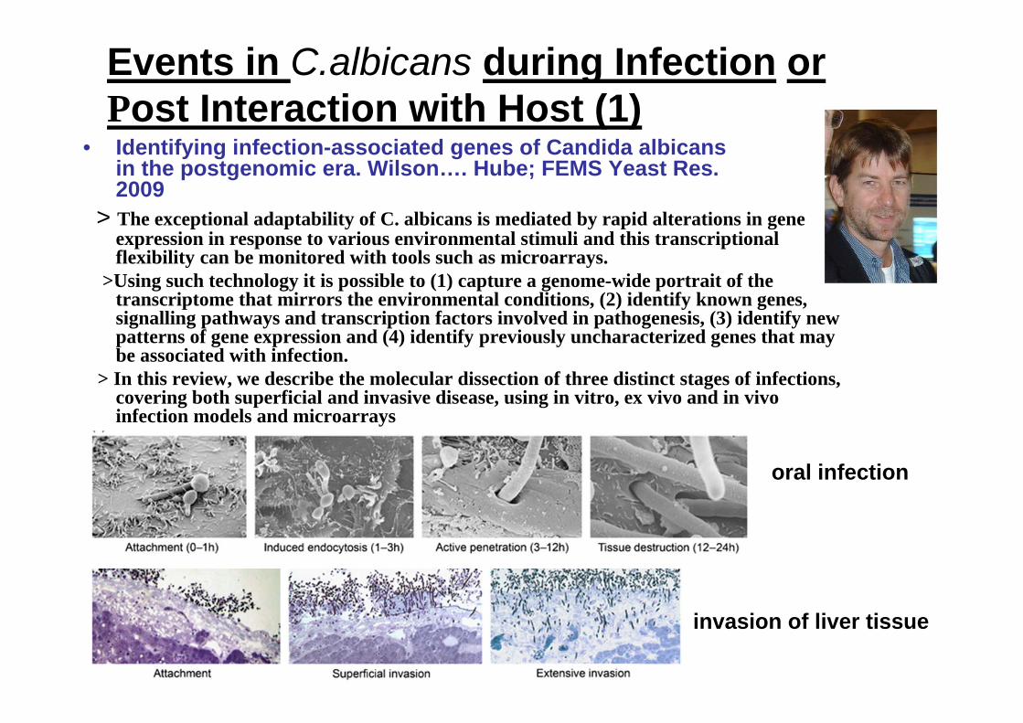
Events in C.albicans during Infection orPost Interaction with Host (1)
• Identifying infection-associated genes of Candida albicansin the postgenomic era. Wilson…. Hube; FEMS Yeast Res. 2009
> The exceptional adaptability of C. albicans is mediated by rapid alterations in gene expression in response to various environmental stimuli and this transcriptional flexibility can be monitored with tools such as microarrays.
>Using such technology it is possible to (1) capture a genome-wide portrait of the transcriptome that mirrors the environmental conditions, (2) identify known genes, signalling pathways and transcription factors involved in pathogenesis, (3) identify new patterns of gene expression and (4) identify previously uncharacterized genes that may be associated with infection.
> In this review, we describe the molecular dissection of three distinct stages of infections, covering both superficial and invasive disease, using in vitro, ex vivo and in vivo infection models and microarrays
oral infection
invasion of liver tissue

Events in C.albicans during Infectionor Post Interaction with Host (2)
• Genome-wide analysis of Candida albicans gene expression patterns during infection of the mammalian kidney. Walker et al. Fungal Genetics and Biology, 2009Abstract:
> In this study we examined the C. albicans SC5314 transcriptome from renal infections in the rabbit.
> Genes involved in adhesion, stress adaptation and the assimilation of alternative carbon sources were up-regulated in these cells compared with control cells grown in RPMI 1640, whereas genes involved in morphogenesis, fermentation and translation were down-regulated.
>When we compared the congenic virulent C. albicans strains NGY152 and SC5314, there was minimal overlap between their transcriptomes during kidney infections.
>This suggests that much of the gene regulation observed during infections is not essential for virulence. Indeed, we observed a poor correlation between the transcriptome and phenome for those genes that were regulated during kidney infection and that have been virulence tested.
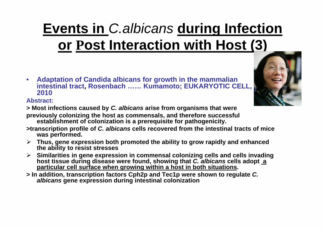
Events in C.albicans during Infectionor Post Interaction with Host (3)
• Adaptation of Candida albicans for growth in the mammalian intestinal tract, Rosenbach …… Kumamoto; EUKARYOTIC CELL, 2010
Abstract:> Most infections caused by C. albicans arise from organisms that werepreviously colonizing the host as commensals, and therefore successful
establishment of colonization is a prerequisite for pathogenicity. >transcription profile of C. albicans cells recovered from the intestinal tracts of mice
was performed. Thus, gene expression both promoted the ability to grow rapidly and enhanced
the ability to resist stresses Similarities in gene expression in commensal colonizing cells and cells invading
host tissue during disease were found, showing that C. albicans cells adopt a particular cell surface when growing within a host in both situations.
> In addition, transcription factors Cph2p and Tec1p were shown to regulate C. albicans gene expression during intestinal colonization

Post Interaction Events in the Host- Segal et alCellular actin is affected by Candida albicans
Actin aggregates
Methodology:•Interaction of FITC labelled Candida with HEp2 cells
•Actin labelled with Rhodamine•Confocal scanning laser microscopy (CLSM)
Results:•Actin surrounds Candida at the site of contact with the HEp2 cells
•Actin is rearranged from the fibrillar form to an aggregate form at the site of contact between the fungus and host cell
Actin rearrangementTsarfaty et al. FEMS Microbiol 2000

A C.albicans metabolite has a similar effect on HEp2 cells:Characteristics of the metabolite
• Contains proteins (10%) • Possesses proteolytic activity• SDS gel chromatography: 9 bands – two (34 kDa & 45
kDa) showed effect on the cytoskeleton
• Proteomics of the two bands: presence of Sap,Als (Als 2, Als 4) & 1,3-beta-glucosidase
• Sap & Als known as molecules involved in recognition processes between fungus and host cells
Sandovsky-Losica H. at al. FEMS Microbiol 2002Sandovsky - Losica H. et al. FEMS Immunol Med Microbiol 2006
Non treated Treated with 25 mg/ml metabolite
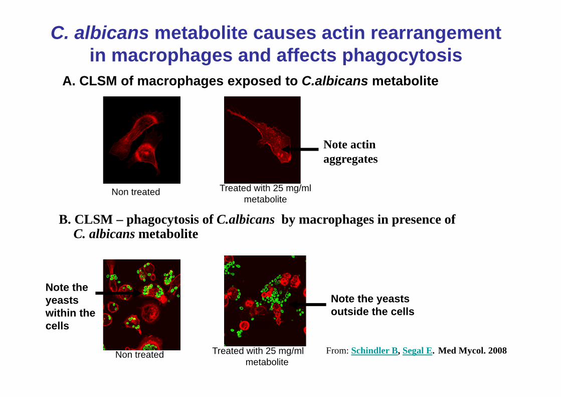
C. albicans metabolite causes actin rearrangement in macrophages and affects phagocytosis
Note actin aggregates
A. CLSM of macrophages exposed to C.albicans metabolite
Non treated
Note the yeasts within the cells
Note the yeasts outside the cells
B. CLSM – phagocytosis of C.albicans by macrophages in presence of C. albicans metabolite
Treated with 25 mg/ml metabolite
From: Schindler B, Segal E. Med Mycol. 2008

Quantitative assessment of phagocytosis of Candida in presence of C. albicans metabolite
Candida metabolite reduces phagocytosis
Conclusion:The data indicate that C. albicans secretesmaterial that affectsphagocytosis of murinemacrophages by changesin the arrangement of actinFrom :Schindler B, Segal E. Med Mycol. 2008
GENERAL CONCLUSIONS• Interaction/Infection of Candida and non-phagocytic or
phagocytic mammalian cells affects the cytoskeleton of the host cells involving reorganization of actin
• Candida probably secretes biomolecules that induce a process involving the cellular signal transduction machinery culminating in actin reorganization
• Concurrently transcriptional changes occur in Candida that may be related to the process in the host cell
Thus, actin plays a role in Candida - host interactionand pathogenesis
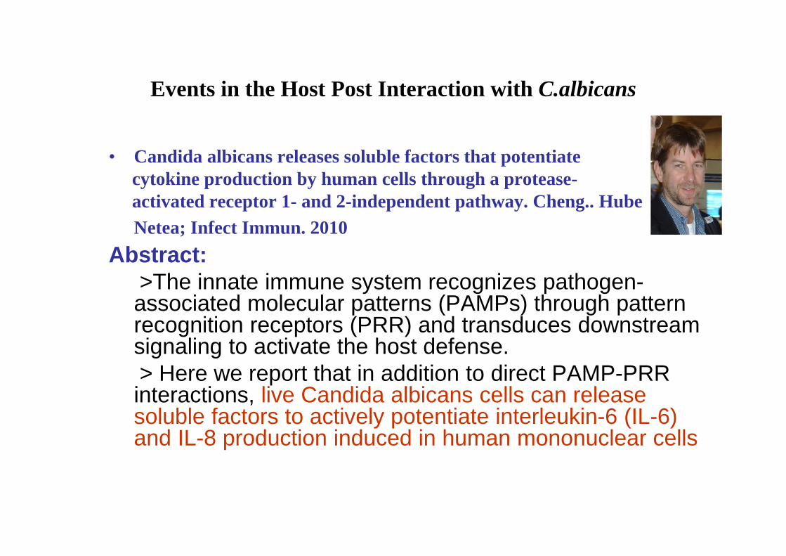
Events in the Host Post Interaction with C.albicans
• Candida albicans releases soluble factors that potentiate cytokine production by human cells through a protease-activated receptor 1- and 2-independent pathway. Cheng.. Hube, Netea; Infect Immun. 2010
Abstract:>The innate immune system recognizes pathogen-associated molecular patterns (PAMPs) through pattern recognition receptors (PRR) and transduces downstream signaling to activate the host defense. > Here we report that in addition to direct PAMP-PRR interactions, live Candida albicans cells can release soluble factors to actively potentiate interleukin-6 (IL-6) and IL-8 production induced in human mononuclear cells

Events in the Host Post Interaction with C.albicans
• The yeast Candida albicans evades human complement attack by secretion of aspartic proteases. Gropp… Hube..Mol Immunol. 2009
Abstract:>Candida albicans, which represents one of the most important humanpathogenic
yeasts, is directly attacked by the host innate immune system upon infection. >However this pathogen has developed multiple strategies to escape host immune defense. >Here, we show that C. albicans secreted proteases interfere and inactivate host innate immune effector components, such as complement proteins.
>Secreted aspartic proteases (Saps) in the culture supernatant of C. albicans cells and also recombinant Sap1, Sap2 and Sap3 degrade host complement components C3b, C4b and C5 and also inhibit terminal complement complex (TCC) formation.
> The complement inhibitory role of Sap1, Sap2 and Sap3 was confirmed in hemolysis assays with rabbit erythrocytes and normal human plasma.
> Secretion of complement degrading proteases provides a highly efficient complement defense response of this human pathogenic yeast that acts after the immediate acquisition of host complement regulators to the cell surface.
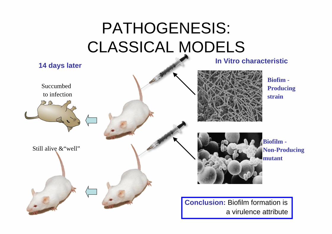
PATHOGENESIS:CLASSICAL MODELS
In Vitro characteristic
Biofim -Producingstrain
Biofilm -Non-Producingmutant
14 days later
Succumbedto infection
Still alive &“well”
Conclusion: Biofilm formation isa virulence attribute

PATHOGENESIS:NOVEL MODELSThe Drosophila melanogaster model
• What is it and why bother about it?Drosophila melanogaster is a fruit fly, a little insect about 3mm long, of the kind that accumulates around spoiled fruit. It is also one of the most valuable of organisms in biological research, particularly in genetics and developmental biology. Drosophila has been used as a model organism for research for almost a century, and today, several thousand scientists are working on many different aspects of the fruit fly. Its importance for human health was recognised by the award of the Nobel prize in medicine/physiology to Ed Lewis, Christiane Nusslein-Volhard and Eric Wieschaus in 1995.
• Why work with Drosophila?Part of the reason people work on it is historical - so much is already known about it that it is easy to handle and well-understood - and part of it is practical: it's a small animal, with a short life cycle of just two weeks, and is cheap and easy to keep large numbers. Mutant flies, with defects in any of several thousand genes are available, and the entire genome has recently been sequenced.
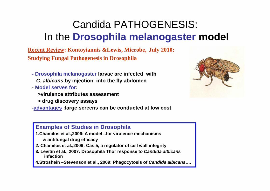
Candida PATHOGENESIS:In the Drosophila melanogaster model
- Drosophila melanogaster larvae are infected withC. albicans by injection into the fly abdomen
- Model serves for:>virulence attributes assessment > drug discovery assays
-advantages :large screens can be conducted at low cost
Examples of Studies in Drosophila1.Chamilos et al.,2006: A model ..for virulence mechanisms
& antifungal drug efficacy2. Chamilos et al.,2009: Cas 5, a regulator of cell wall integrity3. Levitin et al., 2007: Drosophila Thor response to Candida albicans
infection4.Stroshein –Stevenson et al., 2009: Phagocytosis of Candida albicans….
Recent Review: Kontoyiannis &Lewis, Microbe, July 2010:Studying Fungal Pathogenesis in Drosophila

PATHOGENESIS:NOVEL MODELS
The Caenorhabditis elegans model:• Caenorhabditis elegans is a free-living, transparent
nematode (roundworm), about 1 mm in length which lives in temperate soil environments.
• Research into the molecular and developmental biology of C. elegans was begun in 1974 by Sydney Brenner
• It has since been used extensively as a model organism
Laboratory uses• C. elegans is studied as a model organism for a variety of reasons. • It is a multicellular eukaryotic organism that is simple enough to be studied in great detail. • Strains are cheap to breed and can be frozen. When subsequently thawed they remain viable,
allowing long-term storage.• In addition, C. elegans is transparent, facilitating the study of cellular differentiation
and other developmental processes in the intact organism

Candida PATHOGENESIS:In the Caenorhabditis elegans model
Examples of Studies in C.elegans:• Pukila –Vorley et al., .Eukaryot, Cell: 2009: C. albicans
hyphal formation and virulence assessed• Means et al., J Exp Med., 2009: Recognition & innate
immunity to fungal pathogens• Tampakakis et al., Nat.Prot., 2008: A C. elegans whole animal in vivo
screen for the identification of antifungal compounds• Breger et al., PloS Pathog, 2007: Antifungal chemical compounds
identified using a C.elegans pathogenicity assay
Model Used for:• Virulence assessment• Immune response assessment• Antifungal drug research

EPIDEMIOLOGY : A TEACHING SLIDETHE GENUS CANDIDA –Candida species distribution in BSI
The US data –up to 2000• ~ 200 species • ~ 20 species pathogenic
for man• Major pathogen –
C.albicans• During the last decade a
shift in the relative prevalence of species occurred: a decrease in prevalence of C.albicansas cause of infection and an increase in non-albicans species: e.g.C.glabrata, C.krusei, C.parapsilosis
20011989
3952C.albicans
725C.tropicalis
1812C.parapsilosis
349C.glabrata
1.5C. kefyr
1C. krusei
1C.lusitaniae
Adapted from: Vazquez 2004

EPIDEMIOLOGY :THE GENUS CANDIDA –Candida species distribution in BSI
European data – up to 2005
Conclusions:•C. albicans most frequent•Non-CAS- geographic differences From: Tortorano et al. 2006, Inter J AA
ECMM survey: Species causing candidemia.

EPIDEMIOLOGY :THE GENUS CANDIDA –Candida species distribution in BSI
European data : 2005-2010
• FROM: Filioti J et al.Intensive Care Med, 2007: …Candida albicans continues to be the most prevalent isolate, However, an increasing role of non-C. albicans spp., particularly C. parapsilosis,C. tropicalis, account for almost half of pediatric IC cases
• FROM Guery BP et al, Intensive Care Med, 2009 : a review of the literature and a European expert panel discussion (adult ICU). Results: Candida albicans remains the most frequently isolated fungal species followed by C. glabrata.
• FROM Sabino, R. et al, Med. Mycol. 2010: Epidemiology of candidemia in oncology patients: a 6-year survey in a Portuguese hospital. …. The most frequent species was C. albicans (48.7%),followed by C.parapsilosis(20.2%), C. tropicalis (8.4%), C. krusei (6.7%) and C. glabrata (5.0%)
CONCLUSIONS :• Demographic differences• Geographic differences• No significant change : except of increase of C. parapsilosis
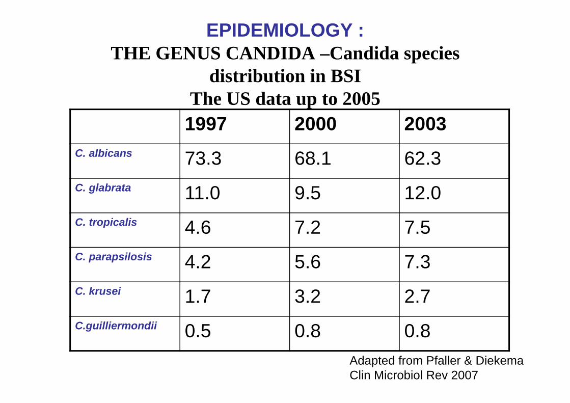
EPIDEMIOLOGY :THE GENUS CANDIDA –Candida species
distribution in BSI The US data up to 20051997 2000 2003
C. albicans 73.3 68.1 62.3C. glabrata 11.0 9.5 12.0C. tropicalis 4.6 7.2 7.5C. parapsilosis 4.2 5.6 7.3C. krusei 1.7 3.2 2.7C.guilliermondii 0.5 0.8 0.8
Adapted from Pfaller & Diekema Clin Microbiol Rev 2007

EPIDEMIOLOGY :THE GENUS CANDIDA –Candida species distribution in BSI
USA data : 2005-2010
• From Horn et al. 2007
Data of 1710 patients with IFIstudied 2006-2007
Candida spp. (%):
C. albicans 47.9
C. glabrata 25.7
C. parapsilosis 14.8
C. tropicalis 6.7
C. krusei 2.6
C. guiliermondii 0.4Conclusion:
Marked increase in non-Candida albicans species
• Neofytos et al., Clin Inf Dis, 2009
• Lewis, Curr Med Res Opin, 2009Similar conclusions
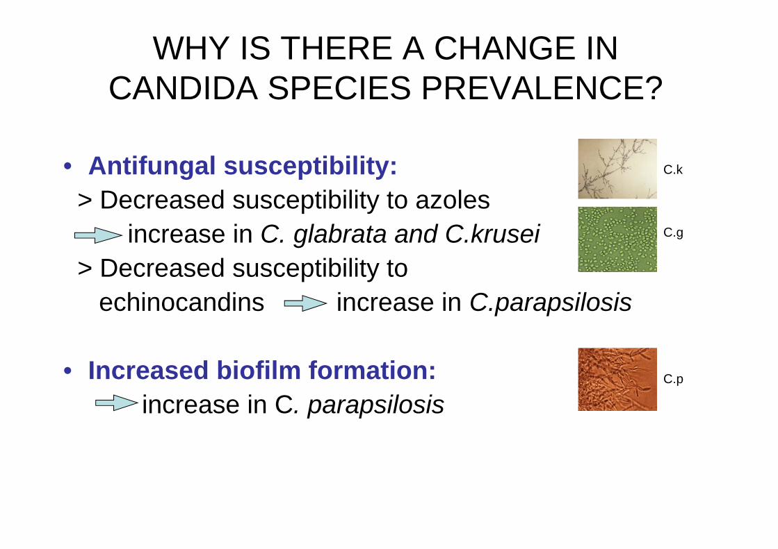
WHY IS THERE A CHANGE IN CANDIDA SPECIES PREVALENCE?
• Antifungal susceptibility:> Decreased susceptibility to azoles
increase in C. glabrata and C.krusei> Decreased susceptibility to
echinocandins increase in C.parapsilosis
• Increased biofilm formation:increase in C. parapsilosis
C.k
C.g
C.p

Are all C.albicans strains the same?
Intra-species strain differentiation in Candida has been a persistent long-time goal –some examples:
•Odds, F., Auger, P., Krogh, P., Neely, A. and E. Segal: Biotyping of Candida albicans. Results of an international collaborative study. Journal of Clinical Microbiology. 1989Abstract: An agar plate system for biotyping isolates of Candida albicans was evaluated in four laboratories for 18 coded yeast isolates, each tested in triplicate on duplicate series of agar plates. The results showed that the biotyping system gave excellent intralaboratory reproducibility. However, because the concordance of data among laboratories was poor, the method must be regarded as suitable only for research applications and not for routine use.
•Merson-Davies LA, Odds FC: A morphology index for characterization of cell shape in Candida albicans. J Gen Microbiol. 1989
•Odds FC, Merson-Davies LA: Colony variations in Candida species. Mycoses. 1989

C.albicans Intra-species strain differentiation - Contemporary Methods (Genotyping):
Multilocus sequence typing (MLST) (Most commonly used)
> MLST based microarrays> Microsattelite typingDNA finger printingElectrophoretic Karyotyping (less common)Single nucleotide polymorphism (SNP) (less
common)Flow Cytometry (less common)

GENOTYPING- Principles of Methods and Examples of Studies
• Genomic Plasticity of the Human Fungal Pathogen Candida albicans. Selmecki, Forche, and Berman; Eukaryot Cell. 2010.
-Electrophoretic Karyotyping: to detect chromosomal rearrangements-Single Nucleotide Polymorphism (SNP): reveals allelic differences between strains-Flow Cytometry: a rapid and accurate method to analyze genome size changes
(ploidy changes).• Property differences among the four major Candida albicans strain clades.
MacCallum, …..Gow, Odds; Eukaryotic Cell, 2009.• PCR melting profile – a new tool for differenetiation of Candida albicans
Krawchyk et al. BMC Infect Dis 2009.• Multilocus sequence typing is a reliable alternative method to
DNA fingerprinting for discriminating among strains of Candida albicansRobles, Koreen, Park and Perlin; J Clin Microbiol. 2004.

GENOTYPING- What do we learn from it: some examples
• Molecular phylogenetics and epidemiology of Candida albicansOdds; Future Microbiology, 2010
- Molecular typing of C. albicans strains shows geographical evolutionary associations .
- Individual patients usually carry a single C. albicans strain type, but this may undergo microvariation leading to detection of mixtures of closely related types
- There are also clade-related associations with lengths of tandem repeats in some cell-surface proteins, but not with virulence or type of infection.
• Progressive loss of echinocandin activity following prolonged use for treatment of Candida albicans oesophagitisLavendiere………Perlin ; J Antimicrob Chemother. 2006
- Antifungal susceptibility profiles on isolates before and during therapy- MLST on isolates before and during therapy- Identical allelic homology for the isolates- Lack of clinical response to micafungin was associated with increases in MICs
to all three echinocandins in association with the acquisition of mutation in FKS1 gene
SUMMARY – What did we learn today?
• Candidosis is an opportunistic fungal disease caused by the human commensal yeast, Candida. It is manifested in a diversity of clinical forms, ranging from muco-cutaneous infection to life threatening systemic disease, which affects primarily immunocompromised and debilitated patients.
• This presentation attempted to overview the state of art of the pathogenesis of candidosis. It highlighted the current view and general understanding on how candidosis evolves, who is particularly at risk to develop the infection and what is the response to it.
• Since it is believed that understanding the pathogenesis of an infection is a basis for a rational approach for diagnosis and therapy, reviewing the advances in the understanding of the pathogenesis of candidosis may contribute to the management of this complex clinical entity.
Thanks to all the students, post-docs & collaboratorscited in this presentation and to the many, many authorsof the studies overviewed today
AND


![Disclaimer - Seoul National University...genes with Enterococcus faecalis in root canal biofilm, thereby potentiating its virulence and antibiotic resistance [6]. Thus, S. gordonii](https://static.fdocuments.net/doc/165x107/60f94d2d6e21554c4560e29a/disclaimer-seoul-national-university-genes-with-enterococcus-faecalis-in-root.jpg)


