Cancer Care - St. Joseph Mercy Ann Arbor
8
INSIDE Cancer Care...............1 Pathology ..................3 Cardiovascular Services ......................4 Orthopedics, Neuro- sciences and Rehabili- tation Services ........... 5 Women’s and Children’s Services.....6 Emergency Medicine .....................7 Imaging Updates ....... 8 May, 2009 Volume 2 Number 1 sjmercyhealth.org Edward Kreske, MD Continued on page 2. St. Joseph Mercy Ann Arbor St. Joseph Mercy Livingston St. Joseph Mercy Saline Saint Joseph Mercy Health System Progress Notes is designed to acquaint you, our partners in health care, with specialists and their expertise, technology and services available to your patients for the major service lines within our health system. These articles were authored by our medical staff, who are available to answer your questions and provide additional information on these topics and much more. To find out more about these physicians and the rest of our medical staff, please visit our Web site at sjmercyhealth.org. Cancer Care Pancreatic Cancer Treatment at St. Joseph Mercy Hospital By Edward Kreske, MD, General Surgery There is a long and rich heritage of pancreatic cancer care at St. Joseph Mercy Hospital (SJMH). This challenging malignancy will be diagnosed in just over 30,000 individuals this year. About the same number of patients will die from the disease during that same time period. Historically, there has been a well-developed interest in the treatment of pancreatic adenocarcinoma across many different specialties at SJMH. That interest continues today with multidisciplinary treatment provided by medical oncologists, radiation oncologists, gastroenterologists, surgeons and interventional radiologists. Pancreatic cancer is ideally treated in a multidisciplinary manner with surgical resection, chemotherapy and often external beam radiation. Unfortunately, of all patients diagnosed with pancreatic cancer, fewer than ten percent will be resectable at the time the diagnosis is made. These patients will have limited options, but they may benefit from palliation with chemotherapy and radiation with subsequent hospice care. If needed, jaundice can be treated with stent placement either endoscopically by our gastroenterology group, or with percutaneous methods available in the interventional radiology department. Intractable pain not responsive to medications can sometimes be relieved with celiac plexus nerve blocks that can be performed by our gastroenterology group using endoscopic ultrasound to guide placement of the block. The transition to hospice care is helpful for these patients and is facilitated with input from the palliative care service at SJMH. For the smaller group of potentially resectable patients, there are more options. The assessment of resectability is based upon a high quality CT scan obtained using multiphasic pancreatic imaging protocols, with 3-D reconstructions if needed. Non-contrast CT, routine abdominal CT and MRI will all be much less useful and generally will only add time and expense to the evaluation. The obvious goal is to limit abdominal exploration to those patients who are likely to be resectable. These patients have Stage I or Stage II disease, and when properly performed, CT scanning should identify these patients with about 90% accuracy. Endoscopic ultrasound is also often used in conjunction with CT scanning to provide additional staging information and when appropriate, to help obtain a tissue diagnosis. When a pancreatic cancer is diagnosed and determined to be resectable, our approach at SJMH is to proceed with surgical resection, followed by adjuvant treatment with gemciabine based chemotherapy and external beam radiation if appropriate. Most often resectable tumors are located in the pancreatic head and the appropriate operation is a Whipple procedure (pancreaticoduodenectomy). SJMH is a high volume institution for pancreatic surgery with the capability of offering patients a number of complex pancreatic surgical procedures, including procedures requiring vascular resection and reconstruction. Continued from cover.
Transcript of Cancer Care - St. Joseph Mercy Ann Arbor

INSIDE
Cancer Care ...............1
Pathology ..................3
Cardiovascular Services ......................4
Orthopedics, Neuro- sciences and Rehabili-tation Services ...........5
Women’s and Children’s Services .....6
Emergency Medicine .....................7
Imaging Updates .......8
May, 2009
Volume 2Number 1
sjmercyhealth.org
Edward Kreske, MD
Continued on page 2.
St. Joseph Mercy Ann ArborSt. Joseph Mercy Livingston
St. Joseph Mercy Saline
Saint Joseph Mercy Health System Progress Notes is designed to acquaint you, our partners in health care, with specialists and their expertise, technology and services available to your patients for the major service lines within our health system. These articles were authored by our medical staff, who are available to answer your questions and provide additional information on these topics and much more. To find out more about these physicians and the rest of our medical staff, please visit our Web site at sjmercyhealth.org.
Cancer CarePancreatic Cancer Treatment at St. Joseph Mercy HospitalBy Edward Kreske, MD, General Surgery
There is a long and rich heritage of pancreatic cancer care at St. Joseph Mercy Hospital (SJMH). This challenging malignancy will be diagnosed in just over 30,000 individuals this year. About the same number of patients will die from the disease during that same time period. Historically, there has been a well-developed interest in the treatment of pancreatic adenocarcinoma across many different specialties at SJMH. That interest continues today with multidisciplinary treatment provided by medical oncologists, radiation oncologists, gastroenterologists, surgeons and interventional radiologists.
Pancreatic cancer is ideally treated in a multidisciplinary manner with surgical resection, chemotherapy and often external beam radiation. Unfortunately, of all patients diagnosed with pancreatic cancer, fewer than ten percent will be resectable at the time the diagnosis is made. These patients will have limited options, but they may benefit from palliation with chemotherapy and radiation with subsequent hospice care. If needed, jaundice can be treated with stent placement either endoscopically by our gastroenterology group, or with percutaneous methods available in the interventional radiology department. Intractable pain not responsive to medications can sometimes be relieved with celiac plexus nerve blocks that can be performed by our gastroenterology group using endoscopic ultrasound to guide placement of the block. The transition to hospice care is helpful for these patients and is facilitated with input from the palliative care service at SJMH.
For the smaller group of potentially resectable patients, there are more options. The assessment of resectability is based upon a high quality CT scan obtained using multiphasic pancreatic imaging protocols, with 3-D reconstructions if needed. Non-contrast CT, routine abdominal CT and MRI will all be much less useful and generally will only add time and expense to the evaluation. The obvious goal is to limit abdominal exploration to those patients who are likely to be resectable. These patients have Stage I or Stage II disease, and when properly performed, CT scanning should identify these patients with about 90% accuracy. Endoscopic ultrasound is also often used in conjunction with CT scanning to provide additional staging information and when appropriate, to help obtain a tissue diagnosis. When a pancreatic cancer is diagnosed and determined to be resectable, our approach at SJMH is to proceed with surgical resection, followed by adjuvant treatment with gemciabine based chemotherapy and external beam radiation if appropriate. Most often resectable tumors are located in the pancreatic head and the appropriate operation is a Whipple procedure (pancreaticoduodenectomy). SJMH is a high volume institution for pancreatic surgery with the capability of offering patients a number of complex pancreatic surgical procedures, including procedures requiring vascular resection and reconstruction.Continued from cover.
Cancer Care continued
Page 2
Eduardo Kleer, MD
Advances in Prostate Cancer TreatmentBy Eduardo Kleer, MD, Urology
St. Joseph Mercy Physician Resource Line: 877-443-0333
Pancreatic Cancer Treatment Continued from cover.An intriguing development in the care of pancreatic cancer patients has been the application of chemotherapy and chemoradiotherapy in a neo-adjuvant fashion. A number of institutions have demonstrated that this approach is safe and feasible when used in the setting of resectable pancreatic cancer. It has been much more difficult to demonstrate improved survival when compared to patients treated with an adjuvant approach, but the results are comparable. Recently another category of patients has been identified. These patients have what is called borderline resectable disease. Typically they have more advanced tumors with local vascular involvement of either the portal venous system (portal vein or superior mesenteric vein) or the arterial system (superior mesenteric artery or celiac arterial branches). Encasement or occlusion of these vessels usually indicates unresectability, but frequently the tumor will involve these structures to a lesser degree. At SJMH, these patients are discussed in a multidisciplinary forum, and then usually treated in a neo-adjuvant fashion. If subsequent restaging demonstrates a treatment response or at least stable disease, then resection is offered. In some of these patients vascular resections with appropriate reconstructions will be required to achieve tumor free surgical margins. Initial results have suggested that borderline resectable patients who are given neo-adjuvant therapy and who ultimately undergo resection will have outcomes comparable to initially resectable groups treated with standard adjuvant approaches.
The results of a study to optimize the administration of LHRH analogues (leuprolide acetate) in the treatment of patients with prostate cancer by lead authors Drs. Silke Greil from the Dept of Internal Medicine and Eduardo Kleer
from the Department of Surgery, Section of Urology has been accepted for publication in the journal Urology. It is currently available in full text in Pub Med. Co-investigators were Eileen Robinson, RN, MS and Bonita Singal MD, PhD from the Clinical Research Dept.
Following informed consent, the investigators prospectively studied patients diagnosed with prostate cancer for eighteen months after starting testosterone suppression therapy with the luteinizing hormone-releasing hormone (LHRH) analogue leuprolide acetate. Leuprolide acetate is usually given as a depot-injection, and for the most commonly used preparation the pharmaceutical companies recommend re-dosing it every four months. By monitoring serum testosterone levels in these patients and redosing only when those
levels exceeded castrate levels (>50 ng/dl) the investigators were able to show that the effects of the drug lasted much longer than the 120 days recommended by the drug manufacturers. The median duration of testosterone suppression was 159, 189, and 163 days for the first, second, and third treatment cycles, respectively. The total number of injections was reduced in all but one participant who completed the 18 month trial. The cost savings from this approach of redosing the leupron injections was $1,400 per patient per year.
This was the first study to monitor patients receiving this type of treatment for three cycles of treatment. On the basis of this and other studies, the authors recommend using testosterone levels to determine the time of re-injecting leupron as opposed to routinely dosing it every four months as recommended by the drug manufacturer. This approach can benefit patients because they need fewer injections to achieve the same level of treatment of their prostate cancer coupled with significant savings to the health care system.
Prostate Cancer ProgramSt. Joseph Mercy Hospital utilizes a multidisciplinary team approach in treating prostate cancer. The Prostate Cancer Program offers:
Guidance for patients from diagnosis through the many treatment options available, including daVinci® robotic minimally invasive surgery and CyberKnife®, Brachytherapy and IMRT
Consultation with a Urologist, Medical Oncologist and Radiation Oncologist Customized treatment plans A Nurse Navigator to help patients coordinate appointments, keep them informed
and connect them to Social Work, Nutrition Services and support groups
To reach our Nurse Navigator for prostate cancer, please call Pam Ceo, NP at 734-712-6813.

Page 3
Phil Stella, MD
Pathology
Clinical LaboratoryAs reported in Laboratory Economics, the Laboratories at St. Joseph Mercy Hospital are ranked 19th in the Top 25 Hospital Lab Facilities by total annual test volume. Outpatient testing comprises approximately 70% of total test volume at SJMHS. This segment of the laboratory business has had a 5% average annual growth rate over the past 5 years.
On April 1, 2009, the Laboratories at St. Joseph Mercy Hospital became the sole laboratory in the State of Michigan to provide histology services to the Organ Procurement Agency of Michigan, also known as Gift of Life. Histology services are now provided on a 24x7x365 basis to Gift of Life and also to SJMH and its client physicians, thus reducing turn-around-time for these services.
To reach the Clinical Laboratory Client Services, please call Kathy Shurtliff at 734-712-5097.
SJMHS participating in National Cancer Institute’s First-Ever Study to Determine if Biomarkers Can Help Guide Treatment for Lung Cancer By Phil Stella, MD, Medical Oncology, Principal investigator of the Michigan Cancer Research Consortium (MCRC) at SJMH
sjmercyhealth.org
Lung cancer is expected to claim 161,840 lives in 2008, and 215,020 people are expected to be diagnosed with the disease this year, making it the number one cancer killer. Non-small cell lung
cancer represents about 85 to 90 percent of all lung cancers. SJMHS recently began offering a large national clinical trial for non-small cell lung cancer, which will help to validate whether a biomarker can predict clinical benefit in the treatment of this disease.
This study, sponsored by the National Cancer Institute (NCI), is called MARVEL (Marker Validation for Erlotinib in Lung Cancer) and will attempt to definitively establish the future value of selecting patients for treatment based on the presence or absence of EGFR activation. Biomarkers, which are molecules found in the body that can signal an abnormal process or disease, would identify a target known as epidermal growth factor receptor (EGFR). This receptor can be increased in some lung cancers due to the presence of extra copies of its coding gene. These extra copies can result in activation of tumor growth, so drugs that block this activation could have a significant impact on lung cancer treatment. Approximately 1,200 lung cancer patients will be tested for the status of this biomarker, and then will be randomly assigned to treatment based on the test results. Both EGFR-positive and EGFR-negative patients will receive either the chemotherapy drugs erlotinib (Tarceva®, Genentech) or pemetrexed (Alimta®, Eli Lilly) after they have received their initial, standard chemotherapy. Erlotinib specifically targets EGFR, whereas pemetrexed blocks tumor cell growth by another mechanism.
It is hypothesized that erlotinib will be superior in the patients with EGFR-positive lung cancer, whereas pemetrexed would be favored in patients with EGFR-negative lung cancer, based on knowledge from earlier, smaller studies. MARVEL will incorporate genetic studies for erlotinib and pemetrexed that will be important to further identify patients with different sensitivity and toxicity profiles to these therapies. Because lung cancer is such a lethal disease and because it is particularly difficult to treat, especially if diagnosed in its later stages, the MARVEL trial is of major importance because it could define, based on a single test, the best therapy for this disease. The future of moving highly targeted agents from the lab to the clinic will be heavily dependent on biomarkers for patient selection.
Both erlotinib and pemetrexed are approved treatments for advanced non-small cell lung cancer. Among the factors that appear to influence responsiveness to erlotinib, in addition to the level of EGFR activation, are whether the resulting cancer cells are classified as adenocarcinomas (as opposed to squamous or other types of cells), female gender, Asian ethnicity, and whether the patient was ever a smoker. However, no forward-looking study has been performed to definitively address which factors are most important.
Please contact Beth LaVasseur, RN, MS at 734-712-5658 or [email protected] for more information on this trial or other clinical trials offered by the MCRC. www.mcrconline.org

Atrial fibrillation is estimated to affect over 2 million US adults. It is the most common arrhythmia in elderly persons and is a potent risk factor for stroke. In those with atrial fibrillation, treatment options include rhythm control versus rate control along with chronic anticoagulation where clinically appropriate. Recent advances have been made in our understanding of atrial fibrillation. Atrial fibrillation usually originates from within or near the areas where the pulmonary veins meet the left atrium. Abnormal electrical
impulses originating from these pulmonary veins, or from the nerves to the heart cause atrial fibrillation in some patients. Better understanding of this pathophysiology has led to the development of advanced techniques for the treatment of atrial fibrillation such as radiofrequency catheter endovascular ablation and the Cox-Maze surgical procedure. Such strategies are being explored, particularly for use in symptomatic patients who do not wish to take long-term antiarrhythmic drug therapy or in patients having recurrent atrial fibrillation despite the use of one or more antiarrhythmic drugs.
Catheter ablation goes by several names, including the Pulmonary Vein Isolation Procedure, Catheter Maze, Pappone Technique, and Wide Area Circumferential Ablation. Catheters are maneuvered from the leg into the left atrium, and ablation of the connections between the left atrium and pulmonary veins is performed. Because the connections are quite complex, atrial fibrillation ablation is much more extensive than a standard ablation procedure, and the procedure can be long. The potential for complications is higher than
Cardiovascular Services
Page 4
Jihn Han, MD
Thorascopically Guided Epicardial Atrial Fibrillation AblationBy Jihn Han, MD, Cardiology and Manak Sood, MD, Cardiothoracic Surgery
St. Joseph Mercy Physician Resource Line: 877-443-0333
New Technique for Deep Vein Thrombosis TreatmentBy Vishal Bhagat, MD, Interventional Radiology
The year 2008 has seen unprecedented changes in the way Deep Vein Thrombosis (DVT) patients are managed - from
increased attention to at-risk prophylaxis
to shifting the paradigm towards endovascular intervention for those who develop a DVT.
Anticoagulation alone prevents thrombus propagation and reduces the risk of embolization. It does not however, actively dissolve thrombus. Residual thrombus and subsequent venous hypertension and/or valvular damage can lead to a debilitating condition called Post-Thrombotic Syndrome (PTS). A recent study by Susan Kahn, et. al. indicates that approximately 40% of patients will develop signs of PTS within one month of a DVT.
An FDA-approved medical device called the Trellis® Peripheral Infusion System is available to remove clots. The Trellis System catheter is inserted into a vein and passed through the clot. Two balloons are inflated on either side of the obstruction and a low dose of clot-busting medication is infused directly into the area. A wire spinning at 3000 rpm helps mix the medicine to help dissolve the clot.
This new technique offers a rapid, effective and safe treatment alternative for patients suffering with acute DVT, and it is available at Saint Joseph Mercy Health System. Outpatient consultations can be scheduled at the Huron Valley Radiology Interventional Clinic by calling 734-712-8350. Inpatient interventional radiology consultations are also being done at St. Joseph Mercy Hospital.
100 Top Hospitals®: Cardiovascular BenchmarksSt. Joseph Mercy Ann Arbor has been recognized among the hospitals listed by Thomas Reuters as a 100 Top Hospitals®: Cardiovascular Benchmarks for Success 2008 and we rank among the top 40 teaching hospitals in the country.We are particularly proud that we exceeded the median performance of national benchmark peer groups in 11 of the 15 data points, including: AMI patient mortality Post-operative infection HF patient mortality Post-operative hemorrhage CABG patient mortality
Vishal Bhagat, MD
Manak Sood, MD
standard ablations, due to the large amount of ablation that is required, and the proximity of the pulmonary veins, the esophagus, and other structures. While catheter ablation has become an important part of the management of atrial fibrillation, success rates vary widely and multiple procedures may be required. The Cox-Maze procedure involves a series of incisions made in the atria intended to stop irregular electrical activity by scar formation. This procedure requires median sternotomy and cardiopulmonary bypass. The “Wolf-Mini-maze” was first reported in 2005. In this procedure, no median sternotomy is required; instead, endoscopes or “mini-thoracotomy” incisions are used (between the ribs). This technique utilizes a multidisciplinary approach, involving both the electrophysiologist and the surgeon. In addition, there is no need for cardiopulmonary bypass; instead, the procedure is performed on the beating heart, with few or no actual incisions into the heart itself. The maze lines are made epicardially by heating the tissue using radiofrequency.
The Wolf-Mini-maze procedure was recently introduced at St. Joseph Mercy Hospital in Ann Arbor as a collaborative effort between cardiothoracic surgery and cardiac electrophysiology. Initial results are very encouraging with respect to both patient outcomes and satisfaction. A recent report announced at the 2009 American College of Cardiology Conference suggests that cure rates with this procedure may be as high as 90% at one-year followup. Patients also have smaller incisions, faster recovery and shorter length of stays as compared to the traditional Cox-Maze procedure. To determine which therapeutic option is right for your patient, a consultation with the Michigan Heart & Vascular Institute (MHVI) outpatient Atrial Fibrillation Clinic can be obtained by calling 734-712-8000.
AMI core measures Procedure volume threshold measure for CABG
for 2006 and 2007 Procedure volume threshold measure for PCI for
2006 and 2006 average cost per case

Page 5
Orthopedics, Neurosciences and Rehabilitation Services
Advances in Shoulder SurgeryBy Steven Acker, DO, Orthopedic Surgery
Steven Acker, DO
Continued on page 6.
sjmercyhealth.org
Over the past five years, new surgical techniques and materials have resulted in significant advances in treating shoulder problems. Although many of the basic scientific surgical principles have remained the same, these advancements have improved our ability to treat a myriad of shoulder afflictions.
Complex proximal humeral fractures that were once deemed non-operative because of poor bone quality or difficult fracture configurations can now be successfully surgically repaired with locking plate implants. Locking plate designs permit the screws that fixate the bone fragments to attach directly to the plate. This fixation technique provides significant strength and stability in difficult fractures. Previously available fixation techniques and orthopedic materials often resulted in unsatisfying outcomes that frustrated orthopedic surgeons and their patients. Advancements in operative techniques and plate design have enabled patients to regain significant use of their shoulder and arm, with improved strength, mobility, and function.
Recent advances in arthroscopic surgery have allowed orthopedic surgeons to diagnosis and treat difficult rotator cuff tears far more successfully. Improved fixation implants such as suture anchors, in conjunction with the use of double row fixation techniques, permit better restoration of the anatomic footprint of the rotator cuff with improved long-term results in cuff repair. Patients are also benefitting from advances in biologics to augment rotator cuff tear repair through the use of extra-cellular or synthetic matrices. Use of these biologics, which are derived from human or animal substrates, improve repair by supporting poor tendon quality and allowing more successful healing and long-term survival of the repair. Further studies in stem cell research are likely to provide even more exciting opportunities for improvement in tissue healing.
Many patients who have massive non-repairable tears of the rotator cuff with severe osteoarthritis have also benefited from recent advances in shoulder replacement surgery. The design of the reverse ball and socket arthroplasty was changed so that the center of rotation of the glenoid component was moved more medial and inferior than in prior designs. This change increases the deltoid lever arm and places decreased forces across the glenoid-implant interface. This design change has dramatically improved patient outcomes, both by allowing many of our patients with rotator cuff arthropathy to lift their arm to an elevation that was previously unattainable, and increasing the durability of the implant by reducing the chance of loosening. The improvement in patient outcomes is profound and patient satisfaction scores after this surgery are extremely high.
Our Department of Orthopedic Surgery is currently implementing these advances in our shoulder surgeries. We continue to study, learn, and employ those techniques and principles to provide the highest quality of care for our patients.
Ankle Arthritis TreatmentBy Robert Mihalich, MD, Orthopedic Surgery
Although not as common as hip or knee arthritis, ankle arthritis can affect many patients and can be just as disabling. The problem can go untreated for a long time due to the relative lack of qualified specialists in the area. Fortunately for these patients, there are many non-operative and operative options for treatment that can result in a significant decrease in pain and improvement in function.
Ankle arthritis primarily occurs due to three main causes. Post-traumatic due to previous fractures, recurrent sprains, or compressive injury is the most common cause. General osteoarthritis that occurs due to wear and tear of the joint, obesity, or misalignment is the second most common type. Rheumatoid arthritis is the third most common cause for degeneration of the joint. The degeneration takes place between the tibia and talus and can result in limited motion, pain with activity, swelling and progressive deformity. Arthritis can also affect the subtalar joint, which moves the foot side to side and may give patients more trouble negotiating uneven ground.
Diagnosis is usually made by history and confirmed with physical examination and plain x-rays. Occasionally, a CT scan will be helpful in determining the extent of the arthritis and involvement of adjacent joints.
Initial treatment is usually focused on anti-inflammatory medications, either orally or now topically, activity modification away from consistent high impact loading and shoe wear modification. Many patients will find a heel lift helpful, especially if they have anterior impingement occurring due to spurring at the front of the ankle. Bracing using anything from a simple lace-up brace to a more rigid ankle-foot orthosis (AFO) can also be very helpful and provide patients with improved function.
Injections using corticosteroid are commonly used and may provide months of relief with a single injection. Hyaluronic acid injections have been very successfully used for the treatment for osteoarthritis and go by the trade names of Supartz, Synvisc, Euflexxa, or Hyalgan. There are ongoing trials that show promise for use in the ankle, however, this treatment is currently not FDA approved which limits insurance coverage. These injections tend to provide longer relief than corticosteroid and do not appear to have any of the harmful effects of repetitive injection than can be seen with corticosteroid.
For patients who fail non operative treatment, operative treatment is divided into three categories. First, debridement of prominent osteophytes and cartilage damage can be done arthroscopically with no limitation of weight bearing or range of motion following surgery. Discrete cartilage lesions or osteochondral defects can be treated with arthroscopy and microfracture techniques where holes are punched into the subchondral bone to stimulate fibrocartilage ingrowth. This has been shown to be 80-90% effective in lesions less than one centimeter in diameter. More advanced treatment Ankle Arthritis Treatment
Robert Mihalich, MD
Women’s and Children’s Services
Page 6 St. Joseph Mercy Physician Resource Line: 877-443-0333
Continued from page 6.
techniques for osteochondral lesions include osteochondral autograft transplant or OATS procedure where plugs of cartilage and bone are harvested from the patient’s knee in a non- articulating area and placed into the ankle to restore the articular surface.
Ankle fusion is the most time-tested treatment for advanced ankle arthritis and provides significant relief of pain and restoration of function. It is true that the motion of the ankle is lost; however, most patients who get to this point have very little true motion in the ankle to start with. For patients with significant loss of motion, obesity, or post-traumatic deformity, fusion is the best option surgically.
Ankle replacement has been available for some time; however, a single prosthesis was the only one available in the United States and it was known to have some limitations. In the last 1-2 years, at least two new prostheses have come on the market, which address some of the shortcomings of the older prosthesis. For the appropriate patient, ankle replacement can be a very successful procedure for relief of pain, improvement of function and maintenance of motion. As experience with the new prostheses increases, it is likely that ankle replacement will become more common and available as an option for patients with ankle arthritis. It is felt that these procedures should only be performed by fellowship-trained orthopedic foot and ankle surgeons or orthopedic surgeons with significant experience taking care of foot and ankle patients.
In July 2008, St. Joseph Mercy Hospital in collaboration with J.P. McCarthy Cord Stem Cell Bank at Karmanos Cancer Institute, received approval from our IRB to implement a Cord Blood Donation program.
Umbilical cord blood transplantation is a recent advancement in medical research. The first cord blood transplant took place in 1986. Initially, all cord blood transplants were from sibling donors. In the mid
1990’s, researchers learned that cord blood from unrelated donors could also work to treat recipients with leukemia and other blood diseases. Most transplants have been done in children, and research is underway to study the use of unrelated cord blood transplantation in adults.
Before birth, a baby’s blood cells move through his/her body, umbilical cord, and placenta. These blood cells carry oxygen and nutrition from the mother’s blood to the baby. After the birth of a baby, the umbilical cord is clamped and cut, separating the baby from the placenta, which is delivered several minutes later and is usually discarded. The placenta contains one-third (1/3) to one-half (1/2) of a cup of blood. Cord blood is rich in
blood stem cells, which is being studied for its usefulness in replacing the blood-forming cells in persons with certain diseases. These transplants are called either cord blood transplants or blood stem cell transplants.
This study of umbilical cord blood banking and transplantation is coordinated by the National Marrow Donor Program® (NMDP). The purposes of this study are:
to examine the safety and efficacy of unrelated donor cord blood transplants,
to evaluate the closeness in tissue type matching between the cord blood unit and recipient,
to study other factors that contribute to transplant recipient survival such as how well the cord blood engrafts (grows in the body of the recipient),
and to examine whether the way the cord blood is collected, processed and stored has any effects on survival and complications in transplant recipients.
Women are now being consented in the AOGP clinic, private practice offices and in Labor and Delivery at SJMH to determine if they would like to donate. Our program began in June 2008 and by December total collections were 161. Collections in January, February and March 2009 have equaled 174 units, with more than half (92) in March alone. We will be expanding this program to St. Joseph Mercy Livingston Hospital in May.
Cord Blood DonationsBy Susan Kheder, LMSW, Service Line Leader, Women and Children’s Services andRobert Stager, MD, Obstetrics and Gynecology and Principle Investigator for Cord Blood Donation Program
Susan Kheder, LMSW
Robert Stager, MD
It’s All Coming Together!Saint Joseph Mercy Health System is redefining health care. Our newly expanded regional health care system has a team of nearly 14,000 nurses and staff, and 2,700 physicians. With hospitals and health centers in Washtenaw, western Wayne, Livingston, Oakland and St. Clair counties, we are coming together to provide a remarkable blend of advanced medicine and compassionate care for patients throughout southeastern Michigan.
Articles written in this issue of Progress Notes are by physicians from St. Joseph Mercy Ann Arbor, St. Joseph Mercy Livingston and St. Joseph Mercy Saline.
St. Joseph Mercy Ann Arbor
St. Joseph Mercy Oakland
St. Mary Mercy Livonia
St. Joseph Mercy Livingston
St. Joseph Mercy Port Huron
Chelsea Community Hospital
St. Joseph Mercy Saline

Therapeutic Hypothermia Used in Emergency Center in Ann ArborBy Stefanie Simmons, MD, Emergency Medicine
The Emergency Department at St. Joseph Mercy Hospital offers a state of the art therapy to improve neurological outcomes after cardiac arrest. Therapeutic hypothermia, the cooling of a survivor’s body for 24 hours after a cardiac arrest, significantly improves the cognitive function of the survivor upon recovery. (NEJM, 2002) This therapy has been adopted for use in our Emergency Department over the past year, showing excellent
neurological function in survivors to discharge.
In cases of cardiac arrest outside of the hospital, survival has been improving due to advances in pre-hospital care and rapid transportation to the Emergency Department. However, survivors often experience brain dysfunction due to anoxic injury during arrest. By cooling the patient in the immediate post-arrest time period, this brain injury can be reduced. Brain protection similar to that seen in accidental cold water immersion can be achieved, but in a controlled medical setting.
By initiating cooling in the Emergency Department soon after survival of cardiac arrest, the maximum benefit is gained. Patients with resuscitation performed at other hospitals can be transferred for initiation of cooling and ICU care. Cooling is accomplished by use of a centrally placed intravascular catheter that contains a closed circuit of cooled saline. Rapid, controlled cooling of the patient can be achieved and maintained with this method. Cooling continues in the ICU setting for 24 hours and the patient is then gradually re-warmed.
This therapy enhances patient outcomes after survival of cardiac arrest and can help survivors achieve a higher quality of life after hospital discharge measured by return to productive work or home compared to long term care. Patient care is tracked and reviewed by a multidisciplinary group of Emergency and Cardiology doctors and nurses, with ongoing process improvements. Collaboration between emergency and ICU teams has made this innovation possible.
Bernard SA, Gray TW, Buist MD et al. Treatment of comatose survivors of out-of-hospital cardiac arrest with induced hypothermia. N Engl J Med. 2002; 346: 557-563
The Hypothermia after Cardiac Arrest Study Group. Mild therapeutic hypothermia to improve the neurologic outcome after cardiac arrest. N Engl J Med. 2002; 346: 549-556
Emergency Medicine
Page 7
Stefanie Simmons, MD
Gynecologic Oncology ServicesBy Angela Kueck, MD, Gynecologic Oncology
sjmercyhealth.org
Gynecologic oncology is a multidisciplinary specialty dedicated to the treatment of women with known or suspected cancer of the female genital tract, including cancers of the following types: Uterine Cervical Ovarian/Fallopian Tube/Primary Peritoneal
Vaginal/Vulvar Gestational Trophoblastic Disease
The Gynecologic oncology division at St. Joseph Mercy Hospital consists of four subspecialists including two on-site faculty members, Carolyn Johnston, MD and Angela Kueck, MD, who provide clinical services in the Cancer Care Center as well as inpatient consultation. Our care encompasses surgery, chemotherapy, radiation implants, clinical trials, surveillance, and management of cancer-related problems.
The treatment of gynecologic malignancies has evolved towards a minimally invasive approach. We offer advanced laparoscopy and da Vinci robot-assisted laparoscopic surgery for indicated endometrial cancer stagings and treatment of cervical cancer. The technology enables us to complete pelvic and para-aortic lymphadenctomies, simple and radical hysterectomies with minimal blood loss, one or two day hospital stay and much shorter recovery time.
In addition to the surgical care, we also prescribe chemotherapy and work in concert with the Radiation Oncology department to provide comprehensive care. Recent data has shown improved survival using intraperitoneal chemotherapy for women with advanced ovarian cancer compared to traditional intravenous chemotherapy. We now place intraperitoneal ports and have well-trained and competent staff to infuse intraperitoneal chemotherapy. We also are an active participant in the Michigan Cancer Research Consortium CCOP and have access to a wide variety of clinical trials. Currently we have nine active trials accruing.
Angela Kueck, MD
Imaging Updates
Physician Relations is a department of Saint Joseph Mercy Health System that is
available to serve you and your office to make referrals and practice with our hospitals
easier. We can provide information on any of our
services and ensure that your office has forms, requisitions,
directories, etc., as well as follow up on any questions or concerns. Please contact us at
734-712-3330.
Progress Notes editor is Sandy Hess, Physician Relations
734-712-7010 or [email protected]
For more information on any Saint Joseph Mercy Health
System physicians and services, please visit our Web site at
sjmercyhealth.org.
Physician Resource Line877-443-0333
5301 East Huron River DriveP.O. Box 995
Ann Arbor, MI 48106
sjmercyhealth.org
Page 8 remarkable medicine. remarkable care.
Screening CT Colonography Now Offered
Screening colonography is available and offered only at St. Joseph Mercy Imaging Center in Ann Arbor, Monday - Friday from 10 a.m. - 4 p.m.
The America Cancer Society recognizes CT colonography as a beneficial option for colon cancer screening; CMS has not approved screening CT colonography as a covered benefit for their beneficiaries.
The charge for the study is $300.00. Payment by check or credit card is required in advance of the procedure as it is not a covered benefit by most insurance companies.
After scheduling, patients will receive instructions by mail to obtain a prep kit at any SJMHS pharmacy. The cost of the kit is included in the charge for the procedure.
New 64 Slice CT Scanner at the Canton Health Center and another coming to St. Joseph Mercy Livingston Hospital
A 64-slice CT scanner is now operational in a newly renovated area at the St. Joseph Mercy Canton Health Center.
This latest CT scanner has some new features, which include a lower table height making access easier for patients in wheelchairs and breathing lights to coach a patient through breath holds.
All CT/CTA exams are offered at Canton with the exception of coronary CT angiography and CT colonography, which are only available at the SJM Imaging Center.
The addition of an MRI scanner is also planned for late 2009 in Canton.
A June start date is anticipated for the 64-slice CT scanner in Livingston Hospital.
Digital mammography and ultrasound exams are also available at the Canton Health Center.
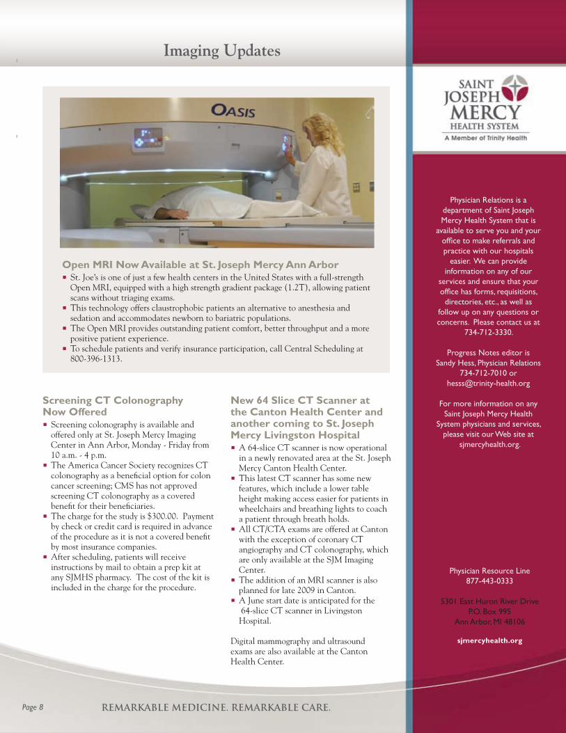
Open MRI Now Available at St. Joseph Mercy Ann Arbor St. Joe’s is one of just a few health centers in the United States with a full-strength
Open MRI, equipped with a high strength gradient package (1.2T), allowing patient scans without triaging exams.
This technology offers claustrophobic patients an alternative to anesthesia and sedation and accommodates newborn to bariatric populations.
The Open MRI provides outstanding patient comfort, better throughput and a more positive patient experience.
To schedule patients and verify insurance participation, call Central Scheduling at 800-396-1313.