Cancer-Associated Fibroblast-Induced Resistance to ...
17
cancers Review Cancer-Associated Fibroblast-Induced Resistance to Chemotherapy and Radiotherapy in Gastrointestinal Cancers In-Hye Ham 1,2,† , Dagyeong Lee 1,3,† and Hoon Hur 1,2,3, * Citation: Ham, I.-H.; Lee, D.; Hur, H. Cancer-Associated Fibroblast- Induced Resistance to Chemotherapy and Radiotherapy in Gastrointestinal Cancers. Cancers 2021, 13, 1172. https://doi.org/10.3390/ cancers13051172 Academic Editor: Corinne Bousquet Received: 3 February 2021 Accepted: 4 March 2021 Published: 9 March 2021 Publisher’s Note: MDPI stays neutral with regard to jurisdictional claims in published maps and institutional affil- iations. Copyright: © 2021 by the authors. Licensee MDPI, Basel, Switzerland. This article is an open access article distributed under the terms and conditions of the Creative Commons Attribution (CC BY) license (https:// creativecommons.org/licenses/by/ 4.0/). 1 Department of Surgery, Ajou University School of Medicine, Suwon 16499, Korea; [email protected] (I.-H.H.); [email protected] (D.L.) 2 Infamm-aging Translational Research Center, Ajou University School of Medicine, Suwon 16499, Korea 3 Department of Biomedical Science, Graduate School of Ajou University, Suwon 16499, Korea * Correspondence: [email protected]; Tel.: +82-31-219-5200; Fax: +82-31-219-5575 † These authors contributed equally to this work. Simple Summary: Gastrointestinal (GI) cancers are primary malignant tumors associated with cancer-related deaths worldwide. Although chemotherapy and radiotherapy are essential modalities to improve patient survival, many patients show resistance to these therapies. Various clinical studies have suggested that cancer-associated fibroblasts (CAFs) play a significant role in this resistance. In this review, we discuss CAF-produced cytokines, chemokines, growth factors, and exosomes, as well as desmoplastic reactions, all of which could be involved in cancer therapy resistance. In the future, the heterogeneity of CAFs should be considered such that CAF subtypes involved in cancer therapy resistance may be identified, thus improving the efficacy of chemotherapy and radiotherapy in GI cancers. Abstract: In the past few decades, the role of cancer-associated fibroblasts (CAFs) in resistance to therapies for gastrointestinal (GI) cancers has emerged. Clinical studies focusing on GI cancers have revealed that the high expression of CAF-related molecules within tumors is significantly correlated with unfavorable therapeutic outcomes; however, the exact mechanisms whereby CAFs enhance resistance to chemotherapy and radiotherapy in GI cancers remain unclear. The cells of origin of CAFs in GI cancers include normal resident fibroblasts, mesenchymal stem cells, endothelial cells, pericytes, and even epithelial cells. CAFs accumulated within GI cancers produce cytokines, chemokines, and growth factors involved in resistance to therapies. CAF-derived exosomes can be engaged in stroma-related resistance to treatments, and several non-coding RNAs, such as miR-92a, miR-106b, CCAL, and H19, are present in CAF-derived exosomes and transferred to GI cancer cells. The CAF-induced desmoplastic reaction interferes with drug delivery to GI cancer cells, evoking resistance to chemotherapy. However, due to the heterogeneity of CAFs in GI cancers, identifying the exact mechanism underlying CAF-induced resistance may be difficult. Recent advancements in single-cell “omics” technologies could offer clues for revealing the specific subtypes and biomarkers related to resistance. Keywords: cancer-associated fibroblasts; resistance; gastrointestinal cancer; chemotherapy; radiotherapy 1. Introduction Cancers originating from the gastrointestinal (GI) tract, including the esophagus, stomach, colorectum, liver, and pancreas, are common malignancies and are the primary cause of cancer-related mortalities worldwide [1]. The core treatment strategy for GI cancers is surgical resection. However, patients with non-resectable or recurrent disease are predominantly treated with chemotherapeutic agents or radiation techniques as a palliative measure [2]. Targeting agents and immunotherapy are recently developed alternatives for improving the survival of GI cancer patients [3]. However, most patients with advanced- Cancers 2021, 13, 1172. https://doi.org/10.3390/cancers13051172 https://www.mdpi.com/journal/cancers
Transcript of Cancer-Associated Fibroblast-Induced Resistance to ...
cancers
Review
Cancer-Associated Fibroblast-Induced Resistance toChemotherapy and Radiotherapy in Gastrointestinal Cancers
In-Hye Ham 1,2,† , Dagyeong Lee 1,3,† and Hoon Hur 1,2,3,*
�����������������
Citation: Ham, I.-H.; Lee, D.; Hur, H.
Cancer-Associated Fibroblast-
Induced Resistance to Chemotherapy
and Radiotherapy in Gastrointestinal
Cancers. Cancers 2021, 13, 1172.
https://doi.org/10.3390/
cancers13051172
Academic Editor: Corinne Bousquet
Received: 3 February 2021
Accepted: 4 March 2021
Published: 9 March 2021
Publisher’s Note: MDPI stays neutral
with regard to jurisdictional claims in
published maps and institutional affil-
iations.
Copyright: © 2021 by the authors.
Licensee MDPI, Basel, Switzerland.
This article is an open access article
distributed under the terms and
conditions of the Creative Commons
Attribution (CC BY) license (https://
creativecommons.org/licenses/by/
4.0/).
1 Department of Surgery, Ajou University School of Medicine, Suwon 16499, Korea;[email protected] (I.-H.H.); [email protected] (D.L.)
2 Infamm-aging Translational Research Center, Ajou University School of Medicine, Suwon 16499, Korea3 Department of Biomedical Science, Graduate School of Ajou University, Suwon 16499, Korea* Correspondence: [email protected]; Tel.: +82-31-219-5200; Fax: +82-31-219-5575† These authors contributed equally to this work.
Simple Summary: Gastrointestinal (GI) cancers are primary malignant tumors associated withcancer-related deaths worldwide. Although chemotherapy and radiotherapy are essential modalitiesto improve patient survival, many patients show resistance to these therapies. Various clinical studieshave suggested that cancer-associated fibroblasts (CAFs) play a significant role in this resistance.In this review, we discuss CAF-produced cytokines, chemokines, growth factors, and exosomes, aswell as desmoplastic reactions, all of which could be involved in cancer therapy resistance. In thefuture, the heterogeneity of CAFs should be considered such that CAF subtypes involved in cancertherapy resistance may be identified, thus improving the efficacy of chemotherapy and radiotherapyin GI cancers.
Abstract: In the past few decades, the role of cancer-associated fibroblasts (CAFs) in resistance totherapies for gastrointestinal (GI) cancers has emerged. Clinical studies focusing on GI cancershave revealed that the high expression of CAF-related molecules within tumors is significantlycorrelated with unfavorable therapeutic outcomes; however, the exact mechanisms whereby CAFsenhance resistance to chemotherapy and radiotherapy in GI cancers remain unclear. The cells oforigin of CAFs in GI cancers include normal resident fibroblasts, mesenchymal stem cells, endothelialcells, pericytes, and even epithelial cells. CAFs accumulated within GI cancers produce cytokines,chemokines, and growth factors involved in resistance to therapies. CAF-derived exosomes can beengaged in stroma-related resistance to treatments, and several non-coding RNAs, such as miR-92a,miR-106b, CCAL, and H19, are present in CAF-derived exosomes and transferred to GI cancer cells.The CAF-induced desmoplastic reaction interferes with drug delivery to GI cancer cells, evokingresistance to chemotherapy. However, due to the heterogeneity of CAFs in GI cancers, identifyingthe exact mechanism underlying CAF-induced resistance may be difficult. Recent advancements insingle-cell “omics” technologies could offer clues for revealing the specific subtypes and biomarkersrelated to resistance.
Keywords: cancer-associated fibroblasts; resistance; gastrointestinal cancer; chemotherapy; radiotherapy
1. Introduction
Cancers originating from the gastrointestinal (GI) tract, including the esophagus,stomach, colorectum, liver, and pancreas, are common malignancies and are the primarycause of cancer-related mortalities worldwide [1]. The core treatment strategy for GIcancers is surgical resection. However, patients with non-resectable or recurrent disease arepredominantly treated with chemotherapeutic agents or radiation techniques as a palliativemeasure [2]. Targeting agents and immunotherapy are recently developed alternatives forimproving the survival of GI cancer patients [3]. However, most patients with advanced-
Cancers 2021, 13, 1172. https://doi.org/10.3390/cancers13051172 https://www.mdpi.com/journal/cancers
Cancers 2021, 13, 1172 2 of 17
stage GI cancers are resistant to these treatment modalities; thus, their survival ratesremain dismal.
Several studies have investigated the mechanisms underlying resistance to therapyin cancers originating from the GI tract. These studies have focused on the tumor cellsthemselves, such as drug efflux through transmembrane transport proteins and anti-apoptotic protein activation [4,5]. However, to date, agents that block these pathways havenot yet been applied in clinical settings. Moreover, numerous studies have revealed thatthe tumor microenvironment (TME) may play a pivotal role in resistance to chemotherapyand radiotherapy [6,7]. The TME of solid cancers comprises various non-cancerous cells,the extracellular matrix, and soluble factors [8,9] that enhance tumorigenesis, invasion,metastasis, and therapy resistance in cancer cells. Therefore, targeting agents that blockthe interaction between cancer cells and the TME may improve treatment outcomes in GIcancer patients [10].
Cancer-associated fibroblasts (CAFs) constitute a significant component of the TMEin GI cancers. They are involved in cancer invasion and tumor growth through theirinteraction with cancer cells and immune microenvironments [11,12]. Numerous studieshave reported that CAFs can trigger the resistance of cancer cells to treatments [13–16].Therefore, CAFs have emerged as a novel treatment target to improve the efficacy ofchemotherapy and radiotherapy in GI cancers. However, drugs targeting CAFs have notyet been administered to patients.
Herein, we introduce clinical evidence for CAF-induced resistance to treatmentsand describe the activity of CAFs in GI cancers. Furthermore, we summarize currentresearch regarding the possible mechanism through which CAFs may evoke resistance tochemoradiotherapy in GI cancers.
2. Clinical Evidence for the Role of CAFs in Chemotherapy and RadiotherapyResistance in GI Cancer
The desmoplastic reaction developed by the recruited fibroblasts is prominently ob-served in progressed GI cancers [17], and this reaction has been considered a major causeof resistance to chemoradiotherapy [18]. Some clinical studies have demonstrated that highdesmoplasia is significantly correlated with poor clinical outcomes in patients with GI can-cers, such as pancreatic ductal adenocarcinoma (PDAC) and colorectal cancer (CRC) [19–21].Therefore, treatment strategies targeting tumor desmoplasia have mainly tried to improvethe survival of patients with advanced GI cancers [11]. For example, the monoclonalantibody for fibroblast activation protein (anti-FAP mAb) showed some therapeutic effectsin CRC without severe toxicity in the early phase of a clinical trial [22]. Additionally, recentphase II clinical trials testing angiotensin I receptor blockers as inhibitors of CAF activationand pegvorhyaluronidase alfa as a decomposer of hyaluronan accumulated by CAFs; thesetrials have described improved outcomes in PDAC patients [23,24]. Although these agentshave not yet been approved as a treatment of choice for GI cancers, accumulating evidencesuggests that targeting CAFs in GI cancers is promising (Table 1).
It has been confirmed through immunohistochemistry (IHC) that CAF accumulationin GI cancers is related to chemotherapy resistance. Ma et al. performed IHC for alpha-smooth muscle actin (α-SMA) in paraffin-embedded formalin-fixed (PEFF) tissues of gastriccancer (GC) patients treated with chemotherapy. The results showed that the GC tissues ofpatients showing resistance to chemotherapy contained more α-SMA-positive CAFs thanthe chemosensitive patients [25]. Other CRC studies also reported a significant correlationbetween a high proportion of α-SMA-expressing CAFs and resistance to 5-fluorouracil plusoxaliplatin-based chemotherapy [26].
Cancers 2021, 13, 1172 3 of 17
Table 1. Clinical studies investigating the role of CAFs in resistance to chemotherapy and radiation therapy ingastrointestinal cancers.
Patients Methods Marker Resistance to Results References
30 pts withgastric cancer IHC α-SMA Chemotherapy High expression in the
non-response group [25]
71 pts withcolorectal cancer IHC α-SMA Palliative 5-FU
and oxaliplatin5.5 (high) vs. 15.0 (low)
months (p = 0.005) [26]
53 pts withrectal cancer RT-PCR
High expression of CXCL12mRNA from microdissection for
the stromal region
Neoadjuvant 5-FU and20-45 cGy radiation
Positive CXCL12: pooroverall survival (p < 0.01) [27]
52 pts withrectal cancer RT-PCR
High expression of CXCL12 andFAP mRNA from microdissection
for the stromal region
Neoadjuvant 5-FU and20-45 cGy radiation
High two genes: poor overallsurvival (p < 0.05) [28]
141 pts withesophageal cancer IHC CXCL1 in CAF Chemoradiation High expression: HR 3.347
(p = 0.001) [29]
130 pts withesophageal cancer IHC TGF-β in CAF Chemoradiation High expression: poor overall
survival (p = 0.002) [30]
68 pts withesophageal cancer IHC PAI-1 in CAF Cisplatin
High expression: poorprogression-free survival
(p = 0.0267)[31]
10 pts withgastric cancer NanoString ECM-related gene set in
pretreated biopsy tissues5-FU based palliative
chemotherapySignificantly high innon-response group [15]
Pts: patients; IHC: immunohistochemistry; CAF: cancer-associated fibroblast; HR: hazard ratio; ECM: extracellular matrix.
The expression of CAF-derived molecules in human GI cancer tissues could be inves-tigated to provide clinical evidence. Some researchers have reported a direct correlationbetween biomarkers originating in stromal cells and response to neoadjuvant treatment.The expression of the two markers FAP-α and C-X-C motif chemokine ligand (CXCL) 12,known as stromal cell-derived factor 1 (SDF-1), was positively associated with poor clinicaloutcomes in rectal cancer patients who underwent neoadjuvant chemoradiation [27,28]. Al-though chemoradiotherapy is the most popular modality for esophageal cancers (ESOCs),patients have frequently exhibited resistance to therapies, resulting in poor outcomes [32].One study described the expression of CXCL1 in ESOC tissue specimens biopsied afterchemoradiation. We concluded that the upregulation of CXCL1 in CAFs was an indepen-dent prognostic factor in these patients [29]. In addition, positive transforming growthfactor-beta (TGF-β) expression in CAFs of ESOC tissues was significantly correlated withpoor survival outcomes in patients treated with chemoradiotherapy [30]. Another groupreported that high PAI-1 expression in CAFs led to considerably worse progression-freesurvival in ESOC patients treated with cisplatin [31].
Large-scale cancer genome studies using high-throughput technologies have providedcomprehensive molecular profiling information for solid cancers [33]. The Cancer GenomeAtlas (TCGA) consortium has suggested molecular subgroups and treatment targets basedon a genome-scale analysis using bulk tumors of large cohorts [34–38]. However, con-sidering the role of non-cancerous cells in the bulk tumors on cancer progression andtherapeutic efficacy, the meanings of these cell fractions should be investigated. Algo-rithms including ESTIMATE [39], CIBERSORT [40], EPIC [41], and MCP-counter [42] canpredict the proportion of stromal or immune cells in bulk cancer tissues. Consequently,the implications of the accumulation of these cells in GI cancer patient prognosis can beinferred. Recent high-throughput transcriptome analyses of GI cancers have highlightedthat stroma-related genes have unfavorable outcomes in patients with various types ofGI cancers, including GC, CRC, PDAC, and hepatocellular carcinoma [43–47]. However,these results were obtained using surgical specimens from patients who underwent cu-rative resection, with or without subsequent adjuvant systemic treatment. To define thecorrelation between gene expression and response to chemotherapy, expression analyses inpretreated samples from patients subjected to preoperative chemotherapy can undoubtedly
Cancers 2021, 13, 1172 4 of 17
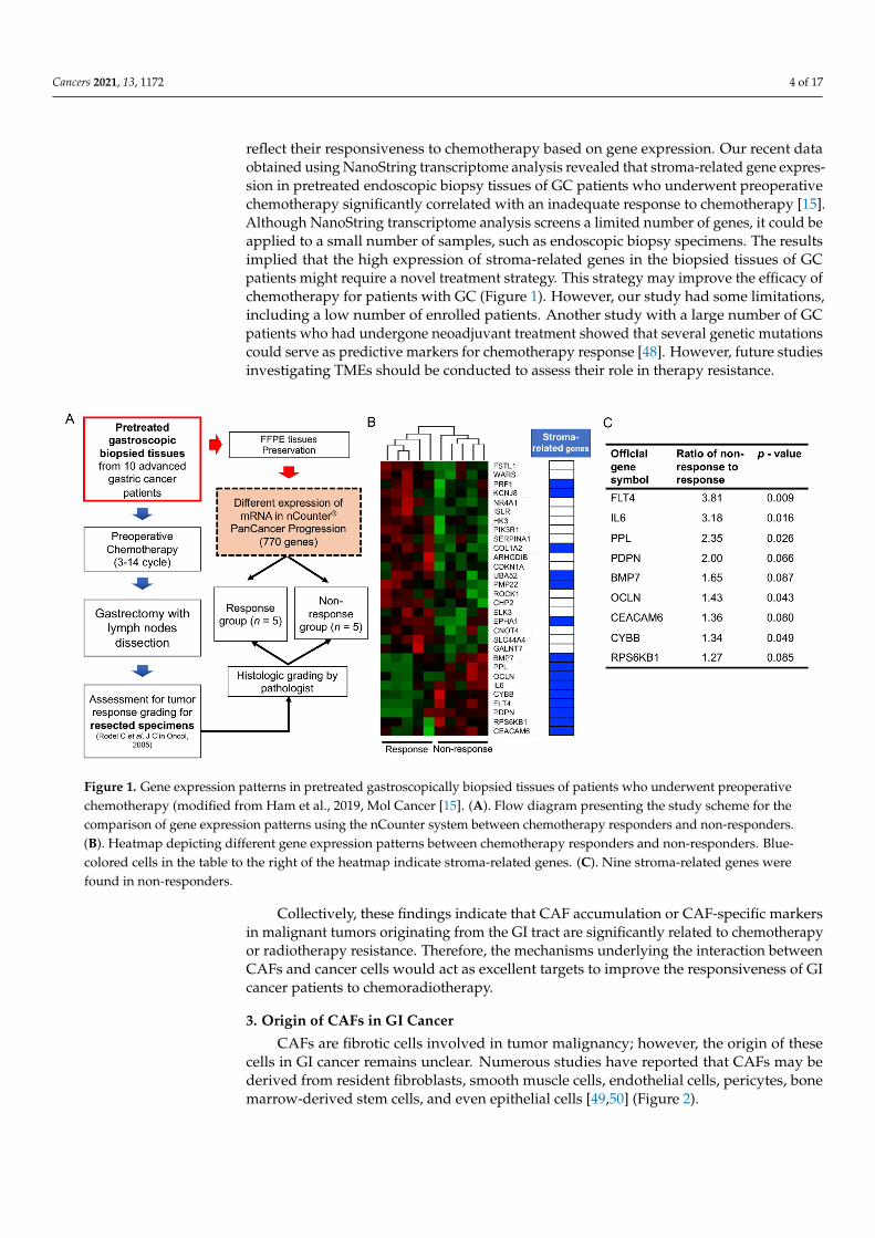
reflect their responsiveness to chemotherapy based on gene expression. Our recent dataobtained using NanoString transcriptome analysis revealed that stroma-related gene expres-sion in pretreated endoscopic biopsy tissues of GC patients who underwent preoperativechemotherapy significantly correlated with an inadequate response to chemotherapy [15].Although NanoString transcriptome analysis screens a limited number of genes, it could beapplied to a small number of samples, such as endoscopic biopsy specimens. The resultsimplied that the high expression of stroma-related genes in the biopsied tissues of GCpatients might require a novel treatment strategy. This strategy may improve the efficacy ofchemotherapy for patients with GC (Figure 1). However, our study had some limitations,including a low number of enrolled patients. Another study with a large number of GCpatients who had undergone neoadjuvant treatment showed that several genetic mutationscould serve as predictive markers for chemotherapy response [48]. However, future studiesinvestigating TMEs should be conducted to assess their role in therapy resistance.
Cancers 2021, 13, x 4 of 17
recent data obtained using NanoString transcriptome analysis revealed that stroma-re-lated gene expression in pretreated endoscopic biopsy tissues of GC patients who under-went preoperative chemotherapy significantly correlated with an inadequate response to chemotherapy [15]. Although NanoString transcriptome analysis screens a limited num-ber of genes, it could be applied to a small number of samples, such as endoscopic biopsy specimens. The results implied that the high expression of stroma-related genes in the biopsied tissues of GC patients might require a novel treatment strategy. This strategy may improve the efficacy of chemotherapy for patients with GC (Figure 1). However, our study had some limitations, including a low number of enrolled patients. Another study with a large number of GC patients who had undergone neoadjuvant treatment showed that several genetic mutations could serve as predictive markers for chemotherapy re-sponse [48]. However, future studies investigating TMEs should be conducted to assess their role in therapy resistance.
Figure 1. Gene expression patterns in pretreated gastroscopically biopsied tissues of patients who underwent preoperative chemotherapy (modified from Ham et al., 2019, Mol Cancer [15]. (A). Flow diagram presenting the study scheme for the comparison of gene expression patterns using the nCounter system between chemotherapy responders and non-respond-ers. (B). Heatmap depicting different gene expression patterns between chemotherapy responders and non-responders. Blue-colored cells in the table to the right of the heatmap indicate stroma-related genes. (C). Nine stroma-related genes were found in non-responders.
Collectively, these findings indicate that CAF accumulation or CAF-specific markers in malignant tumors originating from the GI tract are significantly related to chemother-apy or radiotherapy resistance. Therefore, the mechanisms underlying the interaction be-tween CAFs and cancer cells would act as excellent targets to improve the responsiveness of GI cancer patients to chemoradiotherapy.
3. Origin of CAFs in GI Cancer CAFs are fibrotic cells involved in tumor malignancy; however, the origin of these
cells in GI cancer remains unclear. Numerous studies have reported that CAFs may be derived from resident fibroblasts, smooth muscle cells, endothelial cells, pericytes, bone marrow-derived stem cells, and even epithelial cells [49,50] (Figure 2).
Figure 1. Gene expression patterns in pretreated gastroscopically biopsied tissues of patients who underwent preoperativechemotherapy (modified from Ham et al., 2019, Mol Cancer [15]. (A). Flow diagram presenting the study scheme for thecomparison of gene expression patterns using the nCounter system between chemotherapy responders and non-responders.(B). Heatmap depicting different gene expression patterns between chemotherapy responders and non-responders. Blue-colored cells in the table to the right of the heatmap indicate stroma-related genes. (C). Nine stroma-related genes werefound in non-responders.
Collectively, these findings indicate that CAF accumulation or CAF-specific markersin malignant tumors originating from the GI tract are significantly related to chemotherapyor radiotherapy resistance. Therefore, the mechanisms underlying the interaction betweenCAFs and cancer cells would act as excellent targets to improve the responsiveness of GIcancer patients to chemoradiotherapy.
3. Origin of CAFs in GI Cancer
CAFs are fibrotic cells involved in tumor malignancy; however, the origin of thesecells in GI cancer remains unclear. Numerous studies have reported that CAFs may bederived from resident fibroblasts, smooth muscle cells, endothelial cells, pericytes, bonemarrow-derived stem cells, and even epithelial cells [49,50] (Figure 2).
Cancers 2021, 13, 1172 5 of 17Cancers 2021, 13, x 5 of 17
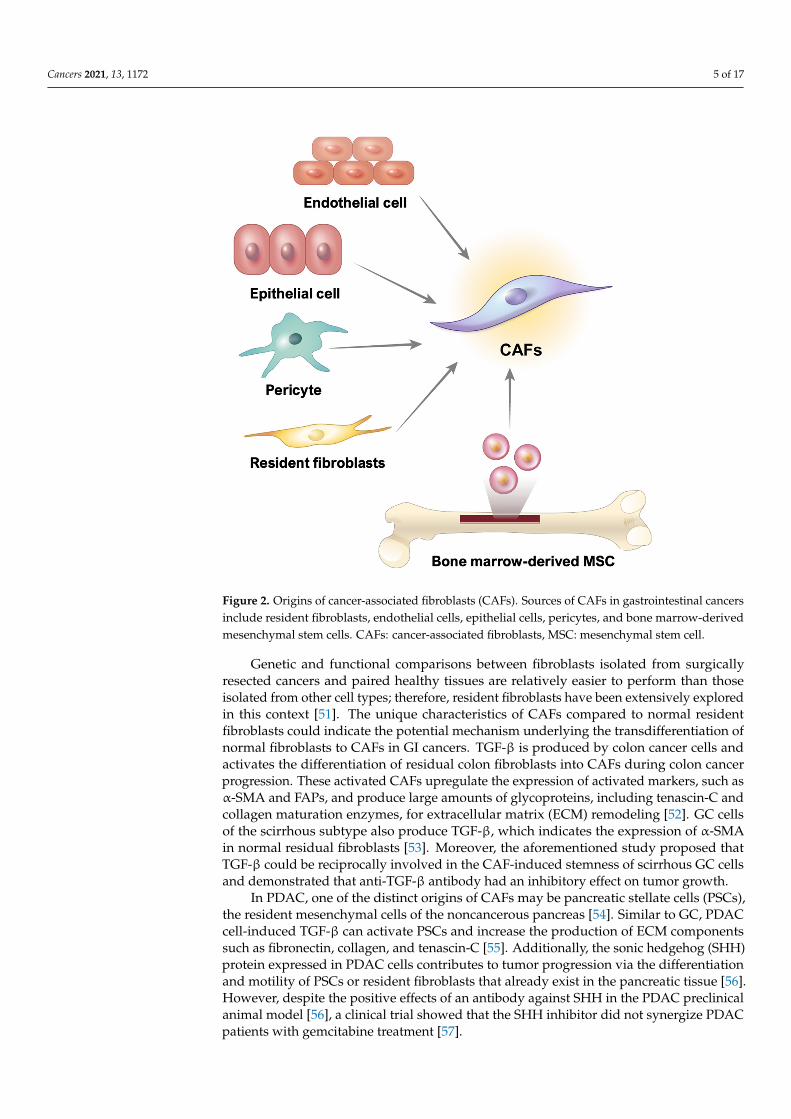
Figure 2. Origins of cancer-associated fibroblasts (CAFs). Sources of CAFs in gastrointestinal cancers include resident fibroblasts, endothelial cells, epithelial cells, pericytes, and bone marrow-derived mesenchymal stem cells. CAFs: cancer-associated fibroblasts, MSC: mesenchymal stem cell.
Genetic and functional comparisons between fibroblasts isolated from surgically re-sected cancers and paired healthy tissues are relatively easier to perform than those isolated from other cell types; therefore, resident fibroblasts have been extensively explored in this context [51]. The unique characteristics of CAFs compared to normal resident fibroblasts could indicate the potential mechanism underlying the transdifferentiation of normal fibro-blasts to CAFs in GI cancers. TGF-β is produced by colon cancer cells and activates the dif-ferentiation of residual colon fibroblasts into CAFs during colon cancer progression. These activated CAFs upregulate the expression of activated markers, such as α-SMA and FAPs, and produce large amounts of glycoproteins, including tenascin-C and collagen maturation enzymes, for extracellular matrix (ECM) remodeling [52]. GC cells of the scirrhous subtype also produce TGF-β, which indicates the expression of α-SMA in normal residual fibroblasts [53]. Moreover, the aforementioned study proposed that TGF-β could be reciprocally in-volved in the CAF-induced stemness of scirrhous GC cells and demonstrated that anti-TGF-β antibody had an inhibitory effect on tumor growth.
In PDAC, one of the distinct origins of CAFs may be pancreatic stellate cells (PSCs), the resident mesenchymal cells of the noncancerous pancreas [54]. Similar to GC, PDAC cell-induced TGF-β can activate PSCs and increase the production of ECM components such as fibronectin, collagen, and tenascin-C [55]. Additionally, the sonic hedgehog (SHH) protein expressed in PDAC cells contributes to tumor progression via the differentiation and motility of PSCs or resident fibroblasts that already exist in the pancreatic tissue [56]. However, despite the positive effects of an antibody against SHH in the PDAC preclinical animal model [56], a clinical trial showed that the SHH inhibitor did not synergize PDAC patients with gemcitabine treatment [57].
Figure 2. Origins of cancer-associated fibroblasts (CAFs). Sources of CAFs in gastrointestinal cancersinclude resident fibroblasts, endothelial cells, epithelial cells, pericytes, and bone marrow-derivedmesenchymal stem cells. CAFs: cancer-associated fibroblasts, MSC: mesenchymal stem cell.
Genetic and functional comparisons between fibroblasts isolated from surgicallyresected cancers and paired healthy tissues are relatively easier to perform than thoseisolated from other cell types; therefore, resident fibroblasts have been extensively exploredin this context [51]. The unique characteristics of CAFs compared to normal residentfibroblasts could indicate the potential mechanism underlying the transdifferentiation ofnormal fibroblasts to CAFs in GI cancers. TGF-β is produced by colon cancer cells andactivates the differentiation of residual colon fibroblasts into CAFs during colon cancerprogression. These activated CAFs upregulate the expression of activated markers, such asα-SMA and FAPs, and produce large amounts of glycoproteins, including tenascin-C andcollagen maturation enzymes, for extracellular matrix (ECM) remodeling [52]. GC cellsof the scirrhous subtype also produce TGF-β, which indicates the expression of α-SMAin normal residual fibroblasts [53]. Moreover, the aforementioned study proposed thatTGF-β could be reciprocally involved in the CAF-induced stemness of scirrhous GC cellsand demonstrated that anti-TGF-β antibody had an inhibitory effect on tumor growth.
In PDAC, one of the distinct origins of CAFs may be pancreatic stellate cells (PSCs),the resident mesenchymal cells of the noncancerous pancreas [54]. Similar to GC, PDACcell-induced TGF-β can activate PSCs and increase the production of ECM componentssuch as fibronectin, collagen, and tenascin-C [55]. Additionally, the sonic hedgehog (SHH)protein expressed in PDAC cells contributes to tumor progression via the differentiationand motility of PSCs or resident fibroblasts that already exist in the pancreatic tissue [56].However, despite the positive effects of an antibody against SHH in the PDAC preclinicalanimal model [56], a clinical trial showed that the SHH inhibitor did not synergize PDACpatients with gemcitabine treatment [57].
Cancers 2021, 13, 1172 6 of 17
Bone marrow-derived mesenchymal stem cells (MSCs) may act as a potential source ofCAFs in inflammation-induced GC [58]. The Helicobacter-induced GC mouse model revealsthat CAFs are derived from α-SMA-positive myofibroblasts in the bone marrow, and theseCAFs can form a tumor niche in the gastric wall and undergo tumor progression. TheMSCs recruited from the bone marrow may act as a source of CAFs in PDAC and pancreaticendocrine tumors [54,59]. MSCs exposed to PDAC cells are activated into CAF-secretingtumor-promoting proteins such as hepatocyte growth factor (HGF), epidermal growthfactor (EGF), and interleukin-6 (IL-6). These proteins stimulate microvascularization,changes in the composition of the stromal framework, and tumor growth through theparacrine system [54].
Other noncancerous cells, such as endothelial cells, pericytes, and even epithelialcells, which accumulate in GI cancer, can be transformed into CAFs through cell transitionmechanisms. A study using a pancreatic islet tumor mouse model revealed that fibroblast-specific protein 1 (FSP1) and CD31 double-positive cells exist in the TME. Previous studieshave reported that TGF-β mediates the transition from endothelial cells to mesenchymalcells in cardiac tissues [60]. Since abundant TGF-β expression was also apparent in thistumor, the authors suggested that TGF-β-exposed pancreatic endothelial cells could be asource of CAFs [61].
Vascular pericytes are multifunctional mural cells that surround endothelial cells [62],and they are crucial in the neoangiogenesis and survival of endothelial cells during tumori-genesis [63]. Emerging evidence has indicated that neural/glial antigen 2 (NG2)-expressingpericytes are transformed into CAFs through platelet-derived growth factor-BB (PDGF-BB)stimulation in a CRC xenograft model [64]. Moreover, the expression of PDGFB and FSP1in various types of solid tumors, including CRC, is significantly correlated with poorpatient prognosis [64].
Furthermore, epithelial cells of GI organs could be a source of CAFs during carcino-genesis. In genetic PDAC mouse models, pancreatic epithelial cells are transformed intomesenchymal cells through epithelial–mesenchymal transition; these cells have a fibroblast-like phenotype, express FSP1, and are deeply involved in tumor formation. However,although these FSP1-expressing cells are similar to CAFs, it is still unclear whether thesecells could be a significant source of CAFs in PDAC tumors [65]. Therefore, further studiesare required to verify whether epithelial cells are a crucial source of CAFs in GI cancers.
4. Factors Related to CAF-induced Resistance to Cancer Treatment4.1. Cytokines and Chemokines
Cytokines and chemokines are inflammatory mediators secreted by cancer cells ortumor stromal cells in the TME. They can stimulate tumor-promoting pathways, includingproliferation, metastasis, and progression in an autocrine or paracrine manner [66]. More-over, the cytokines and chemokines in the TME are deeply related to chemoresistance andpoor prognosis in cancer patients [67,68]. The CAFs in GI cancers could act as a source ofvarious TME cytokines and chemokines.
IL-6, a multifaceted cytokine related to infection or injury response, plays a pre-dominant role in cancer progression [69]. Recent studies have suggested that IL-6 ismainly secreted by CAFs, and CAF-derived IL-6 can induce an inadequate response tochemotherapy in GI cancers, including CRC, esophageal cancer (ESOC), and GC [15,70–72].Qiao et al. [70] demonstrated that IL-6 from CAFs contributes to chemoresistance by ac-tivating the signal transducer and activator of transcription 3 (STAT3)/nuclear factor-κB(NF-κB) pathway and subsequently upregulating C-X-C motif chemokine receptor (CXCR)7 expression in ESOC cells. Additionally, other studies demonstrated that stromal IL-6increases the expression of cancer stem cell (CSC) markers and consequently inducesresistance to chemoradiotherapy in ESOC patients [72]. Moreover, we demonstrated thatCAF-derived IL-6 stimulates the Janus kinase 1 (JAK1)/STAT3 pathway in GC cells ina paracrine manner [15]. Furthermore, in human GC tissues, high expression of stroma-related genes, including IL-6, is significantly correlated with resistance to chemotherapy.
Cancers 2021, 13, 1172 7 of 17
Eventually, we found that the IL-6 receptor monoclonal antibody, tocilizumab, rescuedCAF-induced resistance to chemotherapy in various experimental models [15]. Takentogether, these studies show that CAFs may act as a source of IL-6 in the GI cancer mi-croenvironment; as such, IL-6 inhibition could be a novel therapeutic strategy to decreaseCAF-induced resistance to cancer therapies.
The human chemokine CXCL1, termed the GRO-1 oncogene, specifically binds toCXCR2, a member of the G-protein-coupled receptor family [73]. Zhang et al. [29] reportedthat the expression of CXCL1 in CAFs isolated from ESOC tissues was higher than thatin normal fibroblasts. CAF-derived CXCL1 could be involved in tumor radiotherapyresistance by activating the MEK/ERK pathway.
CXCL12, also known as SDF-1, is mainly secreted from the stromal cells of solidtumors and is a primary ligand of the membrane receptor CXCR4 [74]. The role of CXCL12has been explored in PDAC; several studies have reported that PSCs secrete CXCL12 intothe TME, which promotes the resistance of PDAC cells to chemotherapy in a paracrinemanner [75–77]. Secreted CXCL12 may activate the FAK/ERK1/2/AKT signaling path-ways in PDAC cells, thereby inducing resistance to gemcitabine [75,77]. CXCL12-inducedactivation of this signaling pathway increases the transcriptional activities of β-cateninand NF-κB, thus leading to an elevated expression of survival proteins such as Bcl-2 [77].Moreover, these CXCL12-activated pathways increased the secretion of IL-6 in PDAC cellsrelated to chemoresistance [75]. Therefore, the small-molecule CXCR4 antagonist plerixaforhas been used to abolish CXCL12-induced PDAC growth and chemoresistance [75,77]. Arecent clinical trial demonstrated that the combination of plerixafor and chemotherapy in-creased the response rate of conventional chemotherapy in a hematological malignancy [78];hence, this combination may be an effective chemosensitizer for GI cancer. Radiotherapyhas been perioperatively administered to patients with PDAC. PDAC patients who un-dergo curative resection can be treated with radiotherapy to suppress cancer recurrence.Radiotherapy for inoperable PDAC patients can be used for symptom palliation [79]. Onestudy concluded that CAF-derived CXCL12 promotes PDAC cell resistance to radiotherapythrough CXCR4 activation [80]. This result suggests that CAF-induced CXCL12/CXCR4signaling could be a novel therapeutic target to improve the effectiveness of radiation [80].
Chemotherapeutic agents can stimulate the production of various secretory proteinsin CAFs. In experimental models of CRC, chemotherapy-stimulated CAFs enhance thesecretion of specific cytokines such as IL-17A, and increased serum levels of IL-17 havebeen observed in CRC patients with chemoresistance. CAF-secreted IL-17A promoteschemoresistance in cancer-initiating cells (CICs) through the NF-κB pathway and increasesCIC self-renewal, invasion, and tumor growth in vivo [81].
4.2. Growth Factors
Cancer cells usually express various receptor tyrosine kinases (RTKs) that can medi-ate downstream signaling pathways, such as mitogen-activated protein kinase (MAPK)and phosphatidylinositol-3-OH kinase, which can contribute to therapy resistance [82,83].Although RTKs are highly activated through genetic mutations in various cancers, growthfactor stimulation is a crucial mechanism for RTK-induced inadequate therapeutic re-sponses [84]. In particular, if growth factors are secreted from CAFs, they can act asmessengers for cell–cell communication.
In addition, cancer-secreted TGF-β can enhance the transition of resident fibroblastsinto CAFs, as mentioned in Section 3, and CAF-secreted TGF-β is involved in cancertherapy resistance in GI cancer cells. In ESOC, CAF-conditioned media includes a higherconcentration of TGF-β1 than the conditioned media from normal fibroblasts [30]. Con-sequently, CAF-derived TGF-β1 enhances resistance to cisplatin and taxol, and TGF-β1expression in CAFs is significantly related to poor prognosis in ESOC patients subjectedto chemoradiotherapy [30]. Another study showed that miR-27a/b converts normal fi-broblasts to CAFs in ESOC, and the converted CAFs enhance resistance to cisplatin bysecreting TGF-β1 [85]. Both studies demonstrated that the TGF-β1 inhibitor LY2157299
Cancers 2021, 13, 1172 8 of 17
could improve the response of ESOC cells to various chemotherapeutic agents both in vivoand in vitro.
The insulin-like growth factor (IGF) family plays a crucial role in regulating cellproliferation and apoptosis by activating transmembrane receptors; thus, it contributes toresistance to GI cancer therapies [86]. Ireland et al. [87] suggested that CAFs could be asource of IGF-1 and IGF-2 in PDAC and consequently activate insulin/IGF receptors onPDAC cells. They also demonstrated that the inhibition of IGFs sensitizes PDAC cells togemcitabine. The mechanism underlying the upregulation of IGF-1 expression in PDACCAFs was evaluated by Xiao et al., [88] who demonstrated that the PDAC-enhancedmethylation of suppressor of cytokine signaling 1 (SOCS1) plays a pivotal role in thetransition of normal fibroblasts to CAFs. In turn, SOCS1 downregulation was associatedwith IGF-1 expression in CAFs. Moreover, radiotherapy may trigger the secretion of IGF1,which could be involved in the resistance of rectal cancer to radiotherapy. Radiation-activated CAFs promote CRC cell survival by activating the IGF-1 receptor; thereafter, theneutralization of this receptor in a CRC cancer animal model reduces metastasis [89].
HGF is a major secretory protein of CAFs in solid tumors that promotes cancercell survival and provides therapeutic resistance [90]. CAF-secreted HGF increases theproportion of tumor-initiating cells of HCC through c-MET activation. Activated c-MET intumor-initiating cells further activates the ERK/FRA1/HEY1 cascade, which is related tochemotherapy resistance [91].
Cetuximab, an EGF receptor (EGFR) monoclonal antibody, is a molecular targetedagent that improves the survival of patients with CRC without Kras mutation [92].Luraghi et al. [93] reported that CAF-secreted HGF activates resistance to EGFR inhibitorsin experimental models. CAF-induced HGF could also play a pivotal role in radiotherapyresistance. One study demonstrated that the levels of secreted HGF in irradiated fibroblastsisolated from ESOC were higher than those in non-irradiated controls. HGF derived fromirradiated fibroblasts increases wound healing, migration, and invasion [94].
4.3. Exosomes
Various studies have examined the role of exosomes in cancer progression. The exo-some, a microvesicle of endocytic origin with a diameter of 30–150 nm, is secreted by manycells. As exosomes comprise a lipid bilayer containing various bioactive molecules, such asDNA, microRNAs (miRNAs), proteins, long non-coding RNAs (lncRNAs), circular RNAs,and lipids, they function as natural vehicles in cell–cell communication by transferringgenetic messages. Exosomes secreted from various cells within tumors enable communica-tion among tumor cells surrounding the TME and in distant organs or tissues, leading tothe promotion of metastasis and therapy resistance [95]. Therefore, exosomes generatedfrom CAFs would be suitable messengers to enhance the resistance of GI cancer cellsto therapy.
The function of CAF-derived exosomes in cancer therapy resistance was initiallyinvestigated in CRC. Hu et al. [96] reported that CAF-derived exosomes promote drugresistance by mediating the activation of the Wnt signaling pathway in CSCs in CRC. Next,the kinds of elements included in CAF-derived exosomes that can enhance resistance totherapies have recently been evaluated. Non-coding RNAs (ncRNAs) such as miRNAs andlncRNAs have been suggested as crucial molecules for exosome-mediated communicationbetween CAFs and GI cancer cells [97]. miRNAs are a subtype of ncRNAs that contain17–25 bp of ncRNA and usually suppress messenger RNA translation by targeting the 3′-UTR; thus, miRNAs facilitate the epigenetic regulation of gene expression and can controlthe pathological process in cancers [98]. Notably, several miRNAs present in exosomes areinvolved in drug resistance in cancers [99]. Regarding GI cancers, CAFs of CRC secretemiR-92a-3p-enriched exosomes into the TME. When exosomal miR-92a-3p is transferred toCRC cells, it promotes migration, invasion, metastasis, stemness, and drug resistance. Thus,blocking the function of exosomal miR-92a-3p secreted by CAFs could be used as an alterna-tive modality for therapy resistance in CRC [100]. In GC, Zhang et al. [101] demonstrated
Cancers 2021, 13, 1172 9 of 17
that CAF-secreted exosomal miR-522 regulates arachidonate lipoxygenase 15 (ALOX15)expression and is closely related to lipid reactive oxygen species (ROS) production. Theysuggested that blocking lipid-ROS production might be a novel mechanism for acquireddrug resistance. Thus, targeting exosomal miR-522 could be a modality to increase thesensitivity of GC patients to chemotherapy [101]. Furthermore, when miR-106b in thePDAC CAF-derived exosomes is transferred into PDAC cells, it mediates resistance togemcitabine [102]. Therefore, detecting miRNAs in CAF-derived exosomes of GI cancercould provide efficient biomarkers to predict chemotherapy response. Moreover, targetingthe function of these miRNAs could serve as a promising tool for improving the drugresponse in GI cancers.
Exosomes can also contain lncRNAs, which are nucleotide transcripts over 200 bp inlength that are not translated into proteins [103]. The function of lncRNAs in the resistanceof various cancers to therapies has recently been proposed; this could be the centralmechanism related to CAF exosome-derived drug resistance in GI cancer. Deng et al. [104]demonstrated that CAF-derived exosomes express CRC-associated lncRNA (CCAL) morehighly than normal fibroblasts, and these CCAL-enriched exosomes may drive cancercells to oxaliplatin resistance. In addition, the transfer of CCAL could function as anoncogenic lncRNA and induce Wnt/β-catenin pathway activation in CRC cells. Therefore,CCAL may represent a biomarker and druggable target for CRC chemoresistance. AnotherCAF-derived exosomal lncRNA, H19, also promotes stemness and chemoresistance in CRCcells [105]. Transferred H19 could activate the β-catenin pathway in CRC cells by blockingthe function of miR-141, which could inhibit stemness.
Furthermore, CAF-derived exosomes may also be associated with resistance to ra-diotherapy. Liu et al. [106] reported that CAF-derived exosomes confer robust radiationresistance in CRC cells by activating the TGF-β signaling pathway.
4.4. Other Mechanisms
The other secretory materials produced from CAFs and CAF-induced intratumoralpressure escalation could be involved in the resistance to GI cancer therapies.
Plasminogen activator inhibitor-1 (PAI-1) is a secreted protein that not only enhancesangiogenesis, but also promotes the invasion and metastasis of certain cancer cells [107,108].PAI-1 is secreted from CAFs, and one study reported that cisplatin-treated CAFs increasePAI-1 secretion [31]. CAF-secreted PAI-1 enhances progression and chemoresistance byactivating the AKT/ERK1/2 signaling pathway and inhibiting caspase-3 activity and ROSaccumulation in ESOC cells. Moreover, the high expression of PAI-1 in CAFs is correlatedwith poor prognosis in ESOC patients; consistently, the PAI-1 inhibitor tiplaxtinin presentssynergistic effects with cisplatin both in vitro and in vivo [31].
CAF-secreted perlecan (heparin sulfate proteoglycan 2, HSPG2) plays a critical role inresistance to chemotherapy in PDAC. CAFs isolated from genetic PDAC mouse modelswere reprogrammed in mouse PDAC cells with a P53 mutation. The reprogrammedCAFs increased the stromal deposition of HSPG2, which created a prometastatic andchemoresistant environment in pancreatic cancer cells [109]. Another CAF-induced secretedprotein that protects PDAC cells from gemcitabine is laminin A1. Although PDAC cellssecrete transglutaminase, they do not increase the cytotoxicity of gemcitabine directly;however, transglutaminase enhances the secretion of laminin A1 from CAFs, and secretedlaminin A1 secreted in the TME could protect PDAC cells from chemotherapeutic agentssuch as gemcitabine [110].
CAF-secreted T-lymphoma invasion and metastasis-inducing protein-1 (TIAM1) is akey regulator of chemoresistance in CRC cells [111]. CAF-derived conditioned media in-creased resistance to chemotherapy through TIAM1 overexpression, and TIAM1-associateddrug sensitivity was validated using a xenograft mouse model.
CAFs are the center of desmoplastic reactions in GI cancer as well as a source ofsecreted proteins; thus, they interfere with drug delivery by collapsing the peritumoralcapillaries and increasing intratumoral interstitial pressure. This concept has been suitably
Cancers 2021, 13, 1172 10 of 17
evaluated in a mouse model of PDAC, wherein GI tumors exhibit profuse desmoplasticreactions. The accumulation of hyaluronic acid (HA) produced from CAFs during PDACprogression was found to be responsible for enhancing intratumoral pressure, thus actingas a barrier for drug diffusion in the mouse model. Provenzano et al. [112] suggestedthat enzymatic dissolution of stromal HA could increase the efficacy of cancer drugs byremodeling PDAC stromal lesions.
5. Heterogeneity of CAFs in GI Cancers
Tumor heterogeneity has recently been considered a crucial factor underlying resis-tance to antitumor therapies, including both non-cancerous stromal cells and cancer cells.In addition, various subtypes of CAFs exist [113–115]. Therefore, clarifying the mechanismunderlying CAF heterogeneity may provide crucial information on GI cancer progressionand would enable the development of novel therapeutic approaches.
Of all the GI cancers, CAF heterogeneity is best understood in PDAC. Ohlund et al. [116]reported the existence of distinct subtypes of CAFs based on their localization withinthe primary tumor. The α-SMAhigh CAF subtype is in direct contact with cancer cells,whereas α-SMAlow CAFs are located distally from cancer cells, releasing proinflamma-tory cytokines [116]. Other studies have also explored the function of α-SMAhigh CAFsubtypes. Genetically engineered PDAC mouse models with α-SMA-negative fibroblastsresult in more aggressive tumors and gemcitabine resistance [117,118]. Presumably, α-SMA-expressing CAFs may suppress tumor immunity and increase tumor vascularization.
These findings suggest that the CAF subtype can be characterized and identifyingthe specific subtypes of CAFs that play a crucial role in GI cancer progression couldpresent novel targets for therapy. The recent development of single-cell transcriptometechnology for solid tumors has shed light on the composition of various cancerous and non-cancerous tissues, as well as the heterogeneous population of accumulated cells, throughgene expression patterns [119,120]. Elyada et al. [121] conducted a single-cell analysis ofPDAC CAFs and found the following three subtypes: myofibroblastic, inflammatory, andantigen-presenting. Although they did not demonstrate the function of these subtypes inchemoradiotherapy resistance, this advanced technology can provide detailed informationregarding CAF heterogeneity in GI cancers.
6. Conclusions and Future Perspectives
The role of CAFs in GI cancer progression has been explored extensively over the pastdecade [122,123]. However, in the current review, we have focused on a substantial amountof evidence related to the correlation between CAFs and chemotherapy and radiotherapyresistance in GI cancers (Figure 3, Table 2). CAFs accumulated in GI cancers secreteIL-6 or CXLC12, which can activate signal transduction with respect to drug resistance.Inhibitors of IL-6 and CXCL12, such as tocilizumab and plerixafor, respectively, exertchemosensitizing effects on GI cancers. Growth factors, such as TGF-β1, are crucial inCAF-induced resistance to therapies; however, therapeutic strategies to target CAFs forGI cancer treatment have not yet been applied in clinical settings. More complicatedmechanisms may be involved in the communication between CAFs and GI cancer cells.Recent studies have demonstrated that small extracellular vesicles, such as exosomescontaining miRNAs and lncRNAs, can control the epigenetic regulation of genes related todrug response. Nevertheless, exosome-based controls for improving therapeutic responsesremain underdeveloped. Another factor that complicates this avenue of research is theheterogeneity of CAFs in GI cancer. Heterogeneous CAF populations must be preciselydefined to determine the specific subtypes related to therapy resistance, but this remainsa challenge. Recent advances in technologies, such as single-cell “omics,” will aid theexploration of CAF subpopulations and novel biomarkers related to chemotherapy andradiotherapy resistance in GI cancers.
Cancers 2021, 13, 1172 11 of 17
Cancers 2021, 13, x 11 of 17
challenge. Recent advances in technologies, such as single-cell “omics,” will aid the explo-ration of CAF subpopulations and novel biomarkers related to chemotherapy and radio-therapy resistance in GI cancers.
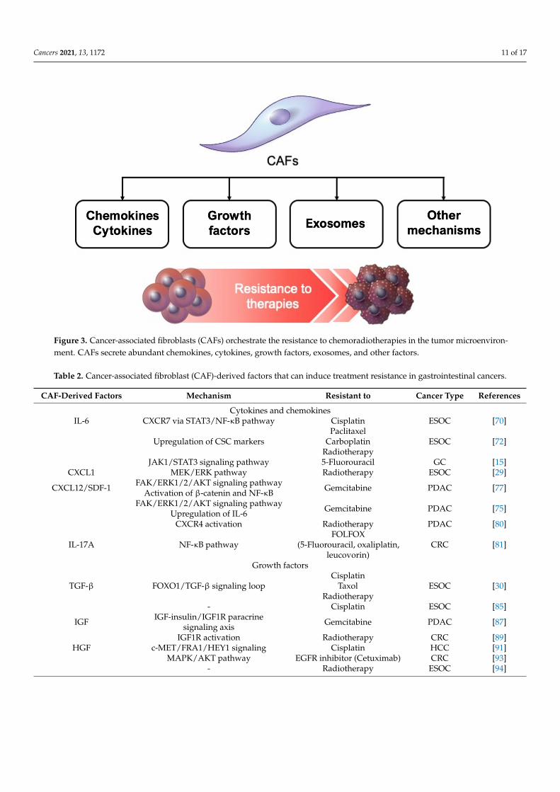
Figure 3. Cancer-associated fibroblasts (CAFs) orchestrate the resistance to chemoradiotherapies in the tumor microenvi-ronment. CAFs secrete abundant chemokines, cytokines, growth factors, exosomes, and other factors.
Table 2. Cancer-associated fibroblast (CAF)-derived factors that can induce treatment resistance in gastrointestinal can-cers.
CAF-Derived Factors Mechanism Resistant to Cancer Type References Cytokines and chemokines
IL-6 CXCR7 via STAT3/NF-κB pathway Cisplatin ESOC [70]
Upregulation of CSC markers Paclitaxel
Carboplatin Radiotherapy
ESOC [72]
JAK1/STAT3 signaling pathway 5-Fluorouracil GC [15] CXCL1 MEK/ERK pathway Radiotherapy ESOC [29]
CXCL12/SDF-1 FAK/ERK1/2/AKT signaling pathway
Activation of β-catenin and NF-κB Gemcitabine PDAC [77]
FAK/ERK1/2/AKT signaling pathway
Upregulation of IL-6 Gemcitabine PDAC [75]
CXCR4 activation Radiotherapy PDAC [80]
IL-17A NF-κB pathway FOLFOX
(5-Fluorouracil, oxali-platin, leucovorin)
CRC [81]
Growth factors
TGF-β FOXO1/TGF-β signaling loop Cisplatin
Taxol Radiotherapy
ESOC [30]
- Cisplatin ESOC [85] IGF IGF-insulin/IGF1R paracrine signaling axis Gemcitabine PDAC [87]
IGF1R activation Radiotherapy CRC [89] HGF c-MET/FRA1/HEY1 signaling Cisplatin HCC [91]
MAPK/AKT pathway EGFR inhibitor (Cetuxi-
mab) CRC [93]
- Radiotherapy ESOC [94] Exosomes
- Wnt signaling pathway 5-Fluorouracil CRC [96]
Figure 3. Cancer-associated fibroblasts (CAFs) orchestrate the resistance to chemoradiotherapies in the tumor microenviron-ment. CAFs secrete abundant chemokines, cytokines, growth factors, exosomes, and other factors.
Table 2. Cancer-associated fibroblast (CAF)-derived factors that can induce treatment resistance in gastrointestinal cancers.
CAF-Derived Factors Mechanism Resistant to Cancer Type References
Cytokines and chemokinesIL-6 CXCR7 via STAT3/NF-κB pathway Cisplatin ESOC [70]
Upregulation of CSC markersPaclitaxel
CarboplatinRadiotherapy
ESOC [72]
JAK1/STAT3 signaling pathway 5-Fluorouracil GC [15]CXCL1 MEK/ERK pathway Radiotherapy ESOC [29]
CXCL12/SDF-1 FAK/ERK1/2/AKT signaling pathwayActivation of β-catenin and NF-κB Gemcitabine PDAC [77]
FAK/ERK1/2/AKT signaling pathwayUpregulation of IL-6 Gemcitabine PDAC [75]
CXCR4 activation Radiotherapy PDAC [80]
IL-17A NF-κB pathwayFOLFOX
(5-Fluorouracil, oxaliplatin,leucovorin)
CRC [81]
Growth factors
TGF-β FOXO1/TGF-β signaling loopCisplatin
TaxolRadiotherapy
ESOC [30]
- Cisplatin ESOC [85]
IGF IGF-insulin/IGF1R paracrinesignaling axis Gemcitabine PDAC [87]
IGF1R activation Radiotherapy CRC [89]HGF c-MET/FRA1/HEY1 signaling Cisplatin HCC [91]
MAPK/AKT pathway EGFR inhibitor (Cetuximab) CRC [93]- Radiotherapy ESOC [94]
Cancers 2021, 13, 1172 12 of 17
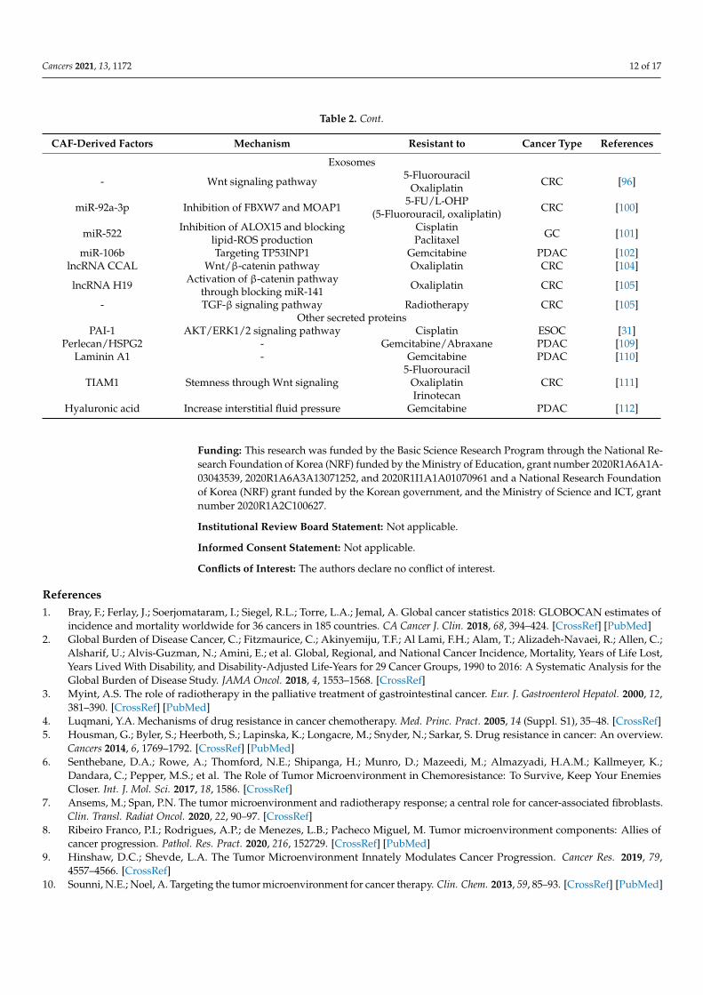
Table 2. Cont.
CAF-Derived Factors Mechanism Resistant to Cancer Type References
Exosomes
- Wnt signaling pathway 5-FluorouracilOxaliplatin CRC [96]
miR-92a-3p Inhibition of FBXW7 and MOAP1 5-FU/L-OHP(5-Fluorouracil, oxaliplatin) CRC [100]
miR-522 Inhibition of ALOX15 and blockinglipid-ROS production
CisplatinPaclitaxel GC [101]
miR-106b Targeting TP53INP1 Gemcitabine PDAC [102]lncRNA CCAL Wnt/β-catenin pathway Oxaliplatin CRC [104]
lncRNA H19 Activation of β-catenin pathwaythrough blocking miR-141 Oxaliplatin CRC [105]
- TGF-β signaling pathway Radiotherapy CRC [105]Other secreted proteins
PAI-1 AKT/ERK1/2 signaling pathway Cisplatin ESOC [31]Perlecan/HSPG2 - Gemcitabine/Abraxane PDAC [109]
Laminin A1 - Gemcitabine PDAC [110]
TIAM1 Stemness through Wnt signaling5-Fluorouracil
OxaliplatinIrinotecan
CRC [111]
Hyaluronic acid Increase interstitial fluid pressure Gemcitabine PDAC [112]
Funding: This research was funded by the Basic Science Research Program through the National Re-search Foundation of Korea (NRF) funded by the Ministry of Education, grant number 2020R1A6A1A-03043539, 2020R1A6A3A13071252, and 2020R1I1A1A01070961 and a National Research Foundationof Korea (NRF) grant funded by the Korean government, and the Ministry of Science and ICT, grantnumber 2020R1A2C100627.
Institutional Review Board Statement: Not applicable.
Informed Consent Statement: Not applicable.
Conflicts of Interest: The authors declare no conflict of interest.
References1. Bray, F.; Ferlay, J.; Soerjomataram, I.; Siegel, R.L.; Torre, L.A.; Jemal, A. Global cancer statistics 2018: GLOBOCAN estimates of
incidence and mortality worldwide for 36 cancers in 185 countries. CA Cancer J. Clin. 2018, 68, 394–424. [CrossRef] [PubMed]2. Global Burden of Disease Cancer, C.; Fitzmaurice, C.; Akinyemiju, T.F.; Al Lami, F.H.; Alam, T.; Alizadeh-Navaei, R.; Allen, C.;
Alsharif, U.; Alvis-Guzman, N.; Amini, E.; et al. Global, Regional, and National Cancer Incidence, Mortality, Years of Life Lost,Years Lived With Disability, and Disability-Adjusted Life-Years for 29 Cancer Groups, 1990 to 2016: A Systematic Analysis for theGlobal Burden of Disease Study. JAMA Oncol. 2018, 4, 1553–1568. [CrossRef]
3. Myint, A.S. The role of radiotherapy in the palliative treatment of gastrointestinal cancer. Eur. J. Gastroenterol Hepatol. 2000, 12,381–390. [CrossRef] [PubMed]
4. Luqmani, Y.A. Mechanisms of drug resistance in cancer chemotherapy. Med. Princ. Pract. 2005, 14 (Suppl. S1), 35–48. [CrossRef]5. Housman, G.; Byler, S.; Heerboth, S.; Lapinska, K.; Longacre, M.; Snyder, N.; Sarkar, S. Drug resistance in cancer: An overview.
Cancers 2014, 6, 1769–1792. [CrossRef] [PubMed]6. Senthebane, D.A.; Rowe, A.; Thomford, N.E.; Shipanga, H.; Munro, D.; Mazeedi, M.; Almazyadi, H.A.M.; Kallmeyer, K.;
Dandara, C.; Pepper, M.S.; et al. The Role of Tumor Microenvironment in Chemoresistance: To Survive, Keep Your EnemiesCloser. Int. J. Mol. Sci. 2017, 18, 1586. [CrossRef]
7. Ansems, M.; Span, P.N. The tumor microenvironment and radiotherapy response; a central role for cancer-associated fibroblasts.Clin. Transl. Radiat Oncol. 2020, 22, 90–97. [CrossRef]
8. Ribeiro Franco, P.I.; Rodrigues, A.P.; de Menezes, L.B.; Pacheco Miguel, M. Tumor microenvironment components: Allies ofcancer progression. Pathol. Res. Pract. 2020, 216, 152729. [CrossRef] [PubMed]
9. Hinshaw, D.C.; Shevde, L.A. The Tumor Microenvironment Innately Modulates Cancer Progression. Cancer Res. 2019, 79,4557–4566. [CrossRef]
10. Sounni, N.E.; Noel, A. Targeting the tumor microenvironment for cancer therapy. Clin. Chem. 2013, 59, 85–93. [CrossRef] [PubMed]
Cancers 2021, 13, 1172 13 of 17
11. Sahai, E.; Astsaturov, I.; Cukierman, E.; DeNardo, D.G.; Egeblad, M.; Evans, R.M.; Fearon, D.; Greten, F.R.; Hingorani, S.R.;Hunter, T.; et al. A framework for advancing our understanding of cancer-associated fibroblasts. Nat. Rev. Cancer 2020, 20,174–186. [CrossRef]
12. Melissari, M.T.; Chalkidi, N.; Sarris, M.E.; Koliaraki, V. Fibroblast Reprogramming in Gastrointestinal Cancer. Front. Cell Dev. Biol.2020, 8, 630. [CrossRef]
13. Orimo, A.; Weinberg, R.A. Stromal fibroblasts in cancer: A novel tumor-promoting cell type. Cell Cycle 2006, 5, 1597–1601. [CrossRef]14. Mueller, M.M.; Fusenig, N.E. Friends or foes - bipolar effects of the tumour stroma in cancer. Nat. Rev. Cancer 2004, 4,
839–849. [CrossRef]15. Ham, I.H.; Oh, H.J.; Jin, H.; Bae, C.A.; Jeon, S.M.; Choi, K.S.; Son, S.Y.; Han, S.U.; Brekken, R.A.; Lee, D.; et al. Targeting
interleukin-6 as a strategy to overcome stroma-induced resistance to chemotherapy in gastric cancer. Mol. Cancer 2019, 18, 68.[CrossRef] [PubMed]
16. Duluc, C.; Moatassim-Billah, S.; Chalabi-Dchar, M.; Perraud, A.; Samain, R.; Breibach, F.; Gayral, M.; Cordelier, P.; Delisle, M.B.;Bousquet-Dubouch, M.P.; et al. Pharmacological targeting of the protein synthesis mTOR/4E-BP1 pathway in cancer-associatedfibroblasts abrogates pancreatic tumour chemoresistance. EMBO Mol. Med. 2015, 7, 735–753. [CrossRef] [PubMed]
17. Kalluri, R.; Zeisberg, M. Fibroblasts in cancer. Nat. Rev. Cancer 2006, 6, 392–401. [CrossRef]18. Liu, H.; Ma, Q.; Xu, Q.; Lei, J.; Li, X.; Wang, Z.; Wu, E. Therapeutic potential of perineural invasion, hypoxia and desmoplasia in
pancreatic cancer. Curr. Pharm. Des. 2012, 18, 2395–2403. [CrossRef]19. Vennin, C.; Murphy, K.J.; Morton, J.P.; Cox, T.R.; Pajic, M.; Timpson, P. Reshaping the Tumor Stroma for Treatment of Pancreatic
Cancer. Gastroenterology 2018, 154, 820–838. [CrossRef] [PubMed]20. Ueno, H.; Jones, A.M.; Wilkinson, K.H.; Jass, J.R.; Talbot, I.C. Histological categorisation of fibrotic cancer stroma in advanced
rectal cancer. Gut 2004, 53, 581–586. [CrossRef] [PubMed]21. Sis, B.; Sarioglu, S.; Sokmen, S.; Sakar, M.; Kupelioglu, A.; Fuzun, M. Desmoplasia measured by computer assisted image analysis:
An independent prognostic marker in colorectal carcinoma. J. Clin. Pathol. 2005, 58, 32–38. [CrossRef]22. Hofheinz, R.D.; Al-Batran, S.E.; Hartmann, F.; Hartung, G.; Jager, D.; Renner, C.; Tanswell, P.; Kunz, U.; Amelsberg, A.;
Kuthan, H.; et al. Stromal antigen targeting by a humanised monoclonal antibody: An early phase II trial of sibrotuzumab inpatients with metastatic colorectal cancer. Onkologie 2003, 26, 44–48. [CrossRef]
23. Murphy, J.E.; Wo, J.Y.; Ryan, D.P.; Clark, J.W.; Jiang, W.; Yeap, B.Y.; Drapek, L.C.; Ly, L.; Baglini, C.V.; Blaszkowsky, L.S.; et al. TotalNeoadjuvant Therapy With FOLFIRINOX in Combination With Losartan Followed by Chemoradiotherapy for Locally AdvancedPancreatic Cancer: A Phase 2 Clinical Trial. JAMA Oncol. 2019, 5, 1020–1027. [CrossRef]
24. Hingorani, S.R.; Zheng, L.; Bullock, A.J.; Seery, T.E.; Harris, W.P.; Sigal, D.S.; Braiteh, F.; Ritch, P.S.; Zalupski, M.M.; Bahary, N.; et al.HALO 202: Randomized Phase II Study of PEGPH20 Plus Nab-Paclitaxel/Gemcitabine Versus Nab-Paclitaxel/Gemcitabine inPatients With Untreated, Metastatic Pancreatic Ductal Adenocarcinoma. J. Clin. Oncol. 2018, 36, 359–366. [CrossRef]
25. Ma, J.; Song, X.; Xu, X.; Mou, Y. Cancer-Associated Fibroblasts Promote the Chemo-resistance in Gastric Cancer through SecretingIL-11 Targeting JAK/STAT3/Bcl2 Pathway. Cancer Res. Treat. 2019, 51, 194–210. [CrossRef]
26. Gu, J.; Li, Z.; Zhou, J.; Sun, Z.; Bai, C. Response prediction to oxaliplatin plus 5-fluorouracil chemotherapy in patients withcolorectal cancer using a four-protein immunohistochemical model. Oncol. Lett. 2019, 18, 2091–2101. [CrossRef] [PubMed]
27. Saigusa, S.; Toiyama, Y.; Tanaka, K.; Yokoe, T.; Okugawa, Y.; Kawamoto, A.; Yasuda, H.; Inoue, Y.; Miki, C.; Kusunoki, M. StromalCXCR4 and CXCL12 expression is associated with distant recurrence and poor prognosis in rectal cancer after chemoradiotherapy.Ann. Surg. Oncol. 2010, 17, 2051–2058. [CrossRef] [PubMed]
28. Saigusa, S.; Toiyama, Y.; Tanaka, K.; Yokoe, T.; Okugawa, Y.; Fujikawa, H.; Matsusita, K.; Kawamura, M.; Inoue, Y.; Miki, C.; et al.Cancer-associated fibroblasts correlate with poor prognosis in rectal cancer after chemoradiotherapy. Int. J. Oncol. 2011, 38,655–663. [CrossRef]
29. Zhang, H.; Yue, J.; Jiang, Z.; Zhou, R.; Xie, R.; Xu, Y.; Wu, S. CAF-secreted CXCL1 conferred radioresistance by regulating DNAdamage response in a ROS-dependent manner in esophageal squamous cell carcinoma. Cell Death Dis. 2017, 8, e2790. [CrossRef]
30. Zhang, H.; Xie, C.; Yue, J.; Jiang, Z.; Zhou, R.; Xie, R.; Wang, Y.; Wu, S. Cancer-associated fibroblasts mediated chemore-sistance by a FOXO1/TGFbeta1 signaling loop in esophageal squamous cell carcinoma. Mol. Carcinog 2017, 56, 1150–1163.[CrossRef] [PubMed]
31. Che, Y.; Wang, J.; Li, Y.; Lu, Z.; Huang, J.; Sun, S.; Mao, S.; Lei, Y.; Zang, R.; Sun, N.; et al. Cisplatin-activated PAI-1 secretion in thecancer-associated fibroblasts with paracrine effects promoting esophageal squamous cell carcinoma progression and causingchemoresistance. Cell Death Dis. 2018, 9, 759. [CrossRef]
32. Luo, Y.; Mao, Q.; Wang, X.; Yu, J.; Li, M. Radiotherapy for esophageal carcinoma: Dose, response and survival. Cancer Manag. Res.2018, 10, 13–21. [CrossRef] [PubMed]
33. Malone, E.R.; Oliva, M.; Sabatini, P.J.B.; Stockley, T.L.; Siu, L.L. Molecular profiling for precision cancer therapies. Genome Med.2020, 12, 8. [CrossRef] [PubMed]
34. Ally, A.; Balasundaram, M.; Carlsen, R.; Chuah, E.; Clarke, A.; Dhalla, N.; Holt, R.A.; Jones, S.J.M.; Lee, D.; Ma, Y.; et al.Comprehensive and Integrative Genomic Characterization of Hepatocellular Carcinoma. Cell 2017, 169, 1327–1341. [CrossRef]
35. Raphael, B.J.; Hruban, R.H.; Aguirre, A.J.; Moffitt, R.A.; Yeh, J.J.; Stewart, C.; Robertson, A.G.; Cherniack, A.D.; Gupta, M.; Getz, G.Integrated Genomic Characterization of Pancreatic Ductal Adenocarcinoma. Cancer Cell 2017, 32, 185–203. [CrossRef] [PubMed]
Cancers 2021, 13, 1172 14 of 17
36. The Cancer Genome Atlas Research Network. Integrated genomic characterization of oesophageal carcinoma. Nature 2017, 541,169–175. [CrossRef] [PubMed]
37. The Cancer Genome Atlas Research Network. Comprehensive molecular characterization of gastric adenocarcinoma. Nature2014, 513, 202–209. [CrossRef]
38. The Cancer Genome Atlas Network. Comprehensive molecular characterization of human colon and rectal cancer. Nature 2012,487, 330–337. [CrossRef]
39. Yoshihara, K.; Shahmoradgoli, M.; Martinez, E.; Vegesna, R.; Kim, H.; Torres-Garcia, W.; Trevino, V.; Shen, H.; Laird, P.W.;Levine, D.A.; et al. Inferring tumour purity and stromal and immune cell admixture from expression data. Nat. Commun. 2013, 4,2612. [CrossRef] [PubMed]
40. Newman, A.M.; Steen, C.B.; Liu, C.L.; Gentles, A.J.; Chaudhuri, A.A.; Scherer, F.; Khodadoust, M.S.; Esfahani, M.S.; Luca, B.A.;Steiner, D.; et al. Determining cell type abundance and expression from bulk tissues with digital cytometry. Nat. Biotechnol. 2019,37, 773–782. [CrossRef] [PubMed]
41. Racle, J.; de Jonge, K.; Baumgaertner, P.; Speiser, D.E.; Gfeller, D. Simultaneous enumeration of cancer and immune cell typesfrom bulk tumor gene expression data. Elife 2017, 6. [CrossRef]
42. Becht, E.; Giraldo, N.A.; Lacroix, L.; Buttard, B.; Elarouci, N.; Petitprez, F.; Selves, J.; Laurent-Puig, P.; Sautes-Fridman, C.;Fridman, W.H.; et al. Estimating the population abundance of tissue-infiltrating immune and stromal cell populations using geneexpression. Genome Biol. 2016, 17, 218. [CrossRef] [PubMed]
43. Wu, Y.; Grabsch, H.; Ivanova, T.; Tan, I.B.; Murray, J.; Ooi, C.H.; Wright, A.I.; West, N.P.; Hutchins, G.G.; Wu, J.; et al.Comprehensive genomic meta-analysis identifies intra-tumoural stroma as a predictor of survival in patients with gastric cancer.Gut 2013, 62, 1100–1111. [CrossRef] [PubMed]
44. Guinney, J.; Dienstmann, R.; Wang, X.; de Reynies, A.; Schlicker, A.; Soneson, C.; Marisa, L.; Roepman, P.; Nyamundanda, G.;Angelino, P.; et al. The consensus molecular subtypes of colorectal cancer. Nat. Med. 2015, 21, 1350–1356. [CrossRef]
45. Calon, A.; Lonardo, E.; Berenguer-Llergo, A.; Espinet, E.; Hernando-Momblona, X.; Iglesias, M.; Sevillano, M.; Palomo-Ponce, S.;Tauriello, D.V.; Byrom, D.; et al. Stromal gene expression defines poor-prognosis subtypes in colorectal cancer. Nat. Genet. 2015,47, 320–329. [CrossRef] [PubMed]
46. Moffitt, R.A.; Marayati, R.; Flate, E.L.; Volmar, K.E.; Loeza, S.G.; Hoadley, K.A.; Rashid, N.U.; Williams, L.A.; Eaton, S.C.; Chung,A.H.; et al. Virtual microdissection identifies distinct tumor- and stroma-specific subtypes of pancreatic ductal adenocarcinoma.Nat. Genet. 2015, 47, 1168–1178. [CrossRef]
47. Ji, J.; Eggert, T.; Budhu, A.; Forgues, M.; Takai, A.; Dang, H.; Ye, Q.; Lee, J.S.; Kim, J.H.; Greten, T.F.; et al. Hepatic stellate cell andmonocyte interaction contributes to poor prognosis in hepatocellular carcinoma. Hepatology 2015, 62, 481–495. [CrossRef]
48. Li, Z.; Gao, X.; Peng, X.; May Chen, M.J.; Li, Z.; Wei, B.; Wen, X.; Wei, B.; Dong, Y.; Bu, Z.; et al. Multi-omics characterization ofmolecular features of gastric cancer correlated with response to neoadjuvant chemotherapy. Sci. Adv. 2020, 6, eaay4211. [CrossRef]
49. Liu, T.; Han, C.; Wang, S.; Fang, P.; Ma, Z.; Xu, L.; Yin, R. Cancer-associated fibroblasts: An emerging target of anti-cancerimmunotherapy. J. Hematol. Oncol. 2019, 12, 86. [CrossRef]
50. Kadel, D.; Zhang, Y.; Sun, H.R.; Zhao, Y.; Dong, Q.Z.; Qin, L.X. Current perspectives of cancer-associated fibroblast in therapeuticresistance: Potential mechanism and future strategy. Cell Biol. Toxicol. 2019, 35, 407–421. [CrossRef]
51. Yoshida, G.J.; Azuma, A.; Miura, Y.; Orimo, A. Activated Fibroblast Program Orchestrates Tumor Initiation and Progression;Molecular Mechanisms and the Associated Therapeutic Strategies. Int. J. Mol. Sci. 2019, 20, 2256. [CrossRef]
52. De Wever, O.; Nguyen, Q.D.; Van Hoorde, L.; Bracke, M.; Bruyneel, E.; Gespach, C.; Mareel, M. Tenascin-C and SF/HGF producedby myofibroblasts in vitro provide convergent pro-invasive signals to human colon cancer cells through RhoA and Rac. FASEB J.2004, 18, 1016–1018. [CrossRef]
53. Fuyuhiro, Y.; Yashiro, M.; Noda, S.; Kashiwagi, S.; Matsuoka, J.; Doi, Y.; Kato, Y.; Hasegawa, T.; Sawada, T.; Hirakawa, K.Upregulation of cancer-associated myofibroblasts by TGF-beta from scirrhous gastric carcinoma cells. Br. J. Cancer 2011, 105,996–1001. [CrossRef] [PubMed]
54. Spaeth, E.L.; Dembinski, J.L.; Sasser, A.K.; Watson, K.; Klopp, A.; Hall, B.; Andreeff, M.; Marini, F. Mesenchymal stem celltransition to tumor-associated fibroblasts contributes to fibrovascular network expansion and tumor progression. PLoS ONE2009, 4, e4992. [CrossRef]
55. Apte, M.V.; Haber, P.S.; Darby, S.J.; Rodgers, S.C.; McCaughan, G.W.; Korsten, M.A.; Pirola, R.C.; Wilson, J.S. Pancreatic stellatecells are activated by proinflammatory cytokines: Implications for pancreatic fibrogenesis. Gut 1999, 44, 534–541. [CrossRef]
56. Bailey, J.M.; Swanson, B.J.; Hamada, T.; Eggers, J.P.; Singh, P.K.; Caffery, T.; Ouellette, M.M.; Hollingsworth, M.A. Sonic hedgehogpromotes desmoplasia in pancreatic cancer. Clin. Cancer Res. 2008, 14, 5995–6004. [CrossRef] [PubMed]
57. Catenacci, D.V.; Junttila, M.R.; Karrison, T.; Bahary, N.; Horiba, M.N.; Nattam, S.R.; Marsh, R.; Wallace, J.; Kozloff, M.;Rajdev, L.; et al. Randomized Phase Ib/II Study of Gemcitabine Plus Placebo or Vismodegib, a Hedgehog Pathway Inhibitor, inPatients With Metastatic Pancreatic Cancer. J. Clin. Oncol. 2015, 33, 4284–4292. [CrossRef]
58. Quante, M.; Tu, S.P.; Tomita, H.; Gonda, T.; Wang, S.S.; Takashi, S.; Baik, G.H.; Shibata, W.; Diprete, B.; Betz, K.S.; et al. Bonemarrow-derived myofibroblasts contribute to the mesenchymal stem cell niche and promote tumor growth. Cancer Cell 2011, 19,257–272. [CrossRef]
59. Direkze, N.C.; Hodivala-Dilke, K.; Jeffery, R.; Hunt, T.; Poulsom, R.; Oukrif, D.; Alison, M.R.; Wright, N.A. Bone marrowcontribution to tumor-associated myofibroblasts and fibroblasts. Cancer Res. 2004, 64, 8492–8495. [CrossRef] [PubMed]
Cancers 2021, 13, 1172 15 of 17
60. Zeisberg, E.M.; Tarnavski, O.; Zeisberg, M.; Dorfman, A.L.; McMullen, J.R.; Gustafsson, E.; Chandraker, A.; Yuan, X.; Pu, W.T.;Roberts, A.B.; et al. Endothelial-to-mesenchymal transition contributes to cardiac fibrosis. Nat. Med. 2007, 13, 952–961.[CrossRef] [PubMed]
61. Zeisberg, E.M.; Potenta, S.; Xie, L.; Zeisberg, M.; Kalluri, R. Discovery of endothelial to mesenchymal transition as a source forcarcinoma-associated fibroblasts. Cancer Res. 2007, 67, 10123–10128. [CrossRef]
62. Birbrair, A.; Zhang, T.; Wang, Z.M.; Messi, M.L.; Mintz, A.; Delbono, O. Pericytes at the intersection between tissue regenerationand pathology. Clin. Sci. 2015, 128, 81–93. [CrossRef] [PubMed]
63. von Tell, D.; Armulik, A.; Betsholtz, C. Pericytes and vascular stability. Exp. Cell Res. 2006, 312, 623–629. [CrossRef]64. Hosaka, K.; Yang, Y.; Seki, T.; Fischer, C.; Dubey, O.; Fredlund, E.; Hartman, J.; Religa, P.; Morikawa, H.; Ishii, Y.; et al. Pericyte-
fibroblast transition promotes tumor growth and metastasis. Proc. Natl. Acad. Sci. USA 2016, 113, E5618–E5627. [CrossRef]65. Rhim, A.D.; Mirek, E.T.; Aiello, N.M.; Maitra, A.; Bailey, J.M.; McAllister, F.; Reichert, M.; Beatty, G.L.; Rustgi, A.K.; Vonderheide,
R.H.; et al. EMT and dissemination precede pancreatic tumor formation. Cell 2012, 148, 349–361. [CrossRef]66. Landskron, G.; De la Fuente, M.; Thuwajit, P.; Thuwajit, C.; Hermoso, M.A. Chronic inflammation and cytokines in the tumor
microenvironment. J. Immunol. Res. 2014, 2014, 149185. [CrossRef] [PubMed]67. Reyes, M.E.; de La Fuente, M.; Hermoso, M.; Ili, C.G.; Brebi, P. Role of CC Chemokines Subfamily in the Platinum Drugs
Resistance Promotion in Cancer. Front. Immunol. 2020, 11, 901. [CrossRef]68. Jones, V.S.; Huang, R.Y.; Chen, L.P.; Chen, Z.S.; Fu, L.; Huang, R.P. Cytokines in cancer drug resistance: Cues to new therapeutic
strategies. Biochim. Biophys. Acta 2016, 1865, 255–265. [CrossRef] [PubMed]69. Kumari, N.; Dwarakanath, B.S.; Das, A.; Bhatt, A.N. Role of interleukin-6 in cancer progression and therapeutic resistance.
Tumour Biol. 2016, 37, 11553–11572. [CrossRef] [PubMed]70. Qiao, Y.; Zhang, C.; Li, A.; Wang, D.; Luo, Z.; Ping, Y.; Zhou, B.; Liu, S.; Li, H.; Yue, D.; et al. IL6 derived from cancer-associated
fibroblasts promotes chemoresistance via CXCR7 in esophageal squamous cell carcinoma. Oncogene 2018, 37, 873–883. [CrossRef]71. Huynh, P.T.; Beswick, E.J.; Coronado, Y.A.; Johnson, P.; O’Connell, M.R.; Watts, T.; Singh, P.; Qiu, S.; Morris, K.; Powell, D.W.; et al.
CD90(+) stromal cells are the major source of IL-6, which supports cancer stem-like cells and inflammation in colorectal cancer.Int. J. Cancer 2016, 138, 1971–1981. [CrossRef]
72. Ebbing, E.A.; van der Zalm, A.P.; Steins, A.; Creemers, A.; Hermsen, S.; Rentenaar, R.; Klein, M.; Waasdorp, C.; Hooijer, G.K.J.;Meijer, S.L.; et al. Stromal-derived interleukin 6 drives epithelial-to-mesenchymal transition and therapy resistance in esophagealadenocarcinoma. Proc. Natl. Acad. Sci. USA 2019, 116, 2237–2242. [CrossRef]
73. Balkwill, F.R. The chemokine system and cancer. J. Pathol. 2012, 226, 148–157. [CrossRef]74. Meng, W.; Xue, S.; Chen, Y. The role of CXCL12 in tumor microenvironment. Gene 2018, 641, 105–110. [CrossRef]75. Zhang, H.; Wu, H.; Guan, J.; Wang, L.; Ren, X.; Shi, X.; Liang, Z.; Liu, T. Paracrine SDF-1alpha signaling mediates the effects
of PSCs on GEM chemoresistance through an IL-6 autocrine loop in pancreatic cancer cells. Oncotarget 2015, 6, 3085–3097.[CrossRef] [PubMed]
76. Sleightholm, R.L.; Neilsen, B.K.; Li, J.; Steele, M.M.; Singh, R.K.; Hollingsworth, M.A.; Oupicky, D. Emerging roles of theCXCL12/CXCR4 axis in pancreatic cancer progression and therapy. Pharmacol. Ther. 2017, 179, 158–170. [CrossRef] [PubMed]
77. Singh, S.; Srivastava, S.K.; Bhardwaj, A.; Owen, L.B.; Singh, A.P. CXCL12-CXCR4 signalling axis confers gemcitabine resistance topancreatic cancer cells: A novel target for therapy. Br. J. Cancer 2010, 103, 1671–1679. [CrossRef]
78. Ghobrial, I.M.; Liu, C.J.; Zavidij, O.; Azab, A.K.; Baz, R.; Laubach, J.P.; Mishima, Y.; Armand, P.; Munshi, N.C.; Basile, F.; et al.Phase I/II trial of the CXCR4 inhibitor plerixafor in combination with bortezomib as a chemosensitization strategy in re-lapsed/refractory multiple myeloma. Am. J. Hematol. 2019, 94, 1244–1253. [CrossRef] [PubMed]
79. Venkatesulu, B.P.; Hsieh, C.E.; Sanders, K.L.; Krishnan, S. Recent advances in radiation therapy of pancreatic cancer. F1000Research2018, 7. [CrossRef] [PubMed]
80. Li, D.; Qu, C.; Ning, Z.; Wang, H.; Zang, K.; Zhuang, L.; Chen, L.; Wang, P.; Meng, Z. Radiation promotes epithelial-to-mesenchymal transition and invasion of pancreatic cancer cell by activating carcinoma-associated fibroblasts. Am. J. Cancer Res.2016, 6, 2192–2206.
81. Lotti, F.; Jarrar, A.M.; Pai, R.K.; Hitomi, M.; Lathia, J.; Mace, A.; Gantt, G.A., Jr.; Sukhdeo, K.; DeVecchio, J.; Vasanji, A.; et al.Chemotherapy activates cancer-associated fibroblasts to maintain colorectal cancer-initiating cells by IL-17A. J. Exp. Med. 2013,210, 2851–2872. [CrossRef]
82. Moritz, A.; Li, Y.; Guo, A.; Villen, J.; Wang, Y.; MacNeill, J.; Kornhauser, J.; Sprott, K.; Zhou, J.; Possemato, A.; et al. Akt-RSK-S6kinase signaling networks activated by oncogenic receptor tyrosine kinases. Sci. Signal. 2010, 3, ra64. [CrossRef]
83. Engelman, J.A.; Settleman, J. Acquired resistance to tyrosine kinase inhibitors during cancer therapy. Curr. Opin. Genet. Dev. 2008,18, 73–79. [CrossRef] [PubMed]
84. Regad, T. Targeting RTK Signaling Pathways in Cancer. Cancers 2015, 7, 1758–1784. [CrossRef] [PubMed]85. Tanaka, K.; Miyata, H.; Sugimura, K.; Fukuda, S.; Kanemura, T.; Yamashita, K.; Miyazaki, Y.; Takahashi, T.; Kurokawa, Y.;
Yamasaki, M.; et al. miR-27 is associated with chemoresistance in esophageal cancer through transformation of normal fibroblaststo cancer-associated fibroblasts. Carcinogenesis 2015, 36, 894–903. [CrossRef] [PubMed]
86. LeRoith, D.; Baserga, R.; Helman, L.; Roberts, C.T., Jr. Insulin-like growth factors and cancer. Ann. Intern. Med. 1995, 122,54–59. [CrossRef]
Cancers 2021, 13, 1172 16 of 17
87. Ireland, L.; Santos, A.; Ahmed, M.S.; Rainer, C.; Nielsen, S.R.; Quaranta, V.; Weyer-Czernilofsky, U.; Engle, D.D.; Perez-Mancera, P.A.;Coupland, S.E.; et al. Chemoresistance in Pancreatic Cancer Is Driven by Stroma-Derived Insulin-Like Growth Factors. Cancer Res.2016, 76, 6851–6863. [CrossRef]
88. Xiao, Q.; Zhou, D.; Rucki, A.A.; Williams, J.; Zhou, J.; Mo, G.; Murphy, A.; Fujiwara, K.; Kleponis, J.; Salman, B.; et al. Cancer-Associated Fibroblasts in Pancreatic Cancer Are Reprogrammed by Tumor-Induced Alterations in Genomic DNA Methylation.Cancer Res. 2016, 76, 5395–5404. [CrossRef]
89. Tommelein, J.; De Vlieghere, E.; Verset, L.; Melsens, E.; Leenders, J.; Descamps, B.; Debucquoy, A.; Vanhove, C.; Pauwels, P.;Gespach, C.P.; et al. Radiotherapy-Activated Cancer-Associated Fibroblasts Promote Tumor Progression through Paracrine IGF1RActivation. Cancer Res. 2018, 78, 659–670. [CrossRef]
90. Kalluri, R. The biology and function of fibroblasts in cancer. Nat. Rev. Cancer 2016, 16, 582–598. [CrossRef]91. Lau, E.Y.; Lo, J.; Cheng, B.Y.; Ma, M.K.; Lee, J.M.; Ng, J.K.; Chai, S.; Lin, C.H.; Tsang, S.Y.; Ma, S.; et al. Cancer-Associated
Fibroblasts Regulate Tumor-Initiating Cell Plasticity in Hepatocellular Carcinoma through c-Met/FRA1/HEY1 Signaling. Cell Rep.2016, 15, 1175–1189. [CrossRef] [PubMed]
92. Chan, D.L.H.; Segelov, E.; Wong, R.S.; Smith, A.; Herbertson, R.A.; Li, B.T.; Tebbutt, N.; Price, T.; Pavlakis, N. Epidermalgrowth factor receptor (EGFR) inhibitors for metastatic colorectal cancer. Cochrane Database Syst. Rev. 2017, 6, CD007047.[CrossRef] [PubMed]
93. Luraghi, P.; Reato, G.; Cipriano, E.; Sassi, F.; Orzan, F.; Bigatto, V.; De Bacco, F.; Menietti, E.; Han, M.; Rideout, W.M., 3rd; et al.MET signaling in colon cancer stem-like cells blunts the therapeutic response to EGFR inhibitors. Cancer Res. 2014, 74, 1857–1869.[CrossRef] [PubMed]
94. Patel, Z.S.; Grugan, K.D.; Rustgi, A.K.; Cucinotta, F.A.; Huff, J.L. Ionizing radiation enhances esophageal epithelial cell migrationand invasion through a paracrine mechanism involving stromal-derived hepatocyte growth factor. Radiat Res. 2012, 177, 200–208.[CrossRef] [PubMed]
95. Kahlert, C.; Kalluri, R. Exosomes in tumor microenvironment influence cancer progression and metastasis. J. Mol. Med. 2013, 91,431–437. [CrossRef] [PubMed]
96. Hu, Y.; Yan, C.; Mu, L.; Huang, K.; Li, X.; Tao, D.; Wu, Y.; Qin, J. Fibroblast-Derived Exosomes Contribute to Chemoresistancethrough Priming Cancer Stem Cells in Colorectal Cancer. PLoS ONE 2015, 10, e0125625. [CrossRef]
97. Xie, Y.; Dang, W.; Zhang, S.; Yue, W.; Yang, L.; Zhai, X.; Yan, Q.; Lu, J. The role of exosomal noncoding RNAs in cancer. Mol. Cancer2019, 18, 37. [CrossRef]
98. Lin, S.; Gregory, R.I. MicroRNA biogenesis pathways in cancer. Nat. Rev. Cancer 2015, 15, 321–333. [CrossRef]99. Bach, D.H.; Hong, J.Y.; Park, H.J.; Lee, S.K. The role of exosomes and miRNAs in drug-resistance of cancer cells. Int. J. Cancer
2017, 141, 220–230. [CrossRef]100. Hu, J.L.; Wang, W.; Lan, X.L.; Zeng, Z.C.; Liang, Y.S.; Yan, Y.R.; Song, F.Y.; Wang, F.F.; Zhu, X.H.; Liao, W.J.; et al. CAFs secreted
exosomes promote metastasis and chemotherapy resistance by enhancing cell stemness and epithelial-mesenchymal transition incolorectal cancer. Mol. Cancer 2019, 18, 91. [CrossRef]
101. Zhang, H.; Deng, T.; Liu, R.; Ning, T.; Yang, H.; Liu, D.; Zhang, Q.; Lin, D.; Ge, S.; Bai, M.; et al. CAF secreted miR-522 suppressesferroptosis and promotes acquired chemo-resistance in gastric cancer. Mol. Cancer 2020, 19, 43. [CrossRef]
102. Fang, Y.; Zhou, W.; Rong, Y.; Kuang, T.; Xu, X.; Wu, W.; Wang, D.; Lou, W. Exosomal miRNA-106b from cancer-associatedfibroblast promotes gemcitabine resistance in pancreatic cancer. Exp. Cell Res. 2019, 383, 111543. [CrossRef] [PubMed]
103. St Laurent, G.; Wahlestedt, C.; Kapranov, P. The Landscape of long noncoding RNA classification. Trends Genet. 2015, 31, 239–251.[CrossRef] [PubMed]
104. Deng, X.; Ruan, H.; Zhang, X.; Xu, X.; Zhu, Y.; Peng, H.; Zhang, X.; Kong, F.; Guan, M. Long noncoding RNA CCAL trans-ferred from fibroblasts by exosomes promotes chemoresistance of colorectal cancer cells. Int. J. Cancer 2020, 146, 1700–1716.[CrossRef] [PubMed]
105. Ren, J.; Ding, L.; Zhang, D.; Shi, G.; Xu, Q.; Shen, S.; Wang, Y.; Wang, T.; Hou, Y. Carcinoma-associated fibroblasts promote thestemness and chemoresistance of colorectal cancer by transferring exosomal lncRNA H. Theranostics 2018, 8, 3932–3948. [CrossRef]
106. Liu, L.; Zhang, Z.; Zhou, L.; Hu, L.; Yin, C.; Qing, D.; Huang, S.; Cai, X.; Chen, Y. Cancer associated fibroblasts-derived exosomescontribute to radioresistance through promoting colorectal cancer stem cells phenotype. Exp. Cell Res. 2020, 391, 111956.[CrossRef] [PubMed]
107. Hirahata, M.; Osaki, M.; Kanda, Y.; Sugimoto, Y.; Yoshioka, Y.; Kosaka, N.; Takeshita, F.; Fujiwara, T.; Kawai, A.; Ito, H.; et al.PAI-1, a target gene of miR-143, regulates invasion and metastasis by upregulating MMP-13 expression of human osteosarcoma.Cancer Med. 2016, 5, 892–902. [CrossRef] [PubMed]
108. Geis, T.; Doring, C.; Popp, R.; Grossmann, N.; Fleming, I.; Hansmann, M.L.; Dehne, N.; Brune, B. HIF-2alpha-dependent PAI-1induction contributes to angiogenesis in hepatocellular carcinoma. Exp. Cell Res. 2015, 331, 46–57. [CrossRef]
109. Vennin, C.; Melenec, P.; Rouet, R.; Nobis, M.; Cazet, A.S.; Murphy, K.J.; Herrmann, D.; Reed, D.A.; Lucas, M.C.; Warren, S.C.; et al.CAF hierarchy driven by pancreatic cancer cell p53-status creates a pro-metastatic and chemoresistant environment via perlecan.Nat. Commun. 2019, 10, 3637. [CrossRef]
110. Lee, J.; Yakubov, B.; Ivan, C.; Jones, D.R.; Caperell-Grant, A.; Fishel, M.; Cardenas, H.; Matei, D. Tissue TransglutaminaseActivates Cancer-Associated Fibroblasts and Contributes to Gemcitabine Resistance in Pancreatic Cancer. Neoplasia 2016, 18,689–698. [CrossRef]
Cancers 2021, 13, 1172 17 of 17
111. Izumi, D.; Toden, S.; Ureta, E.; Ishimoto, T.; Baba, H.; Goel, A. TIAM1 promotes chemoresistance and tumor invasiveness incolorectal cancer. Cell Death Dis. 2019, 10, 267. [CrossRef]
112. Provenzano, P.P.; Cuevas, C.; Chang, A.E.; Goel, V.K.; Von Hoff, D.D.; Hingorani, S.R. Enzymatic targeting of the stroma ablatesphysical barriers to treatment of pancreatic ductal adenocarcinoma. Cancer Cell 2012, 21, 418–429. [CrossRef]
113. Yoshida, G.J. Regulation of heterogeneous cancer-associated fibroblasts: The molecular pathology of activated signaling pathways.J. Exp. Clin. Cancer Res. 2020, 39, 112. [CrossRef] [PubMed]
114. Pereira, B.A.; Vennin, C.; Papanicolaou, M.; Chambers, C.R.; Herrmann, D.; Morton, J.P.; Cox, T.R.; Timpson, P. CAF Subpopula-tions: A New Reservoir of Stromal Targets in Pancreatic Cancer. Trends Cancer 2019, 5, 724–741. [CrossRef]
115. Helms, E.; Onate, M.K.; Sherman, M.H. Fibroblast Heterogeneity in the Pancreatic Tumor Microenvironment. Cancer Discov. 2020,10, 648–656. [CrossRef] [PubMed]
116. Ohlund, D.; Handly-Santana, A.; Biffi, G.; Elyada, E.; Almeida, A.S.; Ponz-Sarvise, M.; Corbo, V.; Oni, T.E.; Hearn, S.A.; Lee,E.J.; et al. Distinct populations of inflammatory fibroblasts and myofibroblasts in pancreatic cancer. J. Exp. Med. 2017, 214,579–596. [CrossRef]
117. Rhim, A.D.; Oberstein, P.E.; Thomas, D.H.; Mirek, E.T.; Palermo, C.F.; Sastra, S.A.; Dekleva, E.N.; Saunders, T.; Becerra, C.P.;Tattersall, I.W.; et al. Stromal elements act to restrain, rather than support, pancreatic ductal adenocarcinoma. Cancer Cell 2014, 25,735–747. [CrossRef] [PubMed]
118. Ozdemir, B.C.; Pentcheva-Hoang, T.; Carstens, J.L.; Zheng, X.; Wu, C.C.; Simpson, T.R.; Laklai, H.; Sugimoto, H.; Kahlert, C.;Novitskiy, S.V.; et al. Depletion of carcinoma-associated fibroblasts and fibrosis induces immunosuppression and acceleratespancreas cancer with reduced survival. Cancer Cell 2014, 25, 719–734. [CrossRef] [PubMed]
119. Tirosh, I.; Izar, B.; Prakadan, S.M.; Wadsworth, M.H., 2nd; Treacy, D.; Trombetta, J.J.; Rotem, A.; Rodman, C.; Lian, C.;Murphy, G.; et al. Dissecting the multicellular ecosystem of metastatic melanoma by single-cell RNA-seq. Science 2016, 352,189–196. [CrossRef]
120. Puram, S.V.; Tirosh, I.; Parikh, A.S.; Patel, A.P.; Yizhak, K.; Gillespie, S.; Rodman, C.; Luo, C.L.; Mroz, E.A.; Emerick, K.S.; et al.Single-Cell Transcriptomic Analysis of Primary and Metastatic Tumor Ecosystems in Head and Neck Cancer. Cell 2017, 171,1611–1624. [CrossRef]
121. Elyada, E.; Bolisetty, M.; Laise, P.; Flynn, W.F.; Courtois, E.T.; Burkhart, R.A.; Teinor, J.A.; Belleau, P.; Biffi, G.; Lucito, M.S.;et al. Cross-Species Single-Cell Analysis of Pancreatic Ductal Adenocarcinoma Reveals Antigen-Presenting Cancer-AssociatedFibroblasts. Cancer Discov. 2019, 9, 1102–1123. [CrossRef] [PubMed]
122. Yashiro, M.; Matsuoka, T.; Ohira, M. The significance of scirrhous gastric cancer cell lines: The molecular characterization usingcell lines and mouse models. Hum. Cell 2018, 31, 271–281. [CrossRef] [PubMed]
123. Kasashima, H.; Yashiro, M.; Nakamae, H.; Kitayama, K.; Masuda, G.; Kinoshita, H.; Fukuoka, T.; Hasegawa, T.; Nakane, T.;Hino, M.; et al. CXCL1-Chemokine (C-X-C Motif) Receptor 2 Signaling Stimulates the Recruitment of Bone Marrow-DerivedMesenchymal Cells into Diffuse-Type Gastric Cancer Stroma. Am. J. Pathol. 2016, 186, 3028–3039. [CrossRef] [PubMed]