Can COVID-19 pandemic boost the epidemic of ...
8
REVIEW Open Access Can COVID-19 pandemic boost the epidemic of neurodegenerative diseases? Alexei Verkhratsky 1,2* , Qing Li 3 , Sonia Melino 4 , Gerry Melino 4 and Yufang Shi 3,5* Abstract The pandemic of Coronavirus Disease 2019 (COVID-19) presents the world with the medical challenge associated with multifactorial nature of this pathology. Indeed COVID-19 affects several organs and systems and presents diversified clinical picture. COVID-19 affects the brain in many ways including direct infection of neural cells with SARS-CoV-2, severe systemic inflammation which floods the brain with pro-inflammatory agents thus damaging nervous cells, global brain ischaemia linked to a respiratory failure, thromboembolic strokes related to increased intravascular clotting and severe psychological stress. Often the COVID-19 is manifested by neurological and neuropsychiatric symptoms that include dizziness, disturbed sleep, cognitive deficits, delirium, hallucinations and depression. All these indicate the damage to the nervous tissue which may substantially increase the incidence of neurodegenerative diseases and promote dementia. Keywords: SARS-Cov-2, COVID-19, Systemic inflammation, Brain, Cognitive deficits, neurodegeneration The second wave of COVID-19 pandemic engulfs the world with number of people infected with SARS-Cov-2 raised over 56 millions with virus claiming more than 1.4 millions of lives [1–4]. These numbers are au pare with another epidemic that slowly but certainly swamps the world – the epidemic (as it was defined by Robert Katzman in 1976 [5]) of neurodegenerative diseases, which propagate through our rapidly ageing population. Along with the rapid changes in living environment and lifestyles, the number of people suffering from neurode- generative disorders, that include vascular dementia, Alzheimer’s and Parkinson disease, frontotemporal de- mentia, various tauopathies and so forth are estimated at 60–70 millions worldwide and these numbers are rising with projection of doubling within next 20 years [6–8]. The numbers of death from AD alone increased by 146% between year 2000 and year 2018 [9]. Although the main battlefield of neurodegenerative disorders is the brain, these diseases are title connected with the overall body state, with the onset and course of neurode- generative diseases being substantially affected by life- style and somatic pathologies. Major peripheral disease, including trauma, sepsis, gastrointestinal disorders, kid- ney pathologies metabolic abnormalities and infections associated with systemic inflammation exacerbate the evolution of neurodegeneration [10–13]. Furthermore neurodegenerative disorders are linked to psychological stress, sleep disturbances, and to depression, all of which may accelerate the onset and evolution of neurodegener- ation. The COVID-19 being generally manifested as a viral pneumonia with respiratory distress and prominent systemic inflammation is likely to modify the course of neurodegenerative pathologies. One common feature shared by neurodegeneration and COVID-19 is age: age is the major risk factor for neurodegenerative diseases [14], while old patients infected with SARS-Cov-2 © The Author(s). 2020 Open Access This article is licensed under a Creative Commons Attribution 4.0 International License, which permits use, sharing, adaptation, distribution and reproduction in any medium or format, as long as you give appropriate credit to the original author(s) and the source, provide a link to the Creative Commons licence, and indicate if changes were made. The images or other third party material in this article are included in the article's Creative Commons licence, unless indicated otherwise in a credit line to the material. If material is not included in the article's Creative Commons licence and your intended use is not permitted by statutory regulation or exceeds the permitted use, you will need to obtain permission directly from the copyright holder. To view a copy of this licence, visit http://creativecommons.org/licenses/by/4.0/. The Creative Commons Public Domain Dedication waiver (http://creativecommons.org/publicdomain/zero/1.0/) applies to the data made available in this article, unless otherwise stated in a credit line to the data. * Correspondence: [email protected]; [email protected]; [email protected] 1 Faculty of Biology, Medicine and Health, The University of Manchester, Manchester M13 9PT, UK 3 CAS Key Laboratory of Tissue Microenvironment and Tumor, Shanghai Institute of Nutrition and Health, Chinese Academy of Sciences, 320 Yueyang Road, Shanghai 200031, China Full list of author information is available at the end of the article Verkhratsky et al. Biology Direct (2020) 15:28 https://doi.org/10.1186/s13062-020-00282-3
Transcript of Can COVID-19 pandemic boost the epidemic of ...

REVIEW Open Access
Can COVID-19 pandemic boost theepidemic of neurodegenerative diseases?Alexei Verkhratsky1,2* , Qing Li3, Sonia Melino4 , Gerry Melino4 and Yufang Shi3,5*
Abstract
The pandemic of Coronavirus Disease 2019 (COVID-19) presents the world with the medical challenge associatedwith multifactorial nature of this pathology. Indeed COVID-19 affects several organs and systems and presentsdiversified clinical picture. COVID-19 affects the brain in many ways including direct infection of neural cells withSARS-CoV-2, severe systemic inflammation which floods the brain with pro-inflammatory agents thus damagingnervous cells, global brain ischaemia linked to a respiratory failure, thromboembolic strokes related to increasedintravascular clotting and severe psychological stress. Often the COVID-19 is manifested by neurological andneuropsychiatric symptoms that include dizziness, disturbed sleep, cognitive deficits, delirium, hallucinations anddepression. All these indicate the damage to the nervous tissue which may substantially increase the incidence ofneurodegenerative diseases and promote dementia.
Keywords: SARS-Cov-2, COVID-19, Systemic inflammation, Brain, Cognitive deficits, neurodegeneration
The second wave of COVID-19 pandemic engulfs theworld with number of people infected with SARS-Cov-2raised over 56 millions with virus claiming more than1.4 millions of lives [1–4]. These numbers are au parewith another epidemic that slowly but certainly swampsthe world – the epidemic (as it was defined by RobertKatzman in 1976 [5]) of neurodegenerative diseases,which propagate through our rapidly ageing population.Along with the rapid changes in living environment andlifestyles, the number of people suffering from neurode-generative disorders, that include vascular dementia,Alzheimer’s and Parkinson disease, frontotemporal de-mentia, various tauopathies and so forth are estimated at60–70 millions worldwide and these numbers are risingwith projection of doubling within next 20 years [6–8].
The numbers of death from AD alone increased by146% between year 2000 and year 2018 [9]. Althoughthe main battlefield of neurodegenerative disorders isthe brain, these diseases are title connected with theoverall body state, with the onset and course of neurode-generative diseases being substantially affected by life-style and somatic pathologies. Major peripheral disease,including trauma, sepsis, gastrointestinal disorders, kid-ney pathologies metabolic abnormalities and infectionsassociated with systemic inflammation exacerbate theevolution of neurodegeneration [10–13]. Furthermoreneurodegenerative disorders are linked to psychologicalstress, sleep disturbances, and to depression, all of whichmay accelerate the onset and evolution of neurodegener-ation. The COVID-19 being generally manifested as aviral pneumonia with respiratory distress and prominentsystemic inflammation is likely to modify the course ofneurodegenerative pathologies. One common featureshared by neurodegeneration and COVID-19 is age: ageis the major risk factor for neurodegenerative diseases[14], while old patients infected with SARS-Cov-2
© The Author(s). 2020 Open Access This article is licensed under a Creative Commons Attribution 4.0 International License,which permits use, sharing, adaptation, distribution and reproduction in any medium or format, as long as you giveappropriate credit to the original author(s) and the source, provide a link to the Creative Commons licence, and indicate ifchanges were made. The images or other third party material in this article are included in the article's Creative Commonslicence, unless indicated otherwise in a credit line to the material. If material is not included in the article's Creative Commonslicence and your intended use is not permitted by statutory regulation or exceeds the permitted use, you will need to obtainpermission directly from the copyright holder. To view a copy of this licence, visit http://creativecommons.org/licenses/by/4.0/.The Creative Commons Public Domain Dedication waiver (http://creativecommons.org/publicdomain/zero/1.0/) applies to thedata made available in this article, unless otherwise stated in a credit line to the data.
* Correspondence: [email protected];[email protected]; [email protected] of Biology, Medicine and Health, The University of Manchester,Manchester M13 9PT, UK3CAS Key Laboratory of Tissue Microenvironment and Tumor, ShanghaiInstitute of Nutrition and Health, Chinese Academy of Sciences, 320 YueyangRoad, Shanghai 200031, ChinaFull list of author information is available at the end of the article
Verkhratsky et al. Biology Direct (2020) 15:28 https://doi.org/10.1186/s13062-020-00282-3

present the most severe clinical picture with prolongedcourse of the disease [15].The neurological and psychiatric complications of
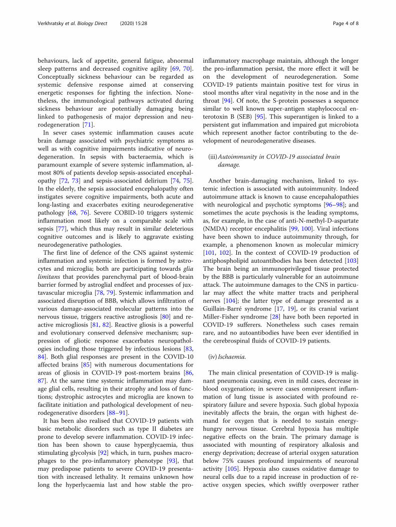
COVID-19 are widely reported; these include encephal-itis, cerebral infarction, delirium, depression, delirium,psychosis Guillain-Barré syndrome [16–20], Miller-Fisher syndrome, and etcetera [19, 21–28]. In at leastthree cases the COVID-19 brought with it symptoms ofclinical Parkinsonism demonstrating therefore a poten-tial direct link between the SARS-CoV-2 infection andneurodegeneration (see [29] for details). These acuteparkinsonian symptoms may be related to an acute dam-age to the dopaminergic system being thus distinct fromsporadic classical Parkinson disease, and yet such associ-ation required serious consideration. How COVID-19may affect the neurodegenerative process and what arethe underlying mechanisms? Below we shall try to over-view several possible scenarios (Fig. 1).
(i) Direct infection of neural cells with SARS-Cov-2.
The common way of the SARS-Cov-2 virus into thecell proceeds through binding of the RBD domain of theS-protein upon cleavage by furin to the receptorangiotensin-converting enzyme 2 (ACE2) with subse-quent internalisation of the virus probably by endocytosisin either clathrin- or pH-dependent manner, which mayalso involve endosomal proton pump and NAADP-sensitive intracellular two-pore channel 2 [30–33]. The
angiotensin system is operational in the nervous tissueand many cells of the brain including neurones andneuroglia express its components including ACE2 andfurin [34–36]. In particular, ACE2 expressing neuralcells are located in the brain stem, in the circumventri-cular organs (CVOs), the subfornical organ, paraven-tricular nucleus (PVN), nucleus of the tractus solitarius(NTS), and rostral ventrolateral medulla, all these struc-tures having high vascularisation and physiologicallyleaky blood-brain barrier [37], which permits directcontact with blood-borne viral particles. An alterna-tive pathways for SARS-CoV-2 entry into the brainthrough nasal epithelium with subsequent retrogradeand trans-synaptic penetration through axons of olfac-tory neurones; this may bring the virus into the olfac-tory bulb [38, 39].The virulence of SARS-CoV-2 may also involve
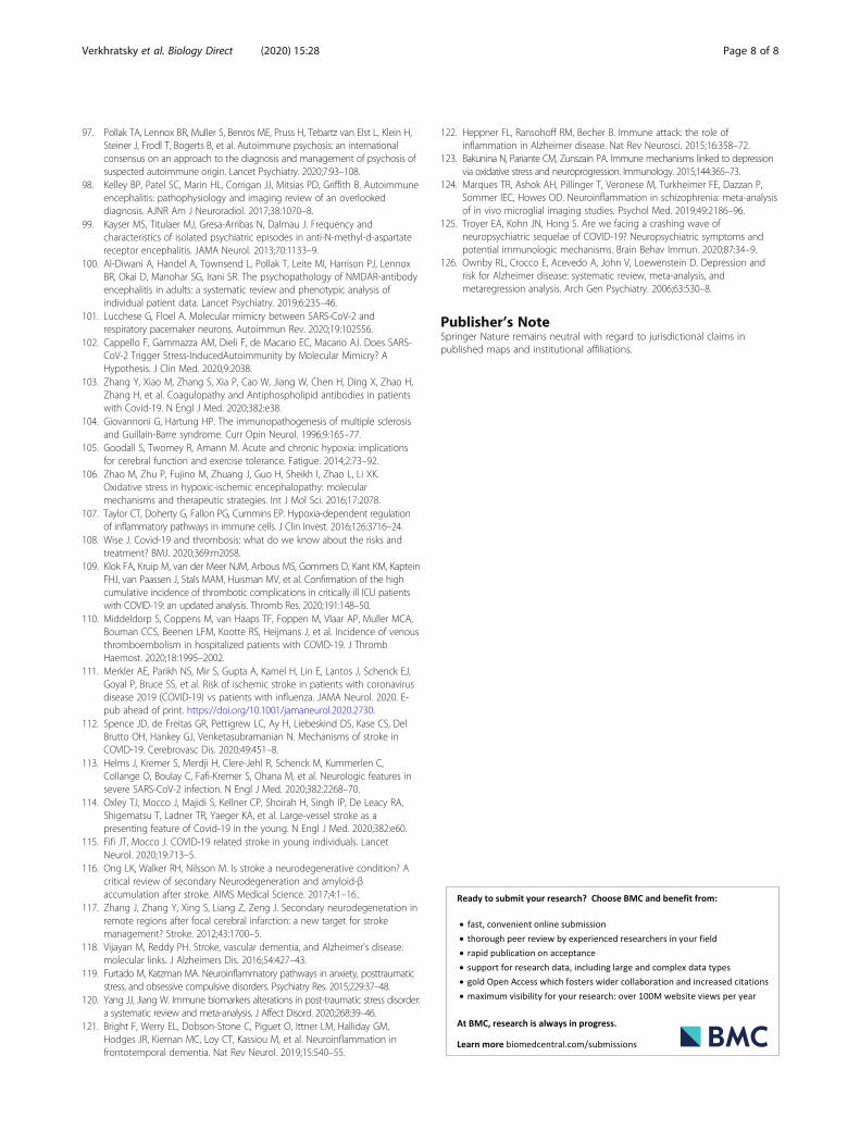
neuropilin-1, known to bind furin-cleaved substrates [40,41]. It appeared that spike coronavirus protein shows apolybasic Arg-Arg-Ala-Arg carboxyl-terminal sequenceon the cleaved fragment of S1 that matches the pre-dicted C-end rule (CendR) motif for physical interactionwith neuropilins. The structure of neuropilin-1 has beenresolved and the coordination of the extracellulardomains 1–4 (a1a2b1b2) is shown in Fig. 2 [42]. It tunesout that the domain B1 is able to bind the small inhibi-tor molecule EG00229 [43], which suppresses the infect-ivity of SARS-CoV-2 [41]. Similarly, monoclonalantibody against neuropilin-1 significantly reduces viral
Fig. 1 COVID-19 damages the brain: possible links to neurodegeneration. See text for explanations and details
Verkhratsky et al. Biology Direct (2020) 15:28 Page 2 of 8

infectivity [40]. In the latter case, post-mortem autopsiesof the olfactory neuronal detected neuropilin-1 at theentry site for the virus. These data offer a significantpotential intervention pathway for the treatment of theinfection, including its involvement of the centralnervous system [44]. Prediction of clinical outcome isessential for medical practice [45–53]; unfortunately,pathobiology of COVID-19 is still missing identifiablemolecular determinants of disease progression and clin-ical outcome.Infection of neurones and neuroglial cells have been
documented in vitro, in particular in 2D cultures andbrain organoids derived from human stem cells [54–57].The viral particles have been also found in the post-mortem brain tissues obtained from COVID-19 victims;the viral load was found in 30–50% of all specimens [58,59]. Can these viruses damage neural cells beyond repairand instigate neurodegenerative process? This is difficultto assess at the moment; it is known that influenza canbe associated (rarely) with encephalitis leading to a sub-stantial damage to the brain tissue [60]. This damage hasbeen detected at the cellular level; in particular suchdamage was manifested by clasmatodenrosis, indicativeof severe degeneration of astrocytes [61]. Hitherto, suchpronounced degenerative change in the SARS-CoV-2 in-fected brains has not been characterised. In addition, the
persistence of SARS-CoV-2 in the brain after viral clear-ance from the respiratory system and blood remains tobe characterised.
(ii) Systemic inflammation.
The systemic inflammation is the main feature of se-vere cases of COVID-19; the “cytokine storm” reflectingmassive increase of pro-inflammatory factors in theblood, is a singular feature of COVID-19 pathogenesis[62]. It should be noted that in severe COVID-19 pa-tients, T cells are often lost and the inflammation ischaracterized by innate immune responses [63]. How in-nate cytokines affect the central nervous system in ab-sence of adaptive cytokines is not clear. The linkbetween systemic inflammation and neurological as wellas neuropsychiatric diseases is universally acknowledgedwith both innate and adaptive immune responses affect-ing the brain [64–67]. Cytokines, chemokines or evenactivated blood-borne immune cells can enter the brainthrough subfornical organs; in addition cytokines cancompromise the BBB thus opening an alternative entryroute for pro-inflammatory agents [68]. Even at low in-tensity of systemic inflammation, invasion of pro-inflammatory factors initiates sickness behaviour, awide-spread syndrome characterised by depressive-like
Fig. 2 Structural constrains of neuropilin-1. a. Secondary structure of the extracellular domains 1–4 (a1a2b1b2) of mouse Neuropilin-1, PDBassignment 4GZ9 (DOI: https://doi.org/10.2210/pdb4GZ9/pdb) [42]. α’- β-D-mannopyranose-(1–4)-2-acetamido-2-deoxy-β-D-glucopyranose-(1–4)-2-acetamido-2-deoxy-beta-Dglucopyranose. a’’ = 1,2-ethanediol. b. Surface structure of neuropilin-1 shown by hydrophobicity (same source ofpanel A). c. Binding of the small inhibitor molecule EG00229 on the B1 domain of human neuropilin-1. PDB assignment 3I97 (DOI:https://doi.org/10.2210/pdb3I97/pdb) [43]
Verkhratsky et al. Biology Direct (2020) 15:28 Page 3 of 8
behaviours, lack of appetite, general fatigue, abnormalsleep patterns and decreased cognitive agility [69, 70].Conceptually sickness behaviour can be regarded assystemic defensive response aimed at conservingenergetic responses for fighting the infection. None-theless, the immunological pathways activated duringsickness behaviour are potentially damaging beinglinked to pathogenesis of major depression and neu-rodegeneration [71].In sever cases systemic inflammation causes acute
brain damage associated with psychiatric symptoms aswell as with cognitive impairments indicative of neuro-degeneration. In sepsis with bacteraemia, which isparamount example of severe systemic inflammation, al-most 80% of patients develop sepsis-associated encephal-opathy [72, 73] and sepsis-associated delirium [74, 75].In the elderly, the sepsis associated encephalopathy ofteninstigates severe cognitive impairments, both acute andlong-lasting and exacerbates exiting neurodegenerativepathology [68, 76]. Severe COBID-10 triggers systemicinflammation most likely on a comparable scale withsepsis [77], which thus may result in similar deleteriouscognitive outcomes and is likely to aggravate existingneurodegenerative pathologies.The first line of defence of the CNS against systemic
inflammation and systemic infection is formed by astro-cytes and microglia; both are participating towards glialimitans that provides parenchymal part of blood-brainbarrier formed by astroglial endfeet and processes of jux-tavascular microglia [78, 79]. Systemic inflammation andassociated disruption of BBB, which allows infiltration ofvarious damage-associated molecular patterns into thenervous tissue, triggers reactive astrogliosis [80] and re-active microgliosis [81, 82]. Reactive gliosis is a powerfuland evolutionary conserved defensive mechanism; sup-pression of gliotic response exacerbates neuropathol-ogies including those triggered by infectious lesions [83,84]. Both glial responses are present in the COVID-10affected brains [85] with numerous documentations forareas of gliosis in COVID-19 post-mortem brains [86,87]. At the same time systemic inflammation may dam-age glial cells, resulting in their atrophy and loss of func-tions; dystrophic astrocytes and microglia are known tofacilitate initiation and pathological development of neu-rodegenerative disorders [88–91].It has been also realised that COVID-19 patients with
basic metabolic disorders such as type II diabetes areprone to develop severe inflammation. COVID-19 infec-tion has been shown to cause hyperglycaemia, thusstimulating glycolysis [92] which, in turn, pushes macro-phages to the pro-inflammatory phenotype [93], thatmay predispose patients to severe COVID-19 presenta-tion with increased lethality. It remains unknown howlong the hyperlycaemia last and how stable the pro-
inflammatory macrophage maintain, although the longerthe pro-inflammation persist, the more effect it will beon the development of neurodegeneration. SomeCOVID-19 patients maintain positive test for virus instool months after viral negativity in the nose and in thethroat [94]. Of note, the S-protein possesses a sequencesimilar to well known super-antigen staphylococcal en-terotoxin B (SEB) [95]. This superantigen is linked to apersistent gut inflammation and impaired gut microbiotawhich represent another factor contributing to the de-velopment of neurodegenerative diseases.
(iii)Autoimmunity in COVID-19 associated braindamage.
Another brain-damaging mechanism, linked to sys-temic infection is associated with autoimmunity. Indeedautoimmune attack is known to cause encepahalopathieswith neurological and psychotic symptoms [96–98]; andsometimes the acute psychosis is the leading symptoms,as, for example, in the case of anti-N-methyl-D-aspartate(NMDA) receptor encephalitis [99, 100]. Viral infectionshave been shown to induce autoimmunity through, forexample, a phenomenon known as molecular mimicry[101, 102]. In the context of COVID-19 production ofantiphospholipid autoantibodies has been detected [103]The brain being an immunoprivileged tissue protectedby the BBB is particularly vulnerable for an autoimmuneattack. The autoimmune damages to the CNS in particu-lar may affect the white matter tracts and peripheralnerves [104]; the latter type of damage presented as aGuillain-Barré syndrome [17, 19], or its cranial variantMiller-Fisher syndrome [28] have both been reported inCOVID-19 sufferers. Nonetheless such cases remainrare, and no autoantibodies have been ever identified inthe cerebrospinal fluids of COVID-19 patients.
(iv) Ischaemia.
The main clinical presentation of COVID-19 is malig-nant pneumonia causing, even in mild cases, decrease inblood oxygenation; in severe cases omnipresent inflam-mation of lung tissue is associated with profound re-spiratory failure and severe hypoxia. Such global hypoxiainevitably affects the brain, the organ with highest de-mand for oxygen that is needed to sustain energy-hungry nervous tissue. Cerebral hypoxia has multiplenegative effects on the brain. The primary damage isassociated with mounting of respiratory alkalosis andenergy deprivation; decrease of arterial oxygen saturationbelow 75% causes profound impairments of neuronalactivity [105]. Hypoxia also causes oxidative damage toneural cells due to a rapid increase in production of re-active oxygen species, which swiftly overpower rather
Verkhratsky et al. Biology Direct (2020) 15:28 Page 4 of 8

limited brain antioxidative defences [106]. Brain hypoxiais also directly linked to activation or exacerbation of in-flammatory response by stimulating hypoxia induciblefactors and the NF-κB signalling cascade, which in turnprompt the release of pro-inflammatory factors [107]. Insummary, severe and/or prolonged hypoxia may causewidespread damage to brain structures being thus dir-ectly linked to neurodegeneration and cognitive deficits.
(v) Thrombosis and stroke.
The systemic inflammation accompanying COVID-19increases blood levels of fibronectin, arguably throughstimulating its liver synthesis [108]. Increased fibronectinfacilitates clot formation and 20–50% of COVID patientsdemonstrate thrombotic and thromboembolic complica-tions [109, 110]. Among these complications stroke hasbeen described relatively frequently with numbers vary-ing between 1.6% and up to 20% of hospitalised patients[111–113], including people of young ages [114, 115].The link between stroke and neurodegeneration is welldocumented. Stroke is associated with stroke-inducedsecondary neurodegeneration [116, 117] as well as withincreased risk of Alzheimer’s disease [118]. Covid-19 as-sociated thrombosis therefore can be directly linked toneurodegenerative diseases.
(vi)Psychological stress.
Patients hospitalised with severe forms of COVID-19are exposed to a prolonged and malignant stress associ-ated with the gravity of their conditions, with extendedperiod lung ventilation, with grave atmosphere of the in-tensive care unit and with periods of delirium, uncon-sciousness and, sometimes, coma. This aversiveexperience amounts to the trauma likely to induce thepost-traumatic stress disorder, which is also linked toimmune pathology [119, 120]. In addition, maladaptivestress response (linked to powerful and long lastingstressors) exacerbates both systemic inflammation andinflammatory damage to the brain through activation ofthe hypothalamic-pituitary-adrenal axis with increase ingluocorticoids. Neuroinflammation is deeply associatedwith several neuropsychiatric and neuro-cognitive dis-eases, including depression, psychosis and neurodegen-eration [121–124]. Previous analysis of SARS-Cov-1infection revealed alarmingly high prevalence of neuro-psychiatric sequalae with 40% of patients suffering frompost-traumatic stress disorder and 36% form depressionin 50–80 months after their hospitalisation [125]. Psy-chological stress affects not only COVID-19 patients butalso general population due to lockdown, self isolationand fear; these factors are especially prominent betweenold people. Depression is a well known risk factor of
dementia and psychological burden of COVID-19 mayincrease the rate of neurodegenerative diseases in theaftermath of the pandemic [126].
RecapitulationThe pandemic of Coronavirus Disease 2019 (COVID-19)presents the world with the medical challenge associatedwith multifactorial nature of this pathology. COVID-19affects the brain in many ways; often the COVID-19 ismanifested by neurological and neuropsychiatric symp-toms that include dizziness, disturbed sleep, cognitivedeficits, delirium, hallucinations and depression. Allthese signal the damage to the nervous tissue which maysubstantially increase the incidence of neurodegenerativediseases and promote dementia.
AcknowledgementsThe authors thank Eleonora Candi and Richard Knight for helpful andconstructive criticisms.
Authors’ contributionsAV, GM and YS conceived the project, AV, GM, SM, QL and YS wrote themanuscript; QL prepared Fig. 1; GM prepared Fig. 2. All of the Authors haveapproved this submitted version.
FundingThis work has been supported by the Associazione Italiana per la Ricercacontro il Cancro (AIRC) to GM (IG#20473; 2018–2022), Ministry of Health &MAECI Italy-China Science and Technology Cooperation (#PGR00961) to GM.National Key R&D Program of China (2018YFA0107500) to YS, Suzhou 2020Emergency Innovation Funding on COVID-19 Infection to YS, and the Na-tional Natural Science Foundation of China (81530043, 81861138015,31771581 and 81571612) to YS.
Availability of data and materialsNot applicable.
Ethics approval and consent to participateNot applicable.
Consent for publicationNot applicable.
Competing interestsThe Authors declare that they have no competing interests.
Author details1Faculty of Biology, Medicine and Health, The University of Manchester,Manchester M13 9PT, UK. 2Achucarro Center for Neuroscience, IKERBASQUE,48011 Bilbao, Spain. 3CAS Key Laboratory of Tissue Microenvironment andTumor, Shanghai Institute of Nutrition and Health, Chinese Academy ofSciences, 320 Yueyang Road, Shanghai 200031, China. 4University of RomeTor Vergata, via Cracovia 1, 00133 Rome, Italy. 5State Key Laboratory ofRadiation Medicine and Protection, The First Affiliated Hospital of SoochowUniversity, Institutes for Translational Medicine, Soochow University MedicalCollege, Suzhou 215123, Jiangsu, China.
Received: 18 November 2020 Accepted: 19 November 2020
References1. Chen J, Lu H, Melino G, Boccia S, Piacentini M, Ricciardi W, Wang Y, Shi Y,
Zhu T. COVID-19 infection: the China and Italy perspectives. Cell Death Dis.2020;11:438.
2. Shi Y, Wang Y, Shao C, Huang J, Gan J, Huang X, Bucci E, Piacentini M,Ippolito G, Melino G. COVID-19 infection: the perspectives on immuneresponses. Cell Death Differ. 2020;27:1451–4.
Verkhratsky et al. Biology Direct (2020) 15:28 Page 5 of 8

3. Sharma A, Kumar Sharma S, Shi Y, Bucci E, Carafoli E, Melino G,Bhattacherjee A, Das G. BCG vaccination policy and preventive chloroquineusage: do they have an impact on COVID-19 pandemic? Cell Death Dis.2020;11:516.
4. Li X, Wang Y, Agostinis P, Rabson A, Melino G, Carafoli E, Shi Y, Sun E. Ishydroxychloroquine beneficial for COVID-19 patients? Cell Death Dis. 2020;11:512.
5. Katzman R. Editorial: the prevalence and malignancy of Alzheimer disease. Amajor killer. Arch Neurol. 1976;33:217–8.
6. Mayeux R, Stern Y. Epidemiology of Alzheimer disease. Cold Spring HarbPerspect Med. 2012;2:a006239.
7. Collaborators GD. Global, regional, and national burden ofAlzheimer's disease and other dementias, 1990–2016: a systematicanalysis for the global burden of disease study 2016. The LancetNeurol. 2019;18:88–106.
8. Tysnes OB, Storstein A. Epidemiology of Parkinson's disease. J NeuralTransm (Vienna). 2017;124:901–5.
9. Association A. Alzheimer's disease facts and figures. Alzheimers Dement.2020;16:391–460.
10. Giridharan VV, Masud F, Petronilho F, Dal-Pizzol F, Barichello T. Infection-induced systemic inflammation is a potential driver of Alzheimer's diseaseprogression. Front Aging Neurosci. 2019;11:122.
11. Holmes C. Review: systemic inflammation and Alzheimer's disease.Neuropathol Appl Neurobiol. 2013;39:51–68.
12. Lim SL, Rodriguez-Ortiz CJ, Kitazawa M. Infection, systemic inflammation,and Alzheimer's disease. Microbes Infect. 2015;17:549–56.
13. Walker KA, Ficek BN, Westbrook R. Understanding the role of systemicinflammation in Alzheimer's disease. ACS Chem Neurosci. 2019;10:3340–2.
14. Hou Y, Dan X, Babbar M, Wei Y, Hasselbalch SG, Croteau DL, Bohr VA.Ageing as a risk factor for neurodegenerative disease. Nat Rev Neurol. 2019;15:565–81.
15. Koff WC, Williams MA. Covid-19 and immunity in aging populations - a newresearch agenda. N Engl J Med. 2020;383:804–5.
16. Zhao H, Shen D, Zhou H, Liu J, Chen S. Guillain-Barre syndromeassociated with SARS-CoV-2 infection: causality or coincidence? LancetNeurol. 2020;19:383–4.
17. Sedaghat Z, Karimi N. Guillain Barre syndrome associated with COVID-19infection: a case report. J Clin Neurosci. 2020;76:233–5.
18. Padroni M, Mastrangelo V, Asioli GM, Pavolucci L, Abu-Rumeileh S, PiscagliaMG, Querzani P, Callegarini C, Foschi M. Guillain-Barre syndrome followingCOVID-19: new infection, old complication? J Neurol. 2020;267:1877–9.
19. Arnaud S, Budowski C, Ng Wing Tin S, Degos B. Post SARS-CoV-2 Guillain-Barre syndrome. Clin Neurophysiol. 2020;131:1652–4.
20. Tiet MY, AlShaikh N. Guillain-Barre syndrome associated with COVID-19infection: a case from the UK. BMJ Case Rep. 2020;13. E-pub head of print.https://doi.org/10.1136/bcr-2020-236536.
21. Ellul MA, Benjamin L, Singh B, Lant S, Michael BD, Easton A, Kneen R, DefresS, Sejvar J, Solomon T. Neurological associations of COVID-19. LancetNeurol. 2020;19:767–83.
22. Wang Q, Xu R, Volkow ND. Increased risk of COVID-19 infection andmortality in people with mental disorders: analysis from electronic healthrecords in the United States. World Psychiatry. 2020. E-pub ahead of print.https://doi.org/10.1002/wps.20806.
23. Steardo L Jr, Steardo L, Verkhratsky A. Psychiatric face of COVID-19. TranslPsychiatry. 2020;10:261.
24. Steardo L, Steardo L Jr, Zorec R, Verkhratsky A. Neuroinfection maycontribute to pathophysiology and clinical manifestations of COVID-19. ActaPhysiol (Oxf). 2020;229:e13473.
25. Paterson RW, Brown RL, Benjamin L, Nortley R, Wiethoff S, Bharucha T,Jayaseelan DL, Kumar G, Raftopoulos RE, Zambreanu L, et al. The emergingspectrum of COVID-19 neurology: clinical, radiological and laboratoryfindings. Brain. 2020;143:3104–20. .
26. Pergolizzi JV Jr, Raffa RB, Varrassi G, Magnusson P, LeQuang JA, Paladini A,Taylor R, Wollmuth C, Breve F, Chopra M, et al. Potential neurologicalmanifestations of COVID-19: a narrative review. Postgrad Med. 2020. https://doi.org/10.1080/00325481.2020.1837503.
27. Li Z, Liu T, Yang N, Han D, Mi X, Li Y, Liu K, Vuylsteke A, Xiang H, Guo X.Neurological manifestations of patients with COVID-19: potential routes ofSARS-CoV-2 neuroinvasion from the periphery to the brain. Front Med.2020;14:533–41.
28. Gutierrez-Ortiz C, Mendez A, Rodrigo-Rey S, San Pedro-Murillo E,Bermejo-Guerrero L, Gordo-Manas R, de Aragon-Gomez F, Benito-Leon J.Miller fisher syndrome and polyneuritis cranialis in COVID-19. Neurology.2020;95:e601–5.
29. Brundin P, Nath A, Beckham JD. Is COVID-19 a perfect storm for Parkinson'sdisease? Trends Neurosci. 2020. E-pub ahead of print. https://doi.org/10.1016/j.tins.2020.10.009.
30. Bayati A, Kumar R, Francis V, McPherson PS. SARS-CoV-2 uses clathrin-mediated endocytosis to gain access into cells. BioRxiv. 2020; preprint.https://doi.org/10.1101/2020.07.13.201509.
31. Wang H, Yang P, Liu K, Guo F, Zhang Y, Zhang G, Jiang C. SARS coronavirusentry into host cells through a novel clathrin- and caveolae-independentendocytic pathway. Cell Res. 2008;18:290–301.
32. Inoue Y, Tanaka N, Tanaka Y, Inoue S, Morita K, Zhuang M, Hattori T,Sugamura K. Clathrin-dependent entry of severe acute respiratory syndromecoronavirus into target cells expressing ACE2 with the cytoplasmic taildeleted. J Virol. 2007;81:8722–9.
33. Petersen OH, Gerasimenko OV, Gerasimenko JV. Endocytic uptake of SARS-CoV-2: the critical roles of pH, Ca2+, and NAADP Function. 2020;1:zqaa003.https://academic.oup.com/function.
34. Gowrisankar YV, Clark MA. Angiotensin II regulation of angiotensin-converting enzymes in spontaneously hypertensive rat primary astrocytecultures. J Neurochem. 2016;138:74–85.
35. Nemoto W, Yamagata R, Nakagawasai O, Nakagawa K, Hung WY, Fujita M,Tadano T, Tan-No K. Effect of spinal angiotensin-converting enzyme 2activation on the formalin-induced nociceptive response in mice. Eur JPharmacol. 2020;872:172950.
36. Xia H, Lazartigues E. Angiotensin-converting enzyme 2: central regulator forcardiovascular function. Curr Hypertens Rep. 2010;12:170–5.
37. Duvernoy HM, Risold PY. The circumventricular organs: an atlas ofcomparative anatomy and vascularization. Brain Res Rev. 2007;56:119–47.
38. Netland J, Meyerholz DK, Moore S, Cassell M, Perlman S. Severe acuterespiratory syndrome coronavirus infection causes neuronal death inthe absence of encephalitis in mice transgenic for human ACE2. J Virol.2008;82:7264–75.
39. Li K, Wohlford-Lenane C, Perlman S, Zhao J, Jewell AK, Reznikov LR,Gibson-Corley KN, Meyerholz DK, McCray PB Jr. Middle East respiratorysyndrome coronavirus causes multiple organ damage and lethal diseasein mice transgenic for human Dipeptidyl peptidase 4. J Infect Dis. 2016;213:712–22.
40. Cantuti-Castelvetri L, Ojha R, Pedro LD, Djannatian M, Franz J, Kuivanen S,van der Meer F, Kallio K, Kaya T, Anastasina M, et al. Neuropilin-1 facilitatesSARS-CoV-2 cell entry and infectivity. Science. 2020;370:856–60.
41. Daly JL, Simonetti B, Klein K, Chen KE, Williamson MK, Anton-Plagaro C,Shoemark DK, Simon-Gracia L, Bauer M, Hollandi R, et al. Neuropilin-1 is ahost factor for SARS-CoV-2 infection. Science. 2020;370:861–5.
42. Janssen BJ, Malinauskas T, Weir GA, Cader MZ, Siebold C, Jones EY.Neuropilins lock secreted semaphorins onto plexins in a ternary signalingcomplex. Nat Struct Mol Biol. 2012;19:1293–9.
43. Jarvis A, Allerston CK, Jia H, Herzog B, Garza-Garcia A, Winfield N, Ellard K,Aqil R, Lynch R, Chapman C, et al. Small molecule inhibitors of theneuropilin-1 vascular endothelial growth factor a (VEGF-A) interaction.J Med Chem. 2010;53:2215–26.
44. Davies J, Randeva HS, Chatha K, Hall M, Spandidos DA, Karteris E, Kyrou I.Neuropilin1 as a new potential SARSCoV2 infection mediator implicated inthe neurologic features and central nervous system involvement ofCOVID19. Mol Med Rep. 2020;22:4221–6.
45. Jayashree S, Murugavel P, Sowdhamini R, Srinivasan N. Interface residues oftransient protein-protein complexes have extensive intra-proteininteractions apart from inter-protein interactions. Biol Direct. 2019;14:1.
46. Dobon B, Montanucci L, Pereto J, Bertranpetit J, Laayouni H. Geneconnectivity and enzyme evolution in the human metabolic network. BiolDirect. 2019;14:17.
47. Han Y, Ye X, Cheng J, Zhang S, Feng W, Han Z, Zhang J, Huang K.Integrative analysis based on survival associated co-expression genemodules for predicting neuroblastoma patients' survival time. Biol Direct.2019;14:4.
48. Han Y, Ye X, Wang C, Liu Y, Zhang S, Feng W, Huang K, Zhang J. Integrationof molecular features with clinical information for predicting outcomes forneuroblastoma patients. Biol Direct. 2019;14:16.
Verkhratsky et al. Biology Direct (2020) 15:28 Page 6 of 8

49. Kim SY, Jeong HH, Kim J, Moon JH, Sohn KA. Robust pathway-based multi-omics data integration using directed random walks for survival predictionin multiple cancer studies. Biol Direct. 2019;14:8.
50. Baali I, Acar DAE, Aderinwale TW, HafezQorani S, Kazan H. Predicting clinicaloutcomes in neuroblastoma with genomic data integration. Biol Direct.2018;13:20.
51. Polewko-Klim A, Lesinski W, Mnich K, Piliszek R, Rudnicki WR. Integration ofmultiple types of genetic markers for neuroblastoma may contribute toimproved prediction of the overall survival. Biol Direct. 2018;13:17.
52. Suo C, Deng W, Vu TN, Li M, Shi L, Pawitan Y. Accumulation of potentialdriver genes with genomic alterations predicts survival of high-riskneuroblastoma patients. Biol Direct. 2018;13:14.
53. Mihaylov I, Kandula M, Krachunov M, Vassilev D. A novel framework forhorizontal and vertical data integration in cancer studies with application tosurvival time prediction models. Biol Direct. 2019;14:22.
54. Song E, Zhang C, Benjamin Israelow B, Lu-Culligan A, Prado AV, Skriabine S,Lu P, Weizman O, Liu F, Dai Y, et al. Neuroinvasion of SARS-CoV- 2 inhuman and mouse brain. BioRxiv. 2020; Preprint. https://doi.org/10.1101/2020.06.25.169946.
55. Zhang BZ, Chu H, Han S, Shuai H, Deng J, Hu YF, Gong HR, Lee AC, Zou Z,Yau T, et al. SARS-CoV-2 infects human neural progenitor cells and brainorganoids. Cell Res. 2020;30:928–31.
56. Yi SA, Nam KH, Yun J, Gim D, Joe D, Kim YH, Kim HJ, Han JW, Lee J.Infection of brain Organoids and 2D cortical neurons with SARS-CoV-2Pseudovirus. Viruses. 2020;12. E-pub ahead of print. https://doi.org/10.3390/v12091004.
57. Ramani A, Muller L, Ostermann PN, Gabriel E, Abida-Islam P, Muller-Schiffmann A, Mariappan A, Goureau O, Gruell H, Walker A, et al. SARS-CoV-2 targets neurons of 3D human brain organoids. EMBO J. 2020;39:e106230.
58. Puelles VG, Lutgehetmann M, Lindenmeyer MT, Sperhake JP, Wong MN,Allweiss L, Chilla S, Heinemann A, Wanner N, Liu S, et al. Multiorgan andrenal tropism of SARS-CoV-2. N Engl J Med. 2020;383:590–2.
59. Matschke J, Lutgehetmann M, Hagel C, Sperhake JP, Schroder AS, Edler C,Mushumba H, Fitzek A, Allweiss L, Dandri M, et al. Neuropathology ofpatients with COVID-19 in Germany: a post-mortem case series. LancetNeurol. 2020;19:919–29.
60. Hayase Y, Tobita K. Influenza virus and neurological diseases. Psychiatry ClinNeurosci. 1997;51:181–4.
61. Tachibana M, Mohri I, Hirata I, Kuwada A, Kimura-Ohba S, Kagitani-ShimonoK, Fushimi H, Inoue T, Shiomi M, Kakuta Y, et al. Clasmatodendrosis isassociated with dendritic spines and does not represent autophagicastrocyte death in influenza-associated encephalopathy. Brain andDevelopment. 2019;41:85–95.
62. Coperchini F, Chiovato L, Croce L, Magri F, Rotondi M. The cytokine stormin COVID-19: an overview of the involvement of the chemokine/chemokine-receptor system. Cytokine Growth Factor Rev. 2020;53:25–32.
63. Tan L, Wang Q, Zhang D, Ding J, Huang Q, Tang YQ, Wang Q, Miao H.Lymphopenia predicts disease severity of COVID-19: a descriptive andpredictive study. Signal Transduct Target Ther. 2020;5:33.
64. Schwartz M, Deczkowska A. Neurological disease as a failure of brain-immune crosstalk: the multiple faces of Neuroinflammation. TrendsImmunol. 2016;37:668–79.
65. Carson MJ, Doose JM, Melchior B, Schmid CD, Ploix CC. CNS immuneprivilege: hiding in plain sight. Immunol Rev. 2006;213:48–65.
66. Hickey WF, Hsu BL, Kimura H. T-lymphocyte entry into the central nervoussystem. J Neurosci Res. 1991;28:254–60.
67. Varatharaj A, Galea I. The blood-brain barrier in systemic inflammation. BrainBehav Immun. 2017;60:1–12.
68. Sankowski R, Mader S, Valdes-Ferrer SI. Systemic inflammation and the brain:novel roles of genetic, molecular, and environmental cues as drivers ofneurodegeneration. Front Cell Neurosci. 2015;9:28.
69. Capuron L, Lamarque D, Dantzer R, Goodall G. Attentional andmnemonic deficits associated with infectious disease in humans.Psychol Med. 1999;29:291–7.
70. Dantzer R. Cytokine, sickness behavior, and depression. Immunol AllergyClin N Am. 2009;29:247–64.
71. Maes M, Berk M, Goehler L, Song C, Anderson G, Galecki P, Leonard B.Depression and sickness behavior are Janus-faced responses to sharedinflammatory pathways. BMC Med. 2012;10:66.
72. Shulyatnikova T, Verkhratsky A. Astroglia in Sepsis associatedencephalopathy. Neurochem Res. 2020;45:83–99.
73. Ren C, Yao RQ, Zhang H, Feng YW, Yao YM. Sepsis-associatedencephalopathy: a vicious cycle of immunosuppression.J Neuroinflammation. 2020;17:14.
74. Ely EW, Shintani A, Truman B, Speroff T, Gordon SM, Harrell FE Jr, Inouye SK,Bernard GR, Dittus RS. Delirium as a predictor of mortality in mechanicallyventilated patients in the intensive care unit. JAMA. 2004;291:1753–62.
75. Ebersoldt M, Sharshar T, Annane D. Sepsis-associated delirium. IntensiveCare Med. 2007;33:941–50.
76. Iwashyna TJ, Ely EW, Smith DM, Langa KM. Long-term cognitive impairmentand functional disability among survivors of severe sepsis. JAMA. 2010;304:1787–94.
77. Garcia LF. Immune response, inflammation, and the clinical Spectrum ofCOVID-19. Front Immunol. 2020;11:1441.
78. Joost E, Jordao MJC, Mages B, Prinz M, Bechmann I, Krueger M. Microgliacontribute to the glia limitans around arteries, capillaries and veins underphysiological conditions, in a model of neuroinflammation and in humanbrain tissue. Brain Struct Funct. 2019;224:1301–14.
79. Verkhratsky A, Nedergaard M. Physiology of Astroglia. Physiol Rev. 2018;98:239–389.
80. Verkhratsky A, Zorec R, Parpura V. Stratification of astrocytes in healthy anddiseased brain. Brain Pathol. 2017;27:629–44.
81. Sierra A, Beccari S, Diaz-Aparicio I, Encinas JM, Comeau S, Tremblay ME.Surveillance, phagocytosis, and inflammation: how never-resting microgliainfluence adult hippocampal neurogenesis. Neural Plast. 2014;2014:610343.
82. Kettenmann H, Hanisch UK, Noda M, Verkhratsky A. Physiology of microglia.Physiol Rev. 2011;91:461–553.
83. Zorec R, Zupanc TA, Verkhratsky A. Astrogliopathology in the infectiousinsults of the brain. Neurosci Lett. 2019;689:56–62.
84. Pekny M, Pekna M, Messing A, Steinhauser C, Lee JM, Parpura V, Hol EM,Sofroniew MV, Verkhratsky A. Astrocytes: a central element in neurologicaldiseases. Acta Neuropathol. 2016;131:323–45.
85. Tremblay M-E, Madore C, Bordeleau M, Tian L, Verkhratsky A.Neuropathobiology of COVID-19: the role for glia. Front Cell Neurosci. 2020;14:a592214.
86. Kanberg N, Ashton NJ, Andersson LM, Yilmaz A, Lindh M, Nilsson S, PriceRW, Blennow K, Zetterberg H, Gisslen M. Neurochemical evidence ofastrocytic and neuronal injury commonly found in COVID-19. Neurology.2020; E-pub ahead of print. https://doi.org/10.1212/WNL.0000000000010111.
87. Reichard RR, Kashani KB, Boire NA, Constantopoulos E, Guo Y, LucchinettiCF. Neuropathology of COVID-19: a spectrum of vascular and acutedisseminated encephalomyelitis (ADEM)-like pathology. Acta Neuropathol.2020;140:1–6.
88. Streit WJ, Sammons NW, Kuhns AJ, Sparks DL. Dystrophic microglia in theaging human brain. Glia. 2004;45:208–12.
89. Streit WJ, Xue QS, Tischer J, Bechmann I. Microglial pathology. ActaNeuropathol Commun. 2014;2:142.
90. Verkhratsky A, Rodrigues JJ, Pivoriunas A, Zorec R, Semyanov A. Astroglialatrophy in Alzheimer's disease. Pflugers Arch. 2019;471:1247–61.
91. Verkhratsky A, Marutle A, Rodriguez-Arellano JJ, Nordberg A. Glial astheniaand functional paralysis: a new perspective on Neurodegeneration andAlzheimer's disease. Neuroscientist. 2015;21:552–68.
92. Han J, Zhang L, Guo H, Wysham WZ, Roque DR, Willson AK, Sheng X, ZhouC, Bae-Jump VL. Glucose promotes cell proliferation, glucose uptake andinvasion in endometrial cancer cells via AMPK/mTOR/S6 and MAPKsignaling. Gynecol Oncol. 2015;138:668–75.
93. Du L, Lin L, Li Q, Liu K, Huang Y, Wang X, Cao K, Chen X, Cao W, Li F, et al.IGF-2 preprograms maturing macrophages to acquire oxidativephosphorylation-dependent anti-inflammatory properties. Cell Metab. 2019;29:1363–75 e1368.
94. Xu Y, Li X, Zhu B, Liang H, Fang C, Gong Y, Guo Q, Sun X, Zhao D, Shen J,et al. Characteristics of pediatric SARS-CoV-2 infection and potentialevidence for persistent fecal viral shedding. Nat Med. 2020;26:502–5.
95. Cheng MH, Zhang S, Porritt RA, Noval Rivas M, Paschold L, Willscher E,Binder M, Arditi M, Bahar I. Superantigenic character of an insert unique toSARS-CoV-2 spike supported by skewed TCR repertoire in patients withhyperinflammation. Proc Natl Acad Sci U S A. 2020;117:25254–62.
96. Crisp SJ, Kullmann DM, Vincent A. Autoimmune synaptopathies. Nat RevNeurosci. 2016;17:103–17.
Verkhratsky et al. Biology Direct (2020) 15:28 Page 7 of 8
97. Pollak TA, Lennox BR, Muller S, Benros ME, Pruss H, Tebartz van Elst L, Klein H,Steiner J, Frodl T, Bogerts B, et al. Autoimmune psychosis: an internationalconsensus on an approach to the diagnosis and management of psychosis ofsuspected autoimmune origin. Lancet Psychiatry. 2020;7:93–108.
98. Kelley BP, Patel SC, Marin HL, Corrigan JJ, Mitsias PD, Griffith B. Autoimmuneencephalitis: pathophysiology and imaging review of an overlookeddiagnosis. AJNR Am J Neuroradiol. 2017;38:1070–8.
99. Kayser MS, Titulaer MJ, Gresa-Arribas N, Dalmau J. Frequency andcharacteristics of isolated psychiatric episodes in anti-N-methyl-d-aspartatereceptor encephalitis. JAMA Neurol. 2013;70:1133–9.
100. Al-Diwani A, Handel A, Townsend L, Pollak T, Leite MI, Harrison PJ, LennoxBR, Okai D, Manohar SG, Irani SR. The psychopathology of NMDAR-antibodyencephalitis in adults: a systematic review and phenotypic analysis ofindividual patient data. Lancet Psychiatry. 2019;6:235–46.
101. Lucchese G, Floel A. Molecular mimicry between SARS-CoV-2 andrespiratory pacemaker neurons. Autoimmun Rev. 2020;19:102556.
102. Cappello F, Gammazza AM, Dieli F, de Macario EC, Macario AJ. Does SARS-CoV-2 Trigger Stress-InducedAutoimmunity by Molecular Mimicry? AHypothesis. J Clin Med. 2020;9:2038.
103. Zhang Y, Xiao M, Zhang S, Xia P, Cao W, Jiang W, Chen H, Ding X, Zhao H,Zhang H, et al. Coagulopathy and Antiphospholipid antibodies in patientswith Covid-19. N Engl J Med. 2020;382:e38.
104. Giovannoni G, Hartung HP. The immunopathogenesis of multiple sclerosisand Guillain-Barre syndrome. Curr Opin Neurol. 1996;9:165–77.
105. Goodall S, Twomey R, Amann M. Acute and chronic hypoxia: implicationsfor cerebral function and exercise tolerance. Fatigue. 2014;2:73–92.
106. Zhao M, Zhu P, Fujino M, Zhuang J, Guo H, Sheikh I, Zhao L, Li XK.Oxidative stress in hypoxic-ischemic encephalopathy: molecularmechanisms and therapeutic strategies. Int J Mol Sci. 2016;17:2078.
107. Taylor CT, Doherty G, Fallon PG, Cummins EP. Hypoxia-dependent regulationof inflammatory pathways in immune cells. J Clin Invest. 2016;126:3716–24.
108. Wise J. Covid-19 and thrombosis: what do we know about the risks andtreatment? BMJ. 2020;369:m2058.
109. Klok FA, Kruip M, van der Meer NJM, Arbous MS, Gommers D, Kant KM, KapteinFHJ, van Paassen J, Stals MAM, Huisman MV, et al. Confirmation of the highcumulative incidence of thrombotic complications in critically ill ICU patientswith COVID-19: an updated analysis. Thromb Res. 2020;191:148–50.
110. Middeldorp S, Coppens M, van Haaps TF, Foppen M, Vlaar AP, Muller MCA,Bouman CCS, Beenen LFM, Kootte RS, Heijmans J, et al. Incidence of venousthromboembolism in hospitalized patients with COVID-19. J ThrombHaemost. 2020;18:1995–2002.
111. Merkler AE, Parikh NS, Mir S, Gupta A, Kamel H, Lin E, Lantos J, Schenck EJ,Goyal P, Bruce SS, et al. Risk of ischemic stroke in patients with coronavirusdisease 2019 (COVID-19) vs patients with influenza. JAMA Neurol. 2020. E-pub ahead of print. https://doi.org/10.1001/jamaneurol.2020.2730.
112. Spence JD, de Freitas GR, Pettigrew LC, Ay H, Liebeskind DS, Kase CS, DelBrutto OH, Hankey GJ, Venketasubramanian N. Mechanisms of stroke inCOVID-19. Cerebrovasc Dis. 2020;49:451–8.
113. Helms J, Kremer S, Merdji H, Clere-Jehl R, Schenck M, Kummerlen C,Collange O, Boulay C, Fafi-Kremer S, Ohana M, et al. Neurologic features insevere SARS-CoV-2 infection. N Engl J Med. 2020;382:2268–70.
114. Oxley TJ, Mocco J, Majidi S, Kellner CP, Shoirah H, Singh IP, De Leacy RA,Shigematsu T, Ladner TR, Yaeger KA, et al. Large-vessel stroke as apresenting feature of Covid-19 in the young. N Engl J Med. 2020;382:e60.
115. Fifi JT, Mocco J. COVID-19 related stroke in young individuals. LancetNeurol. 2020;19:713–5.
116. Ong LK, Walker RH, Nilsson M. Is stroke a neurodegenerative condition? Acritical review of secondary Neurodegeneration and amyloid-βaccumulation after stroke. AIMS Medical Science. 2017;4:1–16..
117. Zhang J, Zhang Y, Xing S, Liang Z, Zeng J. Secondary neurodegeneration inremote regions after focal cerebral infarction: a new target for strokemanagement? Stroke. 2012;43:1700–5.
118. Vijayan M, Reddy PH. Stroke, vascular dementia, and Alzheimer's disease:molecular links. J Alzheimers Dis. 2016;54:427–43.
119. Furtado M, Katzman MA. Neuroinflammatory pathways in anxiety, posttraumaticstress, and obsessive compulsive disorders. Psychiatry Res. 2015;229:37–48.
120. Yang JJ, Jiang W. Immune biomarkers alterations in post-traumatic stress disorder:a systematic review and meta-analysis. J Affect Disord. 2020;268:39–46.
121. Bright F, Werry EL, Dobson-Stone C, Piguet O, Ittner LM, Halliday GM,Hodges JR, Kiernan MC, Loy CT, Kassiou M, et al. Neuroinflammation infrontotemporal dementia. Nat Rev Neurol. 2019;15:540–55.
122. Heppner FL, Ransohoff RM, Becher B. Immune attack: the role ofinflammation in Alzheimer disease. Nat Rev Neurosci. 2015;16:358–72.
123. Bakunina N, Pariante CM, Zunszain PA. Immune mechanisms linked to depressionvia oxidative stress and neuroprogression. Immunology. 2015;144:365–73.
124. Marques TR, Ashok AH, Pillinger T, Veronese M, Turkheimer FE, Dazzan P,Sommer IEC, Howes OD. Neuroinflammation in schizophrenia: meta-analysisof in vivo microglial imaging studies. Psychol Med. 2019;49:2186–96.
125. Troyer EA, Kohn JN, Hong S. Are we facing a crashing wave ofneuropsychiatric sequelae of COVID-19? Neuropsychiatric symptoms andpotential immunologic mechanisms. Brain Behav Immun. 2020;87:34–9.
126. Ownby RL, Crocco E, Acevedo A, John V, Loewenstein D. Depression andrisk for Alzheimer disease: systematic review, meta-analysis, andmetaregression analysis. Arch Gen Psychiatry. 2006;63:530–8.
Publisher’s NoteSpringer Nature remains neutral with regard to jurisdictional claims inpublished maps and institutional affiliations.
Verkhratsky et al. Biology Direct (2020) 15:28 Page 8 of 8