Camptodactyly: Occurrence in TwoNew Syndromes andits ...
10
Journal of Medical Genetics (1972). 9, 203. Camptodactyly: Occurrence in Two New Genetic Syndromes and its Relationship to Other Syndromes RICHARD M. GOODMAN, MARIASSA BAT-MIRIAM KATZNELSON, and ELI MANOR From the Departments of Human Genetics and Family Medicine, Tel Aviv University Medical School and The Chaim Sheba Medical Center, Tel Hashomer, and the Department of Medicine, Shmuel Harofe Government Hospital, Be'er Ya'acov, Israel The term camptodactyly was coined by Landouzy (1906) to describe a form of contractures of the fingers. The aetiology of this defect is hetero- geneous; it may be either genetic or occur as a sporadic event of unknown cause (Gordon, Davies, and Berman, 1969). It is further known that several independent heritable disorders are associated with camptodactyly (Welch and Temtamy, 1966). Recently we have studied two separate families each of which presented with camptodactyly. The main constellation of findings in addition to campto- dactyly were identical in the affected members of each family, but one family differed markedly from the other. Since we have been unable to find pre- vious reports describing these observations they seem to represent two new genetic syndromes. The purposes of this communication will be to give a detailed clinical account of each syndrome and to discuss the possible significance and relationship of camptodactyly in various genetically determined conditions. FAMILY A Syndrome of Camptodactyly with Muscular Hypoplasia, Skeletal Dysplasia, and Abnormal Palmar Creases Clinical Findings. The proposita is a 17-year- old girl of Jewish Moroccan ancestry who was referred to our clinic for evaluation of multiple congenital anomalies. The patient has been in relatively good health except for having had pre- vious surgery for bilateral clubbed feet. Though she denied a history of any definite muscle weakness, she did state that she was unable to participate in any strenuous activity due to lack of stamina. She is of normal intelligence and is considered to be a Received 10 November 1971. good student. Her menstrual periods are normal and menarche started at age 14j years. Physical examination showed a 17-year-old girl appearing younger than her stated age with obvious malformations. Her height was 148-6 cm. There was asymmetry of the face with the right side being less prominent than the left (Fig. 1A). The mouth was small with an increased philtrum length and there was a high arched palate. She had a prominent forehead with mild frontal bossing. The head was brachycephalic and measured 54 cm in circumference. A moderate degree of ocular hypertelorism was present. TABLE I COMMON CLINICAL FEATURES WITH DEGREE OF SEVERITY NOTED IN MEMBERS OF FAMILY A Location and Defect Stature Short Skull Brachycephaly Prominent forehead Facies Asymmetry Ocular hypertelorism Small mouth High arched palate Increased philtrum length Chest Thoracic scoliosis Winging of scapulae Extremities Camptodactyly Syndactyly Abnormal hand prints (palmar creases) Spindle-shaped fingers Clubbed feet Muscular system Hypoplasia of chest Hypoplasia of pelvis Hypoplasia of limb and hand muscles * + + = More severely affected. Degree of Severity III. 1 (proposita- II1.3 (affected sib;- ; aged 17) d aged 13) I + + +++ + + ++ + + + + + + + + + + ++ + ++ + ++ + ++ + + + + + + + ++ + ++ + ++ 203 on February 20, 2022 by guest. Protected by copyright. http://jmg.bmj.com/ J Med Genet: first published as 10.1136/jmg.9.2.203 on 1 June 1972. Downloaded from
Transcript of Camptodactyly: Occurrence in TwoNew Syndromes andits ...

Journal of Medical Genetics (1972). 9, 203.
Camptodactyly: Occurrence in Two New GeneticSyndromes and its Relationship to Other Syndromes
RICHARD M. GOODMAN, MARIASSA BAT-MIRIAM KATZNELSON, andELI MANOR
From the Departments of Human Genetics and Family Medicine, Tel Aviv University Medical School and TheChaim Sheba Medical Center, Tel Hashomer, and the Department of Medicine, Shmuel Harofe Government Hospital,
Be'er Ya'acov, Israel
The term camptodactyly was coined by Landouzy(1906) to describe a form of contractures of thefingers. The aetiology of this defect is hetero-geneous; it may be either genetic or occur as asporadic event of unknown cause (Gordon, Davies,and Berman, 1969). It is further known that severalindependent heritable disorders are associated withcamptodactyly (Welch and Temtamy, 1966).
Recently we have studied two separate familieseach of which presented with camptodactyly. Themain constellation of findings in addition to campto-dactyly were identical in the affected members ofeach family, but one family differed markedly fromthe other. Since we have been unable to find pre-vious reports describing these observations theyseem to represent two new genetic syndromes.The purposes of this communication will be to givea detailed clinical account of each syndrome and todiscuss the possible significance and relationship ofcamptodactyly in various genetically determinedconditions.
FAMILY A
Syndrome of Camptodactyly with MuscularHypoplasia, Skeletal Dysplasia, and
Abnormal Palmar CreasesClinical Findings. The proposita is a 17-year-
old girl of Jewish Moroccan ancestry who wasreferred to our clinic for evaluation of multiplecongenital anomalies. The patient has been inrelatively good health except for having had pre-vious surgery for bilateral clubbed feet. Thoughshe denied a history of any definite muscle weakness,she did state that she was unable to participate inany strenuous activity due to lack of stamina. She isof normal intelligence and is considered to be a
Received 10 November 1971.
good student. Her menstrual periods are normaland menarche started at age 14j years.
Physical examination showed a 17-year-old girlappearing younger than her stated age with obviousmalformations. Her height was 148-6 cm. Therewas asymmetry of the face with the right side beingless prominent than the left (Fig. 1A). Themouth was small with an increased philtrumlength and there was a high arched palate. She hada prominent forehead with mild frontal bossing.The head was brachycephalic and measured 54 cm incircumference. A moderate degree of ocularhypertelorism was present.
TABLE ICOMMON CLINICAL FEATURES WITH DEGREE OFSEVERITY NOTED IN MEMBERS OF FAMILY A
Location and Defect
StatureShort
SkullBrachycephalyProminent forehead
FaciesAsymmetryOcular hypertelorismSmall mouthHigh arched palateIncreased philtrum length
ChestThoracic scoliosisWinging of scapulae
ExtremitiesCamptodactylySyndactylyAbnormal hand prints
(palmar creases)Spindle-shaped fingersClubbed feet
Muscular systemHypoplasia of chestHypoplasia of pelvisHypoplasia of limb andhand muscles
* + + = More severely affected.
Degree of Severity
III.1 (proposita- II1.3 (affected sib;-; aged 17) d aged 13)
I + +++++ +++ ++ ++ ++ ++ ++ +++ ++
+ +++ +++ ++ ++ ++ +++ ++
+ ++
203
on February 20, 2022 by guest. P
rotected by copyright.http://jm
g.bmj.com
/J M
ed Genet: first published as 10.1136/jm
g.9.2.203 on 1 June 1972. Dow
nloaded from

Goodman, Katznelson, and Manor
FIG. 1. A: the proposita's face showing asymmetry, mild ocular hypertelorism, small mouth, and an increased philtrum length. B and C:poor muscular development involving the upper and lower extremities. D and E: spindle-shaped fingers with camptodactyly and hypo-plasia of the hand muscles.
In general most of the patient's voluntary muscu-lar system was poorly developed and lacked normaltone. Both the upper extremities and lower por-tion of the legs appeared abnormal in contour due topoor muscle development (Fig. 1B and C). It wasdifficult to palpate a definite muscle mass in theseareas. There was 'winging' of the scapulae and theflesh about the arms and shoulders hung loosely(Fig. 1B). Both the hip and gluteal regions alsoshowed poor muscle development. There was amoderate degree of scoliosis to the left involving thethoracic spine.The hands showed bilateral camptodactyly with
greater deformity of the right hand. On the righthand fingers 2, 3, 4, and 5 were flexed, while on theleft only the 5th digit was affected. The thenar andhypothenar eminences were poorly developed as wellas all the hand muscles. All fingers were spindle inshape and the right thumb nail was shorter than theleft (Fig. ID and E). An abnormal dermatoglyphicpattern was grossly visible on the surfaces of the
palms and feet. Despite her previous surgery shecontinued to have a mild clubbed foot deformity.The remainder of the physical examination in-
cluding that of heart, lungs, abdomen, and nervoussystem were all within normal limits.One of the proposita's sibs, a brother aged 13, was
found not only to physically resemble the probandbut in some features he was more severely affected(Fig. 2 A-F). He also appeared younger than hisstated age and showed obvious malformations.His height was 142-2 cm and he had a head cir-cumference of 56 cm. Like his sister he gave nohistory of muscle weakness but could not participatein sports due to lack of endurance. In his earlychildhood he had also been operated on for bi-laterally clubbed feet. Table I (p. 203) lists andcompares the main clinical findings as observed inthese two affected sibs. In addition to theseidentical features listed, the brother also displayedblue sclerae, webbing of the fingers, a grade 2systolic murmur, and a left inguinal hernia.
204
on February 20, 2022 by guest. P
rotected by copyright.http://jm
g.bmj.com
/J M
ed Genet: first published as 10.1136/jm
g.9.2.203 on 1 June 1972. Dow
nloaded from
Camptodactyly: Occurrence in Two New Genetic Syndromes and itsRelationship to Other Syndromes 205
i
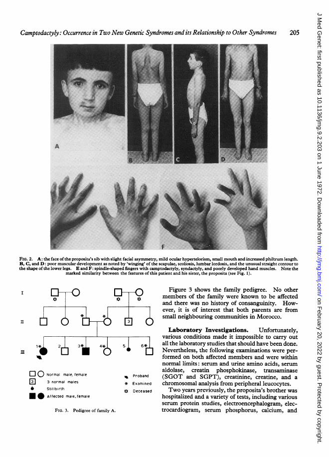
FIG. 2. A: the face of the proposita's sib with slight facial asymmetry, mild ocular hypertelorism, small mouth and increased philtrum length.B, C, and D: poor muscular development as noted by 'winging' of the scapulae, scoliosis, lumbar lordosis, and the unusual straight contour tothe shape ofthe lower legs. E and F: spindle-shaped fingers with camptodactyly, syndactyly, and poorly developed hand muscles. Note the
marked similarity between the features of this patient and his sister, the proposita (see Fig. 1).
Normal male, female
El 3 normal males
* Stillbirth
* Affected male, female
^ Proband
* Examined
St Deceased
FIG. 3. Pedigree of family A.
Figure 3 shows the family pedigree. No othermembers of the family were known to be affectedand there was no history of consanguinity. How-ever, it is of interest that both parents are fromsmall neighbouring communities in Morocco.
Laboratory Investigations. Unfortunately,various conditions made it impossible to carry outall the laboratory studies that should have been done.Nevertheless, the following examinations were per-formed on both affected members and were withinnormal limits: serum and urine amino acids, serumaldolase, creatin phosphokinase, transaminase(SGOT and SGPT), creatinine, creatine, and achromosomal analysis from peripheral leucocytes.Two years previously, the proposita's brother was
hospitalized and a variety of tests, including variousserum protein studies, electroencephalogram, elec-trocardiogram, serum phosphorus, calcium, and
I
II
on February 20, 2022 by guest. P
rotected by copyright.http://jm
g.bmj.com
/J M
ed Genet: first published as 10.1136/jm
g.9.2.203 on 1 June 1972. Dow
nloaded from

TABLE IIDERMATOGLYPHIC FINDINGS IN FAMILY A
Family Members Number of Total Ridge a-b Ridge Breadth Maximal atd Main Line ModificationWhorls Count Ridge Count (microns) Angle Index of Creases
II1.1 (proposita) 8 271* 73 606 110-5 ?+
I11.3 (affected sib) 8 350* 120 536 1010 ? +
III.4 3 135 75 428 76 00 7-1
III.6 4 179 90 385 90 50 5-7
Father 3 163 79 561 710° 6-7
Mother 1 181 82 533 82 00 6-9
* In not all fingers could the triradii be located so that in a few instances the ridge count is only an approximation. Nevertheless, the totalridge count is exceptionally high.
electrolytes were all within normal limits. Nomuscle biopsy or electromyographic studies haveever been done.
X-ray Examinations. Abnormal radiologicalfindings noted in each affected member included thefollowing: an abnormal shaped skull (brachy-cephaly), camptodactyly of the hands, clubbed footdeformity and scoliosis ofthe spine. In addition the
proposita's brother had a double renal collectingsystem on the left.
Dermatoglyphic Findings. Hand prints weretaken from all the members examined while footprints were taken from all except the parents. Asummary of the dermatoglyphic findings is noted inTable II. The most outstanding feature of thetwo affected individuals was that their normal
FIG. 4. Dermatoglyphic features in the proposita in family A (A: right hand) and her affected sib (B: left hand). The following changescan be observed in both handprints: absence of interphalangeal creases, large digital patterns (whorls and loops) with displacement of theirtriradii, the main lines orientated vertically, unusual extension of the a triradii, unusual configuration of D radiant, and wide palmar creases
in the upper portion of the palms.
206 Goodman, Katznelson, and Manor
on February 20, 2022 by guest. P
rotected by copyright.http://jm
g.bmj.com
/J M
ed Genet: first published as 10.1136/jm
g.9.2.203 on 1 June 1972. Dow
nloaded from

Camptodactyly: Occurrence in Two New Genetic Syndromes and its Relationship to Other Syndromes 207
finger creases were completely absent (Fig. 4A andB). Consequently the ridges covering the surfaceof the digits appeared in an unusual pattern. Inaddition they had an increased number of whorls.These whorls differed from normal whorls in thatthey extended beyond the borders of the terminalphalanges and thus the triradii belonging to thewhorls were observed on the middle or even theproximal portions of the digits (Fig. 4A and B). Theunusual size of these whorls further resulted in a hightotal ridge count.The a-b ridge count was very high in the affected
brother. This was due to marked radial displace-ment of the a triradii on both hands. The affectedsister did not show this alteration and thus her abridge count was within normal limits.The values of the maximal atd angle were greater
than normal in the 2 affected sibs. Due to thesevere distortion of the ridges, the main line indexcould not be determined in the 2 affected individ-uals. In addition, the orientation of the palmarridges was vertical in contrast to the usual diagonalarrangement. A mild degree of syndactyly wasnoted in the left palm of the proband and this traitwas definitely observed in both palms of her affectedbrother.Another interesting observation noted in the 2
affected sibs was the unusual exit of the D radiant.It was either aborted at its exit point or it left the dtriradius on the ulnar side of the palm. Sometimesit formed an ulnar loop on this side of the palm.This finding was not observed in the other familymembers and to the best of our knowledge it doesnot appear in the normal population.
As commented upon earlier, there was a completeabsence of phalangeal creases, not only involving thedistal segments as found in cases of trisomy 18(Penrose, 1969) but on all portions of the digits.The palms of these 2 individuals also showed alarge number of coarse creases running horizontally(Fig. 4A and B). The creases were more numerouson the distal portions of the palms and the distancebetween them was relatively small.
Microscopically the ridges in most areas of thepalms were fused and lacked the normal clarity andspacing. Normally appearing ridges were foundin only a few scattered areas of the hypothenarregions. The sweat pores were not well defined.An increased number of whorls was also noted on
the toes of the two affected sibs while their othersibs had no whorls at all. Their soles were identicalto the palms in that there was a complete absence ofdigital creases and many horizontal, coarse creaseswere observed in the distal portions of the soles.
FAMILY B
Syndrome of Camptodactyly with FibrousTissue Hyperplasia and Skeletal DysplasiaClinical Findings. The propositus is a 19-
year-old man of Jewish Iranian ancestry who wasreferred to our clinic for evaluation of possibleMarfan's syndrome. At 7 years of age he wasoperated for ligation of a patent ductus arteriosus.The patient completed schooling to the 10th gradeand appeared to have a lower than normal intelli-gence. Though his body proportions were sug-gestive of Marfan's syndrome there was little else in
FIG. 5. A, B, and C: the facial appearance of the 3 affected sibs of family B. The propositus is shown in C. Note the broad nose and flar-ing of the nostrils. These nasal features are in contrast to most Iranian Jews who have a thin nose with narrow nostrils.
on February 20, 2022 by guest. P
rotected by copyright.http://jm
g.bmj.com
/J M
ed Genet: first published as 10.1136/jm
g.9.2.203 on 1 June 1972. Dow
nloaded from

Goodman, Katznelson, and Manor
the way of family history or physical findings to Tmake this diagnosis.
Physical examination showed a young man appear-ing in good health with an obvious deformity of the ,
m~~~~~
3 4*
112
4* 5* 6*
.J cONormal male, female ^ Proband
- 5 normal females * Examined
~050 Consanguineous marriage 0 Deceased
^ --* * Affected male female
FIG. 7. Pedigree of family B.
hands. His vital signs were within normal limits.He had a height of 175 cm, an arm span of 183 cm,and an upper segment/lower segment ratio of81 cm/94 cm or 0-86. His face showed a broadnose with flaring of the nostrils (Fig. 5C). Hishands and feet were exceptionally large for hisstature. All digits of the hand except the thumbsshowed the changes of camptodactyly, the onset ofwhich was stated to have begun around the age of10 years. Knuckle pads were observed on the 2nd,3rd, and 4th fingers bilaterally (Fig. 6). Hammer
M>;d t̂oes were also present bilaterally. There was amild scoliosis of the thoracic spine.He stated that 2 sisters also had the same abnor-
malities of the hands. All available members of thefamily were subsequently evaluated and the familypedigree is shown in Fig. 7. The proband'sparents are first cousins and were born in Iran.With the exception of the patent ductus bothaffected sisters were identical in their clinicalfeatures to those of the proband. All 3 affectedmembers even stated that they first noted thechanges of camptodactyly appearing around theage of 10 years. This information was confirmedby their mother. Furthermore, the facial featuresof these individuals were quite similar (Fig. 5A andB) and distinct from those of their parents, othersibs and relatives. Table III lists and comparesthe main clinical features observed in these 3affected sibs.
Laboratory Investigations. The followingFIG. 6. A: the propositus' hands with knuckle pad formation on the laboratory studies were performed on these 32nd, 3rd, and 4th fingers. B and C: the extent of the flexion affected individuals and the results were all withindeformity. The thumbs were held in flexed position by the patient al imitinaryamn ais fasting blood(see B and C) but were not involved. normal limits: urinary amino acids, fasting blood
208
on February 20, 2022 by guest. P
rotected by copyright.http://jm
g.bmj.com
/J M
ed Genet: first published as 10.1136/jm
g.9.2.203 on 1 June 1972. Dow
nloaded from

Camptodactyly: Occurrence in Two New Genetic Syndromes and its Relationship to Other Syndromes 209
TABLE IIICOMMON CLINICAL FEATURES WITH DEGREE OFSEVERITY NOTED IN MEMBERS OF FAMILY B
Degree of Severity
Location and Defect IV-7IV-5 gIV-6 (propositusd
(; aLged 24) ( aged 21) dT aged 19)
FaciesBroad noseFlaring nostrilsDull expression withlow normalintelligence
SpineThoracic scoliosis
ExtremitiesLarge hands and feetwith archnodactyly
CamptodactylyKnuckle padsHammer toes
+ ++ +
+I
+
+
* + + = More severely affected.
sugar, serum cholesterol, uric acid, calcium,phosphorus, alkaline phosphatase, protein electro-phoresis, blood urea nitrogen, PBI, and growthhormone assay.
X-ray Examinations. Various radiologicalstudies of the skeletal system were done and 3distinct abnormalities were observed in all 3affected members. These consisted of (1) scoliosisof the thoracic spine, (2) camptodactyly andarchnodactyly of the hands, and (3) a hammer toedeformity of the feet.
Dermatoglyphic Findings. Hand and footprints were taken from all members examined ingenerations III and IV. No specific finding wasobserved in the three affected members nor in theother individuals examined. However, one pointof interest was the wide ridge breadth (612 to 650microns) noted in all family members except themother. This may be an ethnic trait and should beevaluated further.
DiscussionThe distinguishing features in each of these two
syndromes are presented in Tables I-III above.In family A since both sexes are affected and
neither parent showed any of the abnormalitiesnoted in their 2 affected children, it is postulatedthat this syndrome is transmitted as an autosomalrecessive and the parents are heterozygous carriers.Though the parents deny close consanguinity, thefact that both came from neighbouring communitiessuggests the possibility of common ancestry.Neither parent knew of other members of theirfamilies with similar defects as observed in their 2affected offspring. However, one cannot totallyexclude dominant transmission with low penetrance.
In family B the parents are first cousins andneither showed any of the pathological findingsobserved in their affected offspring nor did theyknow of other members with similar findings. Thefact that there is close consanguinity, the parentsare not clinically affected, and those affected areboth male and female in the same sibship makes anautosomal recessive mode of transmission mostprobable.An interesting clinical point is that camptodactyly
occurring alone or in association with other abnor-malities to form a syndrome, has been previouslyknown to be transmitted only as a dominant (Welchand Temtamy, 1966). This mode of inheritance isalso true for the many other genetically determinedmalformations of the hands. The question arisesas to why should we be seeing this defect in 2 syn-dromes whose inheritance seems to be recessive.The answer to this question is not clear but we doknow that a similar situation exists in the Ellis-van-Creveld syndrome. In this syndrome, which is in-herited as an autosomal recessive, polydactyly,which is an integral part of the syndrome, is byitself always transmitted as a dominant (McKusicket al, 1964). Furthermore, there are a few geneticdisorders which can be transmitted as either adominant or a recessive. These include suchconditions as Charcot-Marie-Tooth disease, retini-tis pigmentosa, spastic paraplegia, osteogenesisimperfecta, and achondroplasia (McKusick, 1968).Variations in modes of transmission among certaintraits and diseases raise many interesting questionswith regard to the mechanisms of genetic control.Turning to camptodactyly itself, several terms
(see Table IV) have been used to describe this hand
TABLE IVVARIOUS TERMS USED IN THE LITERATURE TO
DESCRIBE A PERMANENT FLEXION CONTRACTUREOF THE FINGERS AT THE PROXIMAL
INTERPHALANGEAL JOINTS
Term Reference
Congenital contracture of the fingers Tamplin (1846)Congenital Dupuytren's contracture Keen (1882)Camptodactyly Landouzy (1906)Streblomicrodactyly Von Schmidt (1921)Familial finger contracture Murphy (1926)Hammer fingers Whitman (1930)Flexed fingers Spear (1946)Palmar clinodactyly Currarino and Waldman
(1964)
defect which involves a permanent flexion contrac-ture of the fingers at the proximal interphalangealjoints. In contrast to Dupuytren's contracture,Welch and Temtamy (1966) point out that campto-dactyly lacks involvement of the metacarpopha-langeal joint, has slower progression with an
on February 20, 2022 by guest. P
rotected by copyright.http://jm
g.bmj.com
/J M
ed Genet: first published as 10.1136/jm
g.9.2.203 on 1 June 1972. Dow
nloaded from

Goodman, Katznelson, and Manor
earlier age of occurrence, and there is absence ofpuckering of the skin with overt palmar involve-ment. Furthermore, camptodactyly should not beconfused with campylodactyly (Hecht and Beals,1969), which refers to curvature of all fingers at theinterphalangeal joints only when the wrist is dorsi-flexed. For sake of uniformity and clarity we wouldagree with Welch and Temtamy (1966) that theoriginal term camptodactyly be used in all casesfitting the description, no matter which digit is in-volved. In almost all cases this abnormality isbilateral and affects the 5th digit. It may alsoinvolve the 4th, 3rd, and 2nd digits but rarely the1st digit. There is no limitation to further flexionof the fingers but full extension is not possible.This defect may be present at birth as in family A ormore commonly it arises in childhood as in family B.Progression is usually minimal and occurs mainlyduring the first 2 decades of life.
Following the observations of Parish, Horn, andThompson (1963) and of Nevin, Hurwitz and Neill(1966) that in some cases of camptodactyly theremay be an associated aminoaciduria and particu-larly taurinuria, urine from all affected members inboth families was screened for aminoaciduria.Though we did not study 24-hour urine excretion,no abnormalities in the urinary amino acids werenoted. Since some members in these previouslyreported families had camptodactyly withouttaurinuria and taurinuria without camptodactyly,it is difficult at this time to properly assess the re-lationship between these 2 findings. It wouldseem advisable that all familial cases of campto-dactyly should be screened for taurinuria in hopes ofbetter evaluating this observation.
In terms of understanding the basic defect in eachof the syndromes described in this report, the fewbiochemical studies that were performed shed littlelight in this direction. However, in family A thepresence of poor muscle development with normalserum muscle enzyme values suggests a muscularhypoplasia rather than a dystrophy. It is of in-terest that in Maurer's report (1938), of 31 caseswith camptodactyly, 22 individuals had 'winging' orelevation of the scapula. This observationcoincides well with what was observed in ourfamily A in which both affected persons have 'wing-ing' of the scapula. Again, this finding most prob-ably reflects poor muscular development due to ahypoplasia of muscle tissue. In family B the onlysuggestion of a muscle disorder is the presence ofscoliosis in all 3 affected individuals and this may beskeletal rather than muscular in nature.
In 1969, Gordon and his coworkers compiled atable listing the various genetic disorders in which
camptodactyly may be associated. In consideringthese conditions it becomes apparent that they alldisplay a significant defect in tissues of mesodermalorigin, mainly collagen fibres, muscle, and bone.However, at present the precise alteration is notknown for any of these disorders.With regard to camptodactyly, it is not difficult to
conceive that it alone represents some localizeddefect in connective tissue synthesis and when partof a syndrome it may reflect a more diffuse dis-turbance in tissues of mesodermal origin. Campto-dactyly as a localized alteration in connective tissuecan be noted by the longitudinal bands of thickenedpalmar fascia that may be seen or felt extendingfrom the base of the middle phalanges into thepalms.
In patients with camptodactyly one may alsoobserve knuckle pads and flexion contracture of theproximal interphalangeal joints of the toes (ham-mer toes) (Welch and Temtamy, 1966). In orderto produce these pathological changes there must besome alteration in the production of fibrous tissue.We would tend to incriminate the fibroblast cell andstate that when this cell is so programmed, its re-sponse is that of a localized hyperplasia of fibroustissue. When this involves the terminal joints theresulting defect is either camptodactyly or hammertoes or both, and when on the dorsal surface of thefingers it produces knuckle pads.We know that early in embryogenesis cells are
multipotential. If a given mesodermal cell con-tains a mutant gene for a particular disorder ofconnective tissue, it is conceivable that varioustissue of mesodermal origin may exhibit the effectsof this altered gene. Thus, such cells as fibro-blasts, muscle fibres, and bone cells all of meso-dermal origin account for the main sites of tissue in-volvement with regard to syndromes concerningcamptodactyly.Based on the main clinical findings associated with
the various syndromes in which camptodactyly is apart, we would propose that the mutant genes de-termining these syndromes may produce 3 forms ofcellular response: (1) hyperplasia of fibrous tissue,(2) hypoplasia of striated muscle, and (3) dysplasiaof bone and cartilage. Fig. 8 depicts the possiblepathways involved while Table V lists the varioussyndromes associated with camptodactyly includingthe 2 described in this paper.
In order to investigate this hypothesis further, itwould seem worthwhile to culture fibroblasts andmuscle cells from affected sites in these syndromesand compare various aspects and components of cellmetabolism with cells from identical sites inmatched controls.
210
on February 20, 2022 by guest. P
rotected by copyright.http://jm
g.bmj.com
/J M
ed Genet: first published as 10.1136/jm
g.9.2.203 on 1 June 1972. Dow
nloaded from

Camptodactyly: Occurrence in Two New Genetic Syndromes and its Relationship to Other Syndromes 211
Level of Organizational DefectI DNA Mutant gene
Basic defect unknown but of mesodermal originItEmbryonic-------------
Multipoten ial cellb~~~~~m Cellular-----Fibroblattcell Striated muscle cell Bone and carblage cells
1l Tissue - - - Hyperplasia of fibrous tissue Hypoplasia of muscle tissue Dysplasia of tissue
y Anatomic /alterations-Campto- knuckle Hammer Decrease in muscle mass Various skeletal defects
dactyly pads toes
\ / Winging' of the scapulae Skull deformitiesI { Stature alterations
Pectus excavatumKyphoscol iosisBony outgrowth of 2nd metatarsalClubbed foot
M1 Complex of clinical findings fforming a syndrome-- . ----- SYNDROMES
( All of which may hae canp*dactyly pkus various conbirations of te above clinical alterabons )FIG. 8. The possible pathways involved in the formation of syndromes associated with camptodactyly and other connective tissue defects.
TABLE VGENETIC SYNDROMES IN WHICH CAMPTODACTYLY MAY BE OBSERVED
Syndrome and/or Authors Additional Features Transmission
1. Oral-facial-digital (OFD I) (Gorlin, Distinct facies with multiple anomalies of the face and digits X-linked dominantAnderson, and Scott, 1961)
2. Focal dermal hypoplasia, Goltz's Papillomas of the lips, anomalies of the eyes, nose, ears, and X-linked dominant?syndrome (Goltz et al, 1970) digits
3. Nielson's syndrome (Moldenhauer, Pterygium colli, ptosis, and vertebral anomalies X-linked or autosonal1964) dominant
4. Oculo-dento-digital dysplasia (Gorlin, Distinct facies with anomalies of the eyes, teeth, and digits Autosomal dominantMeskin, and St Geme, 1963)
5. Cranio-carpo-tarsal dysplasia, Freeman- Distinct facies with eye and musculo-skeletal anomalies Autosomal dominantSheldon syndrome (Freeman andSheldon, 1938)
6. Marfan syndrome (McKusick, 1966) Skeletal, ocular, and aortic alterations Autosomal dominant
7. Maurer (1938) Ptosis and skeletal anomalies including pectus excavatum Autosomal dominantand kyphoscoliosis
8. Baird (1964) Complete absence of dermal ridges, syndactyly of the toes, Autosomal dominantand transient congenital milia
9. Parish et al (1963) Presence of taurinuria in some cases Autosomal dominant
10. Gordon et al (1969) Cleft palate and club foot Autosomal dominant
11. Present report (Family A) Muscular hypoplasia, peculiar facies, skeletal anomalies, and Autosomal recessive ?markedly abnormal dermatoglyphics
12. Present report (Family B) Broad nose, large hands and feet, and scoliosis Autosomal recessive
on February 20, 2022 by guest. P
rotected by copyright.http://jm
g.bmj.com
/J M
ed Genet: first published as 10.1136/jm
g.9.2.203 on 1 June 1972. Dow
nloaded from

Goodman, Katznelson, and Manor
SummaryTwo new genetic syndromes presenting with
camptodactyly are described. One such disorderhas been termed the syndrome of camptodactyly withmuscular hypoplasia, skeletal dysplasia, and abnormalpalmar creases while the other is referred to as thesyndrome of camptodactyly with fibrous tissue hyper-plasia and skeletal dysplasia. Both are thought to betransmitted as autosomal recessives. Their clinicalfeatures are discussed in detail as well as the possiblesignificance of camptodactyly and its relationship tovarious genetic syndromes involving certain tissuesof mesodermal origin.
We would like to express our appreciation to ProfessorH. Neufeld and Drs Y. Yahini and A. Maimon for re-ferring the families to us for study; to Professor and MrsA. Szeinberg, Dr Y. Zak, and Mrs G. Tauman for per-forming various biochemical studies; to Dr M. Hertz forher radiological assistance; to Mrs E. Molho for herphotographic work; to Mrs R. Grossman and Mrs A.Goodman for their secretarial help, and Dr A. Adam forhis critical comments.
This study was supported in part by a grant from theLester Aronberg Foundation.
REFERENCESBaird, H. W. (1964). Kindred showing congenital absence of the
dermal ridges (fingerprints) and associated anomalies. Journal ofPediatrics, 64, 621-631.
Currarino, G. and Waldman, I. (1964). Camptodactyly. AmericanJournal of Roentgenology, 92, 1312-1321.
Freeman, E. A. and Sheldon, J. A. (1938). Cranio-carpo-tarsaldystrophy. An undescribed congenital malformation. Archivesof Disease in Childhood, 13, 277-283.
Goltz, R. W., Henderson, R. R., Hitch, J. M., and Ott, J. E. (1970).Focal dermal hypoplasia syndrome. A review of the literatureand report of two cases. Archives of Dermatology, 101, 1-11.
Gordon, H., Davies, D., and Berman, M. (1969). Camptodactyly,cleft palate, and club foot. A syndrome showing the autosomal-
dominant pattern of inheritance. Journal of Medical Genetics, 6,266-274.
Gorlin, R. J., Anderson, V. E., and Scott, C. R. (1961). Hyper-trophied frenuli, oligophrenia, familial trembling and anomaliesof the hand; Report of four cases in one family and a forme frustein another. New EnglandJournal of Medicine, 264, 486-489.
Gorlin, R. J., Meskin, L. H., and St Geme, J. W. (1963). Oculo-dentodigital dysplasia. Journal of Pediatrics, 63, 69-75.
Hecht, F. and Beals, R. F. (1969). Inability to open the mouthfully: An autosomal dominant phenotype with facultative cam-pylodactyly and short stature. Birth Defects: Original ArticlesSeries, V, 3, 96. The National Foundation-March of Dimes,New York.
Keen, W. W. (1882). The etiology and pathology of Dupuytren'scontracture of the fingers. Philadelphia Medical Times, 12, 370-378.
Landouzy, L. (1906). Camptodactylie: stigmate organique precocedu neuroarthritisme. Presse Medicale, 14, 251.
McKusick, V. A. (1966). Heritable Disorders of Connective Tissue,3rd ed. C. V. Mosby, St Louis.
McKusick, V. A. (1968). Mendelian Inheritance in Man, 2nd ed.Johns Hopkins, Baltimore.
McKusick, V. A., Egeland, J. A., Eldridge, R., and Krusen, D. E.(1964). Dwarfism in the Amish: I The Ellis-van Creveld syn-drome. Bulletin of the Johns Hopkins Hospital, 115, 306-336.
Maurer, G. (1938). Die Kamptodaktylie. Archiv fur Orthopa-dische und Unfall-Chirurgie, 39, 365-374.
Moldenhauer, E. (1964). Zur Klinik des Nielson-Syndroms.Dermatologische Wochenschrift, 150, 594-601.
Murphy, D. P. (1926). Familial finger contracture and associatedfamilial knee-joint subluxation. Journal of the American MedicalAssociation, 86, 395-396.
Nevin, N. C., Hurwitz, L. J., and Neill, D. W. (1966). Familialcamptodactyly with taurinuria. Journal of Medical Genetics, 3,265-268.
Parish, J. G., Horn, D. B., and Thompson, M. (1963). Familialstreblodactyly with amino-aciduria. British Medical Journal, 2,1247-1250.
Penrose, L. S. (1969). Dermatoglyphics in trisomy 17 or 18.Journal of Mental Deficiency Research, 13, 44-59.
Schmidt, L. von (1921). Die Streckung krumrner Finger. Deutschemedizinische Wochenschrift, 47, 845-848.
Spear, G. S. (1946). The inheritance of flexed fingers. Journal ofHeredity, 37, 189-192.
Tamplin, R. W. (1846). Lectures on the Nature and Treatment ofDeformities. Barrington and Haswell, Philadelphia.
Welch, J. P. and Temtamy, S. A. (1966). Hereditary contracturesof the fingers (camptodactyly). Journal of Medical Genetics, 3,104-113.
Whitman, R. (1930). A Treatise on Orthopedic Surgery, 9th ed.Lea and Febiger, Philadelphia.
212
on February 20, 2022 by guest. P
rotected by copyright.http://jm
g.bmj.com
/J M
ed Genet: first published as 10.1136/jm
g.9.2.203 on 1 June 1972. Dow
nloaded from