Cambrian chaetognaths recognized in Burgess Shale fossils · Cambrian chaetognaths recognized in...
8
Cambrian chaetognaths recognized in Burgess Shale fossils HUBERT SZANIAWSKI Szaniawski, H. 2005. Cambrian chaetognaths recognized in Burgess Shale fossils. Acta Palaeontologica Polonica 50 (1): 1–8. Oesia disjuncta, one of the species of the soft−bodied fauna collected and described by Walcott (1911) from the Middle Cambrian Phyllopod Bed (Burgess Shale, British Columbia, Canada) is recognized as a chaetognath. For anatomical comparisons many specimens of Recent chaetognaths were specially compressed and dried to obtain forms similar to the fossils preserved in shales. The most characteristic features shared by the fossil and Recent specimens include: strongly elongated, transversely striated and very flexible body, large size, and characteristically diversified shape of head, pro− nounced intestine and horizontally oriented caudal fin. Possible traces of other chaetognath structures—grasping appara− tus, lateral fins, seminal vesicles, ventral ganglion, ovaries and anus—are also present but preserved in one specimen only. Among extant genera, those showing the closest similarity to Oesia Walcott, 1911 are the hyperbenthic Archetero− krohnia Casanova, 1986 1 , and Heterokrohnia Ritter−Záhony, 1911, which are considered by some authors as evolutio− narily most primitive. Key words: Chaetognatha, Oesia, soft−bodied fossils, protoconodonts, Cambrian, Burgess Shale, Canada. Hubert Szaniawski [[email protected]], Instytut Paleobiologii, Polska Akademia Nauk, ul. Twarda 51/55, PL−00−818 Warszawa, Poland. Introduction Chaetognaths, commonly known as arrow worms, are a small phylum of exclusively marine, bilaterally symmetrical, invertebrates. They have a rounded or sub−triangular head and strongly elongated body (up to 120 mm in length) (Figs. 1D–F, 4). The head is armed with a feeding apparatus com− posed of strong, chitinous grasping spines and small teeth (Fig. 3A–F, H). The body is circular in cross section, filled with a fluid and usually translucent. Additionally, the ani− mals are equipped with horizontally arranged lateral and tail fins. They live in all marine habitats. Most are planktonic, some are benthic or hyperbenthic but all have a similar struc− ture. Because of their mass occurrence and predatory mode of life, chaetognaths play an important role in the marine food web. Paleontological as well as molecular data indicate their very ancient origin (see Szaniawski 1982, 2002; Tel− ford and Holland 1993, 1997), but despite extensive investi− gations the old statement of Darwin (1844: 1), that the ani− mals are “remarkable for the obscurity of their affinities” is still valid (Bone et al. 1991; Ghirardelli 1994, 1995; Telford 2004). It is not clear even if chaetognaths are closer to protostomes or deuterostomes. It has been long known that during embryogenesis they exhibit deuterostomy and some other features characteristic for the deuterostomes. However, most of the recent molecular studies suggest their closer re− lationship with protostomes (Telford and Holland 1993; Wada and Satoh 1994; Halanych 1996; Helfenbein et al. 2004; Papillon et al. 2004). According to Telford (2004) it is probable that chaetognaths inherited some features directly from a common ancestor of both of the groups, protostomes and deuterostomes, but “[…] more genetic data need to be gathered in the expectation that the picture will continue to become clearer” (Telford 2004: 256). Therefore it is very im− portant to learn more about their origin, evolution and phy− logeny. Unfortunately, the fossilization potential of the deli− cate bodies of chaetognaths is very low, and consequently their fossil record is poor. Only the chitinous grasping appa− ratus has a somewhat greater chance to become fossilized. Such apparatuses and their detached elements, commonly known as protoconodonts, occur quite commonly in rocks of late Proterozoic to Early Ordovician age (Szaniawski 1982, 2002). Recently, they were found also in Carboniferous strata (Doguzhaeva et al. 2002). All of the hitherto known fossil grasping spines of chaetognaths are secondarily phosphatized. Some other, non−phosphatized fossils have been described as possible chaetognath bodies but none of these are really convincing (see Szaniawski 2002). The stratigraphically oldest of these fossils was recently reported from the Lower Cambrian of South China (Chen and Huang 2002). However, preservation of the single specimen, de− scribed as Eognathacantha ercainella Chen and Huang, 2002 is insufficient for certain determination. The most im− portant features suggesting its affinity with chaetognaths are traces of structures resembling grasping spines. However, their nature is uncertain because somewhat similar traces, in addition to those in the head region, also occur near the supposed anus of the specimen. http://app.pan.pl/acta50/app50−001.pdf Acta Palaeontol. Pol. 50 (1): 1–8, 2005 1 According to Kapp (1991) Archeterokrohnia is a junior synonym of Heterokrohnia
Transcript of Cambrian chaetognaths recognized in Burgess Shale fossils · Cambrian chaetognaths recognized in...

Cambrian chaetognaths recognized in Burgess Shale fossils
HUBERT SZANIAWSKI
Szaniawski, H. 2005. Cambrian chaetognaths recognized in Burgess Shale fossils. Acta Palaeontologica Polonica 50 (1):1–8.
Oesia disjuncta, one of the species of the soft−bodied fauna collected and described by Walcott (1911) from the MiddleCambrian Phyllopod Bed (Burgess Shale, British Columbia, Canada) is recognized as a chaetognath. For anatomicalcomparisons many specimens of Recent chaetognaths were specially compressed and dried to obtain forms similar to thefossils preserved in shales. The most characteristic features shared by the fossil and Recent specimens include: stronglyelongated, transversely striated and very flexible body, large size, and characteristically diversified shape of head, pro−nounced intestine and horizontally oriented caudal fin. Possible traces of other chaetognath structures—grasping appara−tus, lateral fins, seminal vesicles, ventral ganglion, ovaries and anus—are also present but preserved in one specimenonly. Among extant genera, those showing the closest similarity to Oesia Walcott, 1911 are the hyperbenthic Archetero−krohnia Casanova, 19861, and Heterokrohnia Ritter−Záhony, 1911, which are considered by some authors as evolutio−narily most primitive.
Key words: Chaetognatha, Oesia, soft−bodied fossils, protoconodonts, Cambrian, Burgess Shale, Canada.
Hubert Szaniawski [[email protected]], Instytut Paleobiologii, Polska Akademia Nauk, ul. Twarda 51/55,PL−00−818 Warszawa, Poland.
Introduction
Chaetognaths, commonly known as arrow worms, are asmall phylum of exclusively marine, bilaterally symmetrical,invertebrates. They have a rounded or sub−triangular headand strongly elongated body (up to 120 mm in length) (Figs.1D–F, 4). The head is armed with a feeding apparatus com−posed of strong, chitinous grasping spines and small teeth(Fig. 3A–F, H). The body is circular in cross section, filledwith a fluid and usually translucent. Additionally, the ani−mals are equipped with horizontally arranged lateral and tailfins. They live in all marine habitats. Most are planktonic,some are benthic or hyperbenthic but all have a similar struc−ture. Because of their mass occurrence and predatory modeof life, chaetognaths play an important role in the marinefood web. Paleontological as well as molecular data indicatetheir very ancient origin (see Szaniawski 1982, 2002; Tel−ford and Holland 1993, 1997), but despite extensive investi−gations the old statement of Darwin (1844: 1), that the ani−mals are “remarkable for the obscurity of their affinities” isstill valid (Bone et al. 1991; Ghirardelli 1994, 1995; Telford2004). It is not clear even if chaetognaths are closer toprotostomes or deuterostomes. It has been long known thatduring embryogenesis they exhibit deuterostomy and someother features characteristic for the deuterostomes. However,most of the recent molecular studies suggest their closer re−lationship with protostomes (Telford and Holland 1993;Wada and Satoh 1994; Halanych 1996; Helfenbein et al.2004; Papillon et al. 2004). According to Telford (2004) it is
probable that chaetognaths inherited some features directlyfrom a common ancestor of both of the groups, protostomesand deuterostomes, but “[…] more genetic data need to begathered in the expectation that the picture will continue tobecome clearer” (Telford 2004: 256). Therefore it is very im−portant to learn more about their origin, evolution and phy−logeny. Unfortunately, the fossilization potential of the deli−cate bodies of chaetognaths is very low, and consequentlytheir fossil record is poor. Only the chitinous grasping appa−ratus has a somewhat greater chance to become fossilized.Such apparatuses and their detached elements, commonlyknown as protoconodonts, occur quite commonly in rocks oflate Proterozoic to Early Ordovician age (Szaniawski 1982,2002). Recently, they were found also in Carboniferousstrata (Doguzhaeva et al. 2002). All of the hitherto knownfossil grasping spines of chaetognaths are secondarilyphosphatized. Some other, non−phosphatized fossils havebeen described as possible chaetognath bodies but none ofthese are really convincing (see Szaniawski 2002). Thestratigraphically oldest of these fossils was recently reportedfrom the Lower Cambrian of South China (Chen and Huang2002). However, preservation of the single specimen, de−scribed as Eognathacantha ercainella Chen and Huang,2002 is insufficient for certain determination. The most im−portant features suggesting its affinity with chaetognaths aretraces of structures resembling grasping spines. However,their nature is uncertain because somewhat similar traces, inaddition to those in the head region, also occur near thesupposed anus of the specimen.
http://app.pan.pl/acta50/app50−001.pdfActa Palaeontol. Pol. 50 (1): 1–8, 2005
1 According to Kapp (1991) Archeterokrohnia is a junior synonym of Heterokrohnia
In most old text books of paleontology, Amiskwia sagitti−formis Wallcott, 1911 has usually been cited as the only fos−silized chaetognath. This species was described in one of theclassic papers of Walcott (1911), among many other excel−lently preserved fossils from the famous Middle CambrianBurgess Shale of British Columbia. However, the systematicposition of the species has been challenged subsequently(Owre and Bayer 1962; Conway Morris 1977; Bieri 1991).
Anatomical−comparative studies of selected fossils fromWalcott’s collection and Recent chaetognaths have confir−med an earlier supposition (Szaniawski 2002) that amongfossil chaetognaths, it is not A. sagittiformis but ratherOesia disjuncta Walcott, 1911, that is most similar to Re−cent chaetognaths and thus should be assigned to this phy−lum. In the original description Oesia was assigned to thepolychaete annelids. Later, Lochmann (1922) interpreted itas a tunicate, but Tarlo (1960) favoured the original deter−mination of Walcott.
Institutional abbreviations.—USNM, U.S. National Museumof Natural History, Smithsonian Institution, Washington, D.C.,USA; ZPAL, Institute of Paleobiology, Polish Academy ofSciences, Warsaw, Poland.
Material and methods
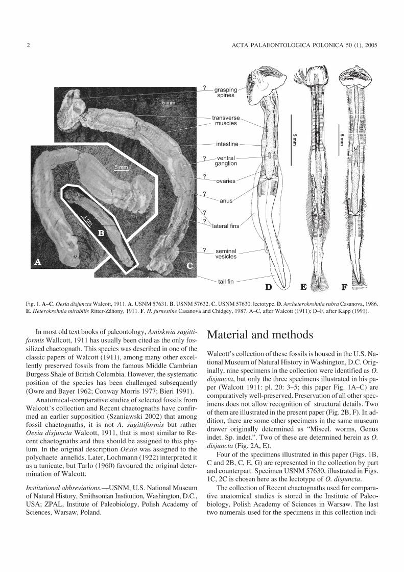
Walcott’s collection of these fossils is housed in the U.S. Na−tional Museum of Natural History in Washington, D.C. Orig−inally, nine specimens in the collection were identified as O.disjuncta, but only the three specimens illustrated in his pa−per (Walcott 1911: pl. 20: 3–5; this paper Fig. 1A–C) arecomparatively well−preserved. Preservation of all other spec−imens does not allow recognition of structural details. Twoof them are illustrated in the present paper (Fig. 2B, F). In ad−dition, there are some other specimens in the same museumdrawer originally determined as “Miscel. worms, Genusindet. Sp. indet.”. Two of these are determined herein as O.disjuncta (Fig. 2A, E).
Four of the specimens illustrated in this paper (Figs. 1B,C and 2B, C, E, G) are represented in the collection by partand counterpart. Specimen USNM 57630, illustrated in Figs.1C, 2C is chosen here as the lectotype of O. disjuncta.
The collection of Recent chaetognaths used for compara−tive anatomical studies is stored in the Institute of Paleo−biology, Polish Academy of Sciences in Warsaw. The lasttwo numerals used for the specimens in this collection indi−
2 ACTA PALAEONTOLOGICA POLONICA 50 (1), 2005
5m
m
5m
m
graspingspines
transversemuscles
intestine
ventralganglion
ovaries
anus
lateral fins
seminalvesicles
tail fin
?
?
?
?
?
?
5 mm
1cm
5 mm
?
Fig. 1. A–C. Oesia disjuncta Walcott, 1911. A. USNM 57631. B. USNM 57632. C. USNM 57630, lectotype. D. Archeterokrohnia rubra Casanova, 1986.E. Heterokrohnia mirabilis Ritter−Záhony, 1911. F. H. furnestine Casanova and Chidgey, 1987. A–C, after Walcott (1911); D–F, after Kapp (1991).

cate the number of the SEM stub and of the specimen on thestub (e.g., 147.5 means specimen no. 5 on the stub no.147).
Studies of the fossils are based on the illustrations pub−lished by Walcott (1911) and on numerous digital photosmade recently using various angles and styles of illumination.
Studies of Recent chaetognaths were based on the richcollection of chaetognaths, gathered during Polish AntarcticExpeditions. About twenty five comparatively large speci−mens of different taxa (Sagitta Quoy and Gaimarad, 1827,
Eukrohnia Ritter−Záhony, 1909, Heterokrohnia, and unde−termined) were selected for comparative anatomical investi−gations. In order to simulate deformations similar to thoseusually observed in soft−bodied fossils preserved in shales,about twenty of the specimens were washed in water and/oralcohol, placed between two pieces of glass (or betweenpieces of paper covered by glass), slightly compressed, andair dried. Some of the specimens before compression werestained for better visibility of the inner structure. The remain−
http://app.pan.pl/acta50/app50−001.pdf
SZANIAWSKI—CAMBRIAN CHAETOGNATHS 3
grasping spines
lateral plate?
Fig. 2. Oesia disjuncta Walcott, 1911. A. USNM 277849. B. USNM 200552, C. The same specimen as on Fig. 1C in slightly different view. D. The samespecimen as on Fig.1A in slightly different view. E.USNM 277842 (E1) and USNM 277841 (E2), part and counterpart of the same specimen. F. USNM203021, whole specimen (F1), and anterior part in higher magnification (F2). G. Anterior part of the counterpart of the specimen illustrated on Fig. 1B. Scalebars 1cm.
der were washed in water, partly dehydrated in alcohol andattached to SEM stubs using sticky, electro conductive tape.Rapid attachment of wet specimens to the sticky tape pro−tected them from strong deformation during drying.
During processing some of the specimens became dam−aged but it was still possible to make some observations andphotograph them under binocular and/or scanning electronmicroscopes.
Results of comparative anatomicalstudiesComparative studies showed numerous close structural simi−larities between Oesia disjuncta and all the experimentallydeformed Recent chaetognaths that are almost certainly notcoincidental.
An anatomical interpretation of O. disjuncta is shown inFig. 1.
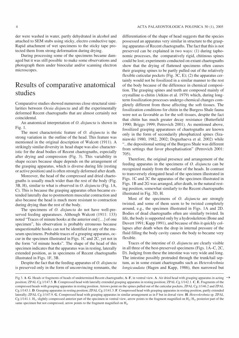
The most characteristic feature of O. disjuncta is thestrong variation in the outline of the head. This feature wasmentioned in the original description of Walcott (1911). Astrikingly similar diversity in head shape was also character−istic for the dead bodies of Recent chaetognaths, especiallyafter drying and compression (Fig. 3). This variability inshape occurs because shape depends on the arrangement ofthe grasping apparatus, which is diverse during life (restingor active position) and is often strongly deformed after death.
Moreover, the head of the compressed and dried chaeto−gnaths is usually much wider than the rest of the body (Fig.3B, H), similar to what is observed in O. disjuncta (Fig. 1A,C). This is because the grasping apparatus often became ex−tended laterally due to postmortem muscular contraction andalso because the head is much more resistant to contractionduring drying than the rest of the body.
The specimens of O. disjuncta do not have well−pre−served feeding apparatuses. Although Walcott (1911: 133)noted “Traces of minute hooks at the anterior end […] of onespecimen”, his observation is probably erroneous becauseunquestionable hooks can not be identified in any of the mu−seum specimens. Probable traces of a grasping apparatus, oc−cur in the specimen illustrated in Figs. 1C and 2C, yet not inthe form “of minute hooks”. The shape of the head of thisspecimen indicates that the apparatus was in resting, laterallyextended position, as in specimens of Recent chaetognathsillustrated in Figs. 1F, 3B.
Despite the fact that the feeding apparatus of O. disjunctais preserved only in the form of unconvincing remnants, the
differentiation of the shape of head suggests that the speciespossessed an apparatus very similar in structure to the grasp−ing apparatus of Recent chaetognaths. The fact that this is notpreserved can be explained in two ways: (1) during tapho−nomic processes, the comparatively rigid, chitinous spinescould be lost; experiments conducted on extant chaetognathsshow that the drying of flattened specimens often causestheir grasping spines to be partly pulled out of the relativelyflexible cuticular pockets (Fig. 3C, E); (2) the apparatus cer−tainly would not be fossilized in a similar manner to the restof the body because of the difference in chemical composi−tion. The grasping spines and teeth are composed mainly ofcrystalline �−chitin (Atkins et al. 1979) which, during long−term fossilization processes undergo chemical changes com−pletely different from those affecting the soft tissues. Thefossilization conditions for chitin in the Burgess Shale faunawere not as favorable as for the soft tissues, despite the factthat chitin has much greater decay resistance (Butterfield1990; Briggs 1999; Petrovich 2001). As mentioned above,fossilized grasping apparatuses of chaetognaths are knownonly in the form of secondarily phosphatized spines (Sza−niawski 1980, 1982, 2002; Doguzhaeva et al. 2002) while“...the depositional setting of the Burgess Shale was differentfrom settings that favor phosphatisation” (Petrovich 2001:705).
Therefore, the original presence and arrangement of thefeeding apparatus in the specimens of O. disjuncta can berecognized mainly from the outline of the heads. In contrastto transversely elongated head of the specimen illustrated inFigs. 1C and 2C the apparatus of the specimen illustrated inFigs. 1B and 2G was arranged, after death, in the natural rest−ing position, somewhat similarly to the Recent chaetognathsillustrated in Fig. 3D, H.
Most of the specimens of O. disjuncta are stronglytwisted, and some of them seem to be twisted completelyaround, e.g., the specimen illustrated in Figs. 1A and 2D.Bodies of dead chaetognaths often are similarly twisted. Inlife, the body is supported only by a hydroskeleton (Bone andDuvert 1991; Kapp 1991), and because of this it quickly col−lapses after death when the drop in internal pressure of thefluid filling the body cavity causes the body to became veryflexible.
Traces of the intestine of O. disjuncta are clearly visiblein all three of the best−preserved specimens (Figs. 1A–C, 2C,D). Judging from these the intestine was very wide and long.The intestine possibly protruded through the trunk/tail sep−tum, as in some extant chaetognaths such as Heterokrohnialongicaudata (Hagen and Kapp, 1986), then narrowed but
4 ACTA PALAEONTOLOGICA POLONICA 50 (1), 2005
Fig 3. A–G. Heads or fragments of heads of undetermined Recent chaetognaths; A–F, in ventral view. A. Air dried head with grasping apparatus in actingposition; ZPAL Cg.1/147.5. B. Compressed head with laterally extended grasping apparatus in resting position; ZPAL Cg.1/142.1. C, E. Fragments of thecompressed heads with grasping apparatus in resting position. Arrows point on the spines pulled out of the cuticular pockets, ZPAL Cg.1/146.2 and ZPALCg.1/143.1. D. Grasping apparatus in resting position; ZPAL Cg.1/141.3. F. Compressed head with grasping apparatus in resting position, partly extendedlaterally; ZPAL Cg.1/147.9. G. Compressed head with grasping apparatus in similar arrangement as in F but in dorsal view. H. Heterokrohnia sp. ZPALCg.1/141.1. H1, slightly compressed anterior part of the specimen in ventral view, arrow points to the fragment magnified on H2; H3, posterior part of thesame specimen but not compressed, arrow points to the fragment magnified on H4.
�
http://app.pan.pl/acta50/app50−001.pdf
SZANIAWSKI—CAMBRIAN CHAETOGNATHS 5

continued far to the posterior.2 The intestine of Recentchaetognaths is also very prominent.
The body of O. disjuncta, or at least part of it, is cross stri−ated (Figs. 1A, C, 2C, D). This striation was interpreted byWalcott (1911) as segmentation. However, the unclear preser−vation suggests that it reflects a rather delicate, most probablyinternal structure. Similar striation occurs in modern chaeto−gnath muscles and is easily visible in their dried bodies (Figs.1D–F, 3H). Striation is especially well−developed in taxa pos−sessing not only longitudinal but also transverse muscles.Such musculature occurs in those genera which, contrary tothe majority of chaetognaths, lead benthic (Paraspadella Sal−vini Plawen, 1987; see Bowman and Bieri 1989), epibenthic(Spadella Langerhans, 1880) or hyperbenthic (Archetero−krohnia, Heterokrohnia, and some Eukrohnia) modes of life.According to some authors (Tokioka 1965; Kassatkina 1980;Casanova 1986; 1996; Casanova and Duvert 1996, 2002),these are evolutionarily primitive forms.
The caudal fin of O. disjuncta, like that of chaetognaths,is horizontally oriented and has a similar shape and size.The fin is comparatively well−preserved in the specimens il−lustrated in Figs. 1C, 2C, F. In other specimens it is stronglydeformed (Fig. 2A, B, E) or insufficiently differentiated(Figs. 1A, B, 2D).
Lateral fins are not well preserved in any of the Oesiaspecimens. Probable remnants of them occur only in thespecimen illustrated in Figs. 1C, 2C. The poor preservationof the fins can be explained by the experiments made on ex−tant chaetognaths, which show that lateral fins are usually
preserved much more poorly than caudal fins. Lateral finsusually become damaged or “glued” to the body and invisi−ble during processing and drying of a specimen. The fins ofchaetognaths are very flexible and delicate and in some spe−cies are completely rayless. It is obvious that their fossiliza−tion cannot be easily accomplished.
Probable seminal vesicles are comparatively well pre−served in the lectotype only (Fig. 1). Interpretation of suchdelicate structures as the ventral ganglion, ovaries and anus,based on the same specimen, is even less certain.
Oesia is compared here to the Recent hyperbenthic gen−era Archeterokrohnia and Heterokrohnia because only thesegenera have transverse muscles developed in both trunk andtail and therefore are considered to be phylogenetically mostprimitive (Casanova 1986; Casanova and Duvert 2002).Benthic genera and some species of the partly hyperbenthicgenus Eukrohnia, possess these muscles in the trunk only,while the majority of chaetognaths lack them altogether.
The hypothesis that Heterokrohnia is primitive is alsosupported by paleontology. Recently described phosphatisedCarboniferous chaetognath grasping spines (Doguzhaeva etal. 2002) are denticulated in a manner similar to the spines ofjuvenile Heterokrohnia longidentata Kapp and Hagen, 1985.In both the fossil and Recent forms the denticles are inclinedtowards the tips of the spines. Among other Recent chaeto−gnaths, similar denticulation of grasping spines occurs onlyin some species of juvenile Eukrohnia, which is probablyclosely related to Heterokrohnia (Casanova and Duvert1996). Furthermore, species of Serratosagitta Tokioka, 1965
6 ACTA PALAEONTOLOGICA POLONICA 50 (1), 2005
Fig. 4. A. Sagitta sp., compressed specimen in transmitted light, dorsal view; ZPAL Cg.1/146. Whole specimen (A1); magnification of the posterior part(A2); magnification of the head (A3). B. ?Sagitta sp., stained and compressed specimen in reflected light, dorsal view; ZPAL Cg.1/146.1.Whole specimen(B1); magnification of the anterior part (B2).
2 According to some authors “Chaetognaths are not tripartite as sometimes suggested” (Bone et al. 1991: 3) and the caudal, transversal septum “... is clearlya secondary separation” (Ghirardelli 1995: 168).
possess spines with denticulation but which are inclined to−ward the base, whereas the spines in the vast majority ofchaetognths are not denticulated.
Obviously there are some differences between Oesia andRecent chaetognaths. The tail of Recent forms gradually nar−rows posteriorly, a feature that cannot be seen in Oesia.However, in some Recent chaetognaths, e.g., FlaccisagittaTokioka, 1965 and Parasagitta Tokioka, 1965, the tail isvery short and does not show a pronounced narrowing. Mostspecies of Recent forms are smaller in size than Oesia, butadult forms of some species are larger. Similarly, most of theRecent forms are comparatively narrow, but some represen−tatives of benthic species are wide. The seminal vesicles ofmost of Recent chaetognaths is small compared to the sup−posed vesicles of Oesia, but in some benthic species (e.g.,Spadella gaetanoi Alvariño, 1978 and Paraspadella schizo−ptera (Conant, 1895)) the vesicles are of a similar size tothose of O. disjuncta.
Archeterokrohnia and Heterokrohnia lead a hyperbenthicmode of life in deep waters (usually 1000–4000m), while mostof the fauna preserved in the Phyllopod Bed of the BurgessShale belong to a benthic community that inhabited moder−ately deep water (Conway Morris 1986; Petrovich 2001).Thus it is very probable that the morphological differences be−tween the fossil and Recent forms compared herein are the re−sults not only of evolutionary development but also of differ−ent environments and modes of life.
It is interesting to note that Walcott when studying Oesiadisjuncta (1911: 133) gained the impression that the species“[…] lived in an irregular tube that was so thin the annelidshows through it”. In fact Recent chaetognaths have a tube−like shape and their body is covered by a thin and translucentepithelium.
AcknowledgementsI am thankful to Dr. Jarosław Stolarski (Institute of Paleobiology, War−saw), for making many digital photos of specimens from the BurgessShale fauna during his visits to the Smithsonian Institution, Washing−ton, D.C. Thanks are also due to Drs. John Repetski (U.S. GeologicalSurvey) and Paul D. Taylor (The Natural History Museum, London) forlinguistic correction and usefull suggestions, as well as to Dr. KrzysztofJażdżewski (University of Łódź, Poland) for providing a large collec−tion of the Recent chaetognaths from the Antarctic region. Finally I amgrateful to all the reviewers, Drs. Stefan Bengtson (Swedish Museum ofNatural History), Elvezio Ghirardelli (University of Trieste, Italy), andMaximilian J. Telford (University College London) for useful andconstructive comments.
ReferencesAtkins, E.D.T., Dlugosz, J. and Foord, S. 1979. Electron diffraction and
electron microscopy of crystalline �−chitin from the grasping spines ofthe marine worm Sagitta. International Journal of Biological Macro−molecules 1: 29–32.
Alvariño, A. 1981. Spadella gaetanoi, a new benthic chaetognath from Ha−waii. Proceedings of the Biological Society of Washington 91: 650–657.
Bieri, R. 1991. Systematics of the Chaetognatha. In: Q. Bone, H. Kapp, andA.C. Pierrot−Bults (eds.), The Biology of Chaetognaths, 122–136. Ox−ford University Press, Oxford.
Bone, Q. and Duvert, M. 1991. Locomotion and buoyancy. In: Q. Bone, H.Kapp, and A.C. Pierrot−Bults (eds.), The Biology of Chaetognaths,32–44. Oxford University Press, Oxford.
Briggs, D.E.G. 1999. Molecular taphonomy of animal and plant cuticles: se−lective preservation and diagenesis. Philosophical Transactions of theRoyal Society London B 354: 7–17.
Butterfield, N.J. 1990. Organic preservation of non−mineralizing organismsand the taphonomy of the Burgess Shale. Palebiology 16: 272–286.
Casanova, J.P. 1986. Archeterokrohnia rubra n. gen., n. sp., nouveauChaetognathe abyssal de l’Atlantique nord−africain: description et posi−tion systématique, hypothèse phylogénétique. Bulletin du Muséum na−tional d’Histoire naturelle, Paris (4 sér.), section A 8 (1): 185–194.
Casanova, J.P. 1996. A new genus and species of deep−benthic chaetognathfrom the Atlantic: a probable link between the families Heterokrohni−idae and Spadellidae. Journal of Natural History 30: 1239–1245
Casanova, J.P. and Duvert, M. 1996. Biodiversity and evolutionary trends inthe phylum Chaetognatha. Buletin de la Societé Zoologique de France12: 77–80.
Casanova, J.P. and Duvert, M. 2002. Comparative studies and evolution ofmuscles in chaetognaths. Marine Biology 141: 925–938.
Casanova, J.P. and Chidgey K.C. 1987. Une nouvelle espèce d’Hetero−krohnia (Chaetognathe) des campagnes du ‘Discovery’ dans l’Atlanti−que nordoriental. Bulletin du Muséum national d’Histoire naturelle.Paris (4 sér.) 9: 879–885.
Chen, J.Y. and Huang, D.Y. 2002. A possible Lower Cambrian Chaetognath(Arrow Worm). Science 298: 187.
Conant, F.S. 1895. Description on two new chaetognaths. Johns HopkinsUniversity Circular 14: 119.
Conway−Morris, S. 1977. A description of the Middle Cambrian wormAmiskwia sagittiformis, Walcott from the Burgess Shale of British Co−lumbia. Palaeontologische Zeitschrift 51: 271–287.
Conway−Morris, S. 1986. The community structure of the Middle CambrianPhyllopod Bed (Burgess Shale). Palaeontology 29: 423–467.
Darwin, C. 1844. Observations on the structure and propagation of the genusSagitta. Annales and Magazine of Natural History, London 13: 1–6.
Doguzhaeva, L.A, Mutvei, H., and Mapes, R.H. 2002. Chaetognath grasp−ing spines from the Upper Mississipian of Arkansas (USA). ActaPalaeontologica Polonica 47: 421–430.
Furnestin, M.L. 1965. Variations morphologiques des crochets au cours dudéveloppement dans le genre Eukrohnia. Revue des Travaux de l’Institutdes Pèche de Castiglione 4: 275–317.
Ghirardelli, E. 1994. The state of knowledge on Chaetognaths. In: R. Argano,C. Cirotto, E. Grassi Milano, and L. Mastrolia (eds.), Contributions to Ani−mal Biology, 237–244. Halocynthia Association, Palermo.
Ghirardelli, E. 1995. Chaetognaths: two unsolved problems: the coelom andtheir affinities. In: G. Lanzavecchia, R. Valvassori, and M.Z. CandiaCarnevali (eds.), Body Cavities: Function and Phylogeny, Selected Sym−posia and Monographs U.Z.I. 8, 167–185. Mucchi, Modena.
Hagen, W. and Kapp, H. 1986. Heterokrohnia longicaudata, a new speciesof Chaetognatha from Antarctic waters. Polar Biology 5: 181–183.
Halanych, K.M. 1996. Testing hypotheses of Chaetognath origins: Longbranches revealed by 18S ribosomal DNA. Systematic Biology 45:223–246.
Helfenbein, K.G., Fourcade, H.M., Vanjani, R.G., and Boore, J.L. 2004.The mitochondrial genome of Paraspadella getoi is highly reduced andreveals that chaetognaths are sister group to protostomes. Proceedingsof the National Academy of Sciences of the united States of America 101(29): 10639–10643.
Kapp, H. 1991. Morphology and anatomy. In: Q. Bone, H. Kapp, and A.C.
http://app.pan.pl/acta50/app50−001.pdf
SZANIAWSKI—CAMBRIAN CHAETOGNATHS 7

Pierrot−Bults (eds.), The Biology of Chaetognaths, 5–17. Oxford Uni−versity Press, Oxford.
Kapp, H. and Hagen, W. 1985. Two new species of Heterokrohnia (Chaeto−gnatha) from Antarctic waters. Polar Biology 4: 53–59.
Kassatkina, A.P. 1980. A new genus Tokiokaispadella and its position in thesystem of Chaetognatha [in Russian]. Zoologičeskij žurnal 59: 1257–1260.
Langerhans, P. 1880. Die Wurmfauna von Madeira. Zeitchrift für wissen−schaftliche Zoologie 34: 1–136.
Lochmann, N.H. 1922. Oesia disjuncta Walcott, eine Appendicularie ausdem Kambrium. Mitteilungen aus dem Zoologischen Staatsinstitut inHamburg 38: 69–75.
Owre, H.B. and Bayer, F.M. 1962. The systematic position of the MiddleCambrian fossil Amiskwia Walcott. Paleontological Notes 36: 1361–1393.
Papillon, D., Perez, Y., Caubit, X., and Le Parco, Y. 2004. Identification ofchaetognaths as protostomes is supported by the analysis of their mito−chondrial genome. Molecular Biology and Evolution doi:10.1093/molbev/msh229.
Petrovich, R. 2001. Mechanisms of fossilization of the soft−bodied andlightly armored faunas of the Burgess Shale and of some other classicallocalities. American Journal of Science 301: 683–726.
Quoy, J.R.C. and Gaimarad, P. 1827. Observations zoologiques faites de“l'Astrolabe” en Mai 1826, dans la Detroit de Gibraltar. Annales de Sci−ence naturelle (Zoologie) 10: 5–239.
Ritter−Záhony, R. 1909. Die Chätognathen der Gazelle Expedition. Zoolo−gischer Anzeiger 34: 783−793.
Ritter−Záhony, R. 1911. Revision der Chaetognathen. Deutsche SüdpolarExpedition 1901–3, 12 (1): 1–72.
Szaniawski, H. 1980. Fused clusters of paraconodonts. In: H.P. Schönlaub(ed.), Abstract for the 2nd European Conodont Symposium (ECOS II).Abhandlungen der Geologischen Bundesanstalt 35: 211.
Szaniawski, H. 1982. Chaetognath gasping spines recognized among Cam−brian protoconodonts. Journal of Paleontology 56: 806–810.
Szaniawski, H. 2002. New evidence for the protoconodont origin of chaeto−gnaths. Acta Palaeontologica Polonica 47: 405–419.
Tarlo, L.B. 1960. The invertebrate origins of the vertebrates. Report of the In−ternational Geological Congress, XXI Session, Norden, 1960, Part XXII,International Paleontological Union. Copenhagen 1960: 113–123.
Telford, M.J. 2004. Affinity for arrow worms. Nature 431: 254–256.Telford, M.J. and Holland, P.W.H. 1993. The Phylogenetic Affinities of the
Chaetognaths: A Molecular Analysis. Molecular Biological Evolution10: 660–676.
Telford, M.J. and Holland, P.W.H. 1997. Evolution of 28S Ribosomal DNAin Chaetognaths: Duplicate Genes and Molecular Phylogeny. Journalof Molecular Evolution 44: 135–144.
Tokioka, T. 1965. The taxonomical outline of chaetognaths. Publications ofthe Seto Marine Biological Laboratory 12: 335–357.
Wada, H. and Satoh, N. 1994. Details of the evolutionary history from inver−tebrates to vertebrates, as deduced from the sequences of 18S rDNA.Proceedings of the National Academy of Sciences of the United States ofAmerica 91 (5): 1801–1804.
Walcott, C.D. 1911. Middle Cambrian annelids. Smithsonian Miscella−neous Collections 57 (2): 109–144.
8 ACTA PALAEONTOLOGICA POLONICA 50 (1), 2005