CALCULOUS CHOLECYSTITISrepository-tnmgrmu.ac.in/7827/1/220100506sivakumar.pdfACKNOWLEDGEMENT I thank...
69
DISSERTATION ON CALCULOUS CHOLECYSTITIS M.S. DEGREE EXAMINATION BRANCH-I GENERAL SURGERY THANJAVUR MEDICAL COLLEGE THE TAMILNADU DR.MGR MEDICAL UNIVERSITY CHENNAI
Transcript of CALCULOUS CHOLECYSTITISrepository-tnmgrmu.ac.in/7827/1/220100506sivakumar.pdfACKNOWLEDGEMENT I thank...
DISSERTATION ON
CALCULOUS CHOLECYSTITIS
M.S. DEGREE EXAMINATION
BRANCH-I
GENERAL SURGERY
THANJAVUR MEDICAL COLLEGE
THE TAMILNADU DR.MGR MEDICAL UNIVERSITY
CHENNAI
CERTIFICATE
This is to certify that this dissertation entitled “CALCULOUS
CHOLECYSTITIS” bonafide record work done by Dr. S SIVAKUMAR
submitted as partial fulfillment for the requirements of M.S. Degree
Examinations Branch I, General Surgery, SEPTEMBER 2006.
Prof. Dr.A.DEVAKUMARI, M.S., Prof.Dr.V.Thirugnanam,M.S.,M.ch.,
Addl. Prof. And Unit Chief, Professor & Head of the Department,
Department of General Surgery, Department of General Surgery,
Thanjavur Medical College, Thanjavur Medical College,
Thanjavur. Thanjavur.
ACKNOWLEDGEMENT I thank to God Almighty without whose help, this work would not have been
possible. I thank profusely The Dean, Thanjavur Medical College Hospital for permitting
me to conduct this study and to use the materials of the Hospital.
I have great pleasure in expressing my deep sense of gratitude to my unit chief.
Prof. Dr. A. Devakumari, M.S., Thanjavur Medical College, Thanjavur., for suggesting
the topic, providing able guidance and constructive criticism. Her interest, special
guidance, constant encouragement and patience contributed much to the making of this
work successfully.
I am very much indebted to Prof. V. Thirugnanam, M.S., M.Ch., Head of the
Department of surgery, Thanjavur Medical College, Thanjavur for providing all
departmental facilities with kind encouragement throughout the period of my work.
I shall be failing in my duty if I do not acknowledge my gratitude to the unit
chiefs of the surgical department of Thanjavur Medical College and Dr. Umadevi,
without whose valuable material and constant help the completion of this dissertation
work would not have been possible.
The kind and valuable criticism by Dr. G.Ravikumar, M.S., M.C.H.,
Dr.Shanthini, M.S., and Dr.M.Elangovan, M.S., Asst. Surgeons, Thanjavur Medical
College, Thanjavur was immensely helpful in completing the dissertation. I thank the
medical record section and the library staff for their help.
I also take this opportunity to thank the professor of pathology,microbiology and bio-
chemistry for their help in conducting this study
INDEX
S.No PARTICULARS
Page No
1. INTRODUCTION 01
2. SURGICAL ANATOMY 02
3. SURGICAL PHYSIOLOGY 06
4. AETIO PATHOGENESIS OF
GALLSTONES
09
5. DIAGNOSIS 19
6. INVESTIGATIONS 22
7. MANAGEMENT 28
8. AIMS OF THIS STUDY 37
9. MATERIALS AND METHODS 38
10. OBSERVATIONS 39
11. DISCUSSION 41
12. SUMMARY AND CONCLUSIONS 48
13. BIBLIOGRAPHY 50
14. PROFORMA 51
15. MASTER CHART 55
INTRODUCTION
Gall Bladder is a pear shaped saccular organ, which stores bile, becomes a
place for formation and growth of Gallstones. The Gallstone disease is more
common in Western world Today the Incidence of cholelithiasis is increasing
considerably in India, Possibly due to change in the dietary habits. Which is
becoming westernized and the life style which is changing. In India, North India
shows 7 times more incidence than that in South India.
The operations on biliary tree and gall bladder rank next only to Hernia
repair and appendicectomy in Northern India. In Southern India, picture is not
clear.
Prevalence of Cholelithasis, in Indian males and females is estimated as 4%
and 6% respectively The exact incidence of cholelithasis is not known.
Prevalence of cholelithasis shows Improved detection due to Imaging modalities
particularly ultrasonogram.
Because of the Extensive studies of Etiology of gallstones and better
understanding of the Pathogenesis in the past two decades, the management has
become more appropriate and effective.
Proliferating research on the minimal invasive surgery especially after 1988
with the advent of Laparoscopic surgeries, Percutaneous removal of stones and
Extracorpereal shock wave lithotripsy has greatly motivated patients for
undergoing early and effective management.
SURGICAL ANATOMY
GALL BLADDER
It is a pear shaped organ 7-10 cm long with a capacity of 30- 50 ml. It is
located in Gall Bladder fossa found at the junction of quadrate Lobe (Segment IV)
and the Right lobe of liver along the line of rex, and is enclosed within its
peritoneal sheath on three sides.
Gall bladder can be divided into Fundus, Body, Infundibulum and neck.
FUNDUS
Projects slightly beyond the free margin of the liver, opposite the upper end
of linea semilunaris. A partial folding of fundus may result in ‘Phyrgian Cap’
deformity. It was suggested that such gall bladders are at higher risk for lithiasis,
but this has not been confirmed.
BODY
Occupies the Gallbladder fossa, covered by peritoneum on 3 surfaces.
Sometimes GB is suspended in a mesentry, off the Inferior surface of the liver –
wandering Gallbladder or rarely embedded deep inside the liver parenchyma
Intrahepatic GB.
Occasionally, several anomalous peritoneal folds from the GB to the
duodenum, colon, or stomach is seen in that order of frequency and are associated
with the pathway of a large gallstone ulcerating from Gallbladder into the
Intestinal tract.
INFUNDIBULUM
It is the angulated posterior portion of the body between the neck and the
point of entrance of Cystic artery. It may show a eccentric bulging on its medial
aspect, called Hartmann’s pouch, and is often associated with Impaction of stone.
NECK
Neck curves up and forward and then sharping back and downward
forming an S to become the cystic duct Mucosal lining shows spiral folds give rise
to ‘spiral valve of heister’ and may interfere with the passage of instrument.
BILE DUCTS
The right and left lobes of liver are drained by ducts originating as bile
canaliculi in the lobules and these canaliculi empty into the canals of Hering in the
Interlobular triads and these canals are collected into ducts, and finally outside the
liver, the Right and left hepatic ducts. Right hepatic duct is formed by the union of
the anterior and posterior segment ducts of the right Lobe of the liver at porta
hepatis. The average length of RHD is 0.9 cm.
Left hepatic duct is formed by the Union of medial and lateral segment
ducts of the left lobe of the liver and average length is 1.7 cm – common hepatic
duct is formed by union of the right and left hepatic duct and average diameter is
about 0.4 cm. Its lower end is defined as its junction with cystic duct.
CYSTIC DUCT
Cystic duct joins the hepatic duct at an angle of about 40o. The length of
cystic duct and the manner in which it joins the hepatic duct vary. Obstruction of
cystic duct leads to hydrops of Gallbladder, contains white bile, composed only of
mucus.
COMMON BILE DUCT
Begins at the union of cystic duct and common hepatic duct and ends at the
papilla of vater in the second part of the duodenun average diameter is 6mm.
CBD is divided into 4 portions
Supraduodenal, retroduodenal, pancreatic, Intramural.
Supraduodenal portion lies in the right free border of lesser onentum, to the
right of the hepatic artery and anterior to the portal vein. Retroduodenal portion
descends behind the 1st part of the duodenum and the pancreatic portion tunnel the
gland substance. Intramural portion takes an oblique path averaging 1.5 cm
though the duodenal wall and receives main pancreatic duct inferiorly. And both
of them end in the ampulla of vater on the posteromedial wall of the second part of
the duodenum.
BLOOD SUPPLY
Cystic artery arises usually from the right hepatic artery, reaches the Gall
bladder behind the common hepatic duct and traverses through the hepato cystic
triangle of calot and branches into Anterior and posterior branch. The Extrahepatic
bile ducts are supplied by right hepatic artery above and gastroduodenal artery
below, with major trunks running along the medial and lateral walls of CBD,
sometimes referred to as 3 o’ clock and 9’o clock position.
ANOMALIES
It should be noted that considerable variations to the above description may
exist. Those which occur most commonly are shown in figure. A knowledge of
such anomalies is of greatest importance to the surgeon, for failure to recognize
them at operation may lead to disaster.
SURGICAL PHYSIOLOGY
BILIARY SECRETION Bile is secreted continuously by the liver cells (hepatocytes) into the biliary
Canaliculi. Daily secretion is 500 – 1000ml per day. Bile is secreted at a pressure
of 150 – 250mm of water. If obstructions occurs, liver continues to secrete upto
the pressure of 300mm of water, there after secretion ceases.
Hepatic bile is slightly alkaline and Gallbladder bile is more acidic than
Hepatic bile.
The primary bile salts are cholate and chenodeoxycholate, conjugated with
taurine and glycine and excreted into the bile. 98% is reabsorbed by enterohepatic
circulation. In the intestine Gut bacteria deconjugates the primary bile salts and
forms secondary bile salts deoxycholate and lithocholate.
Two important functions of Bile salts are
(1) Formation of water soluble complexes with cholesterol, fatty acids and fat
soluble vitamins and their absorption.
(2) Reduction of surface tension and emulsification of fat.
Bile salts are powerful cholerectic which increases hepatic bile production.
Cholesterol and Phospholipids synthesized in the liver are the principal
lipids found. In bile. The colour of the bile is due to the presence of pigment
bilirubin diglucoronide, which is the metabolic product of breakdown of
Hemoglobin in the reticuloendothelial system. In the intestine, bacteria
converts it into urobilinogen which is absorbed and excreted in urine.
FUNCTIONS OF GALLBLADDER
(1) Stores the bile and concentrates it.
(2) Periodically releases bile by contracting in response to meal.
(3) Acidification of hepatic bile.
(4) Production of glyco proteins.
Control of bile flow It is under control of neurogenic, humoral, and chemical stimuli.
Vagal stimulation increases the secretion of bile, while splanchnic nerve
stimulation results in decreased bile flow.
Hydrochloric acid, partly digested proteins and fatty acids in the duodenum
stimulate the release of secretin from the duodenum that in turn increases bile
production and bile flow.
Cholecystokinin also increases the hepatic secretion of bile.
Other substances, which have effect on biliary secretion are VIP, caerulin
and GASTRIN.
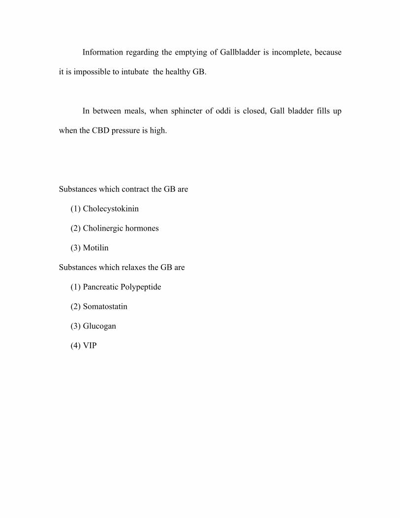
Information regarding the emptying of Gallbladder is incomplete, because
it is impossible to intubate the healthy GB.
In between meals, when sphincter of oddi is closed, Gall bladder fills up
when the CBD pressure is high.
Substances which contract the GB are
(1) Cholecystokinin
(2) Cholinergic hormones
(3) Motilin
Substances which relaxes the GB are
(1) Pancreatic Polypeptide
(2) Somatostatin
(3) Glucogan
(4) VIP
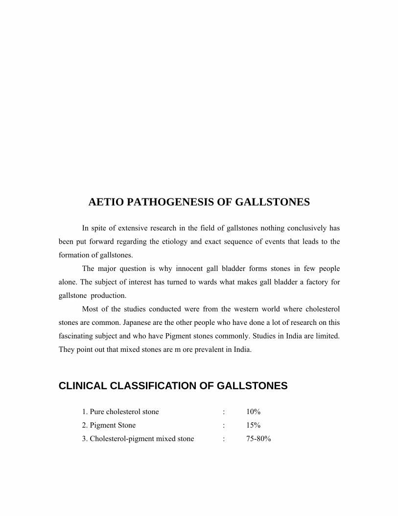
AETIO PATHOGENESIS OF GALLSTONES In spite of extensive research in the field of gallstones nothing conclusively has
been put forward regarding the etiology and exact sequence of events that leads to the
formation of gallstones.
The major question is why innocent gall bladder forms stones in few people
alone. The subject of interest has turned to wards what makes gall bladder a factory for
gallstone production.
Most of the studies conducted were from the western world where cholesterol
stones are common. Japanese are the other people who have done a lot of research on this
fascinating subject and who have Pigment stones commonly. Studies in India are limited.
They point out that mixed stones are m ore prevalent in India.
CLINICAL CLASSIFICATION OF GALLSTONES
1. Pure cholesterol stone : 10%
2. Pigment Stone : 15%
3. Cholesterol-pigment mixed stone : 75-80%
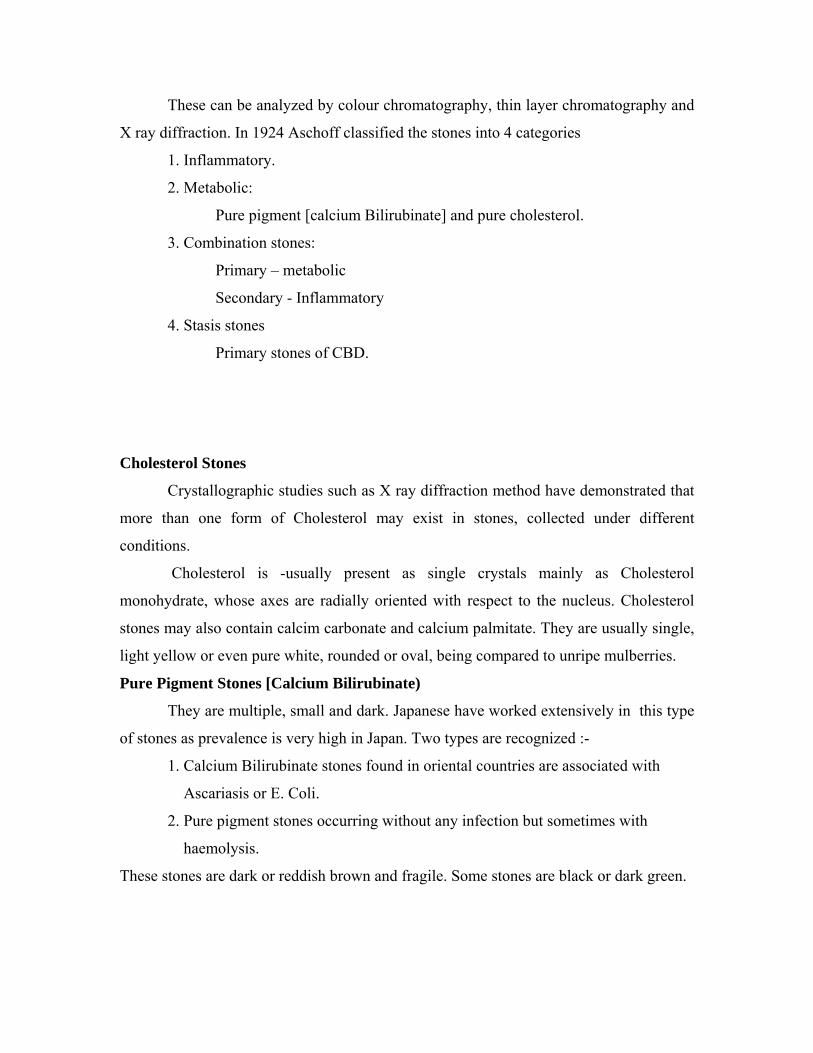
These can be analyzed by colour chromatography, thin layer chromatography and
X ray diffraction. In 1924 Aschoff classified the stones into 4 categories
1. Inflammatory.
2. Metabolic:
Pure pigment [calcium Bilirubinate] and pure cholesterol.
3. Combination stones:
Primary – metabolic
Secondary - Inflammatory
4. Stasis stones
Primary stones of CBD.
Cholesterol Stones
Crystallographic studies such as X ray diffraction method have demonstrated that
more than one form of Cholesterol may exist in stones, collected under different
conditions.
Cholesterol is -usually present as single crystals mainly as Cholesterol
monohydrate, whose axes are radially oriented with respect to the nucleus. Cholesterol
stones may also contain calcim carbonate and calcium palmitate. They are usually single,
light yellow or even pure white, rounded or oval, being compared to unripe mulberries.
Pure Pigment Stones [Calcium Bilirubinate)
They are multiple, small and dark. Japanese have worked extensively in this type
of stones as prevalence is very high in Japan. Two types are recognized :-
1. Calcium Bilirubinate stones found in oriental countries are associated with
Ascariasis or E. Coli.
2. Pure pigment stones occurring without any infection but sometimes with
haemolysis.
These stones are dark or reddish brown and fragile. Some stones are black or dark green.
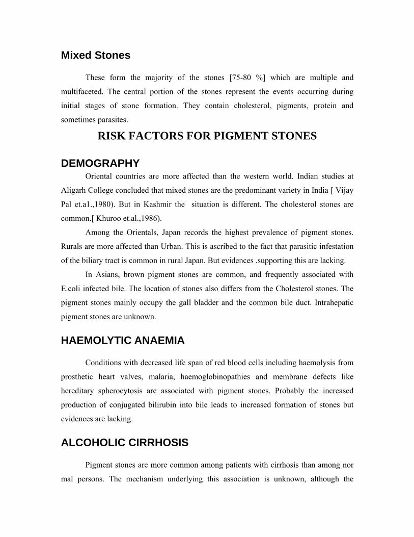
Mixed Stones These form the majority of the stones [75-80 %] which are multiple and
multifaceted. The central portion of the stones represent the events occurring during
initial stages of stone formation. They contain cholesterol, pigments, protein and
sometimes parasites.
RISK FACTORS FOR PIGMENT STONES
DEMOGRAPHY Oriental countries are more affected than the western world. Indian studies at
Aligarh College concluded that mixed stones are the predominant variety in India [ Vijay
Pal et.a1.,1980). But in Kashmir the situation is different. The cholesterol stones are
common.[ Khuroo et.al.,1986).
Among the Orientals, Japan records the highest prevalence of pigment stones.
Rurals are more affected than Urban. This is ascribed to the fact that parasitic infestation
of the biliary tract is common in rural Japan. But evidences .supporting this are lacking.
In Asians, brown pigment stones are common, and frequently associated with
E.coli infected bile. The location of stones also differs from the Cholesterol stones. The
pigment stones mainly occupy the gall bladder and the common bile duct. Intrahepatic
pigment stones are unknown.
HAEMOLYTIC ANAEMIA Conditions with decreased life span of red blood cells including haemolysis from
prosthetic heart valves, malaria, haemoglobinopathies and membrane defects like
hereditary spherocytosis are associated with pigment stones. Probably the increased
production of conjugated bilirubin into bile leads to increased formation of stones but
evidences are lacking.
ALCOHOLIC CIRRHOSIS Pigment stones are more common among patients with cirrhosis than among nor
mal persons. The mechanism underlying this association is unknown, although the
hypersplenism and mild haemolytic anaemia that often accompany cirrhosis might be
suspected of contributing to this increased incidence of gallstones.
INFECTED BILE This is the oldest theory of gallstone formation.
No infection – No stone.
The Naunya's theory has got general support. Hence the nidus of stones is formed
not only by bacteria, but also by inflammatory exudate or cellular exfoliation, parasites,
and ova.
Moynihan has aptly described "Gallstone is a tomb stone erected to the memory
of organism within it".
Bile bathing the gallstones is infected in Japanese. The most common infecting
organism is Escherichia coli, a producer of Beta Glucuronidase which increases bile
saturation by increasing unconjugated water insoluble bilirbin47.
Addition of glucuronidase, to bile in vitro resulted in the precipitation of calcium
bilirubinate. D-glucuronic acid, an inhibitor of glucuronidase prevented the formation of
calcium bilirubinate. Mechanisms other than the deconjugation of bilirubin may also be
involved in the association of the pigment stones with biliary infection. Ascaris
lumbricoides, Round worm eggs are effective nucleating agents for the precipitation of
calcium bilirubinate in vitro and may play similar part in vivo. Over half of the stones
examined in a large series in Japan showed ova of Ascaris lumbricoides. Another
report from Vietnam showed this that roundworm eggs were found in 70% of gallstones
there.
Parasitic infestation causes inflammation of the gall bladder as well as local
chemical changes favourable to the precipitation of calcium salts.
Inflammation does the following
1. Reduces the gall bladder motility.
2. Distorts the intrahepatic bileducts.
3. Interferes with the concentrating ability of the gall bladder and impairs the cholesterol
dissolving capacity of the gall bladder bile.
AGE Like that of Cholesterol stones, the frequency of pigment stones increases with
age. Predominantly seen during the 5th to 7th decade. Before the 1st decade, pigments
stones have been rarely reported in cases of congenital haemolytic diseases.
SEX According to various Western texts, femaleness is not a risk factor for the pigment
stone Indian studies show increased incidence in female sex. [Vijaya Pal et.a1., 1980 ;
Gupta, 1967]
OBESITY Has no definite role in the pigment stone formation. Pancreatitis, Total paraenteral
nutrition and the advanced primary hyperparathyroidism are associated with pigment
stones.
RISK FACTORS FOR CHOLESTEROL STONES
DEMOGRAPHY Rate appears to be the highest in the Scandinavian countries and Northern Europe
while North and South America have higher incidence. Sub sahara and Asia reports very
low incidence .Puma tribes of Arizona has the highest prevalence around 70 % due to its
biological disposition to formation of the gallstones.
INDIAN SITUATION The prevalence of cholesterol Stones is higher in North-lndia.Kashmir in
particular has the highest prevalence of the cholesterol stones which is comparable to
Western Countries.
AGE AND SEX
The greatest incidence occurs between the 5th and 8th decade Incidence is rare
below 20 years old. In females Gallstones tend to occur more than in males, irrespective
of the age, race etc., After puberty the ratio between Females to males is 3:1 to 4:1 Why
females are affected more? Possible hypothesis are
1.Estrogen and its effects
2. Progestrone and its effects.
ESTROGEN
Exogenous Several studies have confirmed that an association between gall stone and use of
exogenous estrogens, whether as oral contraceptives, post menopausal estrogen
replacement or estrogen administered to men.
The possible mechanisms are explained as follows
1. Decreased chenodeoxycholic acid
2. Increased Cholesterol saturation
3. Increased Cholesterol secretion
4. Cholestasis occurring with estrogens
Endogenous A definitely higher prevalence of gall stones among the females are documented
in many studies through out the world. This sex difference appears to be being around the
age of puberty and disappears around menopause
Like oral contraceptives, endogenous estrogen also reduces bile acid pool and increases
cholesterol secretion and the saturation thereby increasing gall stone
formation. Multiparity also shows an increased incidence of gall stones.
PROGESTERONE Saturates bile
Relaxes smooth muscle
Impaired gall bladder emptying
All may predipose to gallstone formation.
FAMLIY HISTORY
Only first degree relatives have two fold risk while others studied showed no
relationship. Siblings have a higher incidence of gall stone disease.
PARITY With increasing parity, the gall stones are more common in young women
probably due to repeated attacks on gall bladder by altered physiology of estrogen or
progesterone on the biliary composition and smooth muscle function of the bilary
apparatus.
OBESITY In untreated obesity, Hepatobiliary tract disease is very much prevalent. The liver
plays a key role in hyperlipidemia Predisposition to the gallstone formation can be
attributed to increased biliary cholesterol secretion in concert with changed nucleating
factors and altered motility Patient who tries to reduce weight by very low calorie diets,
hashigh risk of the gall stone formation .
A large study conducted in obese people [1006 samples] shows in men and wome
n of the 5th decade with obesity an increased. incidence of gallstones -1.7% and 1.8 %
respectively. This is not very high when compared with that in the same age group
without obesity. In these obese patients, Triglycerides were found to be high. Based on
these observations, various mechanisms of gall stone formation in obese individuals are
postulated.
1] Increased saturation of bile in obese individuals due to excessive biliary
secretion of cholesterol.
2] Cholesterol synthesis is related to H M G CO A reductase enzyme.
H M G C 0 A reductase production is related to plasma insulin which is higher in obese
persons and high fat intake also increases this enzyme.
Since none of investigation modalities available can exclude gall blader disease in
morbidly obese patients, a routine cholecystectomy is done in them during other surgical
procedures. The subsequent studies of such removed gall bladder specimens showed 20%
occurrence of gall stones in them [undetectable by any preoperative investigations].95 %
of such patients showed some form of gall bladder disease other than cholelithiasis.
The risk of gall stone formation increases with increase in body mass index
[wt/ht x 100] also called Quetlet index. The results are applicable to both females and
males.
DIET
High calorie diet Increased incidence of gallstones in persons taking high calorie diet was noted in
France.[Sarles,1968]. This relation was obtained when matched for sex, body weight and
the level of physical activity. The results suggested that adiposity and high caloric food
increased cholesterol secretion which predisposed them for gall stone formations
High consumption of simple sugars predisposes individuals to gall stone
formation. An inverse relation to serum cholesterol has been reported [Lefflers, 1946].
Low calorie diet Concern has been expressed that weight reduction diets might favour the
formation of lithogenic bile through a marked reduction in bile acid secretion. This
increase in lithogenic bile occurs through a marked reduction in bile acid secretion.
Possible events that lead to formation of the gallstones in patients who were dieting for
weight reduction is shown in the flow chart.[Table ~ ]
High cholesterol diets The exogenous cholesterol can contributes to the biliary cholesterol pool and
increases its saturation. The main problem in man is that the cholesterol is not converted
to bile acid unlike in animal species ;instead it is reexcreted many times by the liver . The
magnitude of increase is modest, however suggesting that the effects of excess dietary
cholesterol on bile composition are not as great as those associated with increased
cholesterol synthesis.
Poly unsaturated fats [PUSF]
There were conflicting reports regarding the increased incidence of gall stones
associated with intake of food rich in PUSF chronically for years.
Fiber
Lack of dietary vegetable fiber has been suggested as a possible cause of a variety
of gall bladder diseases. However there are no reliable data on this point. Studies of diet
rich fiber versus low demonstrate a reduction in the cholesterol saturation of bile in
pigment stones. But a recent study executed carefully, proved no relation between them
ALCOHOL Again results are controversial. Modest level of alchohol 30-60 miligrams per day
reduces the risk. Alcohol causes increase in high density lipoprotein and reduction in low
density lipoprotein, by which the biliary cholesterol saturation is reduced.
SMOKING Results show inconsistency. It appears there is no relation except in one study by
Layde Jorgenson which shows lower risk of gall stones with smoking. But Norma and
Stemmerma have observed an increased incidence of the gall bladder disease in smokers.
DIABETES A study regarding diabetes and the gallstones reported that the incidence of the
gallstones in diabetics is 30 %. There is progressive increase in the incidence of the
gallstones occurring in each age group beyond the third decade in both female and male
diabetics, white and coloured. In diabetics beyond 50yrs of age, the gallstones were
present in approximately one out of every two white women [Turrill et.aL, 1961].
Increased risk of gallstone formation associated with diabetes is attributed to
1. Supersaturation of bile with Cholesterol.
2. Fatty infiltration of the liver with altered lipid metabolism.
3. Hyperinsulinemia and its effects on lipids
4. Gallbladder dyskinesia due to autonomic neuropathy.
DRUGS
Effects of Cho1esterol lowering drugs
Clofibrate An increased frequency of the gallstones among users of clofibrate has been
shown in two large clinical trials of the efficacy of this drug in heart diseases. The
specific mode of its action on lipids is not known but there is a definite increase in the
incidence of gallstones. These findings are suggestive of increased mobilisation of
Cholesterol from body fat stores by clofibrate which predisposes them for stone
formation.
Bileacid Sequestrants - Cholestyramine and Colestipol
An increased incidence of gallstones among users of bile acid sequestrants has
not been documented. When used alone they have no effect on Cholesterol and bile acid
metabolism .They act by trapping bileacids in the gut and , increasing their fecal loss,
thereby decreasing the total bileacid pool. But compensatory increase in cholic acid in
liver prevents excessive loss of bileacids. When these drugs are combined the loss of
bileacid is marked with increased lithogenicity.
Oral Contraceptives
Its role is described in detail with effects of estrogen [exogenous] on gallstone for
mation. There is a definite increased incidence of gall stones among pill users
EFFECTS OF GASTR0 INTESTINAL DISORDERS AND SURGERIES Ileal disease, resection and bypass
Bileacids are absorbed through out the length of the intestine, but especially in the
ileum, where transport is more active. Bile acids return via portal vein and are resecreted
into the bile in combination with newly synthesised bile acids. Bileacid synthesising
capacity of liver is compromised in most of the patients due to disease process itself,
which limits the normal functions of liver.
Truncal Vagotomy
The gall bladder and the bilary tract are supplied by Celiac plexus, which loses its
connections due to vagotomy. It may alter the physiology of the gall bladder emptying,
leading to stasis. It may lead to formation of the gallstones. This was long debated
matter. But there is no definite increase in the incidence of the gallstones following
truncal vagotomy as pointed out by various studies because the gall bladder becomes
adapted to the situation in a matter of 3 months till then irregular emptying was noted.
Cystic fibrosis with pancreatic insufficiency
An increased prevalence of the gallstones has been noted among children with
cystic fibrosis. The possible mechanism ascribed are
1. Increased mucus production and abnormal mucus
↓
Nucleation of stones
2. Interference with bileflow → stasis→promotes gallstone growth
3. Reduced bileacid pool due to interference of bileacid reabsorbtion due to poorly
secreting pancreas.
DIAGNOSIS CLINICAL FEATURES
It depends on the site of the stone. A stone which is situated in the gall bladder
may remain asymptomatic lifelong. But w hen it tries to move out of the gall bladder may
get obstructed at the neck of the gall bladder resulting in cholecystitis and dull aching
continuous pain. If the gall bladder contracts against obstruction, colicky pain in the right
hypochondrium will result. The obstruction at the neck may become relieved and the
stone may fall back into the gall bladder or passed into the C B D.
In the common bile duct if the stone passes without much of obstruction it will
merely produce mild pain, fever and jaundice. But if it is obstructed surgical jaundice will
result. Intermittent pain, fever, jaundice may ensue. It is called charcot's triad. It is due to
transient attacks of cholangitis. If this is accompanied by CNS disturbances and shock,
then it is called Reynauld's pentad.
The stone that is obstructing the ampulla of Vater may cause pancreatitis in
addition to cholangitis.
SYMPTOMS Silent gallstones: Asymptomatic
Acute cholecystitis:
1. Right hypochondrial continuous dull pain sometimes spreading to entire upper
abdomen.
2. Pain may be referred to right scapula, right shoulder or rarely to left side.
3. Pain lasts for 30-60 mts without relief.
4. Attacks may be precipitated by fatty foods or heavy meals or mere palpation of
abdomen.
. The perspiring sufferer may lie motionless in a curled up posture.
Chronic cholecystitis:
In chronic cases abdominal distension, fullness eructation, flatulent dyspepsia
following fatty meals is common.
SIGNS
Acute cholecystitis:
Abdominal movements with respiration decreases considerably. Local rigidity
and tenderness ensue. cutaneous hyperesthesia is maximal at 8th or 9th right thoracic
segments posteriorly [B0AS SIGN] and right upper abdominal muscles are rigid.
Gallbladder will not be palpable according to Courvoisier's law but it is not always true.
occasionally a tender mass of gall bladder with adherentomentum may be felt. On deep
inspiration when a hand is kept below right hypochondrium, catching of breathing occurs
due to severe tenderness. Liver edges may be tender.
Chronic cholecystitis:
Gallbladder is usually not palpable. Except for right hypochondrial tenderness
nothing is specific
Choledocholithiasis:
1. Charcot's traid may be seen
2. If a large stone obstructs C B D then patient will have surgical jaundice passing
clay colored stool, thick yellow urine and deeply jaundiced.
A stone obstructing the ampulla of Vater may result in epigastric pain, spreading
to back with rigid upper abdomen and peritonitis due to pancreatitis
Differential diagnosis for acute Cholecystilis
GIT 1. Acute Retrocaecal appendicitis/subhepatic appendicitis
2. Leaking duodenal ulcer.
3. Acute pancreatitis [may be a feature of cholelithiasis itself]
4. Intestinal obstruction.
Abdominal wall
5. Bornholm's disease [Epidemic myalgia]
Heart 6. Coronary artery disease [most common D/D]
Lower Abdomen 7. Mesenteric vascular occlusion [rare]
8. Pyelonephritis
9.Salpingitis in Women
Liver 10. Acute Hepatic congestion
11. Hepatic crisis of sickle cell anemia.
12. Hepatitis and Liver abscess.
CNS 13. Radiculitis
LUNG 14. Right lobar pneumonia
Differential diagnosis for chronic cholecystitis
1. Peptic Ulcer: Commonest differential diagnosis and almost all patients would
have had treatment for peptic ulcer.
2. Hiatus Hernia: An associated feature in Saint's traid may mimic chronic
cholecystitis.
Differential diagnosis for Choledocholithiasis with jaundice Biliary stricture and neoplasm
Ampulla of Vater growth
Head of the pancreas growth
Chronic calculous pancreatitis.
Mirrizi syndrome.
INVESTIGATIONS
PLAIN ABDOMINAL RADIOGRAPHS
The films are usually obtained in AP projection. Special view to visualise the gall
bladder like penetrated AP film over the gall bladder area in a suspected case of calculous
Cholecystitis is more contributory to the surgeon.
The gall bladder lies usually parallel to spine at the level of 11th and 12th ribs.
Fundus lies usually in opposition to the duodenal cap and anterior to renal shadow.
Only 10 % of the gallstones are radio opaque, in contrary to renal stones, which
are 90 % radio opaque. 10 -20 % of cholesterol stones and 50 % of pigment stones are
radio opaque. The opacity is due to the presence of calcium greater than 4 % as carbonate
or phosphate. Rarely calcification of the gall bladder wall and presence of air when com
munication exist between the intestine and the gall bladder as fistula can be detected.
ORAL CHOLECYSTOGRAPHY[OCG]
Until mid 1970's oral cholecystogram was the golden standard for the
evaluvation of the gall bladder diseases. With the advent of ultrasonogram and
hepatobiliary scintigraphy the role of cholecystogram has becomevery much limited,
almost virtually eliminated from routine investigations of Cholelithiasis and biliary tract
pathologies.
Graham and Cole [1924] introduced oral cholecystography Iopanoic acid [T
elepaque] and Sodium tyropaonate [Bilipaque] are the dyes commonly used.Standard
dose is 3g [6 tablets of telepaque].Patient is advised to avoid fat for 3 days. At 9 pm
tablets are taken orally. Overnight fast is observed. X -rays are taken on next day
morning at 9 am. A fatty meal is given and X-ray is taken after 45 minutes. This film will
show contractability of the gall bladder. If the gall bladder is not visualized a double dose
[6g] is given and reexamined. If the gall bladder still not visualized, the gall bladder
disease is certain.
Visualisation of the gall bladder depend on both cystic duct patency and the gall
bladder mucosal capacity to absorb water and concentrate the contrast.
Causes for non visualisation of the gall bladder are given below
occlusion of cystic duct
Chronic cholecystitis
Serum Bilirubin more than 2.5 Mg m%
Trapping of the tablet in GIT
Malabsorption syndromes
Diarrhoea
Diminished liver function
Presense of filling defects that seeks gravitational dependancy are diagnostic of
cholelithiasis. Floating stones indicate high cholesterol content. Contrast material adheres
to the surface of the stone and mimics calcified rim [salzmann effect].
Filling defect not moving when patients position is altered is unlikely to be a
gallstone. The accuracy of oral cholecystogram is around 90%.
ULTRASONOGRAM In imaging the gall bladder ,real time ultrasonography has made ultrasonogram a
primary diagnostic technique. The revolution brought about by ultrasonogram is
attributed to its simplicity, repeatability and noninvasiveness. Rapid diagnosis of the gall
bladder and bilary tract pathology without a need of exposure to ionising radiation,
medication, double dose examination etc., are possible with USG. The surgeon feels m
ore secure and definite when operating on a patient with cholelithiasis shown by USG
than with a non visualised gall bladder of oral cholecystography.
Static or real time grey scale B mode equipment with transducers, frequencies
ranging between 2.5 to 5.0 MHz are used. 5 MHz frequency allows best resolution "and
assessment of the size.
Focal zone is the narrowest part of the beam that must be matched to the location
of stone for best image. Fasting for 6-8 hrs prior to examination is required.
Major Criteria for cholelithiasis are
1. Echogenic focus
2. Acoustic shadow
3. Gravitational dependence.
The specificity is 90 %, sensitivity is 85 %, accuracy is 95 % and false negativity is
2-9 %.
In 10-15% cases gallstones may be missed mostly when they are less than 5mm in
size, especially when impacted at the neck of the gall bladder. Sometimes in very obese
and fatty individuals USG has some difficulties. The normal size of the common bile
ductis4-7mm.
CTSCAN This is not a routine investigation. But useful in very fatty persons when USG
fails to give a clear picture. No special preparation is needed other than fasting for 12 hrs.
A series of 8-10 transverse and 5-6 longitudinal scans are performed. It is also useful in
patients with a large amount of gas in the bowel and in jaundice when an associated cause
can be eliminated precisely.
Its sensitivity could never equal that of an ultrasonogram. Also exposure to
radiation is always there.
MAGNETIC RESONANCE 1MAGING At present the value and limitations of magnetic resonane imaging as a diagnostic
modality in gall bladder disease cannot be conclusively determined. M RI imaging of the
gall bladder presently has no indication. The use of contrast both oral and intravenous
administration may offer new prospectives of M RI in the diagnosis of the diseases of the
gall bladder.
INTRAVENOUS CHOLANGIOGRAM With the advent of modern and safe, non invasive diagnostic modalities the role
of intravenous cholangiogram has almost become a history rather than a rare
investigation, which was once upon a time was a routine investigation of the biliary tract
diseases. After 1970 with the advent of P T C, ERCP, USG and C T Scan, nowadays
cholangiogram is rarely done.
The contrast material used is methylglutamine Ioglycamate [cholegraffin or
Biligraffin] 20 ml dose 52 % solution. It is given intravenously as infusion. It appears in
bile in few minutes and permits the radiological visualisation of the bile passages than the
gall bladder. Oral cholecystography is superior in visualising the gall bladder. Indications
• Intolerability to oral contrast materials.
• Post Cholecystectomy syndromes.
Contraindications
Raised Plasma Bilirubin more than 3 mgm/100 ml.
imparied Renal and liver functions
Previous 0 C G within 48 hours.
Paraproteinemias
Thyrotoxicosis
Sensitivity to contrast materials.
Following could be assessed in IV Cholangiogram
Duct size, termination, filling defects in the lumen * Flow of contrast into the
duodenum
Any retrograde filling of the intrahepatic radicles.
PERCUTANEOUS TRANSHEPATIC CHOlANGIOGRAPHY Huard and Doxylon Hop first described the technique of percutaneous
transhepatic cholangiography in 1937. The procedure was sparingly used for 30 years due
to lack of fine chiba needles [23 gauge]. Till the introduction of Chiba needles 18
gauge needles were used which resulted in high incidence of intraperitoneal haem
orraghe and laprotomy subsequently.
Technique
A duct which should be within the liver to lessen the chance of intraperitoneal bile
leakage is chosen. Fluroscopy or image intensifier is ideal both for introduction of the
needle and during the injection of the dye. After preparation and draping, needle is
introduced in the 8th or 9th intercostal space in the midaxillary line parallel to the table.
Failure to enter the biliary tree is common particularly if the ducts are not dilated.
Virtually all dilated ducts are opacified during the procedure.
Indications
obstruction in Biliary tract.
To know the site of obstruction.
Failure of ultrasonogram and C T Scan in showing dilated ducts in a case of
obstructive jaundice case.
Contra Indications and Preventive measures
• Significant coagulopathy from any cause
• Significant Ascites
• History of allergy to contrast material
• Suspected right lobe liver abscess
• Suspected case of Hydatid cyst.
Complications
Sepsis [most common] 3%
Biliary Leak 1-2%
Haemorrhage .2%
Death 0-0.9%
Interpretation In a normal study both common bileducts, right and left hepatic ducts are
visualised. The cystic duct and the gall bladder may not be visualised. But it doesn't
imply cystic duct obstruction always. But in a distal C B D obstruction absense of the gall
bladder indicates cystic duct obstruction or cholecystectomy. The site of obstruction will
be delineated clearly but cause may not be predicted always. Filling defects in the lumen
indicates gallstones.
ENDOSCOPIC RETROGRADE CHOLANGIO PANCREATOGRAPHY
[ERCP]
In 1972 Kasugai et.al, first reported a success rate of 97 % in using fibroscopes
to cannulate the ampulla of Vater. With the advent of side viewing endoscopy, today the
success rate is almost 100 % in cannulating ampulla of Vater and injecting contrast dyes
to visualise the biliary tract and the pancreatic system.
ERCP is far superior to C T Scan and ultrasonogra m study, because it gives an
accurate delineation of the anatomy of the biliary and pancreatic ducts.
Bile duct is cannulated if the cannula tip is directed to wards 11 or 12 0' clock
position approaching from below. If cannulation is difficult a precut sphincterotomy is
useful. The contrast. injection into the ductal system should be done under fluoroscopy to
avoid over injection there by preventing pancreatitis.
A normal cholangiogram shows biliary system with a smooth outline of the CBD
normal CBDmeasures within 7 to 10 mm. Filling defects indicates gallstones.
Indications
• In post-cholecystectomy symptoms to demonstrate any dilatation of C B D due to
stones.
• In case of obstructive jaundice to know the level of obstruction.
• As a pre-therapeutic procedure before removing CBD stones
Contraindications Acute non-gallstone pancreatitis.
Complications • Pancreatitis [0.7 to 7.4 % ]
• Asymptomatic hyperamylasemia [15% cases]
• Cholangitis [0.8 %]
• Bleeding very rarely from the C BD system or the ampulla
• Perforation of the duodenum with cannulation is another rare but potential
complication
Interpretation Radiographically gallstones can be detected as filling defects in the ducts.
Limitations
ERCP is a complex procedure requires an experienced endoscopist, fluoroscopy
control and sideviewing duodenoscopes. The facilities are mostly lacking in smaller
hospitals. So P T C seems superior to ERCP in our country [Nandy, 1988]. Cost of
ERCP is also high. ERCP may be used primarily to evaluate biliary and pancreatic
disease in the absense of jaundice, after cholecystectomy and when percutaneous
transhepatic cholangiography is contraindicated or failed.
RADIOISOTOPE SCANS
A Rose Bengal and 99 m Tc labelled derivatives of iminodiacetic acid dimethyl
[HID A], Diethyl ID A or Isopropyl [DI5ID A] are used. Usual dose is 5 mg. 80 % of the
isotope is excreted in bile and 20 % in the urine. It allows viualisation of the biliary tree
even in hyperbilirubinemia up to 20 m g m/100mI. This detects acute cholecystitis
almost in all cases.
The role of radio isotope scans are very much limited in the diagnosis of
cholelithiasis. So this is not indicated in chronic calculous cholecystitis. But it has a
definite role in Acute Cholecystitis.
MANAGEMENT GALL STONE DISSOLUTION
ORAL DISSOLUTION THERAPY
Thistle & Schoenfield et.aL,.,[1971] were the first to show that oral
administration of chenodeoxycholic acid to women with gallstones produced a significant
rise in the ratio of Cholesterol solubilising agents to cholesterol in bile. These results
were confirmed by Bell, Whitney and Dowling et.aL,., [1972].
Indications Contraindications
Functioning gall bladder Chronic Liver Disease
Radiolucent gallstones Non Functioning gall bladder
Stones<2c m in Diemeter Radio opaque gallstones
Patient unfit for surgery Stones >2c m in Dia meter
Inflammatory Bowel disease
Women of Child bearing age
Pregnancy
DOSE AND DURATIO N
Chenodeoxycholic acid in a dose of 5-25 m g m /kg of body weight has been tried
in various trials, but 15 m g m/kg of body weight is adequate.
Duration of the treatment varies between 6 months and 2yrs depending on the size
of the gallstones. Periodical ultrasonogram is needed to confirm that stones are
dissolving.
SIDE EFFECTS
1. Diarrhoea due to secretion of water and eletrolytes by the colonic mucosa.
2. Hepatotoxicity.
Ursodeoxycholic acid, another agent to cause definite dissolution of gall bladder
gallstones has equal efficacy with lesser diarrhoea and hepatotoxicity [Ma,kino,
1975].
3. Promotion of atherosclerosis.
4. Risk of carcinoima gall bladder & colon.
DRAWBACKS Success rate is only about 40 % . If the treatment is discontinued the · chance of
increase in the size is almost 100% . Even after complete dissolution the recurrence rate
is very high. Oral dissolution is unsuitable for our tropical setup for the following reasons
[Nundy and Tandon, 1988].
1. Expensive and unavailable.
2. Considerable drop out of patients
3. Poor patient compliance
4. Tendency to induce calcification of gallstone during the. treatment.
5. Cholesterol stones are rare in our country.
PERCUTANEOUS CHOLECYSTOLITHOTOMY Recently Kellet and others [1988] have reported that gall stones can be removed
from otherwise normal functioning gall bladders. It may prove to be complimentary to
dissolution therapy or shock wave lithotripsy. This is performed with patient under G. A.
adopting method and instruments used for one stage percutaneous nephrolithotomy.
Potential complications such as bile leakage are likely to limit the use of this procedure.
SHOCK WAVE FOR GALLSTONES Brendel and Enders[1983] from Germany have used shock wave treatments to treat
kidney stones in humans. This same group have used similar shock waves for successful
treatment of gallstones in humans. Just over four fifths of the 200 treated patients had a
solitary stone less than 30 mm in diameter, and the remainder had two or three smaller
stones. Adjuvant treatment with a combination of chenodeoxycholic and ursodeoxycholc
acids was given to dissolve stone fragments. Shock waves were guided by ultrasound.
Patients were immersed in water and given either epidural or intravenous analgesia.
Stones were fragmented in all but two patients. Side effects included mild pancreatitis in
two patients and transient haematuria in 3 percent. Only patients with a radiologically
functioning and contracting gall bladder are suitable for this therapy. This technology is
expensive and will not yet be available in most countries.
DISSOLUTION AND FLUSHING OF BILE DUCT
STONES Flushing may be done with saline, heparinized saline or lignocaine Saline, via the T tube
with pressure less than 30 cm H20 to prevent cholangiovenous reflux and septicemia. The
relaxation of sphincter of oddi may be acquired with synthetic peptide ceruletide.
Dissolution may be tried with Cholate infusions but its efficacy is very 1ow . So
Mono- octanoin which more actively clears the stones is widely used. But the most
effective agent is methylterbutylether [MTBE] which ,is capable of achieving gallstone
dissolution within hours of instillation, in pure cholesterol stones.
OPERATIVE TREATMENT Cholecystectomy is the ideal surgery for symptomatic calculous cholecystitis. It may be
either early or interval. It has got another dimension recently whether open or
laparoscopic. Early cholecystectomy in a carefully prepared patient in younger age
groups has mortality and morbidity similar to elective cholecystectomy done 6-8 weeks
after an acute attack.
CHOLECYSTECTOMY
Technique A right paramedian or right subcostal incision is made. Soon after the abdomen is
opened the whole biliary and pancreatic areas and the liver are examined for congestion,
friability, any signs of ascending cholangitis or stones in the bile ducts.
Exposure of the operative Field All steps of the operation must be carried out under direct vision with careful
packing.
A large abdominal pack is used to push away the colon and a Deaver's retractor
pulls this down and to the left, so that the upper margin of the duodenum is exposed. A
second pack with Deaver's retractor pushes the stomach to the left and slightly up wards.
A long rectangular type of retractor is placed medial to the gall bladder close to
undersurface of the liver in order to rotate the liver slightly upward and thus a better view
of portahepatis is obtained. Division of the visceral peritoneum: The peritoneum over the
free edge of gastro-hepatic omentum is incised for 2-3 c ms near the area of the cystic
duct and the porta hepatis and pushed side wards so that the cystic artery, cystic duct and
bile ducts are exposed well.
Cholecystectomy starting at the cystic duct
This is the more generally accepted procedure. By securing cystic artery first,
three things are accomplished:
i. The subsequent dissection is carried out in a relatively dry field;
ii. After the division of cystic artery, the cystic duct uncoils itself and w ill be
straightened out and clearly defined up to the common bile duct.
iii. It eliminates the danger of serious bleeding from tearing of the cystic artery
through traction upon the gall bladder. Since the cystic artery is a fine vessel, it is better if
it is divided using an aneurysm needle. This artery is closely related to the cystic lymph
gland of Lundh, which may help in identification of it.
Cholecystectomy starting at the fundus
This method is adopted in conditions where the identification of the duct system
is more difficult. Such difficulty will arise in acute or chornic cholecystitis.
Espiner [1982] has described a modification is indicated when the gall bladder is very
much thickened and inflamed, where seperation of the gall bladder bed is carried out in
the sub mucosal plane using a diathermy.
Lahey used finger technique for dissection of Calot's triangle in inflammed friable
gall bladder.
Golden rules in case of difficulty 1. Clear identification of colon, pylorus, and duodenum is a prerequisite.
2. Fine needle aspiration to locate hidden C B D in fibrous tissue.
3. In severe inflammation in the Calot's traingle, open the gall bladder and extract
all the stones and bile, then do either subtotal cholecystectomy, with cauterisation of the
residual mucousal membrane and the cystic duct opening is closed by a catgut suture fro
m within. An alternative is cholecystostomy.
CHOLECYSTOSTOMY A procedure of compromise. A life saving one, which paves the way for safety at
a later date for the performance of a definite operative procedure.
Indications 1.A cute cholecystitis with gall stones,
i) W hen the patient is aged infirm and toxic.
ii) Unusual technical difficulties like anatomic obscurations,extreme obesity.
iii) As a preliminary measure in suppurative cholangitis with obstruction of
commonbile duct.
2. Chronic calculous cholecystitis - when there are risks involved in excising the gall
bladder.
COMMON BILE CUCT EXPLORATION
Indications
Absolute
• If stones are felt in the biliary system
• Patient who is or was recently jaundiced per operatively.
• Patient with a recent history of severe biliary pain or rigors.
• Abnormal liver function tests, in particular a raised alkaline phosphatase.
Relative • Past history of jaundice.
• Single faceted stone
• Multiple small stones
• Biliary sand
• C B D diameter more than 12 mm.
LAPAROSCOPIC CHOLECYSTECTOMY It has become popular in just 5 years after its introduction by Mauret [1987]. The
principle advantages are short hospital stay and early return to normal activity.
Equipments required
High flow C02 insufflator, a xenon light source, a cable to convey the high
monitor video camera, irrigation devices, electrocautery and laparoscopy and
laparoscopic instruments.
Technique Under G A or epidural anaesthesia pneumoperitoneum is established with the
patient in Trendelenberg position. Supra umbilical, epigastric, right mid clavicular and
midaxillary 3-5 c m s incisions are made. C holecystectomy is performed and the gall
bladder is delivered through one of the port usually the epigastric. Clips are used instead
of ligation since it is simple and easy to apply them.
Indications
1. Cholelithiasis and biliary colic
2. Chronic calculous cholecystitis.
3. Symptomatic gall bladder polyps.
4.Resolved gallstone pancreatitis.
Contraindications
Relative Absolute
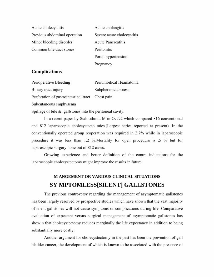
Acute cholecystitis Acute cholangitis
Previous abdominal operation Severe acute cholecystitis
Minor bleeding disorder Acute Pancreatitis
Common bile duct stones Peritonitis
Portal hypertension
Pregnancy
Complications
Perioperative Bleeding Periumbilical Heamatoma
Biliary tract injury Subpherenic abscess
Perforation of gastrointestinal tract Chest pain
Subcutaneous emphysema
Spillage of bile &. gallstones into the peritoneal cavity.
In a recent paper by Stahlschmdt M in Oct'92 which compared 816 conventional
and 812 laparoscopic cholecystecto mies.[Largest series reported at present). In the
conventionally operated group reoperation was required in 2.7% while in laparoscopic
procedure it was less than 1.2 %.Mortality for open procedure is .5 % but for
laparoscopic surgery none out of 812 cases.
Growing experience and better definition of the contra indications for the
laparoscopic cholecystectomy might improve the results in future.
M ANGEMENT OR VARIOUS CLINICAL SITUATIONS
SY MPTOMLESS[SILENT] GALLSTONES The previous controversy regarding the management of asymptomatic gallstones
has been largely resolved by prospective studies which have shown that the vast majority
of silent gallstones will not cause symptoms or complications during life. Comparative
evaluation of expectant versus surgical management of asymptomatic gallstones has
show n that cholecystectomy reduces marginally the life expectancy in addition to being
substantially more costly.
Another argument for cholecystectomy in the past has been the prevention of gall
bladder cancer, the development of which is known to be associated with the presence of
gallstones. However, carcinoma of the gall bladder is rare and the overall operative
mortality with the widespread adoption of prophylactic cholecystectomy in patients with
silent gallstones would certainly exceed that due to cancer of the gall bladder by a
significant margin. The evidence linking cholecystectomy with the development of
colon cancer remains conflicting and cannot be used as a further argument against
prophylactic cholecystectomy. There is no indication for cholecystectomy in the
management of patients with asymptomatic gall stone disease [ C uschieri. A, 1988]
ACUTE CHOLECYSTITIS Initial treatment with nasogastric suction, intravenous fluids and electrolyte
replacement theraphy. Antibiotics and analgesics if required. Two surgical options are
available. They are interval [delayed or elective] cholecystectomy and early
cholecystectomy.
Interval Cholecystectomy
This is the traditional approach where the acute episode is being managed
conservatively and subsequently after the complete resolution of the acute episode,
patients are admitted after 2-3 months for elective cholecystectomy. These rationale for
this treatment is that difficulties are encountered during surgery in the acute inflamatory
episode.
Early Chalecystectomy
This is being performed for acute choecystitis increasingly nowadays. The patient is
operated electively on the next available operating list or within a few days of ad mission.
This must be distinguised from emergency Cholecystectomy, which is done
immediately after admission when gall bladder perforation is suspected. The results of
several prospective clinical trials have shown clearly that early cholecystectomy is
equally safer to elective cholecystectomy. Mortality and morbidity are same for both the
types of operation. But elective cholecystectomy has several disadvantages.
1. Failure of conservative treatment 10-15 %.
2. Premature further episodes while awaiting for the surgery 10-15 % .
3. Patient failing to report 10 %.
4. W hen surgery becomes imperative between second and fourth weeks the incidence of
iatrogenic injuries is very high.
Early cholecystectomy is best performed using the fundus first approach. It is customary
to administer prophylactic antibiotics.
CHRONIC CALCULOUS CHOLECYSTITIS For the treatment of biliary pain non opiate analgesics preferably drugs are
preferred because opiates may cause spasm of sphincter of oddi which cannot be
countered by hyoscine. Antiemetics may be needed to control vomiting.
The definitive treatment of chronic cholecystitis is surgical cholecystectomy open
or laparoscopic. There is little doubt that these patients should have their gall bladder re
moved as approximately 30 % of them will develop complications if surgical treatment is
delayed. The other option is non-surgical gallstone dissloution. It may be oral dissolution
by drugs, extra corporeal shock wave lithotripsy or percutaneous transhepatic
cholecystosomy.
ASSOCIATED DUCTAL CALCULI
The treatment is choledochotomy and cholecystectomy. The stones are extracted
by means of biliary balloon catheters, stone grasping forceps or Dormia basket,
preferably under visual control or with a choledochoscope. A "T" tube is inserted and
choledochotomy wound closed. If CBD is grossly. dilated with papillary stenosis, a
drainage operation is indicated. Choledochoduodenostomy or transduodenal
sphincteroplasty is done. Endoscopic sphincterotomy and removal of stone is the recent
and effective way to manage stones in CBD. Endoscopic sphincterotomy' with extraction
of stone with D0RMIA basket under fluoroscopic control is the best and most effective
method available. If the above fails because of large' stone sphincterotomy and stenting
are done. Later lithotripsy is used to break the stone
AIMS OF THIS STUDY
1. To evaluate the age incidence, the sex incidence, the common etiological and risk
factors of gall stones in this region.
2. To illustrate various types of clinical presentation in calculous cholecystitis.
3. To discuss the usefulness of different diagnostic procedures in a case of calculous
cholecystitis.
4. To study the various modes of management and their results.
5. To study the bacteriology of the bile in calculous gall bladder disease.
6. To assess the incidence of postoperative complications.
7. To analyse the biochemical types of stones prevalent in this part of the country.
8. To study the histopathological changes in calculous gall bladder diseases.
MATERIALS AND METHODS
Forty patients of clearly documented cases of Gallstone diseases of the
Gall bladder and biliary tract admitted in the surgical units and the surgical
Gastroenterology unit of Thanjavur medical college Hospital between January
2005 to Jan’2006. constitute the material of this study.
A detailed History including that about previous treatment was elicited in
all patients and thorough clinical examination was done in them.
Relevant preoperative investigations of blood, Urine, Plain X-ray abdomen
and USG were done in all possible cases. The operative findings and
postoperative complications were recorded and carefully analysed. The Gallstones
removed were sent for Biochemical analysis in 24 cases and bile culture was done
in 16 cases. The Gall bladder specimens of all the cholecystectomy cases were
routinely sent for Histopathological examination.
OBSERVATIONS
Incidence The overall incidence of Gallstone diseases was 0.1% of all hospital
admissions (41,535) between January 2005 to Jan’2006. The 40 patients of gall
stone diseases studied, ranged between 16 and 62 years of age. The mean age by
45 years. The maximum number of cases occurred in the fifth and sixth decades.
The female to male ratio was 3.4 : 1. 37 patients in our series were Hindus, 3
patients belonged to muslim community. 37 (92.5%) belonged to Low Socio
Economic Status. 30 patients were taking mixed diets.
CLINICAL PRESENTATION Abdominal pain, which was localized to right Hypochondrium was the
presenting symptom in 36 patients (90%) 5 cases presented with Jaundice and all
of them were found to have CBD calculi Murphy sign was positive in 14 cases.
Defying the courvoisier’s law, the gall bladder was palpable in none of the cases.
Three of our patients were diabetics.
BIOCHEMICAL INVESTIGATION Four of our patients had elevated blood urea / creatinine levels. Two of
our patients had elevated serum cholesterol level. In our series 5 cases showed,
increased serum bilirubin, with a maximum level upto 7mg %. All the 5 cases of
CBD stones showed increased levels of serum bilirubn and serum alkaline
phosphatase. Prothrombin time was prolonged in one patient, which return to
normal after administration of vit K.
RADIOLOGICAL INVESTIGATIONS Plain X ray abdomen was taken for all cases, and radioopaque stones were
seen in 3 (7.5%) cases.
ULTRASONOGRAM Abdominal ultrasonogram was taken in all cases, except for two patients,
who were taken as emergency 5 cases showed CBD stones with dilatation of CBD.
MANAGEMENT All the 40 cases were operated Of the 40 pts. Cholecystectomy alone was
done in 32 pts, who had only gall bladder stones 4 pts underwent choledochotomy
and the stones were removed followed by ‘T’ tube drainage. In two cases, due to
extensive adhesion, cholecystostomy alone was possible In one case
cholecystectomy was done along with appendicectomy.
INCISIONS Incisions for opening the abdomen for Gallbladder and biliary tract
surgeries was a matter of individual preference right subcostal and right
paramedian incision were commonly used.
Right paramedian 18 case 45%
Right subcostal 19 cases 47.5%
Midiline 3 cases 7.5%
Emergency cholecystectomy.
In our series 2 cases had presented with features of peritonitis. During
laparotomy both cases were found to have gangrenous perforated gall bladder. In
both cases, emergency cholecystectomy was done.
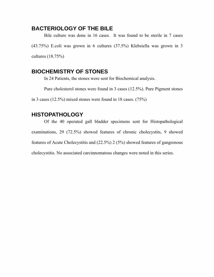
BACTERIOLOGY OF THE BILE Bile culture was done in 16 cases. It was found to be sterile in 7 cases
(43.75%) E.coli was grown in 6 cultures (37.5%) Klebsiella was grown in 3
cultures (18.75%)
BIOCHEMISTRY OF STONES In 24 Patients, the stones were sent for Biochemical analysis.
Pure cholesterol stones were found in 3 cases (12.5%). Pure Pigment stones
in 3 cases (12.5%) mixed stones were found in 18 cases. (75%)
HISTOPATHOLOGY Of the 40 operated gall bladder specimens sent for Histopathological
examinations, 29 (72.5%) showed features of chronic cholecystits, 9 showed
features of Acute Cholecystitis and (22.5%) 2 (5%) showed features of gangrenous
cholecystitis. No associated carcinnomatous changes were noted in this series.
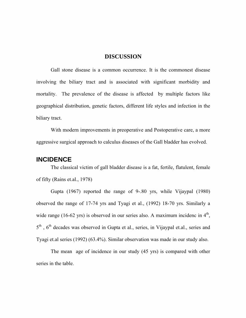
DISCUSSION
Gall stone disease is a common occurrence. It is the commonest disease
involving the biliary tract and is associated with significant morbidity and
mortality. The prevalence of the disease is affected by multiple factors like
geographical distribution, genetic factors, different life styles and infection in the
biliary tract.
With modern improvements in preoperative and Postoperative care, a more
aggressive surgical approach to calculus diseases of the Gall bladder has evolved.
INCIDENCE The classical victim of gall bladder disease is a fat, fertile, flatulent, female
of fifty (Rains et.al., 1978)
Gupta (1967) reported the range of 9-.80 yrs, while Vijaypal (1980)
observed the range of 17-74 yrs and Tyagi et al., (1992) 18-70 yrs. Similarly a
wide range (16-62 yrs) is observed in our series also. A maximum incidenc in 4th,
5th , 6th decades was observed in Gupta et al., series, in Vijaypal et.al., series and
Tyagi et.al series (1992) (63.4%). Similar observation was made in our study also.
The mean age of incidence in our study (45 yrs) is compared with other
series in the table.
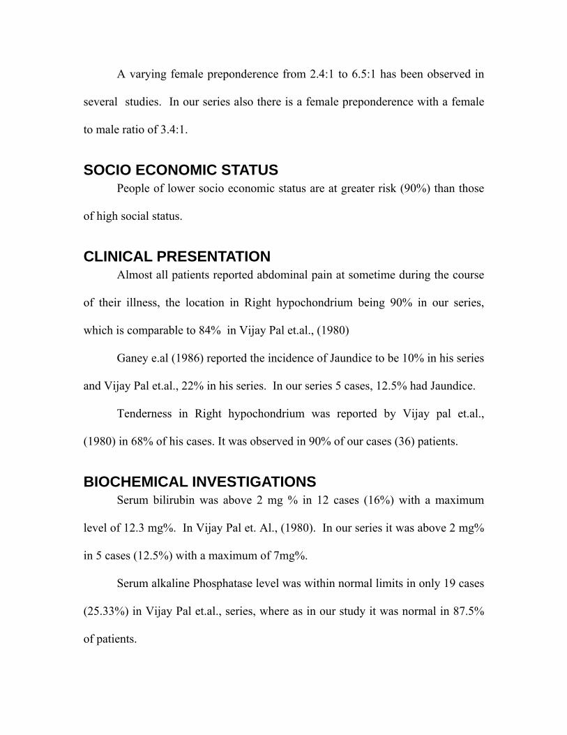
A varying female preponderence from 2.4:1 to 6.5:1 has been observed in
several studies. In our series also there is a female preponderence with a female
to male ratio of 3.4:1.
SOCIO ECONOMIC STATUS People of lower socio economic status are at greater risk (90%) than those
of high social status.
CLINICAL PRESENTATION Almost all patients reported abdominal pain at sometime during the course
of their illness, the location in Right hypochondrium being 90% in our series,
which is comparable to 84% in Vijay Pal et.al., (1980)
Ganey e.al (1986) reported the incidence of Jaundice to be 10% in his series
and Vijay Pal et.al., 22% in his series. In our series 5 cases, 12.5% had Jaundice.
Tenderness in Right hypochondrium was reported by Vijay pal et.al.,
(1980) in 68% of his cases. It was observed in 90% of our cases (36) patients.
BIOCHEMICAL INVESTIGATIONS Serum bilirubin was above 2 mg % in 12 cases (16%) with a maximum
level of 12.3 mg%. In Vijay Pal et. Al., (1980). In our series it was above 2 mg%
in 5 cases (12.5%) with a maximum of 7mg%.
Serum alkaline Phosphatase level was within normal limits in only 19 cases
(25.33%) in Vijay Pal et.al., series, where as in our study it was normal in 87.5%
of patients.
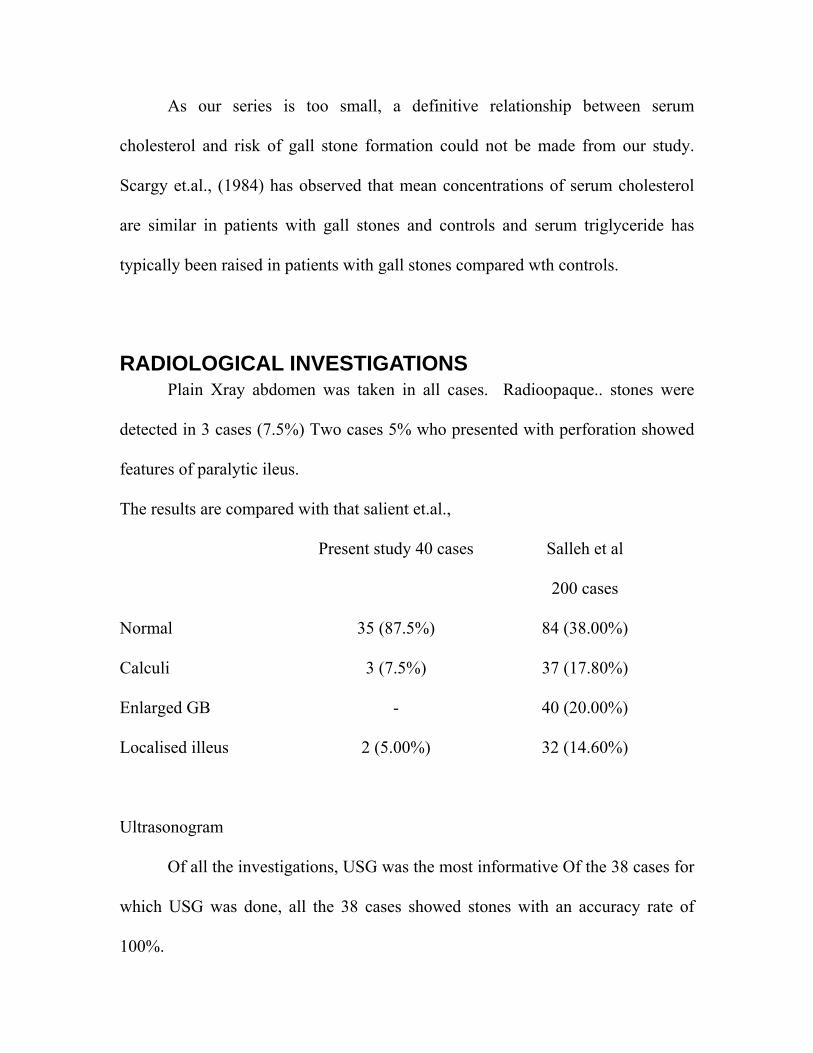
As our series is too small, a definitive relationship between serum
cholesterol and risk of gall stone formation could not be made from our study.
Scargy et.al., (1984) has observed that mean concentrations of serum cholesterol
are similar in patients with gall stones and controls and serum triglyceride has
typically been raised in patients with gall stones compared wth controls.
RADIOLOGICAL INVESTIGATIONS Plain Xray abdomen was taken in all cases. Radioopaque.. stones were
detected in 3 cases (7.5%) Two cases 5% who presented with perforation showed
features of paralytic ileus.
The results are compared with that salient et.al.,
Present study 40 cases Salleh et al
200 cases
Normal 35 (87.5%) 84 (38.00%)
Calculi 3 (7.5%) 37 (17.80%)
Enlarged GB - 40 (20.00%)
Localised illeus 2 (5.00%) 32 (14.60%)
Ultrasonogram
Of all the investigations, USG was the most informative Of the 38 cases for
which USG was done, all the 38 cases showed stones with an accuracy rate of
100%.
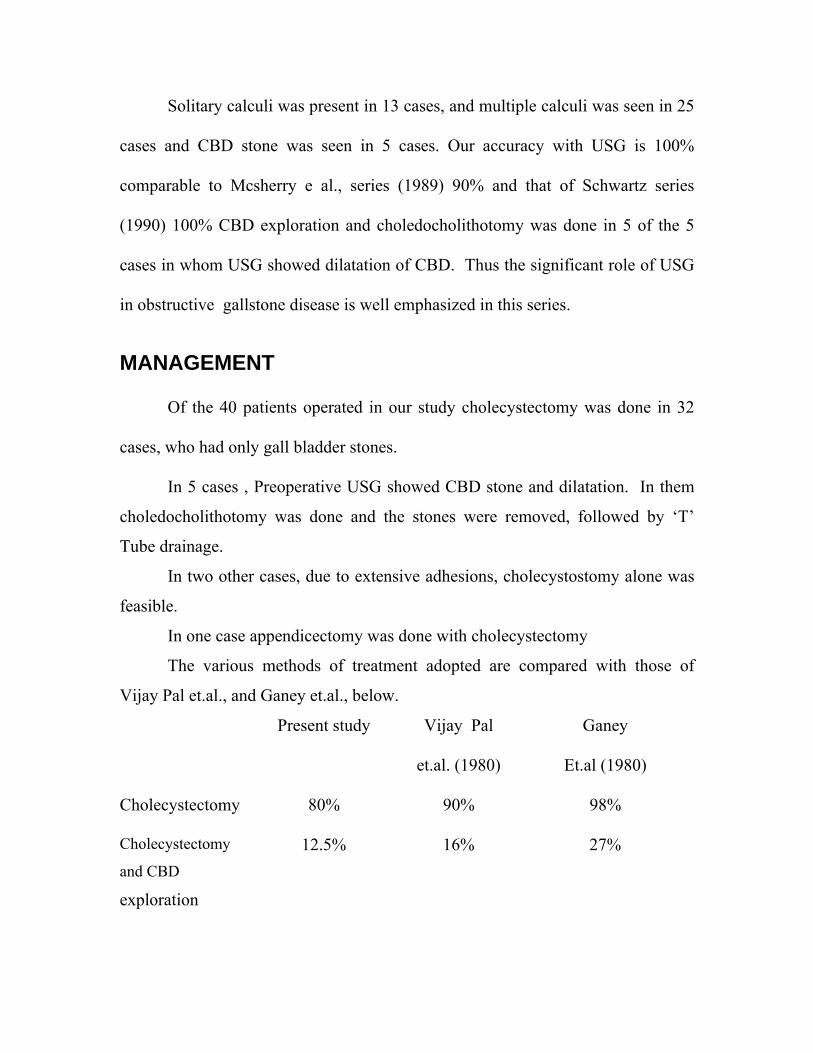
Solitary calculi was present in 13 cases, and multiple calculi was seen in 25
cases and CBD stone was seen in 5 cases. Our accuracy with USG is 100%
comparable to Mcsherry e al., series (1989) 90% and that of Schwartz series
(1990) 100% CBD exploration and choledocholithotomy was done in 5 of the 5
cases in whom USG showed dilatation of CBD. Thus the significant role of USG
in obstructive gallstone disease is well emphasized in this series.
MANAGEMENT Of the 40 patients operated in our study cholecystectomy was done in 32
cases, who had only gall bladder stones.
In 5 cases , Preoperative USG showed CBD stone and dilatation. In them
choledocholithotomy was done and the stones were removed, followed by ‘T’
Tube drainage.
In two other cases, due to extensive adhesions, cholecystostomy alone was
feasible.
In one case appendicectomy was done with cholecystectomy
The various methods of treatment adopted are compared with those of
Vijay Pal et.al., and Ganey et.al., below.
Present study Vijay Pal
et.al. (1980)
Ganey
Et.al (1980)
Cholecystectomy 80% 90% 98%
Cholecystectomy
and CBD
exploration
12.5% 16% 27%
Cholecystostomy 5% - 1%
CHOLECYSTOSTOMY Cholecystostomy is a procedure of comprimise. But in emergency situation,
due to patients general condition, surgeon has to resort to it.
In this series 2 patients had cholecystostomy. The first one had edematous
calot’s triangle and extensive adhesions to sorrounding viscera. Patient had LVF
strain in ECG and elevated Urea / Creatinine. So cholecystostomy was done.
In other patient, who is a old case of CAHD with extensive adhesions,
cholecystostomy was performed and gall stones were removed.
CHOLECYSTECTOMY Cholecystectomy was the commonest procedure in our series.
EMERGENCY CHOLECYSTECTOMY In our series 2 cases had presented with features of perforative. Peritonitis.
During Laparotomy both cases were found to have gangrenous gall bladder with
perforation. The incidence of perforation was 1% in a series reported by J.D.Wig
(1990) in which he has analysed the causes of peritonitis. Incidence of perforated
gall bladder in Ganey et.al., series was 1%. In this study, the incidence is 5%
because of very small number of cases being studied. In both cases,
cholecystectomy was done.
ELECTIVE CHOLECYSTECTOMY This traditional approach which is still popular was followed in 38 cases of
our study. The rationale for this treatment is that in most cases, the raised pressure
within the gallbladder lumen lifts the walls of the organ of the impacted stone,
which then dislodges and falls into the lumen with resolution of the Inflammation,
the view being held that it is safer to operate several weeks after the acute
Inflammatory episode has subsided.
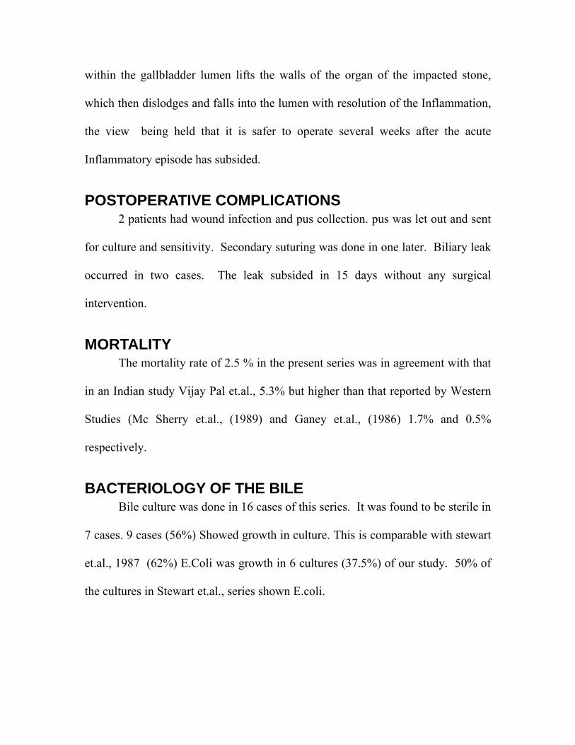
POSTOPERATIVE COMPLICATIONS 2 patients had wound infection and pus collection. pus was let out and sent
for culture and sensitivity. Secondary suturing was done in one later. Biliary leak
occurred in two cases. The leak subsided in 15 days without any surgical
intervention.
MORTALITY The mortality rate of 2.5 % in the present series was in agreement with that
in an Indian study Vijay Pal et.al., 5.3% but higher than that reported by Western
Studies (Mc Sherry et.al., (1989) and Ganey et.al., (1986) 1.7% and 0.5%
respectively.
BACTERIOLOGY OF THE BILE Bile culture was done in 16 cases of this series. It was found to be sterile in
7 cases. 9 cases (56%) Showed growth in culture. This is comparable with stewart
et.al., 1987 (62%) E.Coli was growth in 6 cultures (37.5%) of our study. 50% of
the cultures in Stewart et.al., series shown E.coli.

ANALYSIS OF STONES In this series 75% of the stones were of mixed type. 12.5% of pure
cholesterol and 12.5% of Pigment stones were noted. Similar high incidence of
mixed stones had been observed in both western series Ganey (70%) and also in
Indian series vijay pal 91.30% and Bansali 83.3%.
HISTOPATHOLOGY In our series 29 patients showed features of chronic cholecystits, 9 showed
features of Acute cholecystitis and 2 showed gangrenous changes. No associated
carcinomatous changes were noted in this series.
SUMMARY AND CONCLUSIONS
Gallstone disease is the commonest disease involving biliary tract and is
associated with significant morbidity and mortality. Patients with gallstone are not
a homogenous group. They are now being detected with greater frequencies, with
the advent of Ultrasonogram. 40 cases of well documented calculous cholecystitis
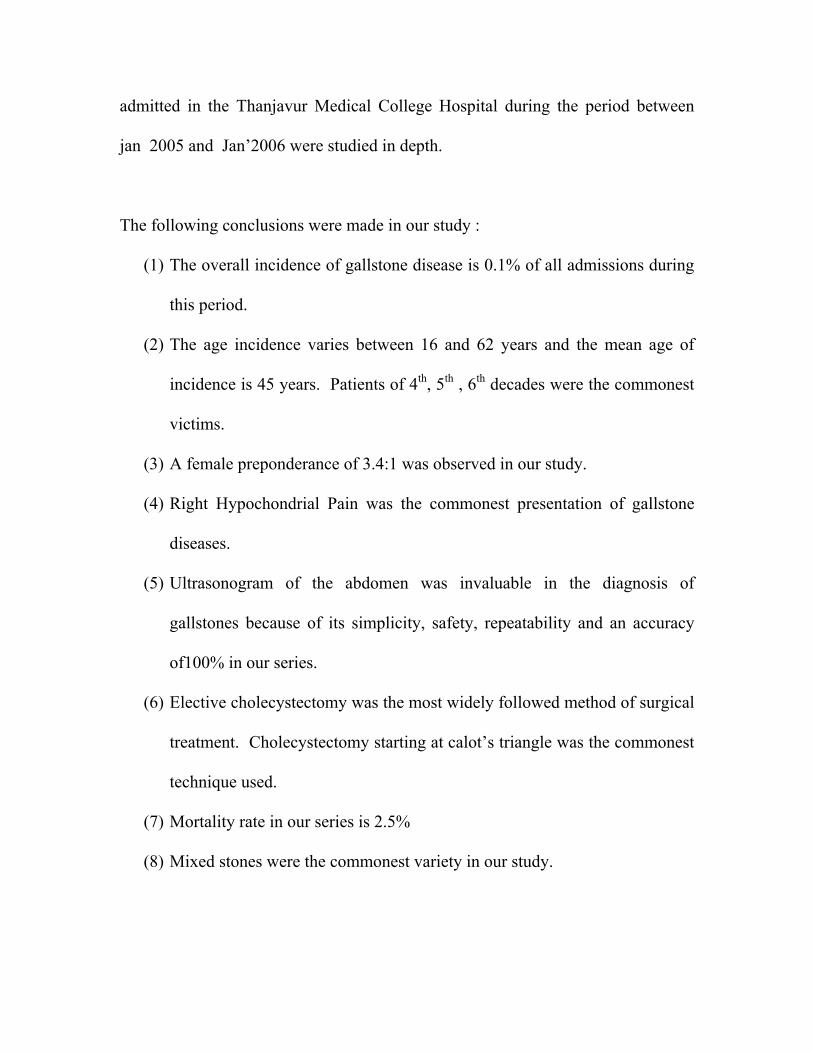
admitted in the Thanjavur Medical College Hospital during the period between
jan 2005 and Jan’2006 were studied in depth.
The following conclusions were made in our study :
(1) The overall incidence of gallstone disease is 0.1% of all admissions during
this period.
(2) The age incidence varies between 16 and 62 years and the mean age of
incidence is 45 years. Patients of 4th, 5th , 6th decades were the commonest
victims.
(3) A female preponderance of 3.4:1 was observed in our study.
(4) Right Hypochondrial Pain was the commonest presentation of gallstone
diseases.
(5) Ultrasonogram of the abdomen was invaluable in the diagnosis of
gallstones because of its simplicity, safety, repeatability and an accuracy
of100% in our series.
(6) Elective cholecystectomy was the most widely followed method of surgical
treatment. Cholecystectomy starting at calot’s triangle was the commonest
technique used.
(7) Mortality rate in our series is 2.5%
(8) Mixed stones were the commonest variety in our study.
(9) The commonest histopathological change associated with gallstone was
chronic cholecystitis. Associated carcinomatous change in calculous
cholecystitis is nil in our series.
Medical dissolution of the stone though theoretical is not very popular with
our Hospital patients, because of the non-availability LAPAROSCOPIC
cholecystectomy is now replacing the open cholecystectomy,. But in our
institution. It is not available. However open cholecystecomy has its value in
smaller hospitals and peripheral centers as the only method of treatment. It is
therefore necessary that a surgeon should have the adequate knowledge and
experience in this field.
In Judicially selected, carefully prepared and operated cases, the results are
bound to be gratifying.
BIBLIOGRAPHY
1. Alexander Mcgreegor 1984 A synopsis of surgical anatomy 11th Edition.
2. Alfred Cusheiri 2002 essentials of surgical practice fourth edition.
3. Alfred cusheiri, Geogrge bersly 1991 Laparoscopic cholecystectomy volume 159;
3 page 273.
4. Anderson.T, Liver & Gallbladder disease before and after very low calorie diets.
A.M.J.. Clinical Nutr. 1992 Jul: 56 2355 – 2395.
5. Bailey and Love 2004 Short practice of surgery 24st Edition.
6. Benhoft 1984 – composition and morophology and classification of gallstones
AJS 148: 77 – 79.
7. Beranrd siegel 1989 radio diagnostic proce durses in surgery 1st Edition.
8. Bowen JC, Brenner HI, Macle.WF Gallstone disease patho physiology
epidemiology, natural history and treatment options. 1992 MCNA. SEP 76 [ 5 ]
1143 – 1157.
9. Farguharson 2003 Text book of operative surgery, 8th edition 381 – 399.
10. Ganong short text book of physiology 1999.
11. Goswitz, bacteria and biliary tract disease 1974 AJS 128: 644 – 646.
12. Gupta Etal Prevalence OF GALL STONE DISEASE 1985 IJS VOL.47
13. Khuroo Etal prevalence of biliary tract disease in India,1986 gut30:201-205.
14. Mcsherry.c.k, 1989 cholesystectomy;the gold standard AJS.206;242-246.
15. North American clinics of gastroenterology 1991.3rd Edn.0885-0889.
16. Nijihawan.S,RR rai, 1992 epidemiology of gallstones in India JAPI vol.40 NUM0
17. Recent advances in surgery 14,15,&16th EDN
18. Rob & Smith operative surgery 1989, abdominal surgeries vol.1 3rd Edn.
19. Rodney maingot’s, 2002 abdominal operations 10th Edn.
20. Sabiston text book of surgery 2005 17th Edn
21. Schwartz principle’s of surgery 2005 8th Edn.
22. Surgical clinics of North America.biliary tract diseases.
23. Tygat.n, maheswari,1992 july JIMA,Morphological changes in diseased
gallbladder.
24. Vijy pal, clinicopathological study of cholecystitis IJS 426-431
PROFORMA
Name D.O.A.
Age D.O.D
Sex D.O.O
IP No. Unit
compliantsPresent History
Pain : Site
Duration
Character
Radiation
Relation to deep breathing
Aggravating factors
Relieving factors :
Vomiting / Nausea
Fever
Duration / Grade / Nature / asso c chills and rigor
Flatulent dyspepsia
Colour of Urine
Colour of stool
Past History
Similar complaints
Jaundice
Fever
Diabetes
Hypertension
Cirrhosis
Personal History
Family History
Menstrual History
DIET :
VEG
MIXED
OILY Food intake
Treatment History :
HIO Any drug intake
OCP
General Examination
Temp :
Pulse Rate :
Jaundice
Anemia
Features of Hyperlipidemia
Examination of abdomen
Inspection
Paipation
Tenderness Site
Murphy sign
Liver
GB
Spleen
Associated conditions
Renal Disease
Colonic motility disorder
Reflux Esopghagitis
Investigations
Urine : Albumin | Sugar
Bile Salt | Bile Pigments
Blood : Hb %
TC DC ESR
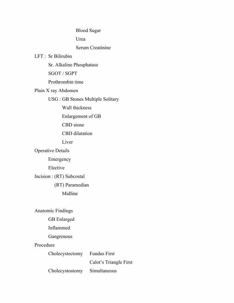
Blood Sugar
Urea
Serum Creatinine
LFT : Sr Bilirubin
Sr. Alkaline Phosphatase
SGOT / SGPT
Prothrombin time
Plain X ray Abdomen
USG : GB Stones Multiple Solitary
Wall thickness
Enlargement of GB
CBD stone
CBD dilatation
Liver
Operative Details
Emergency
Elective
Incision : (RT) Subcostal
(RT) Paramedian
Midline
Anatomic Findings
GB Enlarged
Inflammed
Gangrenous
Procedure
Cholecystectomy Fundus First
Calot’s Triangle First
Cholecystostomy Simultaneous
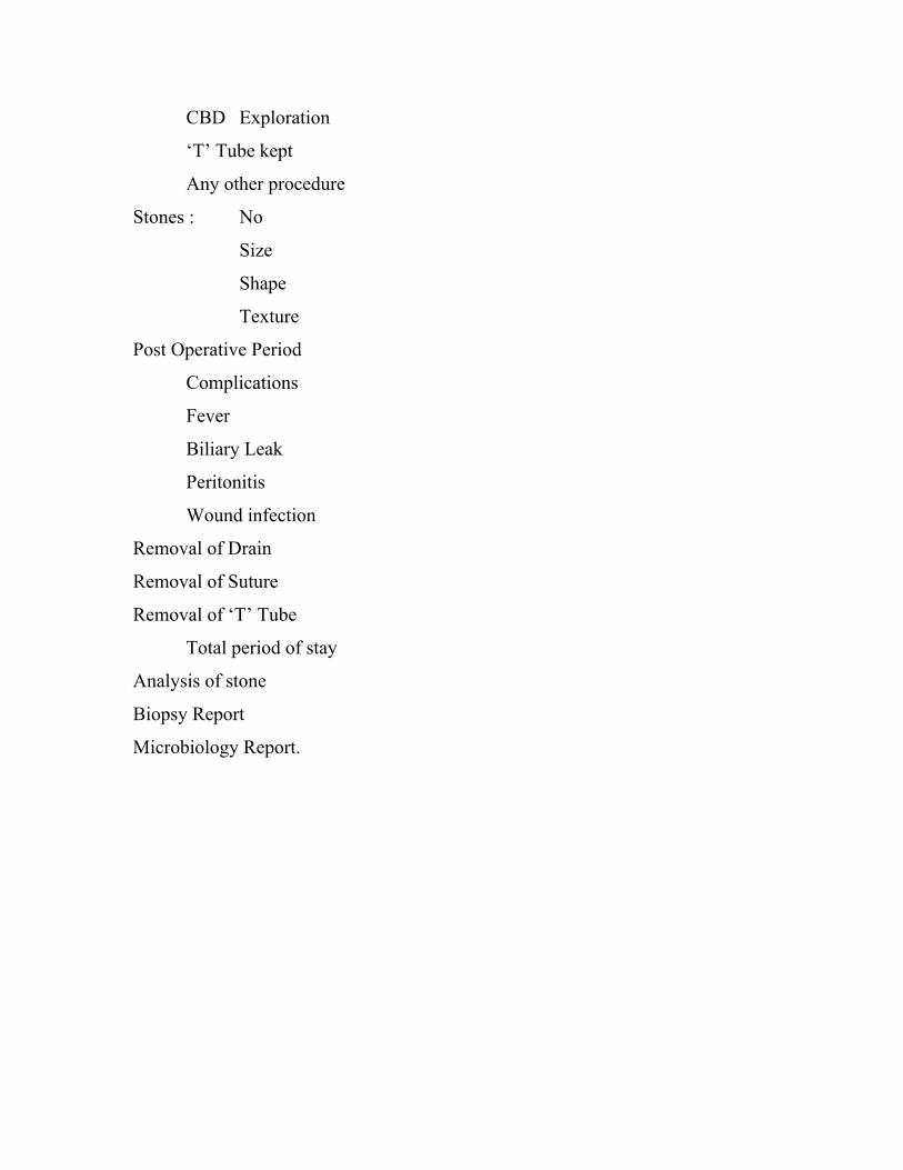
CBD Exploration
‘T’ Tube kept
Any other procedure
Stones : No
Size
Shape
Texture
Post Operative Period
Complications
Fever
Biliary Leak
Peritonitis
Wound infection
Removal of Drain
Removal of Suture
Removal of ‘T’ Tube
Total period of stay
Analysis of stone
Biopsy Report
Microbiology Report.
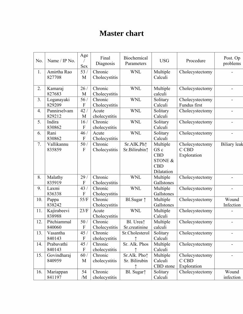
Master chart
No. Name / IP No. Age
/ Sex
Final Diagnosis
Biochemical Parameters USG Procedure Post. Op
problems
1. Amirtha Rao 827708
53 / M
Chronic Cholecystitis
WNL Multiple Calculi
Cholecystectomy -
2. Kamaraj 827683
26 / M
Chronic Cholecystitis
WNL Multiple calculi
Cholecystectomy -
3. Loganayaki 829209
56 / F
Chronic Cholecystitis
WNL Solitary Calculi
CholecystectomyFundus first
-
4. Pannirselvam 829212
42 / M
Acute cholecystitis
WNL Solitary Calculi
Cholecystectomy -
5. Indira 830862
16 / F
Chronic cholecystitis
WNL Solitary Calculi
Cholecystectomy -
6. Rani 830862
46 / F
Acute Cholecystitis
WNL Solitary Calculi
Cholecystectomy -
7. Vallikannu 835859
50 / F
Chronic Cholecystitis
Sr.AIK.Ph↑ Sr.Bilirubin↑
Multiple GS c CBD STONE & CBD Dilatation
Cholecystectomy C CBD Exploration
Biliary leak
8. Malathy 835919
29 / F
Chronic Cholecystitis
WNL Multiple Gallstones
Cholecystectomy -
9. Laxmi 836338
43 / F
Chronic Cholecystitis
WNL Multiple Gallstones
Cholecystectomy -
10. Pappa 838242
55/F Chronic Cholecystitis
Bl.Sugar ↑ Multiple Gallstones
Cholecystectomy Wound Infection
11. Kajirabeevi 838988
23/F Acute Cholecystitis
WNL Multiple Calculi
Cholecystectomy -
12. Pitchiammal 840060
50 / F
Chronic Cholecystitis
Bl. Urea↑ Sr.creatinine
Multiple calculi
Cholecystectomy -
13. Vasantha 840143
45 / F
Chronic cholecystitis
Sr.Cholesterol ↑
Solitary Calculi
Cholecystectomy -
14. Prabavathi 840143
45 / F
Chronic cholecystitis
Sr. Alk. Phos ↑
Multiple Calculi
Cholecystectomy -
15. Govindharaj 840959
60 / M
Chronic cholecystitis
Sr.Alk. Pho↑ Sr. Bilirubin
↑
Multiple Calculi CBD stone
CholecystectomyC CBD Exploration
-
16. Mariappan 841197
54 /M
Chronic cholecystitis
Bl. Sugar↑ Solitary Calculi
Cholecystectomy Wound infection
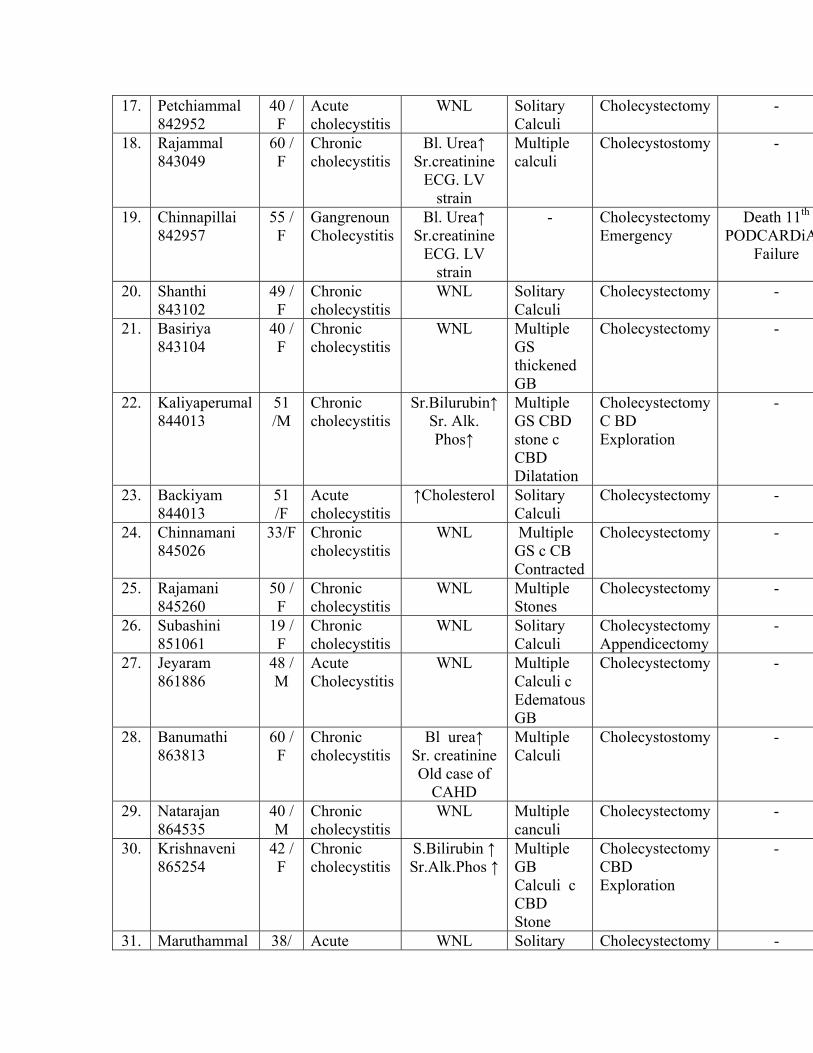
17. Petchiammal 842952
40 / F
Acute cholecystitis
WNL Solitary Calculi
Cholecystectomy -
18. Rajammal 843049
60 / F
Chronic cholecystitis
Bl. Urea↑ Sr.creatinine
ECG. LV strain
Multiple calculi
Cholecystostomy -
19. Chinnapillai 842957
55 / F
Gangrenoun Cholecystitis
Bl. Urea↑ Sr.creatinine
ECG. LV strain
- CholecystectomyEmergency
Death 11th PODCARDiA
Failure
20. Shanthi 843102
49 / F
Chronic cholecystitis
WNL Solitary Calculi
Cholecystectomy -
21. Basiriya 843104
40 / F
Chronic cholecystitis
WNL Multiple GS thickened GB
Cholecystectomy -
22. Kaliyaperumal 844013
51 /M
Chronic cholecystitis
Sr.Bilurubin↑ Sr. Alk. Phos↑
Multiple GS CBD stone c CBD Dilatation
CholecystectomyC BD Exploration
-
23. Backiyam 844013
51 /F
Acute cholecystitis
↑Cholesterol Solitary Calculi
Cholecystectomy -
24. Chinnamani 845026
33/F Chronic cholecystitis
WNL Multiple GS c CB Contracted
Cholecystectomy -
25. Rajamani 845260
50 / F
Chronic cholecystitis
WNL Multiple Stones
Cholecystectomy -
26. Subashini 851061
19 / F
Chronic cholecystitis
WNL Solitary Calculi
CholecystectomyAppendicectomy
-
27. Jeyaram 861886
48 / M
Acute Cholecystitis
WNL Multiple Calculi c Edematous GB
Cholecystectomy
-
28. Banumathi 863813
60 / F
Chronic cholecystitis
Bl urea↑ Sr. creatinine Old case of
CAHD
Multiple Calculi
Cholecystostomy
-
29. Natarajan 864535
40 / M
Chronic cholecystitis
WNL Multiple canculi
Cholecystectomy
-
30. Krishnaveni 865254
42 / F
Chronic cholecystitis
S.Bilirubin ↑ Sr.Alk.Phos ↑
Multiple GB Calculi c CBD Stone
CholecystectomyCBD Exploration
-
31. Maruthammal 38/ Acute WNL Solitary Cholecystectomy -

870307 F cholecystitis Calculi 32. Saraswathy
873321 45 / F
Acute cholecystitis
WNL Solitary Calculi & Thickened GB
CholecystectomyCBD Exploration
-
33. Vijayalakshmi 873703
46 / F
Chronic cholecystitis
WNL Multiple GS
CholecystectomyFundus First
-
34. Chellammal 875530
45/F Chronic cholecystitis
WNL Multiple GS
Cholecystectomy
35. Pattammal 875992
40/F Chronic cholecystitis
WNL Multiple GS
Cholecystectomy
-
36. Rani 877132
55 / F
Chronic cholecystitis
Sr.Bilurubin ↑Sr.Alk.Phos↑
Multiple GS c CBD Dilatation
Cholecystectomyc CBD Exploration
-
37. Abunisha 817246
45 / F
Chronic cholecystitis
WNL Solitary Calculi
CholecystectomyFundus First
-
38. Saroja 866715
40 / F
Chronic cholecystitis
WNL Multiple Calculi
Cholecystectomy
-
39. Parvathy 867377
50 / F
Acute cholecystitis
WNL Multiple Calculi
Cholecystectomy
-
40. Gopal 869513
55 / M
Gangrenous cholecystitis
Bl – Sugar ↑ - Cholecystectomy
-

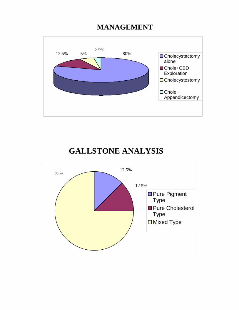
Pure PigmentTypePure CholesterolTypeMixed Type
CholecystectomyaloneChole+CBDExplorationCholecystostomy
Chole +Appendicectomy
12.5% 5%2.5%
80%
75%12.5%
12.5%
MANAGEMENT
GALLSTONE ANALYSIS
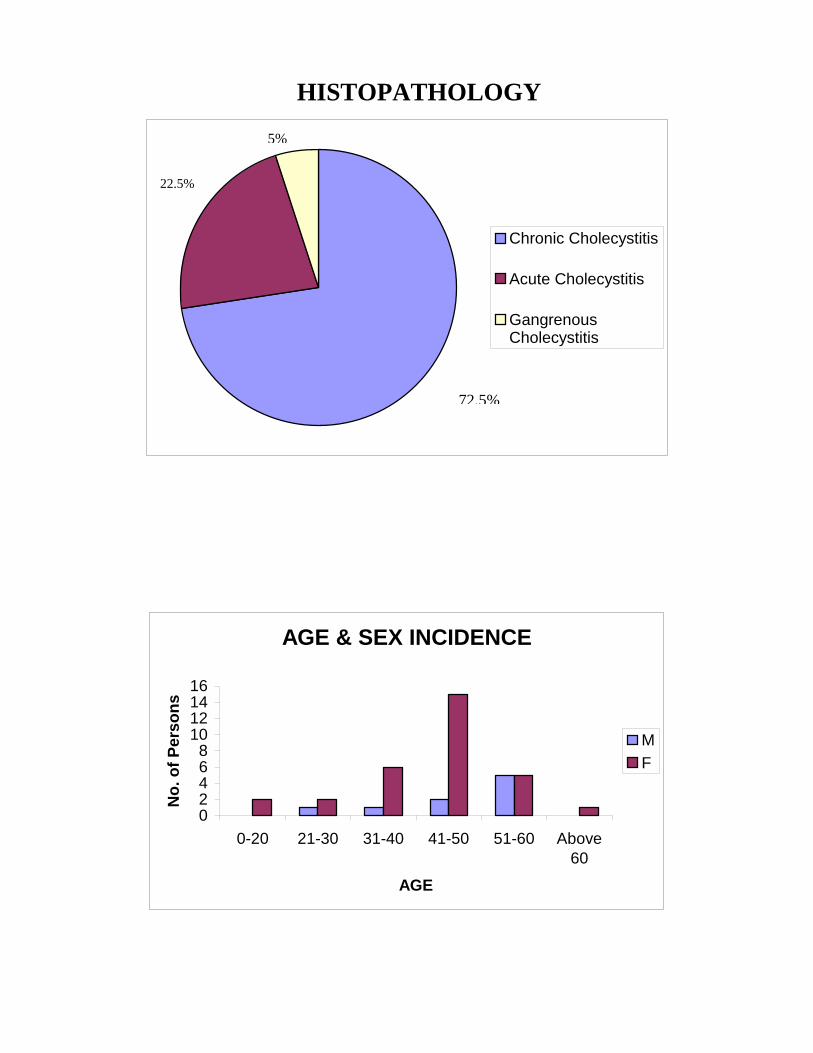
Chronic Cholecystitis
Acute Cholecystitis
GangrenousCholecystitis
AGE & SEX INCIDENCE
02468
10121416
0-20 21-30 31-40 41-50 51-60 Above60
AGE
No.
of P
erso
ns
MF
5%
22.5%
72.5%
HISTOPATHOLOGY
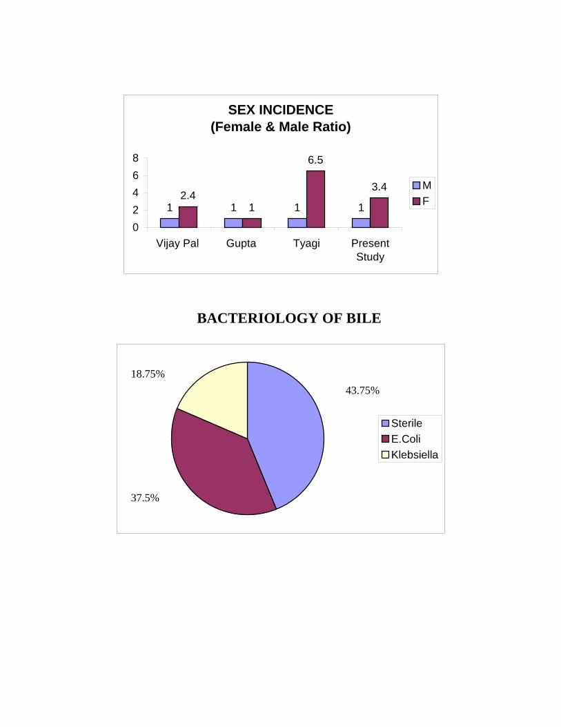
SEX INCIDENCE (Female & Male Ratio)
1 1 1 12.4
1
6.5
3.4
02468
Vijay Pal Gupta Tyagi PresentStudy
MF
SterileE.ColiKlebsiella
43.75%
37.5%
18.75%
BACTERIOLOGY OF BILE