Calcium-Mediated Pore Expansion and Cell Death Following ...
9
Old Dominion University ODU Digital Commons Bioelectrics Publications Frank Reidy Research Center for Bioelectrics 2014 Calcium-Mediated Pore Expansion and Cell Death Following Nanoelectroporation Olga N. Pakhomova Old Dominion University, [email protected] Betsy Gregory Old Dominion University, [email protected] Iurii Semenov Old Dominion University, [email protected] Andrei G. Pakhomov Old Dominion University, [email protected] Follow this and additional works at: hps://digitalcommons.odu.edu/bioelectrics_pubs Part of the Biochemistry Commons , Biomedical Commons , Biophysics Commons , and the Molecular Biology Commons is Article is brought to you for free and open access by the Frank Reidy Research Center for Bioelectrics at ODU Digital Commons. It has been accepted for inclusion in Bioelectrics Publications by an authorized administrator of ODU Digital Commons. For more information, please contact [email protected]. Repository Citation Pakhomova, Olga N.; Gregory, Betsy; Semenov, Iurii; and Pakhomov, Andrei G., "Calcium-Mediated Pore Expansion and Cell Death Following Nanoelectroporation" (2014). Bioelectrics Publications. 195. hps://digitalcommons.odu.edu/bioelectrics_pubs/195 Original Publication Citation Pakhomova, O. N., Gregory, B., Semenov, I., & Pakhomov, A. G. (2014). Calcium-mediated pore expansion and cell death following nanoelectroporation. Biochimica Et Biophysica Acta: Biomembranes, 1838(10), 2547-2554. doi:10.1016/j.bbamem.2014.06.015
Transcript of Calcium-Mediated Pore Expansion and Cell Death Following ...

Old Dominion UniversityODU Digital Commons
Bioelectrics Publications Frank Reidy Research Center for Bioelectrics
2014
Calcium-Mediated Pore Expansion and Cell DeathFollowing NanoelectroporationOlga N. PakhomovaOld Dominion University, [email protected]
Betsy GregoryOld Dominion University, [email protected]
Iurii SemenovOld Dominion University, [email protected]
Andrei G. PakhomovOld Dominion University, [email protected]
Follow this and additional works at: https://digitalcommons.odu.edu/bioelectrics_pubs
Part of the Biochemistry Commons, Biomedical Commons, Biophysics Commons, and theMolecular Biology Commons
This Article is brought to you for free and open access by the Frank Reidy Research Center for Bioelectrics at ODU Digital Commons. It has beenaccepted for inclusion in Bioelectrics Publications by an authorized administrator of ODU Digital Commons. For more information, please [email protected].
Repository CitationPakhomova, Olga N.; Gregory, Betsy; Semenov, Iurii; and Pakhomov, Andrei G., "Calcium-Mediated Pore Expansion and Cell DeathFollowing Nanoelectroporation" (2014). Bioelectrics Publications. 195.https://digitalcommons.odu.edu/bioelectrics_pubs/195
Original Publication CitationPakhomova, O. N., Gregory, B., Semenov, I., & Pakhomov, A. G. (2014). Calcium-mediated pore expansion and cell death followingnanoelectroporation. Biochimica Et Biophysica Acta: Biomembranes, 1838(10), 2547-2554. doi:10.1016/j.bbamem.2014.06.015

Calcium-mediated pore expansion and cell deathfollowing nanoelectroporation
Olga N. Pakhomova ⁎, Betsy Gregory, Iurii Semenov, Andrei G. PakhomovFrank Reidy Research Center for Bioelectrics, Old Dominion University, Norfolk, VA, USA
a b s t r a c ta r t i c l e i n f o
Article history:Received 9 May 2014Received in revised form 19 June 2014Accepted 20 June 2014Available online 28 June 2014
Keywords:Nanosecond pulseElectroporationElectropermeabilizationCalciumNecrosisApoptosis
Opening of long-lived pores in the cell membrane is the principal primary effect of intense, nanosecond pulsedelectric field (nsPEF). Here we demonstrate that the evolution of pores, cell survival, the time and the mode ofcell death (necrotic or apoptotic) are determined by the level of external Ca2+ after nsPEF. We also introduce anovel, minimally disruptive technique for nsEP exposure of adherent cells on indium tin oxide (ITO)-coatedglass coverslips, which does not require cell detachment and enables fast exchanges of bath media. Increasingthe Ca2+ level from the nominal 2–5 μM to 2 mM for the first 60–90 min after permeabilization by 300-nsPEFincreased the early (necrotic) death in U937, CHO, and BPAE cells. With nominal Ca2+, the inhibition of osmoticswelling rescued cells from the early necrosis and increased caspase 3/7 activation later on. However, the inhibi-tion of swelling had a modest or no protective effect with 2 mM Ca2+ in the medium. With the nominal Ca2+,most cells displayed gradual increase in YO-PRO-1 and propidium (Pr) uptake. With 2 mM Ca2+, the initiallylower Pr uptake was eventually replaced by a massive and abrupt Pr entry (necrotic death). It was accompaniedby a transient acceleration of the growth ofmembrane blebs due to the increase of the intracellular osmotic pres-sure. We conclude that the high-Ca2+-dependent necrotic death in nsPEF-treated cells is effected by a delayed,sudden, and osmotically-independent pore expansion (or de novo formation of larger pores), but not by themembrane rupture.
© 2014 Elsevier B.V. All rights reserved.
1. Introduction
Effects of intense nsPEF treatments in mammalian cells include per-meabilization of the plasmamembrane, endoplasmic reticulum (ER),and mitochondria [1–7]; Ca2+ uptake from the outside and releasefrom the ER [2,6,8–10]; destruction of the cytoskeleton [11–13];cell swelling and blebbing [14–16]; and activation of signaling andcell death pathways [10,17–21]. The cytotoxic effects of nsPEF haveattracted particular attention as a novel and promising modality forcancer treatment [22–26].
With the complexity of the cellular response to nsPEF, the mecha-nisms and specific pathways leading to cell death have only been par-tially understood. The early studies focused on the apoptotic response,
and only recently early necrosiswas reported by several groups as a sep-arate or even a predominant mode of nsPEF-induced cell death [15,18].The primary cause of necrosis was the persistent plasma membranepermeabilization to small solutes (b1 nm), which resulted in the os-motic imbalance, water uptake, and cell swelling culminating in themembrane rupture. When the uptake of water was blocked by anisoosmotic addition of a pore-impermeable solute (sucrose [14]),cells were rescued from the necrotic death, but nonetheless diedlater on by apoptosis [15]. However, the cause of the apoptosis incells rescued from the necrosis was not identified.
Ca2+ signaling is critically involved, in manyways, in both the ini-tiation and effectuation of the cell death (see [27] for review). Sever-al studies pointed to the role of mitochondria and Ca2+ increase inthe initiation and execution of the nsPEF-induced apoptosis [3,28],and the increase in the cytosolic Ca2+ is one of the best knownnsPEF effects [2,6,8,9,29,30]. In human pancreatic cancer cells,nsPEF caused Ca2+-dependent production of reactive oxygen spe-cies, indicating that overloading mitochondria with Ca2+ and theirdestruction were part of the apoptotic mechanism [28]. Finally, celllethality increased with increasing extracellular Ca2+ for eithernsPEF or conventional electroporation [31,32]. The injection ofCa2+ into a tumor prior to its electroablation (“Ca2+ electropora-tion”) has increased the treatment efficiency in animals and humans[31].
Biochimica et Biophysica Acta 1838 (2014) 2547–2554
Abbreviations: BPAE, bovine pulmonary artery endothelial cells; ER, endoplasmic re-ticulum; nsPEF, nanosecond pulsed electric field; Pr, propidium; ROS, reactive oxygenspecies⁎ Corresponding author at: Frank Reidy Research Center for Bioelectrics, 4211Monarch
Way, Suite 300, Old Dominion University, Norfolk, VA 23508, USA. Tel.: +1 210 6182824,+1 757 6838006; fax: +1 757 4511010.
E-mail addresses: [email protected], [email protected] (O.N. Pakhomova).
http://dx.doi.org/10.1016/j.bbamem.2014.06.0150005-2736/© 2014 Elsevier B.V. All rights reserved.
Contents lists available at ScienceDirect
Biochimica et Biophysica Acta
j ourna l homepage: www.e lsev ie r .com/ locate /bbamem

Therefore, our experiments were initially aimed at defining the roleof Ca2+ in nsPEF-induced apoptosis in cells rescued from the earlyosmotically-driven necrosis. Instead, we found that the increasedCa2+ facilitated the early necrosis and thereby decreased the cellpopulation that could potentially enter the apoptotic process. TheCa2+-mediated necrotic pathway did not rely on the osmotic swelling,and overrode any protection rendered by the blockage of water uptake.Below we demonstrate that Ca2+-mediated necrosis results from a de-layed, abrupt, irreversible, and osmotically-independent expansion ofpores in the cell membrane.
2. Materials and methods
2.1. Cells and media
We utilized suspension cells U937 (human monocytes), and ad-herent cells CHO-K1 (Chinese hamster ovary) and BPAE (bovinepulmonary artery endothelial). U937 and CHO cells were obtainedfrom the American Type Culture Collection (ATCC, Manassas, VA)and propagated as described previously [1,15,33]. BPAE were akind gift from Dr. J. Catravas (Center for Bioelectrics, ODU). Theywere grown at 37 °C with 5% CO2 in air in a low glucose DMEMwith 10% fetal bovine serum (FBS), 100 IU/ml penicillin, 0.1 mg/mlstreptomycin, and 2.5 μg/ml amphotericin B. The media and its com-ponents were purchased from Thermo Scientific (Waltham, MA),Sigma-Aldrich (St. Louis, MO), and Atlanta Biologicals (Norcross,GA).
2.2. nsPEF exposure and viability of suspension cells
To control for the extracellular Ca2+ during and after nsPEF, weutilized RPMI 1640 medium formulated without Ca2+ (“no Ca2+”
medium). Ca2+ chelators were not used, as their possible entryinto electropermeabilized cells could affect the cytosolic free Ca2+
level and the physiological consequences of electroporation. The ac-tual level of free Ca2+ in this medium was checked by ratiometricfluorescence with Fura-2 [2] and equaled 1–2 μM.
U937 cells were spun and rinsed twice, resuspended in no Ca2+
RPMI at 3 × 10⁶ cells/ml, and then split into two aliquots. In one ofthem, Ca2+ level was raised to 2 mM (“2 Ca2+” medium).
The samples were transferred into 1-mm gap electroporationcuvettes and exposed to 300 pulses of 300-ns duration, at 700 V am-plitude and 10 Hz repetition rate from an AVTECH AVOZ-D2-B-ODAgenerator (AVTECH Electrosystems, Ottawa, Canada) as describedearlier [15,33]. Parallel controls were “sham” exposed. Immediatelyfollowing either nsPEF or sham treatment, all samples were mixedwith a 6× volume of the same medium (0Ca2+ or 2Ca2+) and a 3×volume of an isoosmotic (290 mOsm/kg) water solution of eithersucrose or NaCl [15]. As a nanopore-impermeable solute, sucrosewas shown to prevent the osmotic water uptake in nsPEF-treatedcells [14,15], thereby rescuing them from the osmotically-mediatednecrosis. [15]. NaCl did not render such protection and served as acontrol for the equivalent dilution of the medium [15].
The samples in tested media (no Ca2+ + sucrose; no Ca2+ + NaCl;2 Ca2+ + sucrose; and 2 Ca2+ + NaCl) were aliquoted into a 96-wellplate at 100 μl/well and incubated at 37 °C for 1 h. This time intervalwas chosen to allow either for electropore resealing, or for cell rupturedue to the osmotic imbalance if (most) pores failed to reseal. Next, allsamples were supplemented with 10 μl of FBS and 10 μl of RPMI prop-erly enriched with Ca2+, in order to bring the Ca2+ level in all samplesto 2 mM.
At 1.5, 4, or 24 h after nsPEF, 10 μl of the Presto Blue reagent (LifeTechnologies, Grand Island, NY)were added to thewells. After 1 h of in-cubation at 37 °C with the reagent, the wells were scanned with a Syn-ergy 2 microplate reader (BioTek, Winooski, VT), using excitation at530 nm and detection at 590 nm.
2.3. Caspase 3/7 activity
We utilized a Caspase-Glo 3/7 Assay from Promega (Madison, WI)according to the manufacturer's instructions. The buffer compositions,exposure, and sample handling protocols were the same as describedin the above Section 2.2. In 1.5, 4, or 24 h after nsPEF exposure, cellswere aliquoted at 50 μl/well into a 96-well plate and 50 μl of Caspase-Glo 3/7 reagent was added. The luminescence was measured by theSynergy 2 reader after 40 min of incubation at room temperature.
2.4. A novel concept of nsPEF exposure of adherent cells: the use of indiumtin oxide (ITO)-coated glass coverslips
Electroporation cuvettes are designed for cell suspensions, so adher-ent cells need to be removed from the substrate prior to nsPEF treat-ment. This step alters cell physiology and may affect the survival rate,along with the re-attachment after nsPEF. The greatest challenge is thereplacement of media in electroporated cells, which are too fragile tobe spun (that is whywe use only media dilutions and avoid centrifuga-tion in Section 2.2).
Here, we introduce a novel method of nsPEF exposure of adherentcells in electroporation cuvettes, which utilizes ITO-coated glass cov-erslips and is devoid of stressful cell handling. ITO is a biologicallyinert material which uniquely combines high electrical conductancewith optical transparency. If cells grown on a “regular” glass cover-slip are pulsed in a cuvette, the glass layer shields the cells fromthe E-field. However, the ITO layer cancels this protection, resultingin an efficient and uniform nsPEF exposure of cells over the entirecoverslip surface. The method does not require detachment or re-attachment of cells, and changes of media are accomplished simplyby moving the coverslip into the new medium. NsPEF treatments ofcells on ITO coverslips were highly efficient, requiring about 20-fold fewer pulses than for cells in suspension. Of note, cells grownon a wrong (non-ITO) surface of the coverslip were fully shieldedand could not be damaged by any number of pulses.
2.5. Buffers for nsPEF exposure and post-exposure incubation ofadherent cells
For nsPEF exposure, a 1-mm cuvette was filled with a buffer con-taining either 2 mM Ca2+ or no added Ca2+. Both buffers contained(in mM) 136 NaCl, 5 KCl, 2 MgCl2, 10 HEPES, and 10 Glucose, andwere supplemented with either 2 mM CaCl2 or extra 3 mM NaCl,respectively.
For post-exposure incubation and cell imaging, we either used thesame buffers, or mixed them 7:3 with an isoosmotic sucrose solution(“no Ca2+ + sucrose” and “2 Ca2+ + sucrose”). For the latter buffer,the sucrose also contained 2 mM Ca2+ to prevent Ca2+ dilution. Thepresence of sucrose prevented the colloid-osmotic swelling and mem-brane rupture of nanoporated cells [14,15].
In addition, the post-exposure incubation buffers contained 3 dyes,namely Hoechst (0.5 μg/ml), YO-PRO-1 ( 1 μM), and propidium (Pr) io-dide (50 μg/ml). Cell-permeant Hoechst was used to label nuclei in allcells, whereas poorly permeant YO-PRO-1 and impermeant Pr servedas markers of cell permeabilization. The buffers that were used duringnsPEF exposure were always formulated without the dyes.
The osmolality of all buffers was at 290–310 mOsm/kg, as verifiedwith a freezing point microosmometer (Advanced Instruments, Inc.,Norwood, MA), and their pH was adjusted to 7.4. The chemicals anddyes were purchased from Sigma-Aldrich and from Life Technologies.
2.6. Experiment protocol and monitoring of permeabilization ofadherent cells
Standard glass coverslips (#0 thickness, 8 mm diameter) were cov-ered with ITO by Diamond Coatings (Halesowen, UK). For better cell
2548 O.N. Pakhomova et al. / Biochimica et Biophysica Acta 1838 (2014) 2547–2554

adherence, the ITO surface was treated with poly-L-lysine. CHO or BPAEcellswere seeded at 1–3×10³ cells per coverslip and cultured overnightor longer in the respective growth media.
Immediately prior to the experiment, a coverslip was brieflyrinsed and placed in a glass-bottomed chamber (Warner Instru-ments, Hamden, CT) filled with a chosen incubation buffer. Thechamber was mounted on an Olympus IX81 microscope equippedwith an FV1000 confocal laser scanning system (Olympus America,Center Valley, PA). Several pre-exposure images of cells weretaken, and then the coverslip was moved into a 1-mm gap electropo-ration cuvette filled with an exposure buffer. The cuvette was kepttilted, and the coverslip was laid flat on the bottom electrode(anode), with the glass surface facing down, and the ITO surfacewith cells facing up into the buffer, towards the cathode (Fig. 2A).The coverslip was promptly exposed to 20 pulses of 300 ns duration,600 V, 20 Hz from the AVTECH generator. Within the next 20–30 s,the coverslip was returned to the microscope chamber into the incu-bation buffer. The time lapse imaging began in 1–3 min after the ex-posure. In most experiments, we took one image every 30 s for90 min.
The critical role of the ITO layer in the electroporation of adherentcells can be best appreciated from Fig. 1B, which shows cells grown onITO with an occasional defect of the ITO coating. At 2 h after nsPEF
exposure, all cells on ITO show various degrees of YO-PRO-1 and Pr up-take; many of them no longer have the cell structure and can be consid-ered dead. The only cells that were shielded from the electric field andescaped the electroporation are those within the ITO defect (center).These cells have a healthy appearance, and labeling of their nuclei bythe cell-permeant Hoechst is not quenched by either YO-PRO-1 or Pr.
Hoechst, YO-PRO-1, and Pr were excited at 405, 488, and 543 nm,and the emission was collected at 430–470 nm, 505–525 nm, and560–660 nm, respectively. The lasers were operated in a line sequencemode to avoid the “cross-talking” of the dyes. The sensitivity of theemission detectors was tuned towards better detection of low amountsof the dye uptake, so pixel saturationwith amassive uptakewas expect-ed and acceptable. The detector sensitivity was chosen in preliminaryexperiments and then was kept constant throughout the study. Thebright field images enhanced with the differential interference contrast(DIC) were acquired concurrently.
The image stacks were quantified with MetaMorph Advanced v.7.7.10.0 (Molecular Devices, Foster City, CA). For each of 6 tested combi-nations of the exposure buffer and the incubation buffer, we performed4–9 independent experiments and analyzed up to 800 individual cellsper condition. Statistical analyses included the two-tailed t-test and 2× 2 contingency table/Fisher's exact test when applicable.
3. Results and discussion
3.1. Effect of Ca2+ on cell survival and caspase 3/7 activation
U937 cells exposed to nsPEF in the no-Ca2+ medium showed boththe early (necrotic) and delayed cell loss (Fig. 2A), similar to the effectsobserved in the full growth medium [15] which contained FBS and
ab
c
d
a
b
b
A
B
Fig. 1. Exposure of ITO-coated coverslips in electroporation cuvettes (A) and the criticalrole of the ITO layer in delivering the electric field to adherent cells (B). The schematicin (A) shows the position of the ITO coverslip with cells inside an electroporation cuvette(not to scale). (B): Overlaidfluorescence and DIC images of CHO cells at 2 h after nsPEF ex-posure (20 pulses, 300 ns duration, 20 Hz, 600 V, on ITO coverslip in a 1-mm electropora-tion cuvette). The cells were held in 2 mM Ca2+ buffer with Hoechst (blue), Yo-PRO-1(green), and Pr (red), see Methods for details. The lighter area in the center is a randomdefect in ITO coating. Labels indicate: (a) dead cells with Pr-stained nuclei; (b) dyingcells with Pr-stained nucleus and Yo-PRO-1 stained cytosol; (c) electropermeabilizedbut otherwise healthy cells with YO-PRO-1 staining of cytosol and mixed Hoechst andYo-PRO-1 staining of the nucleus; and (d) healthy, unaffected cells displaying onlyHoechst labeling of the nucleus. Note that the unaffected cells are only found within theITO defect.
B
A
Fig. 2. Effect of extracellular Ca2+ and sucrose on the survival (A) and caspase 3/7 activa-tion (B) in nanoelectroporated U937 cells. Mean values +/- s.e. for 4 independent exper-iments in each group. Cells were exposed in suspension in a 1-mm gap electroporationcuvette (300 pulses, 300-ns duration, 10 Hz, 700 V). The time-matched parallel controldata were taken as 100%. See text for more details on exposure conditions and protocols.
2549O.N. Pakhomova et al. / Biochimica et Biophysica Acta 1838 (2014) 2547–2554

0.8 mMCa2+. Somewhat higher survival in the previous study (~30% at24 h) is consistent with the protective effect of FBS [34].
The presence of the extracellular sucrose in the no Ca2+ mediumcompletely blocked the early necrosis, presumably by preventing theosmotic water uptake, cell swelling, and membrane rupture [15]. Therescued cells nonetheless died later by apoptosis ([15] and Fig. 2B),and the original idea for this study was to test if Ca2+ toxicity was thereason for the apoptosis. However, the experiments showed exactlythe opposite effect, namely a profound increase in the early cell deathand the reduced activation of caspase 3/7 when Ca2+ was added tothe medium (Fig. 2, A,B). Over 90% of cells were dead already at 1.5 h,so there were few cells left to enter the apoptosis even if this pathwaywas activated.
Most surprisingly, sucrose rendered only a partial (albeit statisticallysignificant) protection from the early necrosis in the presence of Ca2+.This incomplete protection could only be explained in two ways:
(a) Pores formed in the presence of 2 mM Ca2+ are larger than thoseformed in the no Ca2+ medium; they are permeable to sucrose, so thepresence of sucrose does not inhibit the cell swelling and cannot pre-vent the membrane rupture, or (b) Ca2+ causes early necrotic celldeath by a different, osmotically-independentmechanism. The next ex-periments were aimed at the detailed analysis of the early pore dynam-ics as affected by Ca2+ and by the inhibition of the osmotic swelling.
3.2. Cell permeabilization by nsPEF on ITO coverslips
For consistency, below we provide the data for BPAE cells, but mostexperiments were performed in CHO cells as well and produced essen-tially the same results.
Cells placed in the incubation buffer and imaged prior to nsPEFexposure expressed only Hoechst staining of the nuclei, but no YO-PRO-1 or Pr uptake (Fig. 3, A, C). Next, cells were exposed to nsPEF
Fig. 3. Ambient Ca2+ determines the membrane permeability in nsPEF-treated BPAE cells. Cells were exposed to nsPEF on ITO coverslips as described in text. Both the pulsing and theincubation buffers contained no added Ca2+ (A, B, E) or 2 mM Ca2+ (C, D, F, G); in G, the incubation buffer also contained 87 mOsm/kg of sucrose. A and C: DIC and fluorescence imagesat selected timepoints before (−2 min) and after nsPEF exposure. In all images, Hoechst emission is shown in blue; Yo-PRO-1 is green, and Pr is red. B and D: representative low-magnification images at 90 min post nsPEF (20 pulses, 300 ns duration, 20 Hz, 600 V, on ITO coverslip in a 1-mm electroporation cuvette). Note random distribution of live (blue) anddead (red) cells over the coverslip surface and higher cell death with 2 mM Ca2+ (D). Also shown are selected line scans from the edge of the coverslip to its center (blue: Hoechst;red: Pr) and areaswith additional 5×magnification (left). E–G:The time dynamics of Pr (top) and Yo-PRO-1 (bottom) emission in nanoporated cells. Each line in the graphs is the emissionof a single, randomly chosen cell, as measured every 30 s. For clarity, the number of cells was limited to 30 per graph. Note different patterns of Pr entry without Ca2+ (E) and with 2 mMCa2+ (F, G), and the lack of protection by sucrose (G).
2550 O.N. Pakhomova et al. / Biochimica et Biophysica Acta 1838 (2014) 2547–2554

in a buffer that contained no dyes, and returned to the incubationbuffer within 20–30 s. Rapid entry of YO-PRO-1 was observed imme-diately, indicating that nsPEF-opened membrane pores remainedopen (which is consistent with earlier observations of long lifetimeof the nanopores [1,35–37]). At the same time, the immediateentry of Pr was rather modest or non-detectable (Figs. 3A, C, E–Gand 4). The uptake of Pr typically became apparent only minutes or
tens of minutes after the exposure; this late Pr uptake reflecteddownstream physiological changes in cells subjected to electropora-tion rather than direct opening of Pr-permeable pores by nsPEF.
The level of extracellular Ca2+ had amajor impact on themembranepermeability in nanoporated cells. Cells that were incubated in the noCa2+ buffer displayed a slow and gradual accumulation of Pr (Fig. 3E),also seen as a pink nuclei color from the colocalization of Hoechst and
A B
Fig. 4.High Ca2+ inhibits the early uptake of YO-PRO-1 (A) and Pr (B) in nanoporated BPAE cells. The uptake of the dyes was quantified at 5 min after nsPEF exposure (20 pulses, 300 nsduration, 20 Hz, 600 V, on ITO coverslip in a 1-mm electroporation cuvette). The bar height is the fraction of cells that display a particular level of Yo-PRO-1 or Pr emission (with the sat-uration level being at 4000 a.u. for both dyes). The exposure and bath buffer both contained either no added Ca2+, or 2mMCa2+. The effect of Ca2+ on dye uptake is significant at p b 0.01for both dyes (200–250 cells from 4–6 independent experiments per group).
Fig. 5. The effect of bath Ca2+ and sucrose on Pr uptake by BPAE cells at 90min after nsPEF (20 pulses, 300 ns duration, 20 Hz, 600 V, on ITO coverslip in a 1-mm electroporation cuvette).The legends indicate the buffer compositions (exposure/incubation). Each group includes 500–800 cells from 4–9 independent experiments. The bar height is the fraction of cells that dis-play a particular level of Pr emission (up to the saturation at 4000 a.u.). Thefirst bar in each chart includes cellswith largely preservedmembrane integrity (without Pr uptake andwith lessthan 400 a.u. uptake). Blue curves are the normal fits. All denoted statistical differences are significant by both the two-tailed t-test and Fisher's exact test. See text for further details.
2551O.N. Pakhomova et al. / Biochimica et Biophysica Acta 1838 (2014) 2547–2554

Pr (Fig. 3A). In a sheer contrast to this pattern, cells incubated with 2mM Ca2+ showed significantly less early uptake of both Yo-PRO-1 andPr (Fig. 4), followed by an abrupt Pr inflow and quenching of bothHoechst and YO-PRO-1 (Fig. 3C, F, G). The likely mechanism ofquenching was the Förster resonance energy transfer (FRET). MassivePr uptake (at or close to the saturation level of the fluorescence detec-tor) was accompanied by characteristic and irreversible changes incell appearance as seen in the DIC channel (granulation, pyknosis, lossof DIC contrast and volume), and could be regarded as a sign of celldeath.
By the end of a 90-min observation period after nsPEF, cell death oc-currence was much higher in cells incubated with 2 mM Ca2+ (Fig. 3B,D). The death occurred in a seemingly random fashion,with no clear de-pendence on the cell density or a specific location on the coverslip sur-face. In isolated experiments (not shown), we observed somewhathigher survival at the edge of the coverslip, which did not impact thefindings reported in this paper.
Of note, sham-exposed control samples showed no considerable Prentry regardless of the external Ca2+ level or the presence of sucrose(data not shown).
3.3. Effects of Ca2+ on Pr uptake and cell survival areosmotically-independent
Sucrose did not change the pattern of Pr uptake in the presence of2 mM Ca2+ (Fig. 3G) and did not improve the 90-min cell survival(Fig. 5). The charts in Fig. 5 summarize the Pr uptake data fromover 40 individual experiments and include all cells that were inthe field of vision during the time-lapse imaging (over 4000 individ-ual cells). Cells without detectable Pr and with a low Pr expression
(b10% of the maximal value) were regarded as short-term survivors.This fraction of cells (the first lighter-color bar in each chart) was sig-nificantly higher in cells incubated without Ca2+ than with 2 mMCa2+. The presence of sucrose blocked cell swelling and increasedthe survivor fraction in the no Ca2+ buffer, but had no protective ef-fect with 2 mM Ca2+.
Importantly, the Pr uptake and the short-term survival were deter-mined by Ca2+ presence in the post-exposure incubation buffer, andmuch less so by Ca2+ in the exposure buffer (Fig. 5, right panels).When cells were exposed to nsPEF in the no Ca2+ buffer and then incu-bated with 2 mM Ca2+, the Pr uptake and cell survival were similar tothe effect of 2 mM Ca2+. Likewise, the incubation without Ca2+ pro-duced high survival levels and low Pr uptake even if the exposure wasdone with 2 mM Ca2+. Thus, Ca2+ entry during and immediately afternsPEF exposure was less impactful than its entry during the subsequentincubation.
3.4. Ca2+ inflicts the cell death by nanopore expansion
In electroporated cells, the osmotic water uptake and cell swellingcan culminate in a sudden membrane rupture, similar to overinflatinga rubber balloon [15,33,38]. Admittedly these studies did not exploreif there was indeed a rupture, but this mechanism fits well with boththe dependence on swelling and the abrupt onset of massive Pr entry.
In this study, we observed the abrupt andmassive Pr entrywhen thebuffer contained 2 mM Ca2+ and swelling was presumably blocked bythe sucrose (Fig. 3G). This result suggested that pores formed with 2mM of Ca2+ could be large enough to admit sucrose and therefore itno longer rendered the protection from swelling. However, if the pro-tection was actually rendered, the only alternative explanation would
A B
C D
Fig. 6. Bleb growth in nanoporated cells accelerates or re-starts with the abrupt Pr uptake in BPAE (A-C) and CHO cells (D). After nsPEF exposure (20 pulses, 300 ns duration, 20 Hz, 600 V,on ITO coverslip in a 1-mm electroporation cuvette) in the 2 mMCa2+ buffer, cells were incubated in the 2 mM Ca2+ buffer (A) or in 2 mM Ca2++sucrose buffer (B–D). The insets showDIC images of the measured blebs at the timepoints which correspond the X axis (A,C,D). In B, the time when images were taken is indicated in the legend. The calibration bar is 10 μm(A) or 5 μm (B–D).
2552 O.N. Pakhomova et al. / Biochimica et Biophysica Acta 1838 (2014) 2547–2554
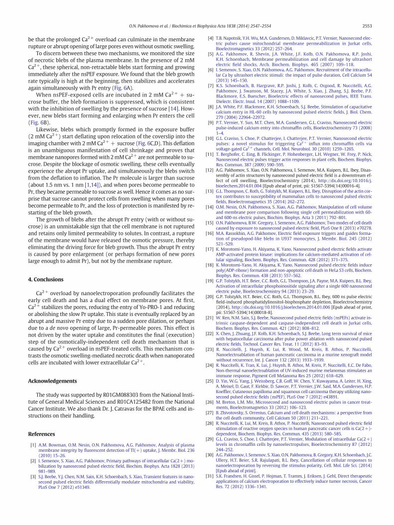
be that the prolonged Ca2+ overload can culminate in the membranerupture or abrupt opening of large pores evenwithout osmotic swelling.
To discern between these two mechanisms, we monitored the sizeof necrotic blebs of the plasma membrane. In the presence of 2 mMCa2+, these spherical, non-retractable blebs start forming and growingimmediately after the nsPEF exposure. We found that the bleb growthrate typically is high at the beginning, then stabilizes and acceleratesagain simultaneously with Pr entry (Fig. 6A).
When nsPEF-exposed cells are incubated in 2 mM Ca2+ + su-crose buffer, the bleb formation is suppressed, which is consistentwith the inhibition of swelling by the presence of sucrose [14]. How-ever, new blebs start forming and enlarging when Pr enters the cell(Fig. 6B).
Likewise, blebs which promptly formed in the exposure buffer(2 mM Ca2+) start deflating upon relocation of the coverslip into theimaging chamber with 2mM Ca2++ sucrose (Fig. 6C,D). This deflationis an unambiguous manifestation of cell shrinkage and proves thatmembrane nanopores formedwith 2mMCa2+ are not permeable to su-crose. Despite the blockage of osmotic swelling, these cells eventuallyexperience the abrupt Pr uptake, and simultaneously the blebs switchfrom the deflation to inflation. The Pr molecule is larger than sucrose(about 1.5 nm vs. 1 nm [1,14]), and when pores become permeable toPr, they became permeable to sucrose aswell. Hence it comes as no sur-prise that sucrose cannot protect cells from swelling when many poresbecome permeable to Pr, and the loss of protection is manifested by re-starting of the bleb growth.
The growth of blebs after the abrupt Pr entry (with or without su-crose) is an unmistakable sign that the cell membrane is not rupturedand retains only limited permeability to solutes. In contrast, a ruptureof the membrane would have released the osmotic pressure, therebyeliminating the driving force for bleb growth. Thus the abrupt Pr entryis caused by pore enlargement (or perhaps formation of new poreslarge enough to admit Pr), but not by the membrane rupture.
4. Conclusions
Ca2+ overload by nanoelectroporation profoundly facilitates theearly cell death and has a dual effect on membrane pores. At first,Ca2+ stabilizes the pores, reducing the entry of Yo-PRO-1 and reducingor abolishing the slow Pr uptake. This state is eventually replaced by anabrupt and massive Pr entry due to a sudden pore dilation, or perhapsdue to a de novo opening of large, Pr-permeable pores. This effect isnot driven by the water uptake and constitutes the final (execution)step of the osmotically-independent cell death mechanism that iscaused by Ca2+ overload in nsPEF-treated cells. This mechanism con-trasts the osmotic swelling-mediated necrotic deathwhen nanoporatedcells are incubated with lower extracellular Ca2+.
Acknowledgements
The study was supported by R01GM088303 from the National Insti-tute of General Medical Sciences and R01CA125482 from the NationalCancer Institute. We also thank Dr. J. Catravas for the BPAE cells and in-structions on their handling.
References
[1] A.M. Bowman, O.M. Nesin, O.N. Pakhomova, A.G. Pakhomov, Analysis of plasmamembrane integrity by fluorescent detection of Tl(+) uptake, J. Membr. Biol. 236(2010) 15–26.
[2] I. Semenov, S. Xiao, A.G. Pakhomov, Primary pathways of intracellular Ca(2+) mo-bilization by nanosecond pulsed electric field, Biochim. Biophys. Acta 1828 (2013)981–989.
[3] S.J. Beebe, Y.J. Chen, N.M. Sain, K.H. Schoenbach, S. Xiao, Transient features in nano-second pulsed electric fields differentially modulate mitochondria and viability,PLoS One 7 (2012) e51349.
[4] T.B. Napotnik, Y.H. Wu,M.A. Gundersen, D. Miklavcic, P.T. Vernier, Nanosecond elec-tric pulses cause mitochondrial membrane permeabilization in Jurkat cells,Bioelectromagnetics 33 (2012) 257–264.
[5] A.G. Pakhomov, R. Shevin, J.A. White, J.F. Kolb, O.N. Pakhomova, R.P. Joshi,K.H. Schoenbach, Membrane permeabilization and cell damage by ultrashortelectric field shocks, Arch. Biochem. Biophys. 465 (2007) 109–118.
[6] I. Semenov, S. Xiao, O.N. Pakhomova, A.G. Pakhomov, Recruitment of the intracellu-lar Ca by ultrashort electric stimuli: the impact of pulse duration, Cell Calcium 54(2013) 145–150.
[7] K.S. Schoenbach, B. Hargrave, R.P. Joshi, J. Kolb, C. Osgood, R. Nuccitelli, A.G.Pakhomov, J. Swanson, M. Stacey, J.A. White, S. Xiao, J. Zhang, S.J. Beebe, P.F.Blackmore, E.S. Buescher, Bioelectric effects of nanosecond pulses, IEEE Trans.Dielectr. Electr. Insul. 14 (2007) 1088–1109.
[8] J.A. White, P.F. Blackmore, K.H. Schoenbach, S.J. Beebe, Stimulation of capacitativecalcium entry in HL-60 cells by nanosecond pulsed electric fields, J. Biol. Chem.279 (2004) 22964–22972.
[9] P.T. Vernier, Y. Sun, M.T. Chen, M.A. Gundersen, G.L. Craviso, Nanosecond electricpulse-induced calcium entry into chromaffin cells, Bioelectrochemistry 73 (2008)1–4.
[10] G.L. Craviso, S. Choe, P. Chatterjee, I. Chatterjee, P.T. Vernier, Nanosecond electricpulses: a novel stimulus for triggering Ca2+ influx into chromaffin cells viavoltage-gated Ca2+ channels, Cell. Mol. Neurobiol. 30 (2010) 1259–1265.
[11] T. Berghofer, C. Eing, B. Flickinger, P. Hohenberger, L.H. Wegner, W. Frey, P. Nick,Nanosecond electric pulses trigger actin responses in plant cells, Biochem. Biophys.Res. Commun. 387 (2009) 590–595.
[12] A.G. Pakhomov, S. Xiao, O.N. Pakhomova, I. Semenov, M.A. Kuipers, B.L. Ibey, Disas-sembly of actin structures by nanosecond pulsed electric field is a downstream ef-fect of cell swelling, Bioelectrochemistry (2014), http://dx.doi.org/10.1016/j.bioelechem.2014.01.004 [Epub ahead of print, pii: S1567-5394(14)00016-4].
[13] G.L. Thompson, C. Roth, G. Tolstykh, M. Kuipers, B.L. Ibey, Disruption of the actin cor-tex contributes to susceptibility of mammalian cells to nanosecond pulsed electricfields, Bioelectromagnetics 35 (2014) 262–272.
[14] O.M. Nesin, O.N. Pakhomova, S. Xiao, A.G. Pakhomov, Manipulation of cell volumeand membrane pore comparison following single cell permeabilization with 60-and 600-ns electric pulses, Biochim. Biophys. Acta 3 (2011) 792–801.
[15] O.N. Pakhomova, B.W. Gregory, I. Semenov, A.G. Pakhomov, Twomodes of cell deathcaused by exposure to nanosecond pulsed electric field, PLoS One 8 (2013) e70278.
[16] M.A. Rassokhin, A.G. Pakhomov, Electric field exposure triggers and guides forma-tion of pseudopod-like blebs in U937 monocytes, J. Membr. Biol. 245 (2012)521–529.
[17] K. Morotomi-Yano, H. Akiyama, K. Yano, Nanosecond pulsed electric fields activateAMP-activated protein kinase: implications for calcium-mediated activation of cel-lular signaling, Biochem. Biophys. Res. Commun. 428 (2012) 371–375.
[18] K. Morotomi-Yano, H. Akiyama, K. Yano, Nanosecond pulsed electric fields inducepoly(ADP-ribose) formation and non-apoptotic cell death in HeLa S3 cells, Biochem.Biophys. Res. Commun. 438 (2013) 557–562.
[19] G.P. Tolstykh, H.T. Beier, C.C. Roth, G.L. Thompson, J.A. Payne, M.A. Kuipers, B.L. Ibey,Activation of intracellular phosphoinositide signaling after a single 600 nanosecondelectric pulse, Bioelectrochemistry 94 (2013) 23–29.
[20] G.P. Tolstykh, H.T. Beier, C.C. Roth, G.L. Thompson, B.L. Ibey, 600 ns pulse electricfield-induced phosphatidylinositol-bisphosphate depletion, Bioelectrochemistry(2014), http://dx.doi.org/10.1016/j.bioelechem.2014.01.006 [Epub ahead of print,pii: S1567-5394(14)00018-8].
[21] W. Ren, N.M. Sain, S.J. Beebe, Nanosecond pulsed electric fields (nsPEFs) activate in-trinsic caspase-dependent and caspase-independent cell death in Jurkat cells,Biochem. Biophys. Res. Commun. 421 (2012) 808–812.
[22] X. Chen, J. Zhuang, J.F. Kolb, K.H. Schoenbach, S.J. Beebe, Long term survival of micewith hepatocellular carcinoma after pulse power ablation with nanosecond pulsedelectric fields, Technol. Cancer Res. Treat. 11 (2012) 83–93.
[23] R. Nuccitelli, J. Huynh, K. Lui, R. Wood, M. Kreis, B. Athos, P. Nuccitelli,Nanoelectroablation of human pancreatic carcinoma in a murine xenograft modelwithout recurrence, Int. J. Cancer 132 (2013) 1933–1939.
[24] R. Nuccitelli, K. Tran, K. Lui, J. Huynh, B. Athos, M. Kreis, P. Nuccitelli, E.C. De Fabo,Non-thermal nanoelectroablation of UV-induced murine melanomas stimulates animmune response, Pigment Cell Melanoma Res 25 (2012) 618–629.
[25] D. Yin, W.G. Yang, J. Weissberg, C.B. Goff, W. Chen, Y. Kuwayama, A. Leiter, H. Xing,A. Meixel, D. Gaut, F. Kirkbir, D. Sawcer, P.T. Vernier, J.W. Said, M.A. Gundersen, H.P.Koeffler, Cutaneous papilloma and squamous cell carcinoma therapy utilizing nano-second pulsed electric fields (nsPEF), PLoS One 7 (2012) e43891.
[26] M. Breton, L.M. Mir, Microsecond and nanosecond electric pulses in cancer treat-ments, Bioelectromagnetics 33 (2012) 106–123.
[27] B. Zhivotovsky, S. Orrenius, Calcium and cell death mechanisms: a perspective fromthe cell death community, Cell Calcium 50 (2011) 211–221.
[28] R. Nuccitelli, K. Lui, M. Kreis, B. Athos, P. Nuccitelli, Nanosecond pulsed electric fieldstimulation of reactive oxygen species in human pancreatic cancer cells is Ca(2+)-dependent, Biochem. Biophys. Res. Commun. 435 (2013) 580–585.
[29] G.L. Craviso, S. Choe, I. Chatterjee, P.T. Vernier, Modulation of intracellular Ca(2+)levels in chromaffin cells by nanoelectropulses, Bioelectrochemistry 87 (2012)244–252.
[30] A.G. Pakhomov, I. Semenov, S. Xiao, O.N. Pakhomova, B. Gregory, K.H. Schoenbach, J.C.Ullery, H.T. Beier, S.R. Rajulapati, B.L. Ibey, Cancellation of cellular responses tonanoelectroporation by reversing the stimulus polarity, Cell. Mol. Life Sci. (2014)[Epub ahead of print].
[31] S.K. Frandsen, H. Gissel, P. Hojman, T. Tramm, J. Eriksen, J. Gehl, Direct therapeuticapplications of calcium electroporation to effectively induce tumor necrosis, CancerRes. 72 (2012) 1336–1341.
2553O.N. Pakhomova et al. / Biochimica et Biophysica Acta 1838 (2014) 2547–2554

[32] B.L. Ibey, C.C. Roth, A.G. Pakhomov, J.A. Bernhard, G.J. Wilmink, O.N. Pakhomova,Dose-dependent thresholds of 10-ns electric pulse induced plasma membrane dis-ruption and cytotoxicity in multiple cell lines, PLoS One 6 (2011) e15642.
[33] O.N. Pakhomova, B.W. Gregory, V.A. Khorokhorina, A.M. Bowman, S. Xiao, A.G.Pakhomov, Electroporation-induced electrosensitization, PLoS One 6 (2011)e17100.
[34] C. Delteil, J. Teissie, M.P. Rols, Effect of serum on in vitro electrically mediated genedelivery and expression in mammalian cells, Biochim. Biophys. Acta 1467 (2000)362–368.
[35] A.G. Pakhomov, A.M. Bowman, B.L. Ibey, F.M. Andre, O.N. Pakhomova, K.H.Schoenbach, Lipid nanopores can form a stable, ion channel-like conduction path-way in cell membrane, Biochem. Biophys. Res. Commun. 385 (2009) 181–186.
[36] A.G. Pakhomov, O.N. Pakhomova, Nanopores: a distinct transmembrane passage-way in electroporated cells, in: A.G. Pakhomov, D.Miklavcic, M.S. Markov (Eds.), Ad-vanced Electroporation Techniques in Biology in Medicine, CRC Press, Boca Raton,2010, pp. 178–194.
[37] A.G. Pakhomov, J.F. Kolb, J.A. White, R.P. Joshi, S. Xiao, K.H. Schoenbach, Long-lastingplasma membrane permeabilization in mammalian cells by nanosecond pulsedelectric field (nsPEF), Bioelectromagnetics 28 (2007) 655–663.
[38] K. Kinosita Jr., T.T. Tsong, Hemolysis of human erythrocytes by transient electricfield, Proc. Natl. Acad. Sci. U. S. A. 74 (1977) 1923–1927.
2554 O.N. Pakhomova et al. / Biochimica et Biophysica Acta 1838 (2014) 2547–2554