CALCIUM AND IRON INCRUSTATION AND OTHER · 2016-06-02 · CALCIUM AND IRON INCRUSTATION AND OTHER...
18
CALCIUM AND IRON INCRUSTATION AND OTHER LESIONS OF THE ELASTIC TISSUE OF THE SPLEEN AND LIVER.* By THOMAS P. SPRUNT, M.D. (From the Laboratory of Pathology, Yohns Hopkins University, Baltimore). PL.A~ES 4 TO 7. This report is intended as a contribution to vascular pathology and to the pathology of elastic tissue. The principal lesion described is evidently an unusual one, since a search through the litera- ture has revealed very few similar observations, and indeed only one in connection with the spleen, namely, that reported by Marini in a case of splenomegaly, which he regarded as an example of Banff's disease. Kockel and Bittrolff have reported analogous but less extensive processes occurring in the lungs in chronic passive congestion and in pernicious anemia. The subject from which the material to be described was obtained was autopsied at the Bay View Hospital on November II, 1910, four hours after death. Only a meagre clinical history is available. The patient was a white man, 59 years old, who for five months had suffered from "stomach trouble," anorexia, loss of weight, weakness, and shortness of breath. Examination showed a large protruding abdomen and marked dis- tention of the superficial veins. The spleen was palpable over a hand's breadth below the costal margin. The abdomen was full of fluid and there was some edema of the lower extremities. The anatomical diagnosis follows. Cholelithiasis (stone in common duct); dilatation of gall bladder and ducts; cholangeitis; cirrhosis of liver; marked dilatation of portal vessels; congestion of area supplied by portal vein with edema of intestines and mesentery; ascites; chronic splenic tumor; rupture of esophageal variees; acute and chronic pan- creatitis; bronchopneumonia; arteriosclerosis, especially of smaller arteries, e. g., coronaries and splenic; chronic diffuse myocarditis; congenital cystic kidneys ; edema and fading purpuric eruption on legs ; emaciation ; slight icterus. Only those organs of especial interest will be described further in this report. * Received for publication, April 27, IgII. 59
Transcript of CALCIUM AND IRON INCRUSTATION AND OTHER · 2016-06-02 · CALCIUM AND IRON INCRUSTATION AND OTHER...

C A L C I U M A N D I R O N I N C R U S T A T I O N A N D O T H E R
L E S I O N S O F T H E E L A S T I C T I S S U E O F T H E
S P L E E N A N D L I V E R . *
By THOMAS P. SPRUNT, M.D.
(From the Laboratory of Pathology, Yohns Hopkins University, Baltimore).
PL.A~ES 4 TO 7.
This repor t is intended as a contr ibut ion to vascular pa thology
and to the pa thology of elastic tissue. The principal lesion described
is evidently an unusual one, since a search th rough the l i tera-
ture has revealed very few similar observat ions, and indeed only
one in connection with the spleen, namely, that repor ted by Mar in i
in a case of splenomegaly, which he regarded as an example of
Banff ' s disease. Kockel and Bit trolff have repor ted analogous but
less extensive processes occurr ing in the lungs in chronic passive
congest ion and in pernicious anemia.
The subject f r o m which the mater ia l to be described was obtained
was autopsied at the Bay View Hosp i t a l on N o v e m b e r I I , 1910,
four hours a f t e r death. Only a meagre clinical h is tory is available.
The patient was a white man, 59 years old, who for five months had suffered from "stomach trouble," anorexia, loss of weight, weakness, and shortness of breath. Examination showed a large protruding abdomen and marked dis- tention of the superficial veins. The spleen was palpable over a hand's breadth below the costal margin. The abdomen was full of fluid and there was some edema of the lower extremities. The anatomical diagnosis follows.
Cholelithiasis (stone in common duct); dilatation of gall bladder and ducts; cholangeitis; cirrhosis of liver; marked dilatation of portal vessels; congestion of area supplied by portal vein with edema of intestines and mesentery; ascites; chronic splenic tumor; rupture of esophageal variees; acute and chronic pan- creatitis; bronchopneumonia; arteriosclerosis, especially of smaller arteries, e. g., coronaries and splenic; chronic diffuse myocarditis; congenital cystic kidneys ; edema and fading purpuric eruption on legs ; emaciation ; slight icterus.
Only those organs of especial interest will be described fu r the r in this report.
* Received for publication, April 27, IgII.
59

60 Calcium and Iron Incrustation of the Liver.
After opening the duodenum, the bile papilla was found to be quite normal in appearance. All the bile passages were much dilated and stained a bright yellow color. Just below the juncture of (he hepatic and cystic ducts, there was an oval concretion measuring 2 by I cm., which was freely movable and which might easily have been pushed up from a position near the ampulla during the examination. The liver was somewhat enlarged, weighing 1,7oo grams; it was greenish yellow, hard, and firm, with a slightly granular surface. The cut surface showed a rather obscured lobulation with thin strands of bluish white tissue between the lobules. Opaque yellowish areas, I or 2 mm. in diameter, with dark red borders were seen here and there. The portal vein and its radicals were enormously dilated and somewhat tortuous.
The spleen was much enlarged, measuring 21 by I3I/2 by 7~ cm., and weigh- ing 775 grams. The surface, especially of the upper pole, was covered with small, elevated, yellowish white nodules. The color of the cut section was brownish red. The pulp was diminished and the interstitial tissue correspondingly in- creased in amount. The consistence was somewhat increased, but there was no suggestion of the rubbery spleen of chronic passive congestion. The Mal- pighian bodies were small and widely spaced. Both capsule and trabeculze were decidedly thickened and prominent and in the latter there were frequently seen small ochre-colored paiches, I or 2 ram. in diameter, in the center of which a small vessel appeared.
IIISTOLOGICAL DESCRIPTION.
Spleen. - -The capsule and trabeculm are much thickened. The
outlines of the sinuses are quite distinct and their endothelial nuclei
unusually prominent. The sinuses contain red blood cells and an
occasional lymphocyte. In the pulp spaces there are very few cells,
consisting of the usual lymphocytes and splenic pulp cells.
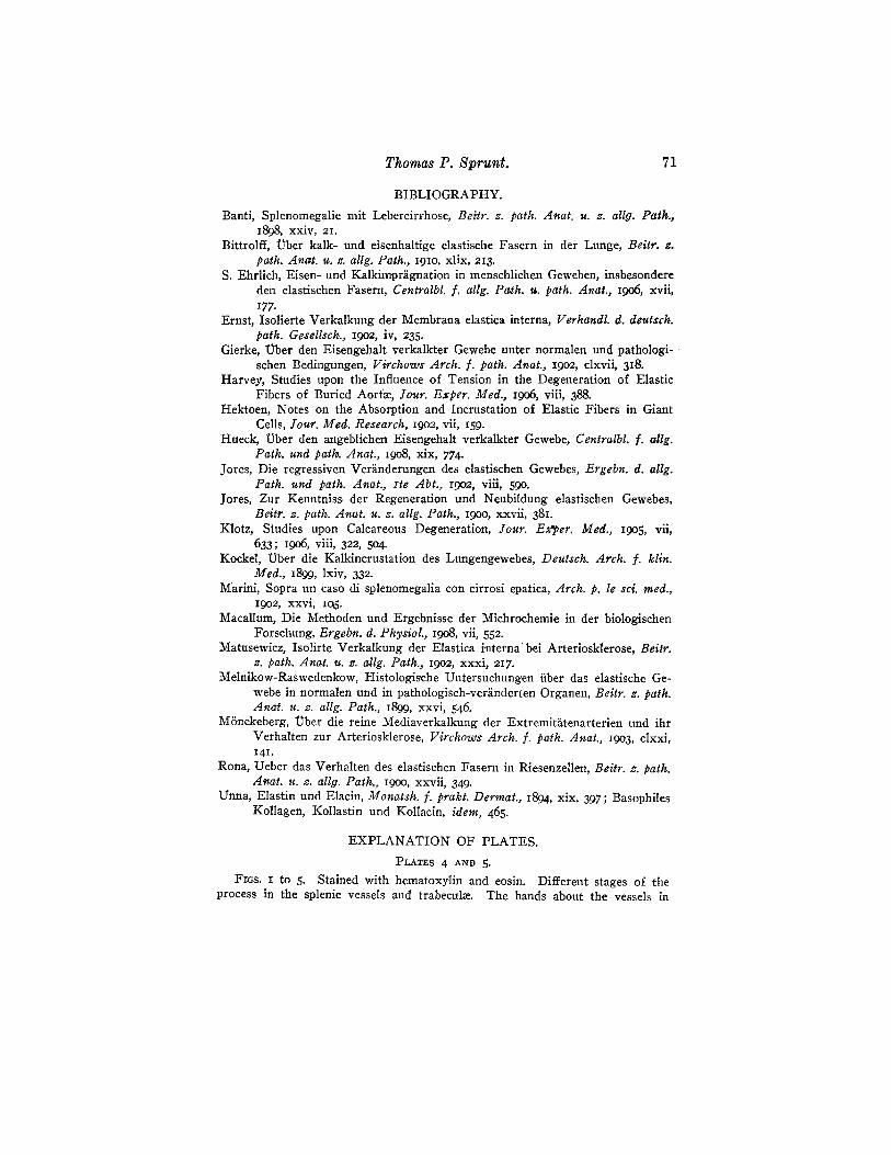
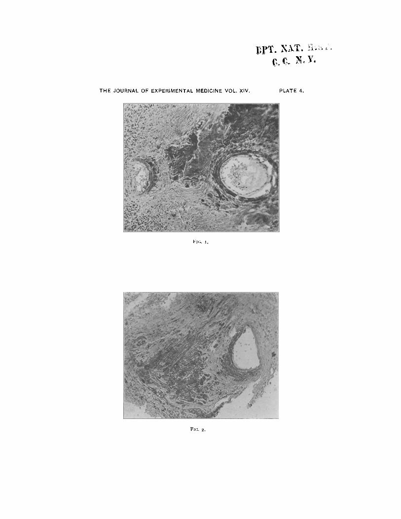
The striking lesion is found in those areas visible to the naked
eye as ochre-colored patches. This impresses one at first as a
peculiar deposit of pigment in the trabecul~e, and especially in the
walls of vessels having a diameter of f rom one tenth to one sixth
of a millimeter (figures I to 4)- In hematoxyl in and eosin prepara-
tions, the lumen of the affected vessels is usually found to be smooth
and regular and lined by a thin intima. Just outside the intima there
are found concentric, circular na r row bands of a glistening, golden
or yellowish green color which may or may not entirely encircle the
vessel. They are not absolutely continuous but appear more as
broken lines or small pieces joined end to end. Beyond this zone
the tissues of the trabeculm have a very peculiar appearance. T h e y
consist of swollen bands or strands, which run sometimes around

Thomas P. Sprunt. 61
the vessel and are sometimes quite irregular in their course, often crossing each other at various angles. They may be broken up into round or oblong masses. Some have the golden, glistening appear- ance already noted, others take a deep hematoxylin stain, either homogeneously or in clumps of granules here and there. They go over rather abruptly into the more normal appearing trabecular tissue. Between the broader bands and masses a delicate blue-stain- ing fibrillar network may occasionally be demonstrated. The lesion is found not only in the vessels of the trabecul~e, but also in many of the arteries within the splenic pulp and often at a point just before they divide into the smaller intralobular branches.
The vessels, which are only partially affected, are particularly instructive, for here it is seen that the elastic tissue is especially concerned. The internal elastic lamella may be traced from the normal wavy, refractile membrane into a less wavy, blue-staining fiber, which goes over directly into the rigid, broken, impregnated structure already described (figure 5). The same transition may be traced in other elastic fibers of the vessel wall, the outer fibers often appearing broken and impregnated, while the immediately adjacent internal lamella is relatively normal. In other places, where the lesion is apparently recent, a vessel is surrounded by two or three concentric bands, and in the trabecula many hematoxylin- staining fibers appear, more or less wavy and a little broader than the refractile elastic fibers with which they are continuous. In addition, there is seen here and there a blue-staining fibrillar net- work and many connective tissue cells filled with a golden, granular pigment.
While, as a rule, the bands or fibers near the lumen of the vessel appear yellowish green and refractile and those further removed stain with hematoxylin, this is by no means invariable. The inner layers may be homogeneousely blue and the outer heavy bands in places possess the glistening greenish characteristics. Either one may be present exclusively. In unstained sections, the impregnated tissues are conspicuous and of a beautiful golden color, which is quite homogeneous in some of the bands, while in others it occurs in globules or even in clumps and strings of small granules. Be-

62 Calcium and Iron Incrustation of the Liver.
tween and around the heavier bands, a delicate golden network sug- gests the elastic fibrils.
M I C R O C H E M I S T R Y .
The affected tissues give a pronounced reaction for iron with ammonium sulphid (figure 6), potassium ferrocyanid and hydro- chloric acid, and with aqueous hematoxylin. With the ferricyanid of potassium, only a very weak reaction is obtained.
The impregnating salts are readily dissolved by dilute mineral acids without the formation of gas bubbles. If sulphuric acid is used, there is a deposit of the needle-shaped crystals of calcium sulphate, and after dissolving out the iron in half-saturated aqueous solution of oxalic acid, the calcium salts may be demonstrated by the violet stain with aqueous hematoxylin. This is found especially in those areas which with hematoxylin and eosin take a deep hema- toxylin stain. After prolonged treatment with oxalic acid, crystals of calcium oxalate are deposited.
Phosphates are easily shown by treating sections with a 3 per cent. solution of silver nitrate. After exposure to light, the en- crusted tissues become black. I f T. 5 per cent. of nitric acid is added to the silver nitrate solution in the attempt to differentiate chlorids, which are insoluble in dilute nitric acid while the phosphates are soluble, the reaction is no longer obtained. A certain amount of pigment in the peripheral parts of the trabeculae remain unchanged. By using the method for demonstrating phosphates recommended by Macallum, the exposure of sections to a solution of ammonium molybdate in nitric acid and subsequent treatment in a dilute solu- tion of phenylhydrazin-hydrochlorid, the reaction is positive throughout most of the impregnated tissues, which become greenish blue in color. The bands which in other preparations did not take the hematoxylin stain behave somewhat differently. Almost imme- diately after immersion in the first fluid, the glistening golden material seems to be partially dissolved and then deposited in small irregularly round masses slightly larger than red blood cells, which, transmitting very little light, appear almost black. Those which are broken up seem to consist of small yellow granules quite similar to the original material. After treatment with phenylhydrazin, they

Thomas P. Sprunt. 63
become bluish in color. Much of the golden material, however, is not so changed and remains in its original position and of the same appearance. So far as may be judged, then, by these reactions, the metals, calcium and iron, are present chiefly as phosphates.
ELASTIC TISSUE.
In studying the elastic tissue, Weigert's, Verhoeff's, and Unna's orcein methods were used. By first treating sections with potas- sium ferrocyanid and hydrochloric acid and then staining with orcein, the iron-containing tissues are colored blue and the elastic tissue becomes a silky brown. By this method the relations between the two are best studied, especially in the less advanced lesions. Here the brown elastic fibers are directly continuous with the blue, iron-containing tissues, some of which preserve something of the characteristic morphology of elastic fibers. I f Perl's stain is not used, the acid content of the Weigert and orcein stains dissolves the salts from the petrified tissues, which then become prominent by reason of their lack of elastic staining. The innermost layer of the int'ernal elastica usually retains the stain, appearing stiff and much thickened without a trace of its usual wavings, and occasion- ally one of the thick bands in the trabecul~e close by, colors intensely with the elective stain. Small elastin granules are sometimes nume- rous. In hematoxylin and eosin preparations, the demineralised tissues have a hyaline appearance.
With Grfibler's polychrome methylene-blue, the incrusted tis- sues retain a deep blue stain after all the other tissues are decol- orised, thus giving the so-called "e lac in" reaction of Unna. The reaction is negative in sections previously treated with acid.
The collagenous and muscular tissues must be affected in the advanced lesion, but from a study of the earlier stages it seems clear that the elastic tissue is primarily concerned.
The elastic tissue of the thickened capsule and trabecul~e generally throughout the sections is not only absolutely but also relatively increased. The network is denser, each fiber being larger and stain- ing more intensely than in the normal spleen. About the intra- lobular art.cries there appears very frequently a pronounced net-

64 Calcium and Iron Incrustation of the Liver.
work, which is also sometimes conspicuous about the periphery of a Malpighian corpuscle.
Retrogressive changes in the elastic tissue, other than the swell- ing and incrustation of its fibers, may be noted. The internal elastic lamella of a vessel is frequently represented by two distinct, thick, wavy lines, but in this case it is often divided into four or five parallel wavy strands. In a few instances in the larger vessels, it is split up into eight or ten fibrils which form a spindle in the course of the elastica. Into this spindle the fibrils run, become straight, more and more beaded, and finally break up into small granules which retain the elective stains. Such a spindle may be a third of a milli- meter in length (figure 7)- Somewhat similar appearances are described by M6nckeberg in the internal elastica of peripheral arteries over calcified areas in the media.
Again, in certain trabeculae it is found that the dense, deeply staining network goes over rather suddenly into an area in which the fibrils are delicate, lightly staining, widely spaced, straighter, and arranged in a less definite network. With the combined Perl's and orcein stain many of these fibrils show blue masses adhering to them or incrusting them, and for a short distance the fibril may stain blue instead of brown. This finding is more frequent and more marked in such atrophic areas than in the more nearly normal trabecuhe.
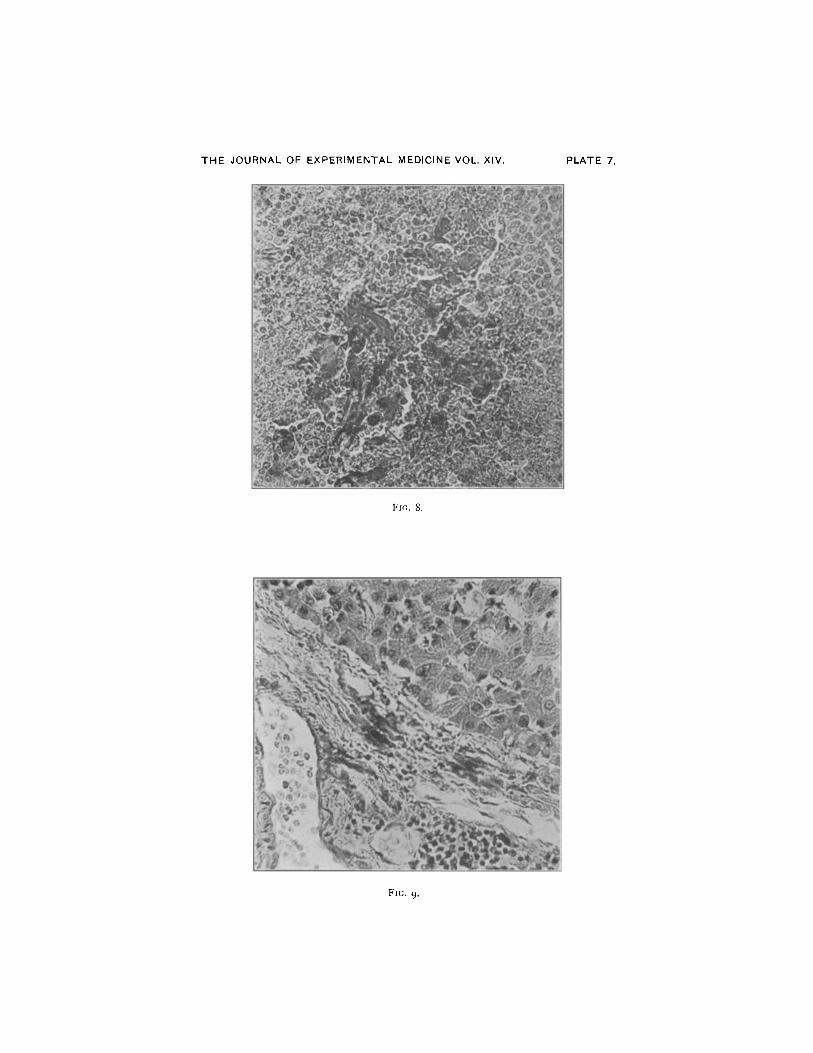
The Malpighian corpuscles are far apart and most of them are normal. Near the center of some of them, the lymphoid cells seem forced apart by a hyaline material staining pink with eosin, which may occur in small, refractile globules or be fused into larger areas half a millimeter in diameter, in which are embedded here and there nuclei of lymphoid cells. With Perl's and orcein stains, a few small, beaded, elastic fibers are seen in direct connection with these areas, which stain, however, a little less intensely with the orcein. Irregu- larly round, iron-containing bodies about six times as large as a lymphoid cell are occasionally found in this hyaline substance (figure 8).
Liver . - -The cirrhosis is an early and, therefore, interesting ex- ample of the type usually known as biliary cirrhosis. The bile ducts are dilated and often filled with polymorphonuclear leucocytes,
Thomas P. Sprunt. 65
which may extend into the sur rounding tissues. The new connec-
tive tissue is cellular and principally localized in the periportal areas,
f rom which fibroblasts may be seen g rowing in between the strands
of liver cells. I n addition, there is a recent, extensive, central
necrosis in many, but not in all, of the lobules. In the denser con-
nective tissue strands there are occasionally seen small, oblong or
rectangular structures which stain intensely with hematoxylin.
Some of these appear as dark blue, wavy fibers (figure 9) .
The elastic tissue is present in increased amounts in the connec-
tive tissue strands, but the fibrils are of ten broken into fine granules.
Apparent ly the same small, oblong or wavy structures stain intensely
b rown with orcein. I n unstained sections, they appear yellowish
and refracfile. In sections treated with silver nitrate, they become
black af te r exposure to light. These calcified elastic fibers in the
liver do riot give an iron reaction.
The selective impregnat ion of the elastic fibers with mineral salts
is in str iking contrast to the calcification usually observed in the
walls o f vessels.
M6nckeberg states that in calcification of the media in peripheral arteries the elastic tissue is the last constituent of the media to be affected. Klotz emphasizes the immunity of elastic fibers in primary medial calcification of arteries of the elastic tissue type. The lesion here described is not confined to the vessels but affects the tissues of the splenic trabeculze to even a greater extent, and, there- fore, may not strictly be compared to the usual forms of vascular disease.
Marini describes a similar but much less extensive change in the elastic tissue of a very large spleen whicl~ weighed 1,9oo grams. There was also an atrophic cirrhosis of the liver. The principal lesions in the spleen were in the Malpighian corpuscles, which were sclerotic from a new growth of both collagenous and elastic fibers. In the arterioles of the follicles in many places, he says, the intima, media, and adventitia had disappeared and were replaced by pieces of a substance which in hematoxylin and eosin preparations appeared yellowish and refractile to light. Tests for calcium and for carbonates were negative. The substance was present only in places frown which the elastic tissue had disappeared; it gave a pronounced iron reaction as well as the reaction for the degenerated material of elastic tissue, which Unna designated as "elacin." Both of these reactions dis- appeared after treating the sections with acid. Marini thought that the process might be explained by the chemical transformation of elastin into elacin, and that the iron might' have been derived from some iron content of the elastin from which the elacin was derived, or that it was due to blood pigment.
Kockel and, recently, Bittrolff have each described a case of calcium and iron incrustation of the elastic fibers in lungs which were the seat of chronic passive congestion. The lesion was present in the walls of small vessels and in the

66 Calcium and Iron Incrustation of the Liver.
elastic tissue of the alveolar walls. It was more extensive in Bittrolff's case, and the appearances described and pictured by him are in many respects quite similar to those reported in this paper. In using Perl's and orcein combination stain, he found that some of the elastic fibers colored brown in part of their course, blue in other parts, and a mixed color in the transitions from the one into the other. He aptly compares the appearance of the broken incrusted elastic fibers to that of chains of anthrax bacilli. A striking feature of his case was the presence of many large, multinucleated giant cells of the foreign body type, each containing broken fragments of the degenerated elastic fibers. Giant cells were present in Kockel's case, but not in such great numbers. Kockel examined a series of lungs and found similar lesions of very slight degree in three with chronic passive congestion.
There are several other observations in the literature of calcification of elastic fibers, and in some of them iron was also demonstrated. Jores, Ernst, and Matusewicz describe isolated, calcification of the membrana elastica interna. Jores considers this of frequent occurrence in the vessels of goitres and has observed it in arteriosclerosis, although in most cases of sclerosis the internal elastic lamella is well preserved.
Rona and Hektoen report calcified and iron-containing elastic fibers in giant cells in various inflammatory processes. In examining a series of cases of lupus, Rona found this lesion in three fourths of the cases. Since he saw no calcified fibers outside of the giant cells, he came to the conclusion that the calcification had occurred within the cells, that the slower metabolism within the giant cells probably accounted for the persistence of the elastic elements enclosed within them, while those outside were early destroyed by the bacterial poisons. In several of his cases iron was also demonstrable in the calcified fibers and usually in those lesions from the lower extremities. He thinks that chronic passive congestion and hemorrhagic inflammations are of importance in deter- mining its presence.
S. Ehrlich reports several instances of simultaneous calcium and iron incrus- tation of elastic fibers in scars of splenic infarctions, in an intestinal polyp, in the alveolar walls of the lungs, and in other lesions. He says his observations show that elastic fibers in the neighborhood of hemorrhages have a special tendency to absorb iron and that such fibers may secondarily become the seat of calcium deposits. He agrees with Schmorl that the iron deposit precedes that of the calcium for which it acts as a kind of mordant.
Gierke found a positive iron reaction not only in many examples of patho- logical calcification but also in certain physiological calcium deposits, notably in fetal bones, and in the embryonic enamel and dentine of guinea pigs.
Hueck, however, protests against the interpretation that such reactions indi- cate that iron occurs along with calcium during life, and he insists that most of the reported findings are artifacts due to the absorption of iron by the calcified tissues from the fluids in which the tissue is fixed or preserved. He points out several sources of error, stating that the reagents obtained from the best manu- facturers are not always iron-free and that, if the tissue contains blood pigment, this may be dissolved by the fluids used and subsequently be absorbed by the calcified areas. He bases his arguments upon his own investigations of fresh material with negative results and upon a rather convincing experiment. He

Thomas P. Sprunt. 67
examined the tissues from young mice in two parallel series, in one of which the sections were cut in paraffin and in the other in celloidin. In the paraffin sections, which had remained in the fluids only a few days, there was no iron. The celloidin sections were exposed to the reagents longer and showed an intense iron reaction only in the calcified areas. On examining the absolute alcohol used for the celloidin tissues, he foun~d that it contained iron.
Hueck's criticisms apply with most force to those reports of positive iron reactions occurring in physiologically calcified tissues. Bittrolff remarks that there seems no good reason why the calcified, presumably necrotic, elastic fibers in his case might not absorb iron during the life of the patient from the fluids rich in iron, in which they were constantly bathed, if calcified tissues show such a predilection for iron after death. It may be added here that in this case the ochre color of the lesion at autopsy, a few hours after death, and the fact that tissue fixed at once in formalin and examined the next day showed as much iron as that examined several months later, indicate clearly that in such lesions the iron and calcium are cofixistent d, uring the life of the subject.
I t is difficult to de termine which process is p r imary . T h e appar -
ent p redominance of the i ron in this case and the occurrence o f i ron
wi thout calcium in some isolated fibers of the splenic trabecul~e
migh t lead one to consider the i ron of p r ime importance. Ehrl ich,
too, claims that iron incrustat ion m a y occur independent ly of calcifi-
cation. On the o ther hand, we know tha t calcified tissues in gen-
eral are usually free f r o m iron or show small t races o f it. I n this
case, we have calcified elastic fibers in t h e liver, but no iron. P rac -
t ically all the repor ted observat ions o f i ron incrus ta t ion of elastic
fibers have been in o rgans or tissues showing ei ther hemor rhages
or m a r k e d congest ion; Kockel ' s and ]3ittrolff 's in congested lungs,
Mar in i ' s and m y case in spleens with cirrhot ic livers, H e k t o e n ' s in
a hemor rho ida l nodule, Ehr l ich ' s in a hemor rhag ic rectal polyp,
splenic infarct ion, etc. R o n a ' s explanat ion, a l ready ment ioned,
fo r the i ron deposited in g iant cells in lupus is the same. I t seems
probable, then, that iron is absorbed secondari ly by the calcium
deposits when the su r round ing tissues are rich in tha t e lement in
inorganic combinat ion, but Mar in i ' s and some o f Ehr l i ch ' s observa-
tions do not pe rmi t us to exclude the possibili ty tha t i ron m a y
incrust elastic fibers wi thout the media t ion o f ca lc ium?
1Since the preparation of this paper, there has come to the author's atten- tion an article by Sumlta, " Zur Frage des Eisenreaction kalkhaltiger Gewebe, insbesondere des Knochens," Virchows Arch. f. path. Anat., 1910, cc, 220. On the basis of careful experiments, Sumita concludes that the deposit of iron precedes that of calcium not only under pathological conditions but also in the physiological formation of bone.

68 Calcium and Iron Incrustation of the Liver.
I f we assume the primary character of the calcium deposits, the reason for their occurrence is quite as obscure as the cause of calci- fication in general. It is a generally accepted principle in pathology that only extremely degenerated or necrobiotic tissue becomes the seat of pathological calcium deposits. In the apparently advancing margins of this lesion, however, it was not possible to demonstrate by the methods used any abnormality of the elastic tissue until after the deposit of the salts. Chemical change in the petrified fibers is shown by their failure to take the selective elastic stains and by the deep blue stain with methylene-blue in alkaline solution, which does not affect the normal elastic tissue. This degenerated substance Unna speaks of as elacin, in contradistinction to the elastin of the normal tissue. This reaction disappeared simultaneously with the iron reaction after treating sections with acids, but it can not depend entirely upon the presence of inorganic iron, since this substance, under other conditions, in tissues near hemorrhages, in hemochro- matosis, etc., does not give the reaction. The intracellular iron pigment in the same sections fails to take the blue stain. Ordinary calcified areas, too, lose the stain when the nuclei are decolorized. In view of the work of Klotz on calcification, a careful search was made for any fatty substances which might be present, but none were demonstrated. Frozen sections of formalin-fixed tissues were stained in Sudan III for twenty-four hours, both before and after dissolving out the salts in different acids.
A puzzling feature of these cases is the occurrence in some of them of great numbers of giant cells, and of their entire absence in others. Kockel, Hektoen, and Bittrolff speak of them as phago- cytic for the degenerated elastic elements. Rona and Unna do not accept any such activity for the giant cells in lupus, and they think the fibers which chance to be enclosed within the cells are protected to a certain extent from the injurious agents which cause the rapid degeneration of those outside the cells. No giant cells were seen in the neighborhood of the incrusted fibers in this case, nor were there any other appearances indicative of a foreign body reaction.
The hyperplasia of elastic tissue in the spleen and liver is in har- mony with the results reported by Melnikow-Raswedenkow, who investigated the elastic tissue of a number of organs under normal

Thomas P. Sprunt. 69
and pathological conditions. The greatest hyperplasia in the spleen was found in chronic splenic tumors occurring as a result of cir- rhosis of the liver. An elastic tissue increase was the rule also in cirrhotic livers. The author referred the growth of elastic tissue to mechanical causes; increased pressure due to congestion, for example. It is possible that in this case the condition of strain resulting from the early stages of congestion was responsible for the hyperplasia of the elastic elements and later, passing into a condition of stress, brought about the regressive changes, which resulted in the calcium and iron incrustation. In this connection the great dilatation and slight sclerosis of the portal and splenic veins should be remembered.
The effect of tension upon degenerative changes of elastic tissue has been studied experimentally by Harvey. He buried guinea pigs' aortas, both collapsed and distended under pressure, within the tissues of other animals of the same species and examined the tissues at different time intervals. He found that the elastic fibers were much more resistant than other portions of the vessel wall and that this power of resistance was much lowered when the tissue was placed under tension. The collapsed aorta, examined after seventy- five days, showed beginning calcification of some of the heavier elastic fibers, and after Io6 days there was extensive calcification. The aortas buried under tension showed complete disappearance of the elastic tissue after fifty days. Those examined after shorter intervals showed no calcification.
The lesion in Marini's case might be explained as a result of the action of the hypothetical toxin which, according to Banti, is respon- sible for all the features of the disease that goes by his name, includ- ing the splenic enlargement, anemia, sclerosis of the splenic and portal veins, and cirrhosis of the liver. It seems possible, if not altogether probable, that the case of this report may also be an example of primary splenomegaly and that the splenic lesions are explicable on this basis. The size and gross appearance of the spleen, the histological picture in general, and the lesions in the Malpighian corpuscles, all support this view. The cirrhosis of the liver is not of the type secondary to splenic enlargement, but seems definitely due to the infectious process in the bile passages, with an

7O Calcium and Iron Incrustation of the Liver.
added terminal central necrosis. However, the cirrhosis seems to be of quite recent origin, the lobulation is well preserved, and the newly formed connective tissue is limited to the periportal areas and is quite cellular. The relatively advanced changes in the spleen seem to be out of all proportion to the grade of cirrhosis in the liver. On the other hand, there was definite portal obstruction with ascites, marked esophageal varices, and dilatation of the superficial abdominal veins. In the absence of a clinical history, we must leave it an open question, whether we have a primary splenomegaly with a superadded biliary cirrhosis or whether the whole condition is the result of the hepatic disease.
For reviews of the subjects of progressive and regressive changes of elastic tissue, the reader is referred to the papers by Jores. I have seen no other reported observation of calcified elastic fibers in the cirrhotic liver.
SUMMARY.
In conclusion, it may be said that the lesion described consists of a selective impregnation of the elastic tissue in the splenic trabeculm and vessels with the phosphates--and perhaps other sal ts--of cal- cium and iron. There is a further chemical change in the elastic fibers which may be expressed by the term "elacin."
Degenerated and, especially, calcified elastic tissue---even more than calcification in general--seems to possess an affinity for iron salts. The iron incrustations occur principally in congested or hemorrhagic tissues.
The etiological agent in this case is obscure. The only other example of this lesion in .the spleen was in a case of Banti's disease. Many features of this case suggest a primary splenomegaly, but the evidence in its favor is not conclusive.
Calcified elastic fibers, without the iron and elacin reactions, were also present in the liver, which was the seat of a recent, apparently biliary or infectious, cirrhosis. The elastic tissue of other organs showed nothing remarkable except in the skin, where the fibers were undergoing the usual senile changes.
T h o m a s P . S p r u n t . 71
BIBLIOGRAPHY'.
Banti, Splenomegalie mit Lebercirrhose, Beitr. z. path. Anat. u. z. allg. Path., 1898, xxiv, 2I.
Bittrolff, tAber kalk- und eisenhaltige elastische Fasern in der Lunge, Beltr. z. path. Anat. u. z. allg. Path., I9IO, xlix, 213.
S. Ehrlich, Eisen- und KalkimpHignation in menschlichen Geweben, insbesondere den elastischen Fasern, Centralbl. f. allg. Path. **. path. Anat., 19o6, xvii, 177.
Ernst, Isolierte Verkalkung der Membraaa elastica interna, Yerhandl. d. deutsch. path. Gesellsch., 19o2, iv, 235.
Gierke, Ober den Eisengehalt verkalkter Gewebe unter normalen und pathologi- schen Bedingungen, Virchows Arch. f. path. Anat., 19o2, clxvii, 318.
Harvey, Studies upon the Influence of Tension in the Degeneration of Elastic Fibers of Buried Aor/ae, ]our. Exper. Med., 19o6, viii, 388.
Hektoen, Notes on the Absorption and Incrustation of Elastic Fibers in Giant Cells, ]our. Med. Research, 19o2, vii, 159.
Hueck, lJber den angeblichen Eisengehalt verkalkter Gewebe, Centralbl. f. allg. Path. und pat& Anat., 19o8, xix, 774.
Jores, Die regressiven Veriinderungen des elastischen Gewebes, Ergebn. d. allg. Path. und path. Anat., Ite Abt., 19o2, viii, 59o.
Jores, Zur Kenntniss der Regeneration und Neubildung elastischen Gewebes, Beitr. z. path. Anat. u. z. allg. Path., 19oo, xxvii, 381.
Klotz, Studies upon Calcareous Degeneration, ]our. Ex'per. Med., 19o5, vii, 633; 19o6, viii, 322, 504.
Kockel, ~ber die Kalkincrustation des Lungengewebes, Deutsch. Arch. f. klln. ivied., 1899, lxiv, 332.
Marini, Sopra un caso di splenomegalia con cirrosi epatica, Arch. p. le sci. reed., 19o2, xxvi, lO,5.
Macallum, Die Methoden und Ergebnisse der Michrochemie in der biologischen Forschung, Ergebn. d. PhysioL, 19o8, vii, 552.
Matusewicz, Isolirte Verkalkung der Elastica iaaterna bei Arteriosklerose, Beitr. z. path. Anat. u. z. allg. Path., 19o2, xxxi, 217.
Melnikow-Raswedenkow, Histologische Untersuohungen fiber das elastische Ge- webe in normalen und in pathologisch-veriinderfen Organen, Beitr. z. path. Anat. u. ~. allg. Path., 1899, xxvi, 546.
M6nckeberg, l~ber die reine Mediaverkalkung der Extremit~itenarterien und ihr Verhalten zur Arteriosklerose, V{rchows Arch. ~. path. Anat., 19o3, clxxi, 141.
Rona, Ueber das Verhalten des elastischen Fasern in Riesenzellen, Be~tr. z. path. Anat. u. z. allg. Path., I9OO, xxvii, 349.
Unna, Elastin und Elacin, Monatsh. f. prakt. Dermat., 1894, xix, 397; Basophiles Kollagen, Kollastin und Kollacin, idem, 465.
EXPLANATION OF PLATES.
PLATES 4 AND 5"
FIGS. I to 5- Stained with hematoxylin and eosin. Different stages of the process in the splenic vessels and trabecul~e. The bands about the vessels in

72 Calcium and Iron Incrustation of the Liver.
figures 2 and 4 and about the smaller vessel in figure I were refractile and yel- lowish green. The rest took the hematoxylin stain in different degrees of in- tensity. Note in figure 3 the fine blue fibrils above and to the right of the vessel and between the heavier bands.
PLATE 6.
FIG. 5. Different degrees of impregnation of the elastica interna. FIG. 6. Splenic vessel and trabecula, showing marked iron reaction with
ammonium sulphid. FIG. 7. Spindle-shaped area of fibrillation and granulation in the course of
the elastica interna of a larger splenic artery, about one millimeter in diameter. Orcein stain.
PLATE 7.
FIG. 8. High power of center of Malpi~hian corpuscle showing the hyaline areas. Orcein combined with Perl's stain. Note the beaded elastic fibers running from the lower left corner toward the center. The round, dark bodies --two of them indicated by arrows--are iron-containing, staining blue.
Fro. 9. Liver. Note the dark, wavy, calcified elastic fibers. Hematoxylin and eosin.
THE JOURNAL OF EXPERIMENTAL MEDICINE VOL. XIV. PLATE 4,
l " l (;. l ,
F I G . 2 .
THE JOURNAL OF EXPERIMENTAL MEDICINE VOL. XIV. PLATE 5.
l~'l~;- 3'
FiG. 4-

T H E JOURNAL OF E X P E R I M E N T A L MEDICINE VOL. XIV. PLATE 6.
FIG. 5.
F ie . 6
F i c . 7.
T H E JOURNAL OF EXPERIMENTAL MEDICINE VOL. XIV. PLATE 7.
FW,. $.
F~G. 9-