Calcitonin Paget's Disease of Bonedm5migu4zj3pb.cloudfront.net/manuscripts/106000/... ·...
14
The Clinical and Metabolic Effects of Porcine Calcitonin on Paget's Disease of Bone FLORENCE SHAI, RICHARD K. BAXm, and STANLEY WALLACH From the Department of Medicine and the U. S. Public Health Service Clinical Research Center, State University of New York, Downstate Medical Center, Brooklyn, New York 11203 A B S T R A C T The clinical and metabolic effects of por- cine calcitonin were assessed in six patients with Paget's disease and two patients with osteoporosis under meta- bolic balance conditions. The administration of calcitonin for 4-17 wk resulted in an amelioration of the clinical phenomena associated with Paget's disease, including bone pain, increased skeletal vascularity, congestive heart failure, and neurologic deficits secondary to skele- tal impingement. The major metabolic effects of calci- tonin in Paget's disease included the induction of posi- tive calcium balance of +50 to +240 mg/day, reduction in hyperphosphatasia and hydroxyprolinuria of 15 to 60%, and a deceleration of radiocalcium turnover by 12 to 46%. Natriuresis, phosphaturia, and reduced urinary calcium excretion were observed, whereas sustained hypocalcemia and hypophosphatemia did not occur. The administration of porcine calcitonin was not associated with adverse objective or subjective reactions, toxic effects, or allergic phenomena. There was no evidence of antibody formation or loss of therapeutic potency. Although the response of individual patients with Paget's disease varied widely, the data indicate that cal- citonin, presumably through its skeletal anti-resorptive action, is able to reduce skeletal turnover and volume in Paget's disease, and thereby improve the associated clinical and metabolic 'abnormalities. Long term thera- peutic studies in progress suggest that prolonged periods of control of the generalized condition may be feasible. In osteoporosis, neither clinical improvement nor This work was performed during a Traineeship in Endo- crine and Metabolic Disease supported by Training Grant 2A-5347 from the National Institute of Arthritis and Meta- bolic Diseases. Dr. Shai is currently an Adjunct in Medicine at the Jewish Hospital and Medical Center of Brooklyn, Brooklyn, New York. Dr. Wallach is Career Scientist, Health Research Council of the City of New York. Received for publication 28 December 1970 and in revised form 12 April 1971. consistent metabolic changes indicative of amelioration of the skeletal disease were observed. INTRODUCTION Paget's disease of the skeleton is associated with ex- treme acceleration of the rates of bone formation and resorption. The resorption process is generally regarded to be a primary consequence of the disease whereas the increased rate of bone formation is considered a sec- ondary event (1). The increased skeletal turnover re- sults in microscopic and gross distortions of bone archi- tecture, and increased vascularity of the skeleton. Important clinical consequences include bone pain and deformity, pathologic fractures, neurologic deficits, and congestive heart failure. Calcitonin, the polypeptide hor- mone of ultimobranchial and thyroid parafollicular cell origin, is a powerful in vivo and in vitro inhibitor of bone resorption (2). The present metabolic and clinical study was undertaken to ascertain whether calcitonin is capable of inhibiting the pathologic bone resorption of generalized Paget's disease. METHODS Six patients with generalized Paget's disease, ranging in age from 55 to 75 yr, were admitted to the hospital for study during treatment with porcine calcitonin. Four of the patients had not received previous therapy for bone disease. One patient (C. E. L.) received sodium fluoride in the distant past and phosphate therapy for 2 yr until 2 wk before admission to the study. One patient (A. V.) re- ceived sodium fluoride for 5 wk but discontinued the drug 3, wk before admission. For comparative purposes, two women, ages 56 and 80 yr with postmenopausal and senile osteoporosis, respectively, were studied under identical con- ditions. One patient (A. D.) had received 2 mg of diethyl- stilbestrol daily and 100 mg of testosterone cyprionate weekly for 3 yr without clinical benefit; these drugs were continued during study. The second patient (J. M.) discon- tinued daily use of sodium fluoride (20 mg) and Premarin, Ayerst Laboratories, N. Y., (1.25 mg) 6 wk before study. The Journal of Clinical Investigation Volume 50 1971 1927
Transcript of Calcitonin Paget's Disease of Bonedm5migu4zj3pb.cloudfront.net/manuscripts/106000/... ·...

The Clinical and Metabolic Effects of Porcine
Calcitonin on Paget's Disease of Bone
FLORENCESHAI, RICHARDK. BAXm, and STANLEYWALLACH
From the Department of Medicine and the U. S. Public Health Service Clinical ResearchCenter, State University of NewYork, Downstate Medical Center,Brooklyn, NewYork 11203
A B S T R A C T The clinical and metabolic effects of por-cine calcitonin were assessed in six patients with Paget'sdisease and two patients with osteoporosis under meta-bolic balance conditions. The administration of calcitoninfor 4-17 wk resulted in an amelioration of the clinicalphenomena associated with Paget's disease, includingbone pain, increased skeletal vascularity, congestiveheart failure, and neurologic deficits secondary to skele-tal impingement. The major metabolic effects of calci-tonin in Paget's disease included the induction of posi-tive calcium balance of +50 to +240 mg/day, reductionin hyperphosphatasia and hydroxyprolinuria of 15 to60%, and a deceleration of radiocalcium turnover by 12to 46%. Natriuresis, phosphaturia, and reduced urinarycalcium excretion were observed, whereas sustainedhypocalcemia and hypophosphatemia did not occur. Theadministration of porcine calcitonin was not associatedwith adverse objective or subjective reactions, toxiceffects, or allergic phenomena. There was no evidenceof antibody formation or loss of therapeutic potency.Although the response of individual patients withPaget's disease varied widely, the data indicate that cal-citonin, presumably through its skeletal anti-resorptiveaction, is able to reduce skeletal turnover and volumein Paget's disease, and thereby improve the associatedclinical and metabolic 'abnormalities. Long term thera-peutic studies in progress suggest that prolonged periodsof control of the generalized condition may be feasible.
In osteoporosis, neither clinical improvement nor
This work was performed during a Traineeship in Endo-crine and Metabolic Disease supported by Training Grant2A-5347 from the National Institute of Arthritis and Meta-bolic Diseases.
Dr. Shai is currently an Adjunct in Medicine at the JewishHospital and Medical Center of Brooklyn, Brooklyn, NewYork. Dr. Wallach is Career Scientist, Health ResearchCouncil of the City of New York.
Received for publication 28 December 1970 and in revisedform 12 April 1971.
consistent metabolic changes indicative of ameliorationof the skeletal disease were observed.
INTRODUCTIONPaget's disease of the skeleton is associated with ex-treme acceleration of the rates of bone formation andresorption. The resorption process is generally regardedto be a primary consequence of the disease whereas theincreased rate of bone formation is considered a sec-ondary event (1). The increased skeletal turnover re-sults in microscopic and gross distortions of bone archi-tecture, and increased vascularity of the skeleton.Important clinical consequences include bone pain anddeformity, pathologic fractures, neurologic deficits, andcongestive heart failure. Calcitonin, the polypeptide hor-mone of ultimobranchial and thyroid parafollicular cellorigin, is a powerful in vivo and in vitro inhibitor ofbone resorption (2). The present metabolic and clinicalstudy was undertaken to ascertain whether calcitonin iscapable of inhibiting the pathologic bone resorption ofgeneralized Paget's disease.
METHODSSix patients with generalized Paget's disease, ranging inage from 55 to 75 yr, were admitted to the hospital forstudy during treatment with porcine calcitonin. Four of thepatients had not received previous therapy for bone disease.One patient (C. E. L.) received sodium fluoride in thedistant past and phosphate therapy for 2 yr until 2 wkbefore admission to the study. One patient (A. V.) re-ceived sodium fluoride for 5 wk but discontinued the drug3, wk before admission. For comparative purposes, twowomen, ages 56 and 80 yr with postmenopausal and senileosteoporosis, respectively, were studied under identical con-ditions. One patient (A. D.) had received 2 mg of diethyl-stilbestrol daily and 100 mg of testosterone cyprionateweekly for 3 yr without clinical benefit; these drugs werecontinued during study. The second patient (J. M.) discon-tinued daily use of sodium fluoride (20 mg) and Premarin,Ayerst Laboratories, N. Y., (1.25 mg) 6 wk before study.
The Journal of Clinical Investigation Volume 50 1971 1927

Metabolic balance determinations were performed accord-ing to the principles outlined by Reifenstein, Albright, andWells (3) and Furman, Howard, Wells, and MacAulay (4).To achieve conditions as ideal as possible, drugs whichmight interfere with study, such as diuretics and hormoneswere discontinued except as noted. Other medications deemednecessary for the well-being of the patients were suppliedfrom a single manufacturer's lot and were administered inconstant doses throughout study.
All patients except A. V. received constant metabolicbalance-diets. A. V. was given a constant calcium and lowhydroxyproline diet. The ranges of daily dietary intakewere 440-670 mg of calcium, 980-1150 mg of phosphorus,12-20 mEq of magnesium, 40-85 mEq of potassium, 25-75mEq of sodium, and 9-12 g of total nitrogen. 5-10 dietaryportions, the rare dietary rejections, and all medicationswere analyzed chemically during each patient's study. Thedaily intakes were calculated as the averages of the analyticvalues for the dietary portions and the administered medi-cations minus those for the dietary rejections. The meta-bolic balance-studies began with a 3 day period of adjust-ment followed by 7-14 days of equilibration to the constantdiet. After equilibration, 2 or 3 wk of control observationswere followed by 4-8 wk of calcitonin administration. Inone osteoporotic patient (J. M.), a 2 wk postcontrol periodfollowed the 4 wk calcitonin period. In most patients, thegelatin vehicle was administered during the last control week.Urinary excretion of inorganic constitutents, total nitrogen,and total hydroxypyroline were determined in 7-day pooledspecimens. Quantitative fecal excretion of the same con-stituents were determined in 7-day pooled specimens cor-responding to the urine pools.
Lyophilized porcine calcitonin (Armour PharmaceuticalCo., Chicago, Ill. AL 0831 H.P.), a partially purified ex-tract of porcine thyroid glands containing 25-35% calcitonin,was reconstituted in 16% gelatin and given intramuscularlyin three equal daily doses at 8-hr intervals. 2 MRC1 (Medi-cal Research Council) U/kg per day was administered dur-ing the first wk, 4 MRCU/kg per day during the 2nd wk,and 8 MRCU/kg per day thereafter. After completion ofthe inpatient study four patients with Paget's disease weregiven single daily intramuscular or subcutaneous inj ectionsof 200 MRCU. One patient (M. B.) was readmitted after 8wk and restudied under metabolic balance conditions whilereceiving the daily 200 MRCU dose. A pagetic patient(A. W.) who did not continue calcitonin was restudied 9months later during the administration of a daily subcutane-ous dose of 200 MRCU. The two osteoporotic patients didnot continue calcitonin after completion of the impatientstudy.
Fasting blood specimens, taken at the beginning of eachcollection period, were analyzed for their content of serumcalcium, magnesium, inorganic phosphorus, creatinine, alka-line phosphatase, and electrolytes. The following additionaltests were performed weekly: blood count, sedimentationrate, platelet count, prothrombin time, urinalysis, urine cul-ture, 24 hr urine protein, fasting blood sugar, blood ureanitrogen, serum protein-bound iodine, bilirubin, uric acid,cholesterol, total protein, albumin and serum protein elec-trophoresis, glutamic oxalacetic transaminase, glutamic py-ruvic transaminase, creatinine phosphokinase, anti-thyroglo-bulin precipitins, Coombs' test, anti-nuclear antibody, andelectrocardiogram. Dr. Alan Josephson periodically deter-mined the level of serum complement (C3 component) by
1 Abbreviations used in this Paper: MRC, Medical ResearchCouncil units; KAU, King-Armstrong units.
radial immunodiffusion (5) and screened sera for precipitinantibodies to the injected calcitonin by the agar diffusion-plate method of Ouchterlony (6). Neurologic evaluation,audiometry, slit lamp examination of the eyes, and skeletalX-ray studies were performed before and at the completionof study. Percutaneous biopsies of bone (iliac crest) wereperformed by Dr. Bernard Gardner with a Nordin needleduring the initial equilibration period and after the meta-bolic balance studies were completed.
Dietary portions and rej ections, medications and fecalpools were prepared for analysis by homogenation in waterand dry-ashed in a muffle furnace. The ash was redissolvedin dilute HC1. Sodium, potassium, chloride, carbon dioxide,inorganic phosphorus, creatinine, and alkaline phosphatasewere determined by modified Technicon AutoAnalyzer(Technicon Instruments Corporation, N. Y.) methods toyield data to ±1%o precision and accuracy. The method forserum alkaline phosphatase-activity utilized phenyl-disodiumphosphate as substrate and was expressed as King-Arm-strong units (KAU). Calcium and magnesium were deter-mined by an automated adaptation of standard atomic ab-sorption spectrophotometry in a Perkin-Elmer model 303spectrophotometer (Perkin-Elmer Corp., Norwalk, Conn.)to a precision and accuracy of ±+1%. Total nitrogen wasdetermined by an automated Kj eldahl micro method andtotal urinary hydroxyproline by an automated modificationof the method of Bergman and Loxley (7, 8), to a pre-cision and accuracy of +2%. Individual analysis of the con-stant diets agreed within +5% of average values.
Radiocalcium turnover studies were performed during thecontrol period and during the 4th to 7th wk of calcitonintherapy. 50 /ACi of 47Ca (Cambridge Nuclear Corp., Cam-bridge, Mass.) was injected intravenously under basal con-ditions between 8:00 and 9:00 a.m. During the following 12hr, multiple heparinized-blood samples were obtained; bloodsamples were taken daily thereafter for 7 days. The '7Cacontent of the plasma specimens, daily urine specimens, 7-day fecal pools, and of appropriately diluted standards pre-pared from the injected material were measured at constantgeometry in a gamma scintillation spectrometer. The iso-topic data were expressed as the per cent of the total doseadministered per liter of sample, and specific activities asthe per cent dose per milliequivalents of calcium. Specificactivity-curves were derived solely from plasma values. Thedata were subjected to digital computer analysis utilizing thefour-compartment open model shown in Fig. 10. Fractional-transport coefficients among the four compartments, com-partment sizes, and total-flux rates were determined by thecomputer analysis.
RESULTS
General. All patients, although severely affected bytheir disease (Table I), tolerated the study without in-cident. No adverse objective or subjective reactionsdeveloped as a result of calcitonin administration andno allergic reactions were noted. Pain was a prominentfeature in five pagetic patients, but in only three (A. W.,M. B., and F. J. C.) was the pain clearly due to pageticbone involvement. One patient (C. E. L.) had markedmotor and sensory neurologic changes and causalgiacould not be ruled out as the cause for lower extremitypain. The fifth patient (P. K.) had severe venous in-sufficiency of the lower extremities and bilateral acetabu-
1928 F. Shai, R. K. Baker, and S. Wallach

TABLE I
Clinical Data of Patients with Paget's Disease and Osteoporosis
Clinical findings*Urinary
*Serum total AbnormalAge, Duration *Serum *Serum Alk. ptase. hydroxy- Bone Bone neuro- Heart
Patient sex of disease Ca phos. KAU proline pain deformity logic failure
yr mg/1OOml KAU mg/24 hrPaget's disease
A. W. I 63, M 19 9.3 3.8 119 662 + + +A. W. II 64, M 20 9.8 3.8 131 586 + + +M. B. 62, M 7 9.5 3.3 46 186 + +A. V. 75, F 7 9.6 3.9 85 249 + +C. E. L. 55, M 19 9.0 4.2 55 347 + + +F. J. C. 62, M 7 9.5 3.3 47 192 + +P. K. 60, M 5 10.3 3.4 53 166 + +
OsteoporosisA. D. 56, F 3 8.9 2.6 6.6 19 +J. M. 80, F 2 9.3 3.0 6.6 26 +
* Normal ranges: Serum Ca: 8.8-10.6 mg per 100 ml; Serum phos: 3.0-4.5 mg per 100 ml; Serum alk. ptase.: < 16 KAU;Urinary total hydroxyproline: < 46 mg/24 hr.
lar protrusion; his pain appeared related to these prob-lems rather than to pagetic bone involvement. A. W.,M. B., and F. J. C. showed significant relief of bonepain within 2 wk of the initiation of calcitonin and thissequence was repeated in A. W. during the secondcourse of treatment. Two patients (A. W. and C. E. L.)had congestive heart failure with edema formation. Whilereceiving constant daily doses of maintenance digoxin,A. W. manifested marked diuresis and return of cardiaccompensation during calcitonin, losing 5.8 kg and 5.1 kgduring two courses. C. E. L. received maintenance dosesof digoxin and hydrochlorothiazide throughout study.He lost 2.2 kg during the 1st 3 days of calcitonin. The
diuresis was not sustained, however, and no further lossof edema occurred. The pagetic tibial deformity presentin A. W. was most marked on the left where a 5-60Fincrease in surface temperature was recorded by com-parison to the right tibia. During calcitonin, a gradualequalization of tibial surface temperature occurred. Thebone deformities present in three other patients wereminimal and not accompanied by a demonstrable in-crease in skin temperature.
Neurologic. All six pagetic patients had audiometricabnormalities with greatest impairment in the higherfrequencies. Improvement in the air conduction com-ponent of greater than 15 decibels was noted in three
TABLE I IPure Tone Audiogram Data-Air Conduction Component
Frequency
Patient Time of test 250 500 750 1000 1500 2000 3000 4000 6000 8000
cycles per secondA. W., II, Before calcitonin 65 70 65 70 85 105
After 6 wk 40 55 60 65 85 85
M. B., Before calcitonin 40 45 45 40 50 55 55 60 85 >110right ear After 17 wk 25 25 30 40 45 45 65 >110
After 29 wk 20 30 30 40 40 50 70 85M. B., Before calcitonin 35 35 35 30 40 45 50 45 75 >110
left ear After 17 weeks 35 35 30 45 50 50 70 85After 29 weeks 25 25 25 40 50 55 70 70
C. E. L., Before calcitonin 55 45 30 50 70 75 85 85right ear After 6 weeks 40 30 35 55 85 80 >110 >110
Values represent decibels required for detection. A change of 15 decibels is considered significant.
Effects of Porcine Calcitonin on Paget's Disease of the Bone 1929

9-
8-
SerumCalcium
mg per 100 ml
'I nI0.-10-
9
8-A.II-
10-
9-
8-
Ion11-
9 -
8-
71
10-
AW. II~
3 -2 -I
AV.
181-
3 -3 -2 -1I
P K.
I I
3 -2 -IM.B.
I I
3 2 -1
A.D.
9.1 |- -9I8-
7 I . .
II-
10.
9-
8-
8
I I I I I
I 2 3 4
A.W.H3
-3 -2 -1 2 3 4 5 6I F.J.C. $0 V9
8-
7 r v-3 -2 -1 0 1 2 3 4 5 6
C.E.L.
8-
-3 -2 -I 0 2 3 4 5 6
. .,, I ,{ . ~-1. I."6 8 10 13 14 15 16 17ifJ.M.
9
. r, , 7- . . .a e- 1 fi __2 _% _ I ^ A e% A e-3 -2 -I U 2 3 4 5 6 7 8
Seven Day Periods-3 -2 -I 0 1 2 3 4
Seven Day PeriodsFIGURE 1 Weekly fasting serum-calcium levels in six patients with Paget's disease (A. W.,A. V., F. J. C., P. K., C. E. L., and M. B.) and two patients with osteoporosis (A. D. andJ. M.) before and during calcitonin. The vertical broken line indicates the initiation of cal-citonin. In J. M., calcitonin was discontinued after 4 wk. The serum calcium increased to theupper limit of normal in three pagetic patients. A. D. was the only patient who developedsustained hypocalcemia during calcitonin.
patients (A. W., M. B., and C. E. L.) after 6 to 17 wkof calcitonin (Table II). In C. E. L., improvement inthe lower frequencies occurred as the higher frequenciesbecame worse. Three pagetic patients had no significantchanges in the audiograms. No patient showed anychange in bone conduction.
Four patients had severe impingement upon the tho-racic spinal cord or corda equina and resultant motorweakness and sensory changes in the lower extremities.In two of the patients with thoracic cord invovement(M. B. and A. V.), muscle atrophy was absent andmarked improvement in muscle power and in the sen-sory deficits was evident by the 4th wk of calcitonin.After 6 wk of calcitonin, both patients left the hospitalwithout the aid of the cane and walker which they hadrequired at admission. One (F. J. C.) of the two pa-tients with a corda equina lesion, sensory deficits, andsevere muscle atrophy, showed a small but significant
improvement in toe and ankle movements and a com-plete reversal of the sensory defect. The other patient(C. E. L.) did not improve neurologically. Three ofthe four patients with neurologic involvement also mani-fested subjective and objective decreases in skin tem-perature in the lower extremities before calcitonin andthese abnormalities disappeared after 1 wk of calcitonintreatment. A detailed study of the response to calcitoninof these four patients and other pagetic patients withneurologic complications will be reported separately.
Blood data. In the patients with Paget's disease notendency to fasting hypocalcemia was observed (Fig. 1)and, in three courses of therapy, the fasting serum cal-cium rose by 0.5 to 1.5 mg per 100 ml during calcitonin.The pattern of hourly serum calcium levels between8 a.m. and 8 p.m. was studied before therapy and duringthe 5th wk of calcitonin, and no differences were noted.The fasting serum inorganic phosphorus levels showed
1930 F. Shai, R. K. Baker, and S. Wallach
I I
10-
I a
1 2 3 4 5 6 7I
11
a I I I I I I---I
6

200
1501100150
1| 2 3 4 5 -75-
50.
25-
A.WII I
*3-2 -I 0 2 3 4 5 6F.J.C.
I
-3 I 0-3 -2 -I 0 1 2 3 4
8100-
75.
50-
25
C.E.L.
V I I I .- . . ~,
-3 -2 -1 0 1 2 3 4 5 6 -3 -2 -1 01
0 I I.MI....
5 6
2 3 4 5 6
-3 -2 -I 0 2 3 4 5 681013 14 15 16 1720 20- J.M.
10 10-
0 0...-_I.0 /__
-3 -2 -1 0 1 2 3 4 5 6 7 8 -3 -2 -1 0 1 2 3 4
Seven Day Periods Seven Day Periods6
FIGuRE 2 Weekly fasting serum alkaline phosphatase levels (King-Armstrong units) inthe pagetic and osteoporotic patients. Format is the same as in Fig. 1. The upper limit ofnormal is 15 KAU. The serum alkaline phosphatase decreased in four of the six pageticpatients during calcitonin.
no significant trend during calcitonin. The serum alka-line phosphatase activity varied from 40 to 150 KAUbefore calcitonin. Decreases of 15-62% occurred in fourpagetic patients during calcitonin, whereas no significantchanges occurred in two patients (Fig. 2).
Urinary hydroxyproline. Urinary total hydroxypro-line was increased 4- to 15-fold above the upper limitof normal (45 mg/24 hr) in all six patients with Paget'sdisease. The average control levels ranged from 168 to619 mg/24 hr. Calcitonin administration caused a 15-55% reduction in urinary hydroxyproline (Figs. 3-8)but in no case was the normal range approached, even
after 17 wk of calcitonin. In A. W., the first course ofcalcitonin (Fig. 3) caused a marked decrease in urinaryhydroxyproline followed by a rebound to initial levels.During the second course at a reduced dose, a less pre-
cipitous decline and no tendency to rebound was ob-served.
Urinary electrolytes. Urinary electrolytes were mea-
sured in 7-day pools and the data represent mean weeklyvalues. Before calcitonin, urinary calcium was slightlyincreased in four pagetic patients, ranging from 140 to210 mg/day on dietary calcium intake of 450-650 mg/day. In two patients, urinary calcium ranged from 60
to 110 mg/day (Figs. 3-8). Urinary phosphorus, so-
dium, potassium, magnesium, and nitrogen were appro-priate to the intake of elements. When calcitonin was
administered, mean calcium excretion decreased by 20to 100 mg/day in six of the seven studies. In M. B., thedecrease in urinary calcium was modest during the 1st-6 wk and became marked after 15 wk of treatment(Fig. 4). Mean phosphorus excretion increased by 20to 140 mg/day in every pagetic patient during the 1stwk of calcitonin (Figs.- 3-8). Subsequently, urinaryphosphorus excretion returned irregularly toward base-line levels. Marked natriuresis was observed in A. W.on two separate occasions, with increases of 16-33 mEq/day. Transient natriuresis of 4-10 mEq/day occurred inthe other pagetic patients, usually during the 1st wk ofcalcitonin. Transient increase in potassium excretion of4-7 mEq/day was observed in four studies during the1st wk of calcitonin. Urinary magnesium and total nitro-gen excretion were not influenced by calcitonin.
Metabolic balance (Figs. 3-7). Calcium balance was
initially negative (- 60 to - 280 mg/day) in all sixpatients. In three studies (A. W. I., A. W. II., andM. B.), sustained reversal of the negative calcium bal-ance occurred during calcitonin, and calcium balance be-
Effects of Porcine Calcitonin on Pagets Disease of the Bone
A.W.I
3 -2 -I
A.V.
2001150-
100-
50-
0
100-
75-
50-
25-
Serum 100
Alkaline 75
-I3 -2P.K.
PhosphatoseK. A. Units per
100 ml
50s
25
5
2
1931

came positive (+ 50 to + 240 mg/day). In a fourthstudy (C. E. L.), calcium balance became transientlypositive (+ 114 mg/day) but then reverted to negative.In all four instances of sustained or transient positivebalance, net gastrointestinal absorption of calcium in-creased by 130-310 mg/day. In two patients, calcitoninhad no influence on calcium balance despite decrease inurinary calcium of 20-100 mg/day.
Calcitonin DoseMRC units/day
Base line phosphorus balance was variable in thepagetic group, varying from - 300 to + 300 mg/day.In the three studies in which a sustained increase incalcium balance occurred, phosphorus balance followedcalcium balance, becoming more positive or less nega-tive by approximately 150 mg/day. The improvementin phosphorus balance during calcitonin occurred de-spite an initial phosphaturia, and was due primarily to
800-400-
II I
4-1
Calcium Balance
g/day
PhosphorusBalance
g/day
Urinary TotalHydroxyproline
mg/day
0.6-
0.4-
0.2-l
DietaryIntake
0.8
0.4
04I600_~
400
200
0.- -c -, v I z a -v c-e. Iv I r a . v
Seven Day Periods of Treatment
FIGURE 3 Calcium and phosphorus balance and urinary total hydroxyprolineexcretion in pagetic patient (A. W., 63 M, Paget's disease) before and duringtwo separate courses of calcitonin (I and II). Fecal excretion (stippled area) isplotted upward from the base line and urinary excretion (crosshatched area) isplotted above fecal excretion. A clear space below the dietary intake line indi-cates positive balance and an extension of the stippled or crosshatched area abovethe intake line indicates negative balance. The vertical broken line indicates initia-tion of calcitonin. A. W. I. received chlorthiazide until 2 wk before the firstperiod of balance (- 3 period), accounting for the low urinary calcium and slightpositive balance during - 3 base line period. Calcium balance then reverted tonegative, a condition the patient was noted to have during previous studies beforechlorthiazide. Note the conversion of negative calcium and phosphorus balance topositive and the reduction in urinary hydroxyproline during two separate coursesof calcitonin at different dose levels. The first course of calcitonin was adminis-tered intramuscularly and the second course subcutaneously.
1932 F. Shai, R. K. Baker, and S. Wallach
-AI I 9 I I
I

Calcitonin Dose 400_MRC units/day O-l---
7
Calcium Balance
g/doy
PhosphorusBalance
g/day
Urinary TotalHydroxyproline
mg/day
FIGURE 4 Calcium and phosphorus balance and urinarytotal hydroxyproline excretion in pagetic patient (M. B.,62 M, Paget's disease) before and during prolonged cal-citonin administration. Format is the same as Fig. 3. After6 wk of intramuscular calcitonin in three daily doses, thedose was decreased to a single intramuscular inj ection of200 MRCU daily until the 15 wk of treatment when thesame dose was continued subcutaneously. Note conversionof negative calcium balance to positive, production of positivephosphorus balance, and a progressive reduction in urinaryhydroxyproline during calcitonin.
improved gastrointestinal absorption of phosphorus. Inthree pagetic patients, phosphorus balance was not in-fluenced by calcitonin.
Except for the natriuresis and consequent negativesodium balance in A. W., sodium balance was not sig-nificantly affected by calcitonin administration. Potas-sium, magnesium, and nitrogen balance were similarlyunaltered.
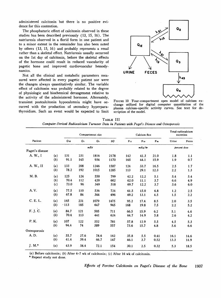
Radiocalcium turnover (Fig. 9). Radiocalcium turn-over, indicated by the decline in the plasma calcium-specific activity curves, was markedly accelerated in allsix pagetic patients. Calcitonin reduced the rate of de-cline of the plasma calcium specific activity in five pa-tients although an accelerated turnover was still evi-dent after 4-16 wk. The turnover data were subjectedto quantitation utilizing a digital computer program anda four-compartment open model (Fig. 10). Compartment
'.61
QiB corresponds to the extracellular calcium pool but innormal individuals it is slightly larger than chemicalestimates of the extracellular calcium, suggesting an ad-ditional component consisting of a small, rapidly ex-changing tissue calcium pool. Pool Qa is visualized as asummated intracellular calcium pool. Pools Q. and Quare the exchangeable and nonexchangeable bone calciumpools, respectively. The sum of QIE, Q., and Qa (AIQ)corresponds to the exchangeable body calcium pool ofother investigators and rarely exceeds 375 mEq innormal individuals studied for 7 days (9-11). Theprecise normal ranges in this laboratory for QIE, Q2,and Q, have not been defined because of the small num-ber of control individuals studied, but liberal upper
800-Colcitonin Dose 400-MRC units/day
f
0-200-
10c
Calcium Balance
g/day
PhosphorusBalance
g/day
Urinary TotalHydroxyproline
mg/day
400-1
200
-3 -2 -I 0 1 2 3 4 5 6
Seven Day Periods of Treatment
FIGURE 5 Calcium and phosphorus balance and urinarytotal hydroxyproline excretion in pagetic patient (C. E. L.,55 M, Paget's disease) before and during calcitonin ad-ministration. Format is the same as Fig. 3. Negative cal-cium balance became positive during the first 3 wk of cal-citonin, but then reverted to negative. Phosphorus balancewas not influenced by calcitonin. Urinary hydroxyprolinedeclined throughout the period of calcitonin administration.
Efects of Porcine Calcitonin on Paget's Disease of the Bone
-2 -1 12 3 4 5 6 14 15 16 17
Seven Day Periods of Treatment
I
1933
800-Calcitonin Dose 400-
MRCunits/dayO
0.8
0.6r
Calcium Balance0.4
g/day0.2
0
1.2 .
Phosphorus 0.8
Balanceg/day 0.4-
F13 were also increased in untreated Paget's disease butthe effects of calcitonin on these parameters were vari-able and inconclusive.
Other Tests. Iliac crest bone biopsy-samples proc-essed by standard histologic techniques failed to showconsistent changes during 4-16 wk of treatment. Therewas also no discernible influence of calcitonin on theroentgenographic appearance of the involved bones.Other tests cited under methods were unchanged fromcontrol values throughout study.
Osteoporotic patients (Figs. 1, 2, 9, 11, 12). PatientA. D. developed persistent hypocalcemia during calci-tonin, achieving levels of 8.0 mg per 100 ml. The fastingserum calcium was unchanged in J. M. The serum phos-phorus, alkaline phosphatase activity, and urinary totalhydroxyproline were unaltered by calcitonin. The rateof decline of the plasma calcium-specific activity after
800-Calcitonin Dose 400_MRCunits/day 0
l0
200Urinary Total
Hydroxyprolinemg/day
100
0-3 -2 -1 0 1 2 3 4 5 6
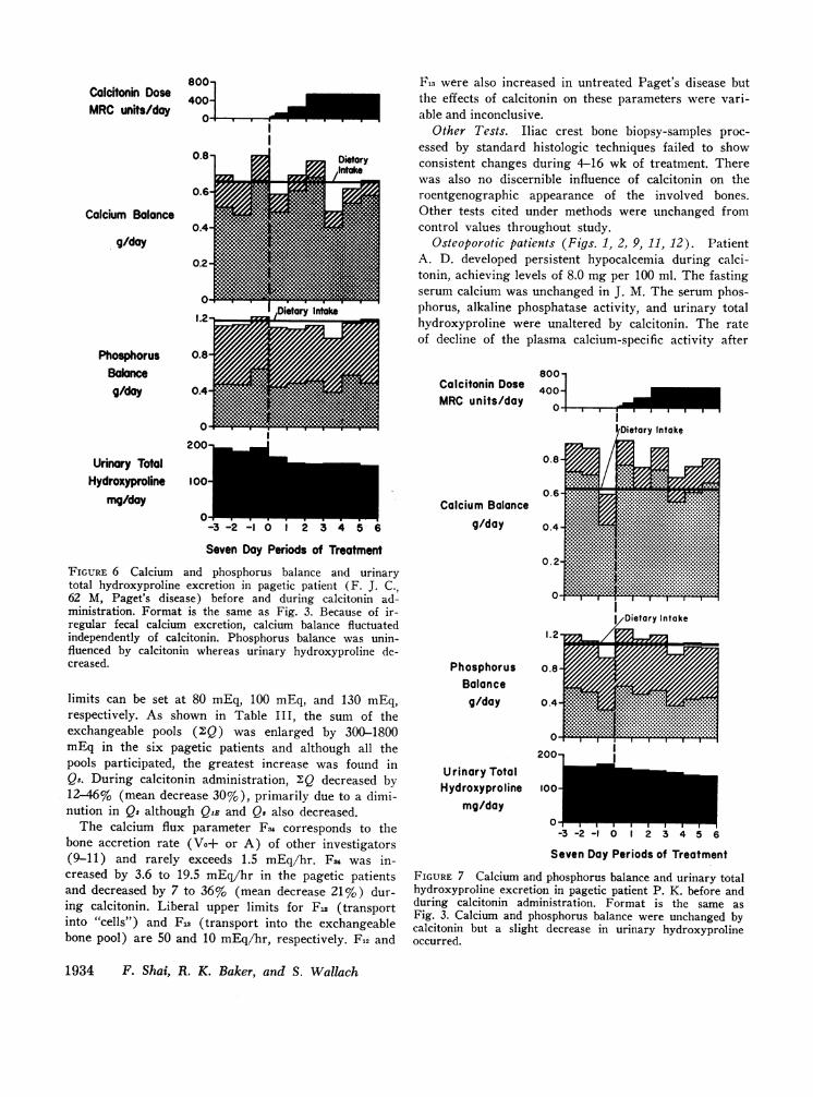
Seven Day Periods of TreotmentFIGURE 6 Calcium and phosphorus balance and urinarytotal hydroxyproline excretion in pagetic patient (F. J. C.,62 M, Paget's disease) before and during calcitonin ad-ministration. Format is the same as Fig. 3. Because of ir-regular fecal calcium excretion, calcium balance fluctuatedindependently of calcitonin. Phosphorus balance was unin-fluenced by calcitonin whereas urinary hydroxyproline de-creased.
limits can be set at 80 mEq, 100 mEq, and 130 mEq,respectively. As shown in Table III, the sum of theexchangeable pools (2;Q) was enlarged by 300-1800mEq in the six pagetic patients and although all thepools participated, the greatest increase was found inQa. During calcitonin administration, 2:Q decreased by12-46% (mean decrease 30%), primarily due to a dimi-nution in Qs although QiB and Qt also decreased.
The calcium flux parameter Fu corresponds to thebone accretion rate (Vo+ or A) of other investigators(9-11) and rarely exceeds 1.5 mEq/hr. F4 was in-creased by 3.6 to 19.5 mEq/hr in the pagetic patientsand decreased by 7 to 36% (mean decrease 21%) dur-ing calcitonin. Liberal upper limits for Fn (transportinto "cells") and F13 (transport into the exchangeablebone pool) are 50 and 10 mEq/hr, respectively. F12 and
Calcium Balanceg/day
PhosphorusBalance
g/day
Urinary TotalHydroxyproline
mg/day
-3 -2 -1 0 1 2 3 4 5 6
Seven Day Periods of Treatment
FIGURE 7 Calcium and phosphorus balance and urinary totalhydroxyproline excretion in pagetic patient P. K. before andduring calcitonin administration. Format is the same asFig. 3. Calcium and phosphorus balance were unchanged bycalcitonin but a slight decrease in urinary hydroxyprolineoccurred.
1934 F. Shai, R. K. Baker, and S. Wallach
I v I I .
II

radiocalcium injection was slow in both patients com-pared to the standard normal range defined by Heaneyet al. (12). In A. D., calcitonin had minimal effects onthe plasma calcium specific activity and on the digitalcomputer-derived parameters of radiocalcium turnover(Table III).
In A. D., calcium balance (Fig. 11) was normal butbecame negative by 100 mg/day during calcitonin. Nitro-gen balance also became negative by 3 g/day. In J. M.,a slight tendency to positive calcium balance occurredduring calcitonin, and calcium balance became distinctlypositive, by 200 mg/day, after calcitonin was stoppedabruptly (Fig. 12). Phosphorus balance became slightlyless positive during calcitonin in both patients. Sodium,potassium, and magnesium balances were not signifi-cantly altered. Iliac crest bone biopsies, skeletal X-rays,and subjective assessments of back and cervical painwere unchanged.
DISCUSSIONThe studies performed provide clinical and metabolicevidence which supports the ability of calcitonin to re-verse the skeletal manifestations of Paget's disease.Skeletal pain, bone vascularity, congestive heart failure,and neurologic impairment were variably but dramati-cally improved. Of the clinical effects, only the pain re-lief was subjective since the improvement in otherclinical parameters was documented by objective changesin physical findings. Prolonged relief of pain was ob-served in these patients and in a series of 20 additionalpatients treated for several months on an outpatientbasis, suggesting that pain relief is a real rather thana psychic result of calcitonin treatment.
Predominant resorption of bone, indicated by nega-tive calcium balance, is frequently present when pageticpatients come to clinical attention. Reversal of negativecalcium balance and a reduction in hydroxyprolinuriacan be related directly to the anti-resorptive functionof calcitonin. Although the turnover of "7Ca and thelevel of serum alkaline phosphatase are generallythought to reflect the rate of bone formation, theseparameters were also favorably affected by calcitonin.The improvement in these two parameters is probablydue to concomitant decrease in bone formation conse-quent to primary inhibition of bone resorption.
Bell, Avery, and Johnston (13) have reported theinduction of positive or less negative calcium balanceof 85-150 mg/day during daily intramuscular adminis-tration of 4-8 MRCU/kg calcitonin to patients withPaget's disease. In their report as well as in the presentstudy, both urinary and fecal calcium decreased. Onthe other hand, Haddad, Birge, and Avioli (14) ob-served small increases in urinary calcium during the
Calcitonin Dose
MRCunits/day
Urinary Calcium
g/day
4C
200-
Urinary TotalHydroxyproline
mg/day100-
O 1
-2 -I o 2 3 4 5 6 7 8
Seven Day Periods of Treatment
FIGURE 8 Urinary calcium and total hydroxyproline ex-cretion in pagetic patient (A. V., 75 F, Paget's disease).The patient was unable to cooperate in metabolic balancestudies because of advanced age and neurologic infirmity.The calcium and hydroxyproline content of the diet wasfixed but the diet was otherwise uncontrolled. Urinary cre-atinine was measured daily. Each week, the two to four 24-hrurines with adequate creatinine levels were selected foranalysis. The data represent averages of these two to fourexcretory values except during the 1st wk of calcitoninwhen urinary losses occurred every day. Note the decreasein urinary calcium and hydroxyproline during calcitoninadministration.
daily administration of 400-1500 MRCU for a longerperiod, but no balance procedures were employed toassess gastrointestinal effects. In these previous studies,urinary hydroxyproline decreased during the 1st wk ofcalcitonin. The present data demonstrate that urinaryhydroxyproline continues to decrease gradually as treat-ment proceeds. In studies of the immediate effects ofintravenous calcitonin, Bijvoet, van der Sluys Veer,and Jansen (15, 16) showed decreases in hydroxypro-line within 2 hr and, in the majority of the patients inthe present report, reductions were manifest on the 1stday of calcitonin. The accumulated data also indicatethat the level of serum alkaline phosphatase is a lesssensitive index of response of Paget's disease to calci-tonin than is urinary hydroxyproline since reductionsare not evident in the early weeks of treatment. Withprolonged treatment, however, the serum alkaline phos-phatase almost invariably decreases. (Unpublished ob-servations.)
The ability of calcitonin to improve net gastroin-testinal absorption of calcium was noted in this seriesof pagetic patients and by Bell, Avery, and Johnston(13) despite previous animal studies indicating that thegastrointestinal tract is not essential for the hypocal-cemic effect of calcitonin (17). The gastrointestinaleffect of calcitonin may not be direct since several stud-
Effects of Porcine Calcitonin on Paget's Disease of the Bone 1935
)o
nV.
10.2- 1
1
0 I I
I

0
0.1
0.06
0.04
0.02
0.010.30.2
0.1
0.06
0.04
0.02
0.01I.0
0.6
0.4
0.22.0
1.0
0 .6
24 48 72 96 120 144 68 0 24 48 72 96 120 144
HOURSAFTER Ca'47 INJECTION
FIGURE 9 Pattern of plasma calcium specific-activity after radiocalcium injection in sixpagetic patients (A. W., A. V., F. J. C., P. K., C. E. L., and M. B.) and two osteoporoticpatients (A. D. and J. M.). Closed circles represent data before calcitonin and open circlesafter 4-16 wk of calcitonin. M. B. had studies during the 5th and 16th wk of calcitonin. InA. W. I., the plasma radioactivity was too low to be measured accurately after 5 days. J. M.was not restudied after calcitonin. Note the individual ordinate scales. Radiocalcium turnoverwas rapid in the pagetic patients and slow in the osteoporotic patients. A decrease in radio-calcium turnover during calcitonin in the pagetic group was evident 1-2 hr after radiocalciuminjection.
ies have been presented in which influences believed tobe directed at skeletal homeostasis have resulted inparallel but secondary effects on calcium absorption(18). The mechanism by which calcium absorption re-
acts to skeletal influence is unknown. Nicolayson, Eeg-
Larsen, and Malm have theorized on the existence ofan "endogenous factor" capable of acting to couple thetwo organs (19). The enhanced absorption of calciummight have been due to increased parathyroid hormonesecretion resulting from the hypocalcemic action of the
1936 F. Shai, R. K. Baker, and S. Wallach
*8efore CalcitoninoAfter Calcitonin
0.2
E 0.2
O 0.1
0.06
0.04
- 0.02
2 0.010.6
i 0.4IL
2 0.2
-J4 0.1
< 0.06U) 0.04
CL0.020.80.6
0.4
0.2
0.1
0.06
0.04
0.02
Fr. .A.
0
M. B. J A.D.
I I I
J.M.
~
168
0.5A. V.
0
I I I I. v..5
administered calcitonin but there is no positive evi-dence for this contention.
The phosphaturic effect of calcitonin observed in thesestudies has been described previously (13, 15, 16). The-natriuresis observed in a florid form in one patient andto a minor extent in the remainder has also been notedby others (13, 15, 16) and probably represents a renalrather than a skeletal effect. Natriuresis usually occurredon the 1st day of calcitonin, before the skeletal effectsof the hormone could result in reduced vascularity ofpagetic bone and improved cardiovascular hemody-namics.
Not all the clinical and metabolic parameters mea-sured were affected in every pagetic patient nor werethe changes always quantitatively similar. The variableeffect of calcitonin was probably related to the degreeof physiologic and biochemical derangement relative tothe activity of the administered hormone. Alternately,transient postcalcitonin hypocalcemia might have oc-curred with the production of secondary hyperpara-thyroidism. Such an event would be expected to limit
URINE FECES t404
FIGURE 10 Four-compartment open model of calcium ex-change utilized for digital computer quantitation of theplasma calcium-specific activity curves. See text for de-scription of the model.
TABLE IIIComputer-Derived Radiocalcium Turnover Data in Patients with Paget's Disease and Osteoporosis
Total radiocalciumCompartment size Calcium flux excretion
Patient QIE Q2 Q Q F12 Fia Fs4 Urine Feces
mEil mEq/hr percent dosePaget's disease
A. W., I (a) 131 231 1816 2178 142 41.3 21.0 1.8 1.1(b) 91.1 145 934 1170 165 44.1 15.9 1.9 0.7
A. W., II (a) 133 208 1246 1587 124 33.7 16.5 2.5 1.7(b) 78.2 192 1015 1285 113 29.1 12.3 2.2 1.3
M. B. (a) 125 124 550 799 42.2 12.2 5.1 5.4 5.4(b) 70.4 112 420 602 62.0 11.1 3.7 6.6 4.9(C) 73.0 96 349 518 69.7 12.2 3.7 3.6 6.0
A. V. (a) 77.5 110 536 724 61.5 15.0 6.8 1.2 2.5(b) 67.8 84 344 496 49.2 13.1 4.5 1.5 2.2
C. E. L. (a) 165 231 1079 1475 95.2 17.4 8.5 2.0 3.5(b) -113 185 647 945 168 19.8 7.5 2.2 5.2
F. J. C. (a) 84.7 121 505 711 66.5 15.9 6.2 5.1 4.6(b) 70.4 113 441 624 64.7 14.9 5.8 2.6 4.2
P. K. (a) 107 122 552 781 57.8 12.9 5.5 4.5 5.3
(b) 94.4 74 389 557 73.6 15.7 4.8 5.6 6.6Osteoporosis
A. D. (a) 55.7 27.6 78.6 162 35.8 5.5 0.41 16.1 14.6(b) 41.4 39.4 66.7 147 46.1 3.7 0.52 13.3 14.9
J. M.* (a) 43.9 38.4 72.1 154 30.1 2.5 0.32 5.3 18.5
(a) Before calcitonin; (b) After 4-7 wk of calcitonin;* Repeat study not done.
(c) After 16 wk of calcitonin.
Effects of Porcine Calcitonin on Paget's Disease of the Bone 1937

PhosphorusBalanceg/day
Urinary TotalHydroxyproline
mg/doy
DietaryI Intaake
0.4
0.2
DietaryIntake
0.8
0.4
200-
0n-
ww'
-3 -2 -I 0 2 3 4 5 6 7 8Seven Day Periods of Treatment
FIGURE 11 Calcium and phosphorus balance and urinarytotal hydroxyproline in osteoporotic patient (A. D., 56 F,postmenopausal osteoporosis.) Format is the same as Fig. 3.Calcitonin administration resulted in negative calcium andless positive phosphorus balance. The slight increase inurinary hydroxyproline during the last base line period (- 1period) before calcitonin is due to injections of the 16%ogelatin vehicle.
the degree of fasting hypocalcemia, positive calciumbalance, reduction in serum alkaline phosphatase-ac-tivity, urinary hydroxyproline excretion, and radiocal-cium turnover induced by calcitonin. The tendency todevelop slightly higher fasting serum calcium levelscould also have resulted from secondary hyperparathy-roidism. No measurements of circulating parathyroidhormone levels were made in these patients. However,elevation of circulating parathyroid hormone levels mea-
sured by radioimmunoassay have been found in pageticpatients given calcitonin (20). The additional observa-tion in two pagetic patients that lower doses of calci-tonin resulted in as good or better clinical and metaboliceffects than the larger dose is also compatible with theproduction of secondary hyperparathyroidism as an un-
desirable side effect of high doses. A third possibilityfor the variable quantitative effect of calcitonin couldrelate to the "escape phenomenon" observed with calci-tonin in vitro (21) and in patients with high circulatinglevels of calcitonin due to medullary carcinoma of thethyroid who do not manifest hypocalcemia (22).
The most dramatic clinical effects observed duringcalcitonin administration related to the neurologic sys-
term. Spinal cord compression injuries usually affect thelarge motor fibers first and the sensory fibers later. Re-covery occurs in reverse order. In the two patients withthoracic cord impingement but no muscle atrophy, neu-rologic improvement followed a similar course. Also, inone patient with a corda equina-type lesion and muscleatrophy, complete restoration of sensory modalities oc-curred despite a minimal return of muscular function.The improved air conduction component of audition,which occurred in three patients, suggests a calcitonin-related change in the pagetic state of the footplate of thestapes. Since most of these neurologic defects have theirbasis in increased skeletal volume with impingement, theobserved reversal of neurologic phenomena probably re-quired a reduction in skeletal volume. Although thereis no supporting histologic data, it is tempting to specu-late that calcitonin is not simply an inhibitor of bone re-sorption in Paget's disease. Because of the inducedreduction in skeletal turnover, significant reductions invascularity and in pathologic remodelling of the skele-
Calcitonin DoseMRC units/day
Calcium Balance
g/day
PhosphorusBalanceg/day
Urinary Totall 'ydroxyproline
mg/day
800-
400-
Dietary Iffl Intak I
I I . I II
20 0:-l
0#-2 - 0 1 2 3 4
Seven Day Periods of Treatment
FIGURE 12 Calcium and phosphorus balance and urinarytotal hydroxyproline in osteoporotic patient (J. M., 80 F,senile osteoporosis) before, during, and after a 4 wk courseof calcitonin. Format is the same as Fig. 3. Despite fluctua-tions in fecal calcium excretion, positive calcium balanceis evident during and after calcitonin. Phosphorus balancebecame less positive during calcitonin and more positiveafter calcitonin was discontinued. The 16% gelatin vehiclewas not administered before calcitonin.
1938 F. Shai, R. K. Baker, and S. Wallach
800-Calcitonin Dose 400-MRC units/day 0- _
0.61
Calcium Balance
g/day

ton occur, which act to reduce the abnormally increasedskeletal volume typical of the disease. A minute reduc-tion in skeletal volume in a strategic area, undetectableby X-ray, would suffice to reduce neurologic impairment.
In contrast to the observations in Paget's disease, theresults of calcitonin administration in two patients withosteoporosis were not dramatic. Several possible rea-sons for the disappointing results may be cited. Theradiocalcium turnover studies suggest that skeletal turn-over was moderately slow in both patients and calci-tonin may be relatively inactive in such situations (23).Second, one patient was on estrogen therapy through-out study whereas the other patient had recently discon-tinued estrogen. In rats, estradiol has been found ca-pable of antagonizing the hypocalcemic action of calci-tonin (24; F. Shai, and S. Wallach, manuscript in prep-aration), suggesting that a similar ability to inhibitcalcitonin might occur in humans receiving estrogens.Lastly, the induction of secondary hyperparathyroidismmight have completely overriden the positive effects ofthe calcitonin. Patient A. D. developed sustained hypo-calcemia and subsequently developed negative calciumand nitrogen balance, findings which are similar tothose observed in hyperparathyroidism (25). The ob-servations in osteoporosis should not be taken as evi-dence of inefflcacy of calcitonin. Lower doses of calci-tonin combined with high calcium, magnesium, and/orphosphorus intake might forestall secondary hyperpara-thyroidism, stimulate bone formation, or enhance theendorgan action of the calcitonin.
The relatively negative data in osteoporosis furtherhighlight the efficacy of calcitonin in ameliorating theclinical and metabolic manifestations of Paget's disease.In studies in progress, it has also been found that out-patient treatment with calcitonin by self-injection, usingsingle daily subcutaneous doses of 100-200 MRCU isequally efficacious. Continuous treatment for more than6 months has not caused allergic reactions, side effects,or evidence of antibody formation or loss of thera-peutic potency. If long term control of this previouslyuntreatable disease can be achieved with calcitonin, thepossibility that early therapy might prevent the seriousconcomitants of the generalized condition becomes anintriguing prospect.
ACKNOWLEDGMENTSThe authors are indebted to Dr. Anthony Nicastri for thepathologic interpretation of the iliac crest-bone biopsies, tothe staff of the United States Public Health Service ClinicalResearch Center for the superb care rendered the patients,and to Doctors James Bastian and James Lesh of theArmour Company for supplying the calcitonin and support-ing this project. Mr. William Siler and the staff of theProgram in Medical Computer Science at the DownstateMedical Center devised the computer program and performed
the computer analyses, operating under the National Insti-tute of Health Special Research Resource Grant RR-00291.
This study was supported by grant (RR-318) from theGeneral Clinical Research Centers Program of the Divisionof Research Resources, National Institutes of Health, UnitedStates Public Health Service Research Contract AM08453from the National Institute of Arthritis and MetabolicDiseases, and by a grant from the Armour Company.
Note added in proof: Since submission of this manuscript,approximately twenty patients have completed 6-12 monthsof treatment with porcine calcitonin at a single daily sub-cutaneous dose of 100-200 MRCU. All have maintained theclinical improvement achieved during the first 6 monthsof treatment. However, in over half the patients, the serumalkaline phosphatase activity, after decreasing during thefirst 6 months, has remained constant in the range 20-50KAU. In addition, the urinary total hydroxyproline, afterdecreasing 50% or more during the first 6 months, has re-turned to or toward base line levels. Possible explanationsinclude secondary hyperparathyroidism, neutralizing antibodyproduction, and the "escape phenomenon" (21). These pos-sibilities are being explored currently.
REFERENCES
1. Nagant de Deuxchaisnes, C., and S. M. Krane. 1964.Paget's disease of bone: clinical and metabolic observa-tions. Medicine (Baltimore). 43: 233.
2. Hirsch, P. F., and P. L. Munson. 1969. Thyrocalcitonin.Physiol. Rev. 49: 548.
3. Reifenstein, E. C., Jr., F. Albright, and S. L. Wells.1945. The accumulation, interpretation, and presentationof data pertaining to metabolic balances, notably thoseof calcium, phosphorus, and nitrogen. J. Clin. Endo-crinol. 5: 367.
4. Furman, R. H., R. P. Howard, S. L. Wells, and S. M.MacAulay. 1968. Techniques for studying anabolism andcatabolism in humans. J. Amer. Med. Woman's Assoc.23: 733.
5. Mancini, G., A. 0. Carbonara, and J. F. Heremans.1965. Immunochemical quantitation of antigens by singleradial immunodiffusion. Immunochemistry. 2: 235.
6. Ouchterlony, 0. 1958. Diffusion-in-gel methods for im-munological analysis. Progr. Allergy. 5: 1.
7. Bergman, I., and R. Loxley. 1963. Two improved andsimplified methods for the spectrophotometric determi-nation of hydroxyproline. Anal. Chem. 35: 1961.
8. Baker, R. K., H. Francis, and A. Posner. 1969. Anautomated method for the estimation of hydroxyproline.Clin. Chem. 15: 817 (Abstr.).
9. Aubert, J. P., F. Bronner, and L. J. Richelle. 1963.Quantitation of calcium metabolism. Theory. J. Clin.Invest. 42: 885.
10. Schwartz, E., V. A. Panariello, and J. Saeli. 1965.Radioactive calcium kinetics during high calcium intakein osteoporosis. J. Clin. Invest. 44: 1547.
11. Neer, R., M. Berman, L. Fisher, and L. E. Rosenberg.1967. Multicompartmental analysis of calcium kinetics innormal adult males. J. Clin. Invest. 46: 1364.
12. Heaney, R. P., G. C. H. Bauer, F. Bronner, J. 'F.Dymling, F. W. Lafferty, B. E. C. Nordin, and C.Rich. 1964. A normal reference standard for radio-calcium turnover and excretion in humans. J. Lab. Clin.Med. 64: 21.
Effects of Porcine Calcitonin on Paget's Disease of the Bone 1939

13. Bell, N. H., S. Avery, and C. C. Johnston, Jr., 1970.Effects of calcitonin in Paget's disease and polyostoticfibrous dysplasia. J. Clin. Endocrinol. Metab. 31: 283.
14. Haddad, J. G. Jr., S. J. Birge, and L. V. Avioli. 1970.Effects of prolonged thyrocalcitonin administration inPaget's disease of bone. N. Engl. J. Med. 283: 549.
15. Bijvoet, 0. L. M., J. van der Sluys Veer, and A. P.Jansen. 1968. Effects of calcitonin on patients withPaget's disease, thyrotoxicosis or hypercalcemia. Lancet.1: 876.
16. Bijvoet, 0. L. M., and A. P. Jansen. 1967. Thyrocal-citonin in Paget's disease. Lancet. 2: 471.
17. Aliapoulios, M. A., and P. L. Munson. 1965. Thyrocal-citonin. Surg. Forum 16: 55.
18., Wasserman, R. H., and A. N. Taylor. 1969. Some as-pects of the intestinal absorption of calcium with specialreference to vitamin D. In Mineral Metabolism, Vol. IIIC. L. Comar, and F. Bronner, editors. Academic PressInc., New York. 321.
19. Nicolaysen, R., N. Eeg-Larsen, and 0. J. Malm. 1953.Physiology of calcium metabolism. Physiol. Rev. 33:424.
20. Arnaud, C. D., H. S. Tsao, and T. Littledike. 1971.Radioimmunoassay of human parathyroid hormone inserum. J. Clin. Invest. 50: 21.
21. Raisz, L. G., W. Y. W. Au, J. Friedman, and I. Nei-mann. 1967. Thyrocalcitonin and bone resorption: studiesemploying a tissue culture bioassay. Amer. J. Med. 43:684.
22. Tashjian, A. H. Jr., and K. E. W. Melvin. 1968. Medul-lary carcinoma of the thyroid gland. Studies of thyr-calcitonin in plasma and tumor extracts. N. Engl. J.Med. 279: 279.
23. Mittleman, R., S. Wallach, A. Chausmer, and J. Bellavia.1967. Interaction of thyrocalcitonin and vitamin D onbone resorption. In Calcitonin, Proceedings of the Sym-posium on Thyrocalcitonin and the C Cells. S. Taylor,editor. Heinemann & Zsolnay Ltd., London. 276.
24. Kaplan, E. L., M. Singh, and C. D. Arnaud. 1970. In-fluence of sex hormones on calcitonin responses in therat. Current Topics Surg. Res. 2: 167.
25. R. L. Landau, and A. Kappas. 1965. Anabolic hormonesin hyperparathyroidism, with observations on the gen-eral catabolic influence of parathyroid hormone in man.Ann. Inter. Med. 62: 1223.
1940 F. Shai, R. K. Baker, and S. Wallach