Overview of Aortic Stenosis and Transcatheter Aortic valve ...

J O U R N A L O F T H E AM E R I C A N C O L L E G E O F C A R D I O L O G Y V O L . 6 6 , N O . 5 , 2 0 1 5
ª 2 0 1 5 B Y T H E AM E R I C A N C O L L E G E O F C A R D I O L O G Y F O U N DA T I O N I S S N 0 7 3 5 - 1 0 9 7 / $ 3 6 . 0 0
P U B L I S H E D B Y E L S E V I E R I N C . h t t p : / / d x . d o i . o r g / 1 0 . 1 0 1 6 / j . j a c c . 2 0 1 5 . 0 5 . 0 6 6
THE PRESENT AND FUTURE
STATE-OF-THE-ART REVIEW
Calcification in Aortic Stenosis
The Skeleton KeyTania A. Pawade, MD,*y David E. Newby, MD, PHD,*y Marc R. Dweck, MD, PHD*yz
ABSTRACT
Fro
yEdIca
Bri
con
by
Lis
Ma
Aortic stenosis is a common, potentially fatal condition that is set to become an increasing public health burden. Once
symptoms develop, there is an inexorable deterioration with a poor prognosis. Despite this, there are no medical ther-
apies capable of modifying disease progression, and the only available treatment is aortic valve replacement, to which
not all patients are suited. Conventional teaching suggests that aortic stenosis is a degenerative condition whereby “wear
and tear” leads to calcium deposition within the valve. Although mechanical stress and injury are important factors, it
is becoming increasingly appreciated that aortic stenosis is instead governed by a highly complex, regulated pathological
process with similarities to skeletal bone formation. This review discusses the pathophysiology of aortic stenosis with an
emphasis on the emerging importance of calcification, how this can be visualized and monitored using noninvasive
imaging, and how our improved knowledge may ultimately translate into novel disease-modifying treatments.
(J Am Coll Cardiol 2015;66:561–77) © 2015 by the American College of Cardiology Foundation.
A ortic stenosis is the most common form ofvalve disease in the Western world and isset to become an ever-increasing public
health burden (1,2). Despite this, there are no medicaltherapies to halt or delay disease progression, and theonly available treatment is aortic valve replacementor implantation, to which not all patients are suited.There is, therefore, a major unmet clinical need toidentify pharmacological treatments capable of modi-fying this disease process.
Aortic stenosis was long considered to be adegenerative condition whereby “wear and tear”resulted in progressive calcium formation within thevalve. Although mechanical stress and injury remaincentral to its pathophysiology, emerging evidence hasindicated that aortic stenosis develops as part of ahighly complex and tightly regulated series of pro-cesses, each of which may be amenable to medicalintervention (3). In particular, aortic stenosis can be
m the *British Heart Foundation/University of Edinburgh Centre for Ca
inburgh Heart Centre, NHS Lothian, Edinburgh, United Kingdom; and
hn School of Medicine at Mount Sinai, New York, New York. Drs. Pawad
tish Heart Foundation (SS/CH/09/002, FS/14/78, and CH/09/002); and
trolled trial Saltire 2: Bisphosphonates and RANKL Inhibition in Aortic Ste
a Wellcome Trust Senior Investigator Award (WT103782AIA).
ten to this manuscript’s audio summary by JACC Editor-in-Chief Dr. Vale
nuscript received February 4, 2015; revised manuscript received May 26,
divided into 2 distinct phases: an early initiationphase dominated by valvular lipid deposition, injury,and inflammation, with many similarities to athero-sclerosis, and a later propagation phase where pro-calcific and pro-osteogenic factors take over andultimately drive disease progression (Figure 1) (4).This review discusses the pathophysiology of aorticstenosis, with an emphasis on the emerging impor-tance of calcification, how this can be imagedwith modern noninvasive techniques, and how ourimproved knowledge might ultimately lead to thedevelopment of novel therapies.
PATHOLOGY OF AORTIC STENOSIS
INFLAMMATION, LIPIDS, AND THE INITIATION
PHASE OF AORTIC STENOSIS. Under normal cir-cumstances, the aortic valve is composed of 3 leaflets,each of which is a thin (<1 mm), smooth, flexible, and
rdiovascular Science, Edinburgh, United Kingdom;
the zTranslational and Molecular Imaging Institute,
e and Dweck and Prof. Newby are supported by the
are the Principal Investigators of the randomized
nosis (NCT02132026). Prof. Newby is also supported
ntin Fuster.
2015, accepted May 26, 2015.

ABBR EV I A T I ON S
AND ACRONYMS
BMP = bone morphogenetic
protein
CT = computed tomography
FDG = fluorodeoxyglucose
LDL = low-density lipoprotein
OPG = osteoprotegerin
PET = positron emission
tomography
RANK = receptor activator of
nuclear factor kappa B
RANKL = receptor activator of
nuclear kappa B ligand
TGF = transforming growth
factor
VIC = valvular interstitial cell
Pawade et al. J A C C V O L . 6 6 , N O . 5 , 2 0 1 5
Calcification in Aortic Stenosis A U G U S T 4 , 2 0 1 5 : 5 6 1 – 7 7
562
mobile structure (3). In aortic stenosis, theseleaflets become thickened, fibrosed, andcalcified, resulting in reduced leaflet mobilityand progressive valvular obstruction.
The early stages of aortic stenosis are inmany ways similar to atherosclerosis.Indeed, the 2 conditions share many commonrisk factors, with large longitudinal studiesconsistently demonstrating that the incidenceof aortic stenosis is linked to factors such assmoking, age, and hypertension (5–7). As inatherosclerosis, endothelial damage due toincreased mechanical stress and reducedshear stress is believed to be the initiatinginjury, perhaps best illustrated by bicuspidvalve disease. The characteristic 2-leafletstructure of these valves results in less effi-cient dissipation of mechanical stress and
accelerated endothelial damage, so that patientsalmost universally develop aortic stenosis anddisplay more rapid disease progression (8).
Following endothelial damage, the same lipidsimplicated in atherosclerosis infiltrate the valve, inparticular, lipoprotein(a) and oxidized low-densitylipoprotein (LDL) cholesterol. Consequently, obser-vational studies have identified cholesterol and itsrelated lipoproteins as independent risk factors forthe development of aortic stenosis (5–7,9). Indeed, astrong genome-wide association was recently estab-lished between a single-nucleotide polymorphism inthe locus of lipoprotein(a) and the incidence of aorticvalve calcification (10). Progressive endothelial injuryand lipid oxidization then establishes an inflamma-tory response within the valve that is characterizedpredominantly by infiltration of macrophages, butalso involves T lymphocytes and mast cells (11).At this early stage, regions of stippled micro-calcification that colocalize with sites of lipid depo-sition are observed (11). The formation of thesemicrocalcifications may be mediated by cell deathand the release of apoptotic bodies in these areas.Such apoptotic bodies are similar to the matrix vesi-cles found in bone, which contain the prerequisitecomponents for calcium crystal deposition (includingcalcium and inorganic phosphate ions) and facilitatethe formation of needle-like crystals of hydroxyapa-tite (4,12). In bone, as these hydroxyapatite crystalsexpand, they pierce the outer membrane of thevesicle and become exposed to the extracellularenvironment, thereby forming nucleation sites forfurther calcium deposition. It is probable that similarprocesses also occur within the valve (13). Further-more, hydroxyapatite deposition evokes further pro-inflammatory responses from macrophages, creating
a positive feedback loop of calcification and inflam-mation in the early stages of disease (14). It seemslikely that these mechanisms underlie early calciumformation in aortic stenosis and its association withlipid and inflammation.
The apparent link between lipid, inflammation,and calcification in these early stages and the patho-logical similarities with atherosclerosis led to thehypothesis that statins might be beneficial in patientswith aortic stenosis. This was supported by encour-aging nonrandomized human data (15) and studies inhypercholesterolemic animal models demonstratingthat lipid deposition and oxidative stress precede theconversion of valvular interstitial cells to those withan osteoblastic phenotype, and that this process isinhibited by atorvastatin (16,17). However, whenstatins were formally tested in 3 independent ran-domized controlled trials of patients with aortic ste-nosis, each demonstrated a failure of this therapy tohalt or retard aortic stenosis progression, despitereducing the serum LDL cholesterol concentrationsby more than one-half (18–20). This has led in-vestigators to re-examine the pathophysiology un-derlying aortic stenosis and to the realization thatalthough inflammation and lipid deposition may beimportant in establishing the disease (the initiationphase), the later stages are instead characterized byan apparently self-perpetuating cycle of calcium for-mation and valvular injury (the propagation phase)(4). Indeed, once this propagation phase has becomeestablished, disease progression is dictated neither byinflammation nor by lipid deposition, but rather bythe relentless accumulation of calcium in the valveleaflets. This may explain the failure of statins tomodify disease progression in aortic stenosis, whichcommonly presents beyond the initiation phase.Moreover, there is some data that statins may even beprocalcific in the vasculature (21,22).CALCIFICATION AND THE PROPAGATION PHASE. Ske-letal bone formation is characterized by the initialdeposition of collagen matrix, which provides ascaffold upon which progressive calcification candevelop. With time, this calcium acquires a more or-dered crystalline structure until the characteristicfeatures of lamellar bone are finally observed. Similarstructural processes are believed to occur in the aorticvalve, with many of the same cell mediators andproteins implicated (23). Indeed, in aortic stenosis,collagen is deposited in anticipation of the procalcificprocesses that subsequently dominate. This fibroticprocess within the valve may be mediated, in part,by reduced nitric oxide expression following endo-thelial injury (24); however, the renin-angiotensinsystem (RAS) is also believed to play a central

FIGURE 1 The Pathophysiology of Aortic Stenosis
8. Apoptotic bodiesform nucleation sitesfor calcium deposition
ApoptoticBody
7. Apoptosis ofVICs and
InflammatoryCells
4. RANKL Bindingto RANK
RANKL RANKValve
Interstitial Cell(VIC)
5. OsteoblasticDifferentiation
• BMP• Notch
• RANK/RANKLOsteoblast
6. Calcium depositionon a collagen scaffold
9. CalciumOrtho-phosphate
11. Activation,differentiation & apoptosis(Wnt/catenin & ENPP1)
1. Endothelialinjury 2. Infiltration of
Inflammatory Cellsand Lipid deposition
Macrophage T lymphocyte
OxidizedLDL
Lipoprotein(a)
Mast cell
3. Release of Inflammatory Mediators
Injury
10. Increasedmechanicalstress and injury
INITIATION PHASE PROPAGATION PHASE
Initiation phase: endothelial injury (1) facilitates the infiltration of oxidized lipids and inflammatory cells (2) into the valve and the release of proinflammatory medi-
ators (3). These trigger the very early stages of valve calcification. The propagation phase: these proinflammatory processes subsequently induce VICs to undergo
osteogenic differentiation (5) via several different mechanisms, including the binding of RANKL to RANK (4). Differentiated cells within the aortic valve first lay down a
collagen matrix and other bone-related proteins causing valvular thickening and stiffening before producing calcium (6). Additionally, apoptotic remnants of some VICs
and inflammatory cells (7) create a nidus for apoptosis-mediated calcification (8). Calcification of the valve (9) induces compliance mismatch, resulting in increased
mechanical stress and injury (10). This results in further calcification via osteogenic differentiation and apoptosis (11). Hence, a self-perpetuating cycle of calcification,
valve injury, apoptosis, and osteogenic activation is established that drives the propagation phase of the disease. BMP ¼ bone morphogenetic protein; ENPP1 ¼ectonucleotide pyrophosphate 1; LDL ¼ low-density lipoprotein; RANK ¼ receptor activator of nuclear kappa B; RANKL ¼ receptor activator of nuclear kappa B ligand;
RAS ¼ renin-angiotensin system; VIC ¼ valvular interstitial cell.
J A C C V O L . 6 6 , N O . 5 , 2 0 1 5 Pawade et al.A U G U S T 4 , 2 0 1 5 : 5 6 1 – 7 7 Calcification in Aortic Stenosis
563
role. Angiotensin-converting enzyme (ACE) is up-regulated in calcific aortic valve disease and is likelyto be delivered to the valve by LDL, its natural vehicle(25). Here it facilitates the conversion of angiotensinI to II, which mediates profibrotic effects via theangiotensin II type 1 (AT1) receptor. Although angio-tensin II is also able to mediate antifibrotic and anti-inflammatory effects via angiotensin II type 2 (AT2)receptors, differential expression of these re-ceptors in favor of AT1 has been demonstrated incalcified aortic valves, so that a profibrotic
profile dominates. Likewise, although angiotensin-converting enzyme type 2 (ACE-2) exerts antifibroticand anti-inflammatory influences via the Ang1-7/Maspathway, this pathway is down-regulated in calcifiedaortic stenosis, with reduced expression of bothACE-2 and Mas receptors in calcified valves comparedwith control subjects (26). Increased RAS expressionis, therefore, implicated in the development offibrosis within the valve. On a systemic level, RASis implicated in the development of hyperten-sion, which often accompanies aortic stenosis and

Pawade et al. J A C C V O L . 6 6 , N O . 5 , 2 0 1 5
Calcification in Aortic Stenosis A U G U S T 4 , 2 0 1 5 : 5 6 1 – 7 7
564
may accelerate its progression given the increasedmechanical stress it imposes upon the valve (27).
Beyond this initial fibrosis, valvular calcification inaortic stenosis ultimately dominates and appearsdependent upon the presence of osteoblast-like cellsthat develop an osteogenic phenotype. In support ofthis hypothesis, gene-profiling studies have demon-strated increased valvular expression of severalosteoblast-specific proteins, including the Cbfa1/Runx2 transcription factor, essential for osteoblasticdifferentiation and regulation of osteoblast function(28,29). A number of other extracellular matrix pro-teins closely associated with osteoblast function andmore commonly associated with skeletal bone for-mation are also up-regulated in calcific aortic valves.These include osteopontin and bone sialoprotein,which are facilitators of the attachment of osteoblaststo the bone matrix, and demonstrate up to a 7-foldelevation in gene expression at sites of developingcalcification (30,31). Importantly, valvular ossificationalso appears to be dependent upon angiogenesis,supporting the hypothesis that this is an active,highly regulated, pathological process (23).
The source of osteoblast-like cells within theaortic valve remains controversial. In vitro, multiplecell types present in the vasculature are capable ofundergoing differentiation into those with anosteoblast-like phenotype. The most likely candi-date appears to be the myofibroblast, a highlyplastic cell that is also commonly referred to as thevalve interstitial cell (VIC) (32). The differentiationof this cell into an osteoblast phenotype is not fullycharacterized, but appears to be a central step in thedevelopment of aortic stenosis and is regulated by arapidly growing list of molecules and complexpathways. In vivo molecular imaging has demon-strated that in the early stages of aortic stenosis,this differentiation appears coordinated by macro-phages (33,34) via the action of proinflammatorycytokines (interleukin [IL]-1b, IL-6, IL-8, tumor ne-crosis factor [TNF]-a, insulin-like growth factor-1,and transforming growth factor [TGF]-b) (4,35,36).However, in the later stages, this differentia-tion again appears to be dominated by calcificpathways, including the Notch, Wnt/b-catenin, andreceptor activator of nuclear factor kappa B (RANK)/receptor activator of nuclear factor kappa B ligand(RANKL)/osteoprotegerin (OPG) pathways, which wediscuss here.
Notch belongs to a family of cell surface receptors(Notch 1 to 4) that are highly expressed in the aorticvalve, playing an important role in its morphologicaldevelopment (37). Individuals with loss-of-functionmutations in Notch-1 have higher rates of
cardiovascular calcification and aortic stenosis. In 2unrelated families with a high incidence of con-genital aortic valve disease, genome-wide linkageanalysis identified loss-of-function Notch-1 muta-tions as the cause (37). In particular, Notch-1 ap-pears to be important in establishing osteogeniccells in the valve via the action of bone morphoge-netic protein (BMP)-2 (38). BMP-2 is a potent oste-ogenic differentiation factor and part of a family ofmultifunctional cytokines belonging to the TGF-bsuperfamily. Expression of BMP-2 is increased incalcified atherosclerotic lesions and aortic valves(29,39), and it appears to have a central role in thedifferentiation of plastic cell populations toward anosteogenic phenotype. Indeed, exposure of normalhuman VICs to BMP-2 induces osteoblastic featuresin these cells (40,41). In addition, binding of Wnt toLDL receptor-related protein 5 receptors may acti-vate the canonical Wnt/b-catenin pathway that isalso implicated in osteogenic cell differentiation(42). Similarly, TGF-b1 is able to induce nucleartranslocation of b-catenin and increased Wntsignaling, stimulating the osteogenic differentiationof mesenchymal progenitor cells (43). The latterprocess can increase in response to mechanical stressand may therefore explain, in part, the self-perpetuating and exponential increase in calcifica-tion activity observed once osteogenic differentiationhas occurred and the propagation phase is estab-lished (Figure 1) (43,44).
Systemic regulators that govern calcificationactivity, both in the bone and in the vasculature,tightly control calcium homeostasis; consequently,there is an inverse correlation between bone mineraldensity and vascular calcification. Osteoporosisis associated with age-independent increases invascular calcification and even cardiovascular mor-tality (45). A prospective study of 25,639 men andwomen demonstrated an inverse correlation betweenbone mineral density and incident aortic stenosisin older women (46). Moreover, other disorders ofbone turnover, including chronic kidney diseaseand Paget’s disease, also manifest changes in thevasculature (47–50). This dichotomy has been termedthe “calcification paradox” and is likely to beexplained by common pathological pathways havingreciprocal effects on the bone and vasculaturesimultaneously.
A potential mechanism for this association lies inthe activity of the RANK/RANKL/OPG pathway(Figure 2A). In bone, RANKL (a member of the TNFcytokine family) binds to RANK (a transmembraneprotein expressed on marrow stromal cells and pre-osteoclasts), acting as a potent inducer of osteoclast

J A C C V O L . 6 6 , N O . 5 , 2 0 1 5 Pawade et al.A U G U S T 4 , 2 0 1 5 : 5 6 1 – 7 7 Calcification in Aortic Stenosis
565
differentiation and activity. This drives demineral-ization of bone, but is policed by osteoprotegerin(OPG), a soluble decoy receptor, which binds RANKLand prevents it from activating RANK (Figure 2). Incontrast, RANKL appears to have the opposite effecton cells in the vasculature, inducing an osteoblasticphenotype in human VIC cells that results inincreased matrix calcification, the formation ofcalcific nodules, and increased expression of alkalinephosphatase and osteocalcin (Figure 2A) (51). RANKLalso promotes the osteogenic properties of vascularsmooth muscle cells, once again via the up-regulationof BMP-2. As a consequence, whilst OPG-deficientmice develop osteoporosis, they simultaneouslyaccelerate vascular calcification in association withincreased expression of RANKL in both regions (52).A potential explanation for the differential effects ofRANK/RANKL/OPG in these 2 tissues is that in bonethere is an abundance of pre-osteoclasts that favorsthe pro-osteoclastic properties of RANKL (36). Incontrast, this pool is absent in the vasculature so thatRANKL’s pro-osteoblastic effects on myofibroblastand smooth muscle cells predominate.
Imbalances in RANKL/OPG signaling have beendemonstrated in calcific aortic valves. In humanvalve tissue taken from patients with aortic stenosis,immunochemistry revealed less OPG-positive cells inareas of focal calcification, whereas western blottingdemonstrated that OPG was not expressed at rele-vant levels in aortic stenosis, but was detectable incontrol subjects. The converse is true of RANKL,with increased levels observed in stenotic aorticvalves (51). In combination, these data support thehypothesis that the RANK/RANKL/OPG axis isimplicated in the development of aortic valve calci-fication and provide 1 explanation for the link be-tween aortic valve calcification and bone mineraldensity. Other investigators have suggested that thedifferential effects of oxidized LDL might also be ofimportance, with this molecule appearing to pro-mote calcification and the osteoblastic differentia-tion of vascular cells in vitro, whilst inhibiting theseprocesses in a bone-derived pre-osteoblast cell line(4,53,54).
Fetuin-A is a circulating protein that canexist in isolation or as a complex with matrixg-carboxyglutamic acid protein (MGP). Both arepowerful guardians against ectopic calcification andsimultaneously inhibit many of the procalcific pro-cesses discussed earlier (55). MGP needs to be bothcarboxylated and phosphorylated to be activated, aprocess dependent on vitamin K. There is speculationthat use of the vitamin K antagonists, such as couma-rins, may be associated with increased vascular
calcification (56). The actions of fetuin-A and MGPinclude inhibition of BMP2 and TGF-b, reduction ofapoptosis-mediated calcification, and direct preven-tion of calcification by binding to calcium crystals.Reduced circulating levels of Fetuin-A and MGP arethought to explain the vascular calcification seen withend-stage renal failure. Moreover, plasma fetuin-Aconcentrations are decreased in aortic stenosisand inversely associated with the rate of disease pro-gression (57,58). Interestingly, this association wasseen only in older patients (>70 years of age) (59).Conversely, increased plasma dephosphorylated(inactive) MGP was a strong independent predictor offaster stenosis progression, but only in younger pa-tients (#57 years of age) (60).WHY DOES CALCIUM BEGET CALCIUM? Once calci-fication is established in the valve, it would appearto initiate further calcium formation. This self-perpetuating cycle of calcification and valve injuryappears to be the central driver of disease progres-sion and the propagation phase of aortic stenosis(Figure 1).
The mechanism for this may, in part, relate to thecompliance mismatch caused by calcific deposits inthe leaflets that results in increased mechanicalstress, injury-induced activation of the Wnt/b-cateninpathway, and further osteoblast differentiation.However, it may also be explained by the actions ofmembrane-bound ectonucleotidases. These are pro-duced by VICs and regulate the extracellular pro-duction of inorganic phosphate (a promoter ofcalcification) and inorganic pyrophosphate, an in-hibitor of pyrophosphate. Ectonucleotide pyrophos-phatase 1 (ENPP1) is highly up-regulated in calcificaortic valve disease, with a polymorphism associatedwith increased transcripts of ENPP1 identified in ste-notic valves (61). Hydrolysis of extracellular adeno-sine triphosphate (ATP) by ENPP1 produces a netincrease in inorganic phosphate, thus favoring calci-fication and promoting the production of furtherENPP1 in a positive feedback loop (61). Moreover,because ATP acts as a cell survival signal for VICs viathe P2Y2 receptor, its depletion also triggers apoptosisof these cells, providing a further key stimulus tocalcification (61). Finally, loss of P2Y2 signalingincreases the secretion of IL-6, a cytokine that pro-motes further osteogenic differentiation of VICs viathe actions of BMP (62). Thus, via these multiplemechanisms, the ectonucleotidase pathway appearsto have a central role in amplifying procalcific pro-cesses within the valve during the propagation phaseof aortic stenosis.
Given that the pathophysiology and the progres-sion of aortic stenosis are dominated by calcification,

FIGURE 2 Mechanisms Underlying the Relationship Between Bone Mineral Density and Valvular Calcification and Potential Mechanisms
of Action for Denosumab and Bisphosphonates
RANKLA
B Bisphosphonates: Mechanism of Action
RANKL
AORTICVALVE
AORTICVALVE
Osteoblast-like cell
OsteoclastPrecursor
OsteoclastPrecursor
Osteoclast
BONE
RANK
RANK
BoneResorption
Osteoclast
BONEBone
Resorption
Calciumdeposition
Calciumdeposition
Calcium &Phosphate
Calcium &Phosphate
Reduced differentiation ofinterstitial cells into osteoblasts
Inhibition of secretion of bonerelated proteins
Reduced calcium &phosphate release
ValveInterstitial Cell
ValveInterstitial Cell
Osteoblast-like cell
Differentiationinto osteoblast
RANK
OsteoclastDifferentiationand Activation
Binding of RANKL to RANK
OPGOPG
OPG
OPG
OPG prevents binding ofRANKL to RANK
Release of calciuminto blood stream
Continued on the next page
Pawade et al. J A C C V O L . 6 6 , N O . 5 , 2 0 1 5
Calcification in Aortic Stenosis A U G U S T 4 , 2 0 1 5 : 5 6 1 – 7 7
566

FIGURE 2 Continued
Denosumab: Mechanism of ActionC
RANKL
AORTICVALVE
OsteoclastPrecursor
RANK
RANK
Denosumab
Denosumab
Denosumab
Denosumab
Osteoclast
BONEBone
Resorption
Calcium
deposition
Calcium &Phosphate
Reduced calcium &phosphate release
Denosumab bindsRANKL therebyinhibitingactivation ofRANK
ValveInterstitial
Cell
Osteoblast-like cell
(A) Schematic representation of the differential effects of the RANK/RANKL/OPG axis upon skeletal bone and the calcifying aortic valve. Aortic
valve: binding of RANKL to RANK on valvular interstitial cells induces osteogenic differentiation and calcium deposition. Bone: binding of
RANKL to RANK on osteoclast precursors induces osteoclastic maturation and differentiation, resulting in bone resorption and increased
availability of calcium and phosphate. Osteoprotegerin binds to RANKL, preventing it from binding to RANK, thus reducing both bone
resorption and calcium deposition within the valve. (B) Proposed mechanism of action of bisphosphonates in the treatment of aortic stenosis.
Bisphosphonates reduce the production of inflammatory cytokines, thereby reducing osteogenic differentiation of VICs. They also inhibit the
production of matrix metalloproteinases and regulate extracellular mineralization within the vasculature. Simultaneously, the prevention
of bone resorption by bisphosphonates also reduces the availability of procalcific substances. In combination, this is likely to result in reduced
calcium deposition in the valve, along with reduced bone resorption. (C) Proposed mechanism of action of denosumab in the treatment of aortic
stenosis. Binding of denosumab to RANKL prevents its binding to RANK, which inhibits the osteogenic differentiation of valvular interstitial
cells. It simultaneously prevents osteoclast differentiation in the bone, and inhibits bone resorption and availability of procalcific substances.
In combination, this is likely to result in reduced calcium deposition in the valve and reduced bone resorption. OPG ¼ osteoprotegerin; other
abbreviations as in Figure 1.
J A C C V O L . 6 6 , N O . 5 , 2 0 1 5 Pawade et al.A U G U S T 4 , 2 0 1 5 : 5 6 1 – 7 7 Calcification in Aortic Stenosis
567
we will next discuss how this process can be imagedto better understand the pathophysiology of aorticstenosis, to predict disease progression and clinicaloutcomes, and to help develop novel treatmentsstrategies for this common and potentially fatalcondition.
CLINICAL IMAGING OF
AORTIC VALVE CALCIFICATION
The burden and activity of aortic valve calcificationcan be measured using noninvasive imaging. Inparticular, echocardiography, computed tomography(CT), and positron emission tomography (PET) can allbe used to provide progressively more detailed as-sessments of the calcific processes occurring within
the valve (Figure 3). These techniques have not onlyinformed our understanding as to the importance ofcalcification in aortic stenosis, but have also aided ourability to assess disease severity and to predict pro-gression and adverse cardiovascular outcomes. Thelatter is of particular importance. Aortic stenosisprogression frequently does not occur in a linear orpredictable manner, making estimation as to whenvalve replacement will be required challenging.Annual or biannual clinical review is generallyrequired, with serial echocardiography performed totrack progressive valve narrowing. The developmentof a noninvasive method capable of predicting thefuture natural history of aortic stenosis and the likelytiming of valve surgery would represent a majoradvance and help streamline patient care. Given

FIGURE 3 Different Methods for Imaging Calcification in a Single Subject
With Aortic Stenosis
(A) 2-dimensional echocardiography. (B) CT calcium scoring. (C) CT angiography.
(D) 18F-fluoride PET-CT. CT ¼ computed tomography; PET ¼ positron emission
tomography.
Pawade et al. J A C C V O L . 6 6 , N O . 5 , 2 0 1 5
Calcification in Aortic Stenosis A U G U S T 4 , 2 0 1 5 : 5 6 1 – 7 7
568
the central role that mineralization plays in diseaseprogression, it is, perhaps, not surprising that as-sessments of aortic valve calcification have, to date,provided the best prediction.
ECHOCARDIOGRAPHY
Echocardiography is a cheap, safe, and widely usedmethod of assessing aortic stenosis severity in theclinical setting. International guidelines recommendgrading aortic stenosis severity using the followinghemodynamic echocardiographic assessments: thepeak velocity, the mean gradient, and the aortic valvearea (63). However, echocardiography can also be usedto categorize valves according to their degree ofvalvular calcification into those with no, mild, mod-erate, and severe calcification. Indeed, in a series of 128patients with severe, asymptomatic aortic stenosis,this semiquantitative assessment provided powerfulprognostic information, acting as a strong indepen-dent predictor of death or aortic valve replacementthat outperformed the more conventional hemody-namic measures (64). Although this observation hasbeen confirmed in another study of 141 asymptomaticpatients (65), the clinical utility of this approach has
been limited by disappointing interobserver agree-ment in grading the calcification (66,67).
CT CALCIUM SCORING
CT provides a much more detailed, reproducible,and accurate assessment of the calcific burden in theaortic valve than echocardiography (66). Using thesame protocols used for coronary calcium scoring,electrocardiography-gated noncontrast CT can pro-vide information with respect to the density, vol-ume, and mass of macroscopic calcium depositswithin the valve (66). However, as in the coronaryarteries, the aortic valve calcium burden is generallydescribed using Agatston units (AU), which take boththe radiodensity and volume of calcium into ac-count. In a series of explanted aortic valves, scoresof 500, 1,100, and 2,000 AU approximated to 300,1,100, and 1,200 mg of aortic valve calcium, respec-tively (66).
Early studies demonstrated that CT calciumscoring of the aortic valve could be used as an alter-native marker of stenosis severity, demonstrating agood relationship with hemodynamic echocardio-graphic assessments (66,68,69). However, untilrecently, we lacked appropriate thresholds that mightdifferentiate patients with and without severe aorticstenosis, thereby limiting its utility (Table 1) (70).These thresholds are now available as a consequenceof a landmark series of papers published by Clavelet al. (71–73). Across 3 sites in Europe and NorthAmerica, they performed both echocardiography andCT calcium scoring in 646 patients with moderate orsevere aortic stenosis and good left ventricular func-tion. In those subjects whose severity of stenosis wasnot in doubt on echocardiography (n ¼ 460), the au-thors examined the optimal CT calcium score fordifferentiating moderate from severe aortic stenosis.Interestingly, female subjects required less calcium todevelop severe hemodynamic stenosis than malesubjects (even after correcting for body surface areaand the left ventricular outflow tract area calculatedby echocardiography), so that the optimal thresholdswere found to be 1,275 AU in women and 2,065 AU inmen. These thresholds then appeared to be of use inadjudicating the severity of the stenosis when echo-cardiographic markers were discordant. More impor-tantly, the authors went on to demonstrate that, in apopulation of 794 patients, these thresholds pre-dicted all-cause mortality independent of all othermarkers of an adverse prognosis (73). Standard he-modynamic parameters on echocardiography wereincluded in this analysis, suggesting that CT canprovide additional, complementary information to

TABLE 1 Studies Attempting to Define Computed Tomography Calcium Scoring Thresholds for the Diagnosis of Aortic Stenosis
First Author(Ref. #) Year n
Method and Criteria for DefiningSevere Aortic Stenosis
Calcium ScoreThreshold (AU) Sensitivity Specificity
PositivePredictive Value
NegativePredictive Value
Cowell et al. (107) 2003 157 Echocardiographyaortic valve velocity >4 m/s
>3,700 100 50 39 100
Messika-Zeitoun et al. (66) 2004 100 Echocardiographyaortic valve area <1 cm2
>500 100 69 57 100
Koos et al. (74) 2004 72 Cardiac catheteraortic valve area <1 cm2
>563 85 92 95 77
Clavel et al. (72) 2013 460 Echocardiographyaortic valve area index
#0.6 cm2/m2, meangradient $40 mm Hg
>1,274 (women)>2,065 (men)
8689
8980
9388
7982
Cueff et al. (108) 2011 179 Echocardiographyaortic valve area <1 cm2
>1,651 82 80 70 88
Cueff et al. (108) 2011 20 Echocardiographylow flow/low gradient severe ASAortic valve area <1 cmand ejection fraction #40%and mean pressure gradient#40 mm Hg2
>1,651 95 89 97 80
Values are % unless otherwise indicated.
AS ¼ aortic stenosis; AU ¼ Agatston units.
J A C C V O L . 6 6 , N O . 5 , 2 0 1 5 Pawade et al.A U G U S T 4 , 2 0 1 5 : 5 6 1 – 7 7 Calcification in Aortic Stenosis
569
that obtained during routine clinical care, as hadpreviously been hinted at by earlier studies (Table 2)(66,73–75).
An expanding body of published data has alsodemonstrated the ability of CT calcium scoring topredict disease progression in aortic stenosis. Initialstudies indicated that the aortic valve CT calciumscore progresses fastest in patients with the highestbaseline calcium burden (76). We have recentlyconfirmed this predictive ability in a large prospectivestudy of patients with the full range of calcific aorticvalve disease. The fastest rates of progression wereagain observed in subjects with the most advanceddisease. Indeed, a good correlation was observed
TABLE 2 Studies Using Aortic Valve Computed Tomography Calcium
First Author (Ref. #) Year nDuration ofFollow-Up
Messika-Zeitoun et al. (66) 2004 100 2.0 � 2.3 yrs Event-fSurviva
angor n
Feuchtner et al. (73) 2006 34 18–24 months Major aSympto
progCardiac
Utsunomiya et al. (109) 2013 64 29 months CardiacCardiac
andurge
Clavel et al. (75) 2014 794 3.1 � 2.6 yrs Mortali
AU ¼ Agatston units; AVC ¼ aortic valve calcium; AVCS ¼ aortic valve calcium score; A
between the baseline calcium score and disease pro-gression at 1 year (r ¼ 0.58; 95% confidence interval[CI]: 0.15 to 0.82; p ¼ 0.01) (77), which strengthenedfurther after 2 years of follow-up (r ¼ 0.90; 95% CI:0.84 to 0.93; p < 0.001) (67). Moderate associationsbetween the baseline CT calcium score and echocar-diographic measures of disease progression were alsoobserved (e.g., change in mean gradient; r ¼ 0.40;95% CI: 0.21 to 0.56; p < 0.001) (67), and very similarobservations were recently reported in a differentpatient population (Figure 4) (78).
In summary, CT calcium would, therefore, appearto be a useful alternative method for gradingdisease severity in aortic stenosis, offering powerful
Scoring to Predict Outcomes
Outcomes Key Findings
ree survivall without dyspnea,ina, syncope, heart failure,eed for surgery
AVC independently predicted event-free survival, withan adjusted relative risk of 1.06 (95% CI: 1.02–1.10)per 100-AU increment (p < 0.001). 5-year event freesurvival rate was 90 � 4% for those with AVC <500 AUvs. 29 � 14% for those with AVC $500 AU (p < 0.0001).
dverse clinical eventms due to hemodynamicressiondeath
AVC strongest predictor of a major adverse clinical event(p < 0.001) among all parameters assessed (1,928 � 789 AUvs. 5,111 � 2,409 AU).
eventsdeath, AVR, nonfatal MI,heart failure requiringnt hospitalization
AVC predictor of cardiac events (HR: 1.09; 95% CI: 1.04–1.15)per 100-AU increment
AVCS $723 (the median value) had significantly worseoutcomes than those with AVCS <723 (p < 0.0001)
ty Severe AVC (defined as $1,274 AU in women and $2,065 AUin men) was an independent predictor of overall mortality(HR: 1.71; 95% CI: 1.12 to 2.62; p ¼ 0.01)
VR ¼ aortic valve replacement; CI ¼ confidence interval; HR ¼ hazard ratio; MI ¼ myocardial infarction.
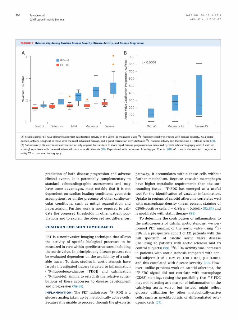
FIGURE 4 Relationship Among Baseline Disease Severity, Disease Activity, and Disease Progression
6 900
p < 0.0001800
700
600
500
400
300
200
100
0Mild AS
Aort
ic V
alve
Cal
cific
atio
n Pr
ogre
ssio
n, A
U/Ye
arModerate AS Severe AS
4
2
0Control Sclerosis Mild
18F-NaF
Max
imum
TBR
Val
ue
18F-FDG
Moderate Severe
A B
(A) Studies using PET have demonstrated that calcification activity in the valve (as measured using 18F-fluoride) steadily increases with disease severity. As a conse-
quence, activity is highest in those with the most advanced disease, and a good correlation exists between 18F-fluoride activity and the baseline CT calcium score (79).
(B) Subsequently, this increased calcification activity appears to translate to more rapid disease progression (as measured by both echocardiography and CT calcium
scoring) in patients with the most advanced forms of aortic stenosis (78). Reproduced with permission from Nguyen V, et al. (78). AS ¼ aortic stenosis; AU ¼ Agatston
units; CT ¼ computed tomography.
Pawade et al. J A C C V O L . 6 6 , N O . 5 , 2 0 1 5
Calcification in Aortic Stenosis A U G U S T 4 , 2 0 1 5 : 5 6 1 – 7 7
570
prediction of both disease progression and adverseclinical events. It is potentially complementary tostandard echocardiographic assessments and mayhave some advantages, most notably that it is notdependent on cardiac loading conditions, geometricassumptions, or on the presence of other cardiovas-cular conditions, such as mitral regurgitation andhypertension. Further work is now required to vali-date the proposed thresholds in other patient pop-ulations and to explain the observed sex differences.
POSITRON EMISSION TOMOGRAPHY
PET is a noninvasive imaging technique that allowsthe activity of specific biological processes to bemeasured in vivo within specific structures, includingthe aortic valve. In principle, any disease process canbe evaluated dependent on the availability of a suit-able tracer. To date, studies in aortic stenosis havelargely investigated tracers targeted to inflammation(18F-fluorodeoxyglucose [FDG]) and calcification(18F-fluoride), aiming to establish the relative contri-butions of these processes to disease developmentand progression (79–81).
INFLAMMATION. The PET radiotracer 18F- FDG is aglucose analog taken up by metabolically active cells.Because it is unable to proceed through the glycolytic
pathway, it accumulates within these cells withoutfurther metabolism. Because vascular macrophageshave higher metabolic requirements than the sur-rounding tissue, 18F-FDG has emerged as a usefultool for the identification of vascular inflammation.Uptake in regions of carotid atheroma correlates wellwith macrophage density (mean percent staining ofCD68-positive cells, r ¼ 0.85; p < 0.0001) (82,83) andis modifiable with statin therapy (84).
To determine the contribution of inflammation tothe pathogenesis of calcific aortic stenosis, we per-formed PET imaging of the aortic valve using 18F-FDG in a prospective cohort of 121 patients with thefull spectrum of calcific aortic valve disease(including 20 patients with aortic sclerosis and 20control subjects) (79). 18F-FDG activity was increasedin patients with aortic stenosis compared with con-trol subjects (1.58 � 0.21 vs. 1.30 � 0.13; p < 0.001),and this correlated with disease severity (79). How-ever, unlike previous work on carotid atheroma, the18F-FDG signal did not correlate with macrophage(CD68) staining, raising the possibility that 18F-FDGmay not be acting as a marker of inflammation in thecalcifying aortic valve, but instead might reflectglucose utilization by other metabolically-activecells, such as myofibroblasts or differentiated oste-ogenic cells (77).

J A C C V O L . 6 6 , N O . 5 , 2 0 1 5 Pawade et al.A U G U S T 4 , 2 0 1 5 : 5 6 1 – 7 7 Calcification in Aortic Stenosis
571
CALCIFICATION. 18F-fluoride has been used safely asa bone tracer for more than 40 years, exchangingwith hydroxyl groups in hydroxyapatite to form flu-oroapatite. Similar hydroxyl bonds are also present inthe different forms of calcium in the vasculature(including hydroxyapatite and amorphous calcium)so that 18F-fluoride binding acts as a marker ofvascular calcification. In particular, the binding of18F-fluoride to calcium appears to be criticallydependent upon the surface area of calcium ortho-phosphate available for incorporation. 18F-fluoride,therefore, preferentially binds regions of newlydeveloping microcalcification (beyond the resolutionof CT), which have a nanocrystalline structure andvery high surface area, rather than to large, estab-lished, macroscopic deposits, where much of thecalcium is internalized and, therefore, not availablefor binding (85). On this basis, increased 18F-fluorideuptake is observed in regions of actively developingcalcification, demonstrating a close association withalkaline phosphatase staining (r ¼ 0.65; p ¼ 0.04) onexcised aortic valve tissue removed at the time ofsurgery (77).
When the same cohort of 121 patients was imagedwith 18F-fluoride, the observed PET signal in theaortic valve was stronger and more clearly demar-cated than was seen with 18F-FDG (Figure 3). More-over, the spatial distribution of the 18F-fluoride signalwas often discrete from the macroscopic calcium de-posits identified by CT, indicating that 18F-fluorideuptake provides distinct, but complementary infor-mation to CT alone. Uptake was increased in patientswith aortic stenosis compared with healthy controlsubjects (2.87 � 0.82 vs. 1.55 � 0.17; p < 0.001) andcorrelated with disease severity (r ¼ 0.73; p < 0.001)(79). Indeed, the highest calcification activity, asmeasured using this tracer, was observed in patientswith the most advanced disease (Figure 4A). Again,this supports the hypothesis that calcification begetscalcification activity in aortic stenosis and wouldexplain the rapid rates of disease progression in thoseat the severe end of the spectrum.
When patients were recalled for repeat CT calciumscoring of the valve at 1 and 2 years, new calciumcould be observed in the areas of increased 18F-fluoride activity seen on the baseline scan (Figure 5).As a consequence, a close correlation was observedbetween the baseline valvular 18F-fluoride uptake andthe progression of the aortic valve CT calcium score(r ¼ 0.80; 95% CI: 0.69 to 0.87; p < 0.001), with PETappearing to offer some additional predictive infor-mation over and above the baseline calcium score.Moreover, this translated into an ability to predictvalve hemodynamic progression, with moderate
correlations also observed between 18F-fluoride ac-tivity and the mean (r ¼ 0.32; 95% CI: 0.13 to 0.50;p ¼ 0.001) and peak (r ¼ 0.32; 95% CI: 0.12 to 0.49;p ¼ 0.002) aortic valve gradients (67). Finally, after amedian of 1,526 days of follow-up, 18F-fluorideemerged as a prognostic marker serving as an inde-pendent predictor of the combined endpoint of aorticvalve replacement and cardiovascular mortality(hazard ratio: 1.55; 95% CI: 1.33 to 1.81, after adjustingfor age and sex; p < 0.001).
In summary, these data highlight the potentialapplication of 18F-fluoride as an immediate, nonin-vasive measure of disease activity in aortic stenosiswith the ability to predict its natural history. Theinstantaneous readout of disease activity holdsparticular promise in assessing the early efficacy ofnovel therapeutic agents, in which treatment effectsare likely to be discernible over a much shorter timeperiod than could be resolved using clinical end-points, echocardiography, or CT.
Should 18F-fluoride PET be used as a clinical tool?Although PET performed well in predicting the nat-ural history of aortic stenosis, the far simpler tech-nique of CT calcium scoring appeared to providealmost equivalent prediction of disease progression.Moreover, in agreement with the work by Clavel et al.(72,73), CT again provided incremental prediction ofclinical outcomes to even echocardiographic assess-ments of hemodynamic severity. Whilst supporting agreater role for CT, this would argue against the use ofPET in the routine clinical arena. It also raises thequestion as to why an anatomic measure of calciumburden can provide such effective prediction offuture disease progression? We believe that thisreflects the close association between calcificationactivity in the valve (as assessed by 18F-fluoride) andthe baseline calcium score (r ¼ 0.80; p < 0.001), andprovides further evidence for the model of calciumbegetting further calcium formation in the propaga-tion phase of the disease (Figure 1). Regardless of themechanism, the close link between calcium burdenand calcification activity in the valve ensures thateven the simplest methods of aortic valve calciumburden provide a surrogate of disease activity andeffective prediction of disease progression.
The imaging techniques described previously allowus to image calcification in the valve in progressivedetail. They have helped to confirm the importantrole that calcification plays in driving aortic stenosisand have allowed us to both characterize the severityof disease and to better predict disease progression.We anticipate that CT calcium scoring will assume agreater clinical role, whereas PET will prove apowerful research tool, in particular as an endpoint

FIGURE 5 Change in Aortic Valve CT Calcium Score and 18F-Sodium Fluoride PET Activity After 1 Year
Baseline CT calcium scores (left) for patients 1 and 2 (top and bottom). Fused coaxial 18F-fluoride PET-CT scans (middle) show fluoride uptake in red and yellow.
The 1-year follow-up (right) suggests that the baseline PET signal predicts the spatial distribution of subsequent macrocalcification (77). Abbreviations as in Figure 3.
Pawade et al. J A C C V O L . 6 6 , N O . 5 , 2 0 1 5
Calcification in Aortic Stenosis A U G U S T 4 , 2 0 1 5 : 5 6 1 – 7 7
572
in clinical trials assessing the efficacy of novel,potentially disease-modifying therapies. Indeed,18F-fluoride PET has the potential to provide bothmechanistic insights and a far more rapid readout ofefficacy than CT calcium scoring or echocardiographicparameters.
POTENTIAL NOVEL
DISEASE-MODIFYING THERAPIES
As our understanding of the pathophysiology of aorticstenosis has improved, the key role that calcificationplays in driving disease progression has led us awayfrom targeting inflammation and lipid deposition andtoward therapies capable of directly halting valvecalcification (86). How might this be achieved? Theclose association between disorders of skeletal bonemetabolism and increased calcification in the vascu-lature offers a potential starting point. A growingbody of pre-clinical and clinical data indicates thattreatments for osteoporosis, such as bisphosphonates
and denosumab, can reduce vascular calcification andthat these agents hold considerable promise as noveltherapies for aortic stenosis (87).
BISPHOSPHONATES. Bisphosphonates are inhibitorsof osteoclast-mediated bone resorption, are welltolerated in elderly patients, and have been widelyused for the treatment of osteoporosis (88). Inter-estingly, bisphosphonates also have important car-diovascular effects, demonstrating a consistentreduction in calcification of the vasculature and theaortic valve (87,89,90). This, in part, appears to be aconsequence of their inhibition of bone resorption,which results in reduced release of calcium andphosphate into the circulation and, therefore, in thereduced systemic availability of these procalcificsubstances (Figure 2B) (87). However, bisphospho-nates also appear to exert direct anticalcific effects onthe aortic valve tissue itself. They reduce the pro-duction of IL-1b, IL-6, and TNF-a (key inflammatorycytokines implicated in the early stages of aorticstenosis [91]) and inhibit the secretion of matrix

J A C C V O L . 6 6 , N O . 5 , 2 0 1 5 Pawade et al.A U G U S T 4 , 2 0 1 5 : 5 6 1 – 7 7 Calcification in Aortic Stenosis
573
metalloproteinases 2 and 9, which remodel the valveas aortic stenosis progresses (Figure 1) (92,93). More-over, nitrogen-containing bisphosphonates act asinorganic pyrophosphate analogs (48), which, as dis-cussed, have powerful anticalcific properties in thevasculature. Finally, bisphosphonates attenuate thedifferentiation of aortic valve myofibroblasts intocells with an osteogenic phenotype (94), the key stepin triggering the propagation phase of aortic stenosis(86). In combination, these data offer support forbisphosphonates as a treatment strategy for aorticstenosis that is increasingly being supported byobservational clinical data. A recent analysis of 3,710women in the MESA (Multi-Ethnic Study of Athero-sclerosis) indicated that bisphosphonate use wasassociated with less valvular and vascular calcifica-tion in older women (users vs. nonusers: aortic valvering calcium 38% vs. 59%; p < 0.0001) (95). Otherstudies appear to support these findings with a directbeneficial effect of these drugs on echocardiographicmeasures of aortic stenosis progression (96–98), aswell as reducing valvular calcification in patients withrenal failure and amongst those with bioprostheticvalves (87,99). Although encouraging, such retro-spective, observational studies are prone to bias,cannot assess cause-and-effect, have provided con-flicting results (100), and are confounded by the un-derlying effects of the osteoporosis for which theseagents were prescribed. Indeed, the true effect ofbisphosphonates in aortic stenosis will only becomeclear within the context of a randomized controlledtrial (101).DENOSUMAB. As discussed, the OPG/RANK/RANKLaxis appears to play a pivotal role in aortic valvecalcification and may provide an explanation for thelink between osteoporosis and increased vascularcalcification. It, therefore, represents an attractivetherapeutic target for reducing vascular calcification(Figure 2C). Denosumab is a human monoclonal anti-body to RANKL that prevents its binding to RANK,thereby recapitulating the actions of OPG. In a trial of7,868 post-menopausal women with osteoporosis,denosumab increased bone mineral density andreduced vertebral fracture rates by 68% over a 3-yearperiod (102). Importantly, denosumab was extremelywell tolerated, with very few adverse side effects andno major excess of adverse events. Given the centralregulatory role of the OPG/RANK/RANKL system invascular and aortic valve calcification, denosumabalso holds considerable promise as a novel treatmentfor aortic stenosis. Again, this is supported by pre-clinical data, with denosumab halving the aorticcalcification observed in a murine model of osteopo-rosis (103). Interestingly, in the same study, this
reduction was closely associated with inhibitedbone resorption from the skeleton, indicatingthat the cardiovascular effects of denosumab are,like bisphosphonates, in part related to reducedcalcium and phosphate release from bone into thecirculation.
FUTURE PERSPECTIVES
Bisphosphonates and denosumab hold promise asnovel treatments for aortic stenosis and are currentlybeing investigated as part of an ongoing randomizedcontrol trial (NCT02132026) (104). However, even ifthese prove ineffective, we believe that future treat-ments should still be directed at the propagationphase and at breaking the self-perpetuating cycle ofvalvular injury, osteogenic differentiation, and cal-cium deposition. As discussed, a rapidly expandinglist of signaling pathways and molecular processesgoverning the pathogenesis of aortic stenosis havebeen elucidated, uncovering many additional targetsat different phases of the disease; these are discussedin the following paragraphs. In addition, furtherinvestigation is warranted to assess whether poten-tially pro-calcific drugs, including calcium supple-ments and coumarins, should be avoided in patientswith aortic stenosis.
Ultimately, many of the procalcific pathways inthe valve appear to converge on the up-regulationof osteogenic differentiation factors (e.g., BMP-2,Wnt-b-catenin) that establish osteoblast-like func-tion within the valve. These factors therefore pro-vide an attractive therapeutic strategy, although,given the overlap in factors governing calcificationin the bone and the valve, the major challenge willbe to slow aortic stenosis progression withoutcompromising bone health. One potential approachwould be to target the upstream cytokines thatactivate BMP, such as using inhibitors of IL-6 orTNF-a (as already used in rheumatoid arthritis).However, once again, it remains unclear whethertargeting inflammation will be effective in thepropagation phase once the procalcific processeshave become established. Targeting ectonucleoti-dases may be more effective, given their apparentlycentral role in establishing the positive feedbackloop by which calcium begets calcium. Ectonucleo-tidase inhibitors have already been tested in thewarfarin rat model and have been shown to preventthe development of calcific aortic valve disease(105). Interest also surrounds P2Y2 receptor antago-nists as a means of reducing VIC apoptosis and thecalcification that this induces. Therapeutic admin-istration of fetuin-A, or a mimetic of MGP, could

CENTRAL ILLUSTRATION The Pathogenesis of Aortic Stenosis in 2 Stages
Endothelial injury
Infiltration of oxidized lipids and inflammatory cells into aortic valve
Lipoprotein(a) and oxidized low-density
lipoprotein cholesterol
Differentiation of valve interstitial cellsto osteoblast-like cells
Deposition ofcollagen and bone-
related proteins
Mechanical stress, injury and apoptosis Micro-calcification
Macro-calcification
Progressive valve obstruction
Cholesterolreduction with:• Lipoprotein(a)
inhibitors • Statins
Calcificationreduction with:• Bisphosphonates• Denosumab• Ectonucleotidase
and ACE inhibitors
• 18F-Fluoride PETto investigate micro-calcification activity in the valve
• Computed Tomography (CT) Calcium scoringto assess the burdenof macroscopic calcium in the valve
• Echocardiographyto assess valve obstruction severity
• Potentially 18F-FDG Positron Emission Tomography (PET)to investigate inflammation
PATHOPHYSIOLOGY
Phas
e 1:
Initi
atio
nPh
ase
2: S
elf p
ropa
gatin
g lo
op
TREATMENT OPTIONSIMAGING OPTIONS
Macrophages, mast cells and T-lymphocytes
Pawade, T.A. et al. J Am Coll Cardiol. 2015; 66(5):561–77.
Initiation phase: similar to the early stages of atherosclerosis, endothelial injury facilitates the infiltration of oxidized lipids and inflammatory cells into the
valve and the release of proinflammatory mediators. These trigger the very early stages of valve calcification so that the incidence of aortic stenosis closely
relates to traditional cardiovascular risk factors for atherosclerosis. Propagation phase: these proinflammatory processes subsequently induce VICs to
undergo osteogenic differentiation. The VICs first lay down a collagen matrix and other bone-related proteins before producing calcium (7,8). Calcification
of the valve induces compliance mismatch, resulting in increased mechanical stress, injury, and apoptosis (9), which triggers further calcification (10).
Hence, a self-perpetuating cycle of calcification, valve injury, and osteogenic activation is established that drives the propagation phase of the disease. As a
consequence, disease progression in aortic stenosis more closely relates to procalcific factors, rather than lipid infiltration or inflammation. The different
stages of calcification in the valve can be imaged using 18F-Fluoride PET (newly developing microcalcification) and CT calcium scoring (macroscopic calcific
deposits). Given the central role of calcification in the propagation phase of aortic stenosis, it is perhaps unsurprising that these imaging techniques provide
important information with respect to prognosis and disease progression. Moreover, calcification represents an important potential therapeutic target, with
the use of drugs, such as denosumab and bisphosphonates, to interrupt the vicious cycle of calcification that drives progressive narrowing of the valve.
ACE ¼ angiotensin-converting enzyme; CT ¼ computed tomography; FDG ¼ fluorodeoxyglucose; PET ¼ positron emission tomography; VIC ¼ valvular
interstitial cell.
Pawade et al. J A C C V O L . 6 6 , N O . 5 , 2 0 1 5
Calcification in Aortic Stenosis A U G U S T 4 , 2 0 1 5 : 5 6 1 – 7 7
574
simultaneously target multiple pathways thought todrive valvular calcification.
The ability of lipoprotein(a)-lowering therapies tomodify aortic stenosis disease progression is likely toform the basis of a future clinical trial. Given thefailure of the statin trials, it will be of great interest todetermine whether a more targeted lipid interventionwill have greater success in reducing disease
progression in the propagation phase (106). On thebasis of the apparent contribution of the RAS to theinitiation of aortic stenosis, it is also not unreasonableto consider ACE inhibitors, or even selective AT1 re-ceptor antagonists or novel renin inhibitors, as noveltreatments. Indeed, these agents are also likely tohave a beneficial effect with respect to hypertensionand left ventricular remodeling in aortic stenosis,

J A C C V O L . 6 6 , N O . 5 , 2 0 1 5 Pawade et al.A U G U S T 4 , 2 0 1 5 : 5 6 1 – 7 7 Calcification in Aortic Stenosis
575
given the role that the RAS system also plays indriving myocardial hypertrophy, fibrosis, and thetransition to heart failure.
CONCLUSIONS
Aortic stenosis is a common condition that is set tobecome an increasing health care burden. We lackeffective medical therapies capable of slowing itsrelentless progression toward major surgery oradverse events. Recent insights into the pathophysi-ology of aortic stenosis have indicated that althoughlipid and inflammation may be important in estab-lishing the disease (initiation phase), it is theself-perpetuating processes of calcification that arepredominantly responsible for driving disease
progression (propagation phase) (Central Illustration).On this basis, imaging modalities capable of quanti-fying aortic valve calcification will be best placed topredict its natural history, whereas novel anticalcifictherapies hold major promise as methods of treat-ment. Randomized controlled trials of such agents,perhaps using imaging endpoints such as CT calciumscoring and 18F-fluoride PET activity, are nowrequired to establish their early efficacy.
REPRINT REQUESTS AND CORRESPONDENCE: Dr.Marc R. Dweck, British Heart Foundation/UniversityCentre for Cardiovascular Science, Room SU 305,Chancellor’s Building, University of Edinburgh, 49Little France Crescent, Edinburgh EH16 4SB, UnitedKingdom. E-mail: [email protected].
RE F E RENCE S
1. Nkomo VT, Gardin JM, Skelton TN, et al. Burdenof valvular heart diseases: a population-basedstudy. Lancet 2006;368:1005–11.
2. Osnabrugge RL, Mylotte D, Head SJ, et al.Aortic stenosis in the elderly: disease prevalenceand number of candidates for transcatheter aorticvalve replacement: a meta-analysis and modelingstudy. J Am Coll Cardiol 2013;62:1002–12.
3. Rajamannan NM, Evans FJ, Aikawa E, et al.Calcific aortic valve disease: not simply a degen-erative process. A review and agenda for researchfrom the National Heart and Lung and BloodInstitute Aortic Stenosis Working Group. Executivesummary: calcific aortic valve disease—2011update. Circulation 2011;124:1783–91.
4. New SE, Aikawa E. Molecular imaging insightsinto early inflammatory stages of arterial andaortic valve calcification. Circ Res 2011;108:1381–91.
5. Thanassoulis G, Massaro JM, Cury R, et al.Associations of long-term and early adultatherosclerosis risk factors with aortic and mitralvalve calcium. J Am Coll Cardiol 2010;55:2491–8.
6. Stewart BF, Siscovick D, Lind BK, et al. Clinicalfactors associated with calcific aortic valve dis-ease. Cardiovascular Health Study. J Am CollCardiol 1997;29:630–4.
7. Stritzke J, Linsel-Nitschke P, Markus MR, et al.Association between degenerative aortic valvedisease and long-term exposure to cardiovascularrisk factors: results of the longitudinal population-based KORA/MONICA survey. Eur Heart J 2009;30:2044–53.
8. Pachulski RT, Chan KL. Progression of aorticvalve dysfunction in 51 adult patients withcongenital bicuspid aortic valve: assessment andfollow up by Doppler echocardiography. Br Heart J1993;69:237–40.
9. Smith JG, Luk K, Schulz CA, et al., for the Co-horts for Heart and Aging Research in GeneticEpidemiology (CHARGE) Extracoronary CalciumWorking Group. Association of low-density
lipoprotein cholesterol-related genetic variantswith aortic valve calcium and incident aortic ste-nosis. JAMA 2014;312:1764–71.
10. Thanassoulis G, Campbell CY, Owens DS, et al.,for the CHARGE Extracoronary Calcium WorkingGroup. Genetic associations with valvular calcifi-cation and aortic stenosis. N Engl J Med 2013;368:503–12.
11. Otto CM, Kuusisto J, Reichenbach DD, et al.Characterization of the early lesion of ’degenera-tive’ valvular aortic stenosis. Histological andimmunohistochemical studies. Circulation 1994;90:844–53.
12. Proudfoot D, Skepper JN, Hegyi L, et al.Apoptosis regulates human vascular calcificationin vitro: evidence for initiation of vascular calcifi-cation by apoptotic bodies. Circ Res 2000;87:1055–62.
13. Kim KM. Calcification of matrix vesicles inhuman aortic valve and aortic media. Fed Proc1976;35:156–62.
14. Nadra I, Mason JC, Philippidis P, et al. Proin-flammatory activation of macrophages by basiccalcium phosphate crystals via protein kinase Cand MAP kinase pathways: a vicious cycle ofinflammation and arterial calcification? Circ Res2005;96:1248–56.
15. Moura LM, Ramos SF, Zamorano JL, et al.Rosuvastatin affecting aortic valve endothelium toslow the progression of aortic stenosis. J Am CollCardiol 2007;49:554–61.
16. Rajamannan NM, Subramaniam M, Springett M,et al. Atorvastatin inhibits hypercholesterolemia-induced cellular proliferation and bone matrixproduction in the rabbit aortic valve. Circulation2002;105:2660–5.
17. Weiss RM, Ohashi M, Miller JD, et al. Calcificaortic valve stenosis in old hypercholesterolemicmice. Circulation 2006;114:2065–9.
18. Cowell SJ, Newby DE, Prescott RJ, et al., forthe Scottish Aortic Stenosis and Lipid LoweringTrial, Impact on Regression (SALTIRE)
Investigators. A randomized trial of intensive lipid-lowering therapy in calcific aortic stenosis. N EnglJ Med 2005;352:2389–97.
19. Chan KL, Teo K, Dumesnil JG, et al., for theASTRONOMER Investigators. Effect of lipidlowering with rosuvastatin on progression ofaortic stenosis: results of the aortic stenosis pro-gression observation: measuring effects of rosu-vastatin (ASTRONOMER) trial. Circulation 2010;121:306–14.
20. Rossebø AB, Pedersen TR, Boman K, et al., forthe SEAS Investigators. Intensive lipid loweringwith simvastatin and ezetimibe in aortic stenosis.N Engl J Med 2008;359:1343–56.
21. Puri R, Nicholls SJ, Shao M, et al. Impact ofstatins on serial coronary calcification duringatheroma progression and regression. J Am CollCardiol 2015;65:1273–82.
22. Capoulade R, Clavel MA, Dumesnil JG, et al.,for the ASTRONOMER Investigators. Impact ofmetabolic syndrome on progression of aortic ste-nosis: influence of age and statin therapy. J AmColl Cardiol 2012;60:216–23.
23. Mohler ER 3rd, Gannon F, Reynolds C, et al.Bone formation and inflammation in cardiacvalves. Circulation 2001;103:1522–8.
24. El Accaoui RN, Gould ST, Hajj GP, et al. Aorticvalve sclerosis in mice deficient in endothelialnitric oxide synthase. Am J Physiol Heart CircPhysiol 2014;306:H1302–13.
25. O’Brien KD, Shavelle DM, Caulfield MT, et al.Association of angiotensin-converting enzymewith low-density lipoprotein in aortic valvularlesions and in human plasma. Circulation 2002;106:2224–30.
26. Peltonen T, Napankangas J, Ohtonen P, et al.(Pro)renin receptors and angiotensin convertingenzyme 2/angiotensin-(1-7)/Mas receptor axis inhuman aortic valve stenosis. Atherosclerosis 2011;216:35–43.
27. Capoulade R, Clavel MA, Mathieu P, et al.Impact of hypertension and renin-angiotensin

Pawade et al. J A C C V O L . 6 6 , N O . 5 , 2 0 1 5
Calcification in Aortic Stenosis A U G U S T 4 , 2 0 1 5 : 5 6 1 – 7 7
576
system inhibitors in aortic stenosis. Eur J ClinInvest 2013;43:1262–72.
28. Ducy P. CBFA1: a molecular switch in osteo-blast biology. Dev Dyn 2000;219:461–71.
29. Rajamannan NM, Subramaniam M, Rickard D,et al. Human aortic valve calcification is associatedwith an osteoblast phenotype. Circulation 2003;107:2181–4.
30. Pohjolainen V, Taskinen P, Soini Y, et al.Noncollagenous bone matrix proteins as a part ofcalcific aortic valve disease regulation. Hum Pathol2008;39:1695–701.
31. Ganss B, Kim RH, Sodek J. Bone sialoprotein.Crit Rev Oral Biol Med 1999;10:79–98.
32. Liu AC, Joag VR, Gotlieb AI. The emerging roleof valve interstitial cell phenotypes in regulatingheart valve pathobiology. Am J Pathol 2007;171:1407–18.
33. Aikawa E, Nahrendorf M, Figueiredo JL, et al.Osteogenesis associates with inflammation inearly-stage atherosclerosis evaluated by molecu-lar imaging in vivo. Circulation 2007;116:2841–50.
34. Aikawa E, Otto CM. Look more closely at thevalve: imaging calcific aortic valve disease. Circu-lation 2012;125:9–11.
35. Watson KE, Bostrom K, Ravindranath R, et al.TGF-beta 1 and 25-hydroxycholesterol stimulateosteoblast-like vascular cells to calcify. J ClinInvest 1994;93:2106–13.
36. Tintut Y, Demer L. Role of osteoprotegerin andits ligands and competing receptors in athero-sclerotic calcification. J Investig Med 2006;54:395–401.
37. Garg V, Muth AN, Ransom JF, et al. Mutationsin NOTCH1 cause aortic valve disease. Nature2005;437:270–4.
38. Nigam V, Srivastava D. Notch1 repressesosteogenic pathways in aortic valve cells. J MolCell Cardiol 2009;47:828–34.
39. Boström K, Watson KE, Stanford WP, et al.Atherosclerotic calcification: relation to develop-mental osteogenesis. Am J Cardiol 1995;75:88B–91.
40. Yang X, Meng X, Su X, et al. Bone morpho-genic protein 2 induces Runx2 and osteopontinexpression in human aortic valve interstitial cells:role of Smad1 and extracellular signal-regulatedkinase 1/2. J Thorac Cardiovasc Surg 2009;138:1008–15.
41. Yang X, Fullerton DA, Su X, et al. Pro-osteogenic phenotype of human aortic valveinterstitial cells is associated with higher levels ofToll-like receptors 2 and 4 and enhanced expres-sion of bone morphogenetic protein 2. J Am CollCardiol 2009;53:491–500.
42. Caira FC, Stock SR, Gleason TG, et al. Humandegenerative valve disease is associated withup-regulation of low-density lipoprotein receptor-related protein 5 receptor-mediated bone forma-tion. J Am Coll Cardiol 2006;47:1707–12.
43. Chen JH, Chen WL, Sider KL, et al. b-cateninmediates mechanically regulated, transforminggrowth factor-b1-induced myofibroblast differen-tiation of aortic valve interstitial cells. ArteriosclThromb Vasc Biol 2011;31:590–7.
44. Rajamannan NM. Oxidative-mechanical stresssignals stem cell niche mediated Lrp5 osteo-genesis in eNOS�/� null mice. J Cell Biochem 2012;113:1623–34.
45. Kado DM, Browner WS, Blackwell T, et al. Rateof bone loss is associated with mortality in olderwomen: a prospective study. J Bone Miner Res2000;15:1974–80.
46. Pfister R, Michels G, Sharp SJ, et al. Inverseassociation between bone mineral density and riskof aortic stenosis in men and women in EPIC-Norfolk prospective study. Int J Cardiol 2015;178:29–30.
47. Aksoy Y, Yagmur C, Tekin GO, et al. Aorticvalve calcification: association with bone mineraldensity and cardiovascular risk factors. CoronArtery Dis 2005;16:379–83.
48. Demer LL, Tintut Y. Vascular calcification:pathobiology of a multifaceted disease. Circula-tion 2008;117:2938–48.
49. Persy V, D’Haese P. Vascular calcification andbone disease: the calcification paradox. TrendsMol Med 2009;15:405–16.
50. Hultgren HN. Osteitis deformans (Paget’sdisease) and calcific disease of the heart valves.Am J Cardiol 1998;81:1461–4.
51. Kaden JJ, Bickelhaupt S, Grobholz R, et al.Receptor activator of nuclear factor kB ligand andosteoprotegerin regulate aortic valve calcification.J Mol Cell Cardiol 2004;36:57–66.
52. Bucay N, Sarosi I, Dunstan CR, et al.Osteoprotegerin-deficient mice develop earlyonset osteoporosis and arterial calcification. GenesDev 1998;12:1260–8.
53. Parhami F, Morrow AD, Balucan J, et al. Lipidoxidation products have opposite effects oncalcifying vascular cell and bone cell differentia-tion. A possible explanation for the paradox ofarterial calcification in osteoporotic patients.Arterioscl Thromb Vasc Biol 1997;17:680–7.
54. Demer LL. Vascular calcification and osteo-porosis: inflammatory responses to oxidized lipids.Int J Epidemiol 2002;31:737–41.
55. Schurgers LJ, Uitto J, Reutelingsperger CP.Vitamin K-dependent carboxylation of matrixGla-protein: a crucial switch to control ectopicmineralization. Trends Mol Med 2013;19:217–26.
56. Tantisattamo E, Han KH, O’Neill WC. Increasedvascular calcification in patients receiving warfarin.Arterioscl Thromb Vasc Biol 2015;35:237–42.
57. Kaden JJ, Reinöhl JO, Blesch B, et al. Systemicand local levels of fetuin-A in calcific aortic valvestenosis. Int J Mol Med 2007;20:193–7.
58. Koos R, Brandenburg V, Mahnken AH, et al.Association of fetuin-A levels with the progressionof aortic valve calcification in non-dialyzed pa-tients. Eur Heart J 2009;30:2054–61.
59. Mohty D, Côté N, Pibarot P, et al. Reducedfetuin a serum level is associated with fasterprogression and increased valvular calcification inelderly patients with aortic stenosis. J Clin ExpCardiol 2011;2:147.
60. Capoulade R, Côté N, Mathieu P, et al., for theASTRONOMER Investigators. Circulating levels ofmatrix GLA protein and progression of aortic
stenosis: a substudy of the Aortic Stenosis Pro-gression Observation: Measuring Effects of rosu-vastatin (ASTRONOMER) trial. Can J Cardiol 2014;30:1088–95.
61. Côté N, El Husseini D, Pépin A, et al. ATP acts asa survival signal and prevents the mineralization ofaortic valve. J Mol Cell Cardiol 2012;52:1191–202.
62. El Husseini D, Boulanger MC, Mahmut A, et al.P2Y2 receptor represses IL-6 expression by valveinterstitial cells through Akt: implication forcalcific aortic valve disease. J Mol Cell Cardiol2014;72:146–56.
63. Baumgartner H, Hung J, Bermejo J, et al.Echocardiographic assessment of valve stenosis:EAE/ASE recommendations for clinical practice.Eur J Echocardiogr 2009;22:1–25.
64. Rosenhek R, Binder T, Porenta G, et al.Predictors of outcome in severe, asymptomaticaortic stenosis. N Engl J Med 2000;343:611–7.
65. Cioffi G, Mazzone C, Faggiano P, et al. Prog-nostic stratification by conventional echocardiog-raphy of patients with aortic stenosis: the“CAIMAN-ECHO score.” Echocardiography 2013;30:367–77.
66. Messika-Zeitoun D, Aubry MC, Detaint D, et al.Evaluation and clinical implications of aortic valvecalcificationmeasured by electron-beamcomputedtomography. Circulation 2004;110:356–62.
67. Jenkins W, Dweck M, Shah A, et al. 18F-NaF isa predictor of progression and outcome in aorticvalve disease (abstr). J Am Coll Cardiol 2014;63Suppl 12:A995.
68. Liu F, Coursey CA, Grahame-Clarke C, et al.Aortic valve calcification as an incidental finding atCT of the elderly: severity and location as pre-dictors of aortic stenosis. AJR Am J Roentgenol2006;186:342–9.
69. Koos R, Kühl HP, Mühlenbruch G, et al. Prev-alence and clinical importance of aortic valvecalcification detected incidentally on CT scans:comparison with echocardiography. Radiology2006;241:76–82.
70. Aggarwal SR, Clavel MA, Messika-Zeitoun D,et al. Sex differences in aortic valve calcificationmeasured by multidetector computed tomographyin aortic stenosis. Circ Cardiovasc Imaging 2013;6:40–7.
71. Dweck MR, Chin C, Newby DE. Small valve areawith low-gradient aortic stenosis: beware the hardhearted. J Am Coll Cardiol 2013;62:2339–40.
72. Clavel MA, Messika-Zeitoun D, Pibarot P, et al.The complex nature of discordant severe calcifiedaortic valve disease grading: new insights fromcombined Doppler echocardiographic and com-puted tomographic study. J Am Coll Cardiol 2013;62:2329–38.
73. Clavel MA, Pibarot P, Messika-Zeitoun D, et al.Impact of aortic valve calcification, as measured byMDCT, on survival in patients with aortic stenosis:results of an international registry study. J Am CollCardiol 2014;64:1202–13.
74. Feuchtner GM, Müller S, Grander W, et al.Aortic valve calcification as quantified withmultislice computed tomography predicts short-term clinical outcome in patients with

J A C C V O L . 6 6 , N O . 5 , 2 0 1 5 Pawade et al.A U G U S T 4 , 2 0 1 5 : 5 6 1 – 7 7 Calcification in Aortic Stenosis
577
asymptomatic aortic stenosis. J Heart Valve Dis2006;15:494–8.
75. Koos R, Mahnken AH, Sinha AM, et al. Aorticvalve calcification as a marker for aortic stenosisseverity: assessment on 16-MDCT. AJR Am JRoentgenol 2004;183:1813–8.
76. Messika-Zeitoun D, Bielak LF, Peyser PA, et al.Aortic valve calcification: determinants and pro-gression in the population. Arterioscl Thromb VascBiol 2007;27:642–8.
77. Dweck MR, Jenkins WS, Vesey AT, et al. 18F-NaF uptake is a marker of active calcification anddisease progression in patients with aortic steno-sis. Circ Cardiovasc Imaging 2014;7:371–8.
78. Nguyen V, Cimadevilla C, Estellat C, et al.Haemodynamic and anatomic progression of aorticstenosis. Heart 2015;101:943–7.
79. Dweck MR, Jones C, Joshi NV, et al. Assess-ment of valvular calcification and inflammation bypositron emission tomography in patients withaortic stenosis. Circulation 2012;125:76–86.
80. Marincheva-Savcheva G, Subramanian S,Qadir S, et al. Imaging of the aortic valve using flu-orodeoxyglucose positron emission tomography:increased valvular fluorodeoxyglucose uptake inaortic stenosis. J Am Coll Cardiol 2011;57:2507–15.
81. Dweck MR, Khaw HJ, Sng GK, et al. Aorticstenosis, atherosclerosis, and skeletal bone: isthere a common link with calcification andinflammation? Eur Heart J 2013;34:1567–74.
82. Tahara N, Kai H, Nakaura H, et al. The preva-lence of inflammation in carotid atherosclerosis:analysis with fluorodeoxyglucose-positron emis-sion tomography. Eur Heart J 2007;28:2243–8.
83. Tawakol A, Migrino RQ, Bashian GG, et al.In vivo 18F-fluorodeoxyglucose positron emissiontomography imaging provides a noninvasivemeasure of carotid plaque inflammation in pa-tients. J Am Coll Cardiol 2006;48:1818–24.
84. Tahara N, Kai H, Ishibashi M, et al. Simvastatinattenuates plaque inflammation: evaluation byfluorodeoxyglucose positron emission tomogra-phy. J Am Coll Cardiol 2006;48:1825–31.
85. Irkle A, Vesey AT, Lewis DY, et al. Identifyingactive vascular micro-calcification by 18F-sodiumfluoride positron emission tomography. Nat Com-mun 2015;6:7495.
86. Otto CM. Calcific aortic stenosis—time to lookmore closely at the valve. N Engl J Med 2008;359:1395–8.
87. Price PA, Faus SA, Williamson MK.Bisphosphonates alendronate and ibandronate
inhibit artery calcification at doses comparable tothose that inhibit bone resorption. ArteriosclThromb Vasc Biol 2001;21:817–24.
88. Fleisch H, Russell RG, Francis MD. Diphosph-onates inhibit hydroxyapatite dissolution in vitroand bone resorption in tissue culture and in vivo.Science 1969;165:1262–4.
89. Rosenblum IY, Flora L, Eisenstein R. The effectof disodium ethane-1-hydroxy-1,1-diphosphonate(EHDP) on a rabbit model of athero-arterioscle-rosis. Atherosclerosis 1975;22:411–24.
90. Kramsch DM, Chan CT. The effect of agentsinterfering with soft tissue calcification and cellproliferation on calcific fibrous-fatty plaques inrabbits. Circ Res 1978;42:562–71.
91. Sansoni P, Passeri G, Fagnoni F, et al. Inhibitionof antigen-presenting cell function by alendronatein vitro. J Bone Miner Res 1995;10:1719–25.
92. Giraudo E, Inoue M, Hanahan D. An amino-bisphosphonate targets MMP-9-expressing mac-rophages and angiogenesis to impair cervicalcarcinogenesis. J Clin Invest 2004;114:623–33.
93. Lai TJ, Hsu SF, Li TM, et al. Alendronateinhibits cell invasion and MMP-2 secretion inhuman chondrosarcoma cell line. Acta Pharmaco-logica Sinica 2007;28:1231–5.
94. Elmariah S, Cimmino G, Fuster V, et al.Bisphosphonates prevent oxidized low- densitylipoprotein-induced expression of osteogenicmarkers in aortic valve myofibroblasts (abstr).J Am Coll Cardiol 2010;55 Suppl 10A:A149.
95. Elmariah S, Delaney JAC, O’Brien KD, et al.Bisphosphonate use and prevalence of valvularand vascular calcification in women: MESA (TheMulti-Ethnic Study of Atherosclerosis). J Am CollCardiol 2010;56:1752–9.
96. Innasimuthu AL, Katz WE. Effect ofbisphosphonates on the progression of degenera-tive aortic stenosis. Echocardiography 2011;28:1–7.
97. Skolnick AH, Osranek M, Formica P, et al.Osteoporosis treatment and progression of aorticstenosis. Am J Cardiol 2009;104:122–4.
98. Sterbakova G, Vyskocil V, Linhartova K.Bisphosphonates in calcific aortic stenosis: asso-ciation with slower progression in mild disease–apilot retrospective study. Cardiology 2010;117:184–9.
99. Rapoport HS, Connolly JM, Fulmer J, et al.Mechanisms of the in vivo inhibition of calcifica-tion of bioprosthetic porcine aortic valve cuspsand aortic wall with triglycidylamine/mercaptobisphosphonate. Biomaterials 2007;28:690–9.
100. Aksoy O, Cam A, Goel SS, et al. Dobisphosphonates slow the progression of aorticstenosis? J Am Coll Cardiol 2012;59:1452–9.
101. Dweck MR, Newby DE. Osteoporosis is amajor confounder in observational studies inves-tigating bisphosphonate therapy in aortic stenosis.J Am Coll Cardiol 2012;60:1027.
102. Cummings SR, San Martin J, McClung MR,et al., for the FREEDOM Trial. Denosumab forprevention of fractures in postmenopausal womenwith osteoporosis. N Engl J Med 2009;361:756–65.
103. Helas S, Goettsch C, Schoppet M, et al. Inhi-bition of receptor activator of NF-kB ligand bydenosumab attenuates vascular calcium deposi-tion in mice. Am J Pathol 2009;175:473–8.
104. University of Edinburgh. Study Investigatingthe Effect of Drugs Used to Treat Osteoporosis onthe Progression of Calcific Aortic Stenosis(SALTIRE II). 2014. Available at: https://clinicaltrials.gov/ct2/show/NCT02132026. Accessed May 27,2015.
105. Côté N, El Husseini D, Pépin A, et al. Inhibi-tion of ectonucleotidase with ARL67156 preventsthe development of calcific aortic valve disease inwarfarin-treated rats. Eur J Pharmacol 2012;689:139–46.
106. Hung MY, Tsimikas S. What is the ultimatetest that lowering lipoprotein(a) is beneficial forcardiovascular disease and aortic stenosis? CurrOpin Lipidol 2014;25:423–30.
107. Cowell SJ, Newby DE, Burton J, et al. Aorticvalve calcification on computed tomography pre-dicts the severity of aortic stenosis. Clin Radiol2003;58:712–6.
108. Cueff C, Serfaty JM, Cimadevilla C, et al.Measurement of aortic valve calcification usingmultislice computed tomography: correlation withhaemodynamic severity of aortic stenosis andclinical implication for patients with low ejectionfraction. Heart 2011;97:721–6.
109. Utsunomiya H, Yamamoto H, Kitagawa T,et al. Incremental prognostic value of cardiaccomputed tomography angiography in asymp-tomatic aortic stenosis: significance of aorticvalve calcium score. Int J Cardiol 2013;168:5205–11.
KEY WORDS aortic valve, calcification of,calcinosis, calcium, computed tomography,diphosphonates, positron emissiontomography