CAD-CAM milled versus rapidly prototyped (3D-printed ...RESEARCH AND EDUCATION CAD-CAM milled versus...
7
RESEARCH AND EDUCATION CAD-CAM milled versus rapidly prototyped (3D-printed) complete dentures: An in vitro evaluation of trueness Nicole Kalberer, Med dent, a Albert Mehl, Dr med dent, Dr rer biol hum, b Martin Schimmel, Dr med dent, MAS, c Frauke Müller, Dr med dent Habil, d and Murali Srinivasan, Dr med dent, BDS, MDS, MBA, MAS e The fabrication of complete dentures by computer-aided design and computer-aided manufacturing (CAD-CAM) methods has become popular in both clinical and laboratory practices in recent years. 1 This increased popularity may be attributed to the improvements in the CAD-CAM tech- niques and the growing awareness of dental practitioners and laboratory technicians, along with an increasing flexi- bility to combine parts of the digital workflow with The authors gratefully acknowledge the sponsorship from the Swiss Dental Association (SSO) for the acquisition of the IScan D103i, Imetric 3D SA, Courgenay, Switzerland. a Research and Teaching Assistant, Division of Gerodontology and Removable Prosthodontics, University Clinics of Dental Medicine, University of Geneva, Geneva, Switzerland. b Professor, Division of Computerized Restorative Dentistry, Clinic of Preventive Dentistry, Periodontology, and Cariology, Center of Dental Medicine, University of Zurich, Zurich, Switzerland. c Professor, Division of Gerodontology, School of Dental Medicine, Bern, Switzerland; and Senior Lecturer, Division of Gerodontology and Removable Prosthodontics, University Clinics of Dental Medicine, University of Geneva, Geneva, Switzerland. d Professor, Division of Gerodontology and Removable Prosthodontics, University Clinics of Dental Medicine, University of Geneva, Geneva, Switzerland; and Professor, Service of Geriatrics, Department of Internal Medicine, Rehabilitation and Geriatrics, University Hospitals of Geneva, Thônex, Switzerland. e Research and Teaching Fellow, Division of Gerodontology and Removable Prosthodontics, University Clinics of Dental Medicine, University of Geneva, Geneva, Switzerland. ABSTRACT Statement of problem. Complete dentures fabricated by computer-aided design and computer-aided manufacturing (CAD-CAM) techniques have become popular. The 2 principal CAD-CAM techniques, milling and rapid prototyping (3D printing), used in the fabrication of complete dentures have been reported to yield clinically acceptable results. However, clinical trials or in vitro studies that evaluated the accuracy of the 2 manufacturing techniques are lacking. Purpose. The purpose of this in vitro study was to compare the differences in trueness between the CAD-CAM milled and 3D-printed complete dentures. Material and methods. Two groups of identical maxillary complete dentures were fabricated. A 3D-printed denture group (3DPD) (n=10) and a milled denture group (MDG) (n=10) from a reference maxillary edentulous model. The intaglio surfaces of the fabricated complete dentures were scanned at baseline using a laboratory scanner. The complete dentures were then immersed in an artificial saliva solution for a period of 21 days, followed by a second scan (after immersion in saliva). A third scan (after the wet-dry cycle) was then made after 21 days, during which the complete dentures were maintained in the artificial saliva solution during the day and stored dry at night. A purpose-built 3D comparison software program was used to analyze the differences in the trueness of the complete dentures. The analyses were performed for the entire intaglio surface and specific regions of interest: posterior crest, palatal vault, posterior palatal seal area, tuberosity, anterior ridge, vestibular flange, and mid-palatal raphae. Independent t tests, ANOVA, and post hoc tests were used for statistical analyses (a=.05). Results. The trueness of the milled prostheses was significantly better than that of the rapid prototyping group with regard to the entire intaglio surface (P<.001), posterior crest (P<.001), palatal vault (P<.001), posterior palatal seal area (P<.001), tuberosity (P<.001), anterior ridge (baseline: P<.001; after immersion in saliva: P=.001; after the wet-dry cycle: P=.011), vestibular flange (P<.001), and mid-palatal raphae (P<.001). Conclusions. The CAD-CAM, milled complete dentures, under the present manufacturing standards, were superior to the rapidly prototyped complete dentures in terms of trueness of the intaglio surfaces. However, further research is needed on the biomechanical, clinical, and patient-centered outcome measures to determine the true superiority of one technique over the other with regard to fabricating complete dentures by CAD-CAM techniques. (J Prosthet Dent 2019;121:637-43) THE JOURNAL OF PROSTHETIC DENTISTRY 637
Transcript of CAD-CAM milled versus rapidly prototyped (3D-printed ...RESEARCH AND EDUCATION CAD-CAM milled versus...
RESEARCH AND EDUCATION
The authors gaResearch anSwitzerland.bProfessor, DZurich, SwitzcProfessor, DUniversity ClidProfessor, DService of GeeResearch an
ABSTRAStatementhave becomdentures h2 manufac
Purpose. Tcomplete d
Material aa milled dewere scann21 days, fothe complesoftware pintaglio surflange, and
Results. Thintaglio sur(baseline: P
Conclusioncomplete dpatient-cendentures b
THE JOURNA
CAD-CAM milled versus rapidly prototyped (3D-printed)complete dentures: An in vitro evaluation of trueness
Nicole Kalberer, Med dent,a Albert Mehl, Dr med dent, Dr rer biol hum,b Martin Schimmel, Dr med dent, MAS,c
Frauke Müller, Dr med dent Habil,d and Murali Srinivasan, Dr med dent, BDS, MDS, MBA, MASe
CTof problem. Complete dentures fabricated by computer-aided design and computer-aided manufacturing (CAD-CAM) techniquese popular. The 2 principal CAD-CAM techniques, milling and rapid prototyping (3D printing), used in the fabrication of complete
ave been reported to yield clinically acceptable results. However, clinical trials or in vitro studies that evaluated the accuracy of theturing techniques are lacking.
he purpose of this in vitro study was to compare the differences in trueness between the CAD-CAM milled and 3D-printedentures.
nd methods. Two groups of identical maxillary complete dentures were fabricated. A 3D-printed denture group (3DPD) (n=10) andnture group (MDG) (n=10) from a reference maxillary edentulous model. The intaglio surfaces of the fabricated complete denturesed at baseline using a laboratory scanner. The complete dentures were then immersed in an artificial saliva solution for a period ofllowed by a second scan (after immersion in saliva). A third scan (after the wet-dry cycle) was then made after 21 days, during whichte dentures were maintained in the artificial saliva solution during the day and stored dry at night. A purpose-built 3D comparisonrogram was used to analyze the differences in the trueness of the complete dentures. The analyses were performed for the entireface and specific regions of interest: posterior crest, palatal vault, posterior palatal seal area, tuberosity, anterior ridge, vestibularmid-palatal raphae. Independent t tests, ANOVA, and post hoc tests were used for statistical analyses (a=.05).
e trueness of the milled prostheses was significantly better than that of the rapid prototyping group with regard to the entireface (P<.001), posterior crest (P<.001), palatal vault (P<.001), posterior palatal seal area (P<.001), tuberosity (P<.001), anterior ridge<.001; after immersion in saliva: P=.001; after the wet-dry cycle: P=.011), vestibular flange (P<.001), and mid-palatal raphae (P<.001).
s. The CAD-CAM, milled complete dentures, under the present manufacturing standards, were superior to the rapidly prototypedentures in terms of trueness of the intaglio surfaces. However, further research is needed on the biomechanical, clinical, andtered outcome measures to determine the true superiority of one technique over the other with regard to fabricating completey CAD-CAM techniques. (J Prosthet Dent 2019;121:637-43)
The fabrication of complete dentures by computer-aideddesign and computer-aided manufacturing (CAD-CAM)methods has become popular in both clinical and laboratorypractices in recent years.1 This increased popularity may be
ratefully acknowledge the sponsorship from the Swiss Dental Association (Sd Teaching Assistant, Division of Gerodontology and Removable Prosthod
ivision of Computerized Restorative Dentistry, Clinic of Preventive Dentistryerland.ivision of Gerodontology, School of Dental Medicine, Bern, Switzerland; annics of Dental Medicine, University of Geneva, Geneva, Switzerland.ivision of Gerodontology and Removable Prosthodontics, University Clinicsriatrics, Department of Internal Medicine, Rehabilitation and Geriatrics, Und Teaching Fellow, Division of Gerodontology and Removable Prosthodontic
L OF PROSTHETIC DENTISTRY
attributed to the improvements in the CAD-CAM tech-niques and the growing awareness of dental practitionersand laboratory technicians, along with an increasing flexi-bility to combine parts of the digital workflow with
SO) for the acquisition of the IScan D103i, Imetric 3D SA, Courgenay, Switzerland.ontics, University Clinics of Dental Medicine, University of Geneva, Geneva,
, Periodontology, and Cariology, Center of Dental Medicine, University of Zurich,
d Senior Lecturer, Division of Gerodontology and Removable Prosthodontics,
of Dental Medicine, University of Geneva, Geneva, Switzerland; and Professor,iversity Hospitals of Geneva, Thônex, Switzerland.s, University Clinics of Dental Medicine, University of Geneva, Geneva, Switzerland.
637
Clinical ImplicationsThis study provides evidence to help in the clinicalselection of appropriate CAD-CAM manufacturingtechniques for fabricating complete dentures.Currently, complete dentures manufactured byCAD-CAM milling technique should be preferredover complete dentures using the rapid prototypingmethod.
638 Volume 121 Issue 4
conventional clinical and laboratory protocols. Two CAD-CAM techniques, a computerized numeric control sub-tractive milling process and a system of rapid prototyping(RP) that is commonly known as 3D printing, an additivemanufacturing process, are available to fabricateCAD-CAMcomplete dentures. Most providers currently use themillingtechnique for commercial production of complete dentures,whereas the RP method is mainly used for fabricatinginterim or evaluation complete dentures and, rarely, defin-itive complete dentures. However, the milling processwastes large quantities of denture base material, and morerecent 3D prototyping promises a more sustainable additiveapproach by using less denture resin.
Complete dentures fabricated with either of the CAD-CAM techniques have been evaluated. When comparedwith conventional complete dentures, CAD-CAM, milledcomplete dentures show similar or better fit of the in-taglio surfaces, equal biocompatibility, and improvedmechanical properties.2e7 High patient and cliniciansatisfaction have also been reported with CAD-CAM,milled complete dentures.8,9 The clinical protocolsconsiderably reduce the chairside time, whereas themanufacturing process may reduce the laboratory fees insome countries.
Complete dentures fabricated using the RP techniquehave also elicited patient satisfaction comparable withthat for conventional complete dentures.10,11 RP hasbeen further used in complete denture fabrication for theprecise reproduction of denture bases and printed waxpatterns.12,13 Although both the techniques are success-ful in fabricating clinically acceptable complete dentures,the authors are unaware of a study that has comparedthe accuracy of the intaglio surface of complete denturesmanufactured by RP (3D printing) and a milledtechnique.
The International Standards Organization uses theterms “trueness” and “precision” to describe the accu-racy of a measurement method. Trueness is defined asthe closeness of agreement between the arithmeticmean of many test results and the true or acceptedreference value. Precision refers to the closeness ofagreement between test results.14 The purpose of thisin vitro study was to compare the trueness of the
THE JOURNAL OF PROSTHETIC DENTISTRY
intaglio surfaces of complete dentures fabricated usingthe CAD-CAM milling technique with that of thosefabricated with the RP (3D printing) technology. Thenull hypothesis was that no difference would be foundin the trueness of the intaglio surfaces of completedentures fabricated either by CAD-CAM RP or millingtechniques.
MATERIAL AND METHODS
This in vitro study was conducted in the Division ofGerodontology and Removable Prosthodontics, Univer-sity Clinics of Dental Medicine, University of Geneva,Switzerland. Ethical approval was not required becauseno patient records or data were used. The best-fit 3D-superimposition color mapping and analysis of the dif-ferences were performed at the Division of ComputerizedRestorative Dentistry, Clinic for Preventive Dentistry,Periodontology and Cariology, Center for Dental Medi-cine, University of Zurich, Switzerland.
The sample size was calculated using a freewareprogram (G*Power 3.1.9.3 for Mac OS X)15 from theresults of a previously published study.3 The effect size(dz=1.5004) and the required sample size were calculatedfor a=.05 and a power of .95 (1−b error probability),assuming a normal distribution. For this study, a samplesize of 9 was obtained, which subsequently increased to10 per group to remain consistent with similar previouslypublished studies and to minimize errors.2,3
A completely edentulous maxillary cobalt-chromiummodel used in a previous experiment3 served as themaster reference model. All the complete denture spec-imens were fabricated using the scan of this referencemodel. A master scan of the reference model was per-formed using a laboratory scanner (IScan D103i; Imetric3D SA). The high-resolution scanner was calibrated to aprecision of 6 mm,16 with a manufacturer-specifiednominal point spacing of 6 to 8 mm and a repeatabilityof 10 mm at an accuracy of 20 mm. The bundle scannersoftware was equipped with an auto-align function thataligned multiple scan sets, and the resultant completesurface was stored in 3D standard tessellation language(STL) format.
The file of this master scan in STL format wastransmitted to the CAD-CAM complete denture pro-vider using a purpose-built software program (AvaDentConnect software; AvaDent Digital Dental SolutionsEurope, Global Dental Science Europe BV). Theanatomic landmarks were identified, and the peripherallimits were marked on a virtual model in the designsoftware, which then served to design the definitivecomplete denture. A digital preview was generated andsent for approval to the investigators before fabrication.Both milled and 3D-printed complete dentures used thesame design.
Kalberer et al
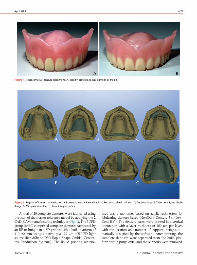
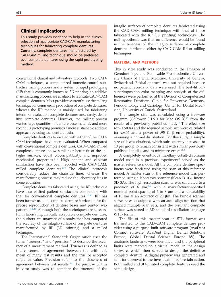
Figure 1. Representative denture specimens. A, Rapidly prototyped (3D printed). B, Milled.
Figure 2. Regions of interests investigated. A, Posterior crest. B, Palatal vault. C, Posterior palatal seal area. D, Anterior ridge. E, Tuberosity. F, Vestibularflange. G, Mid-palatal raphae. H, Total intaglio surface.
April 2019 639
A total of 20 complete dentures were fabricated usingthe scan of the master reference model by applying the 2CAD-CAM manufacturing techniques (Fig. 1). The 3DPDgroup (n=10) comprised complete dentures fabricated byan RP technique in a 3D printer with a build platform of110×62 mm using a native pixel 29-mm 405 LED lightsource (RapidShape D30; Rapid Shape GmbH, Genera-tive Production Systems). The liquid printing material
Kalberer et al
used was a monomer based on acrylic resin esters forfabricating denture bases (NextDent Denture 3+; Next-Dent B.V.). The denture bases were printed in a verticalorientation with a layer thickness of 100 mm per layer,with the location and number of supports being auto-matically designed by the software. After printing, thecomplete dentures were separated from the build plat-form with a putty knife, and the supports were removed.
THE JOURNAL OF PROSTHETIC DENTISTRY
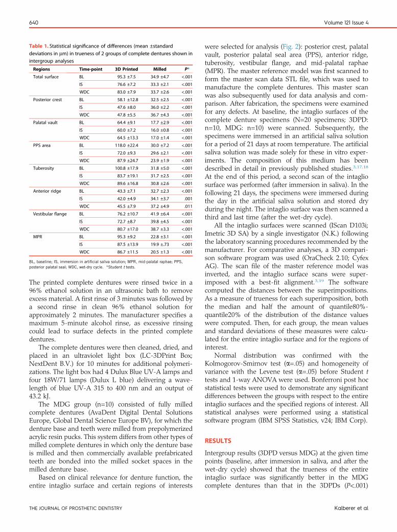
Table 1. Statistical significance of differences (mean ±standarddeviations in mm) in trueness of 2 groups of complete dentures shown inintergroup analyses
Regions Time-point 3D Printed Milled P*
Total surface BL 95.3 ±7.5 34.9 ±4.7 <.001
IS 76.6 ±7.2 33.3 ±2.1 <.001
WDC 83.0 ±7.9 33.7 ±2.6 <.001
Posterior crest BL 58.1 ±12.8 32.5 ±2.5 <.001
IS 47.6 ±8.0 36.0 ±2.2 <.001
WDC 47.8 ±5.5 36.7 ±4.3 <.001
Palatal vault BL 64.4 ±9.1 17.7 ±2.9 <.001
IS 60.0 ±7.2 16.0 ±0.8 <.001
WDC 64.5 ±13.3 17.0 ±1.4 <.001
PPS area BL 118.0 ±22.4 30.0 ±7.2 <.001
IS 72.0 ±9.3 29.6 ±2.1 <.001
WDC 87.9 ±24.7 23.9 ±1.9 <.001
Tuberosity BL 100.8 ±17.9 31.8 ±5.0 <.001
IS 83.7 ±19.1 31.7 ±2.5 <.001
WDC 89.6 ±16.8 30.8 ±2.6 <.001
Anterior ridge BL 43.3 ±7.1 32.7 ±2.3 <.001
IS 42.0 ±4.9 34.1 ±3.7 .001
WDC 45.5 ±7.9 37.2 ±4.9 .011
Vestibular flange BL 76.2 ±10.7 41.9 ±6.4 <.001
IS 72.7 ±8.7 39.8 ±4.5 <.001
WDC 80.7 ±17.0 38.7 ±3.3 <.001
MPR BL 95.3 ±9.2 22.8 ±3.1 <.001
IS 87.5 ±13.9 19.9 ±.73 <.001
WDC 86.7 ±11.5 20.5 ±1.3 <.001
BL, baseline; IS, immersion in artificial saliva solution; MPR, mid-palatal raphae; PPS,posterior palatal seal; WDC, wet-dry cycle. *Student t tests.
640 Volume 121 Issue 4
The printed complete dentures were rinsed twice in a96% ethanol solution in an ultrasonic bath to removeexcess material. A first rinse of 3 minutes was followed bya second rinse in clean 96% ethanol solution forapproximately 2 minutes. The manufacturer specifies amaximum 5-minute alcohol rinse, as excessive rinsingcould lead to surface defects in the printed completedentures.
The complete dentures were then cleaned, dried, andplaced in an ultraviolet light box (LC-3DPrint Box;NextDent B.V.) for 10 minutes for additional polymeri-zations. The light box had 4 Dulux Blue UV-A lamps andfour 18W/71 lamps (Dulux L blue) delivering a wave-length of blue UV-A 315 to 400 nm and an output of43.2 kJ.
The MDG group (n=10) consisted of fully milledcomplete dentures (AvaDent Digital Dental SolutionsEurope, Global Dental Science Europe BV), for which thedenture base and teeth were milled from prepolymerizedacrylic resin pucks. This system differs from other types ofmilled complete dentures in which only the denture baseis milled and then commercially available prefabricatedteeth are bonded into the milled socket spaces in themilled denture base.
Based on clinical relevance for denture function, theentire intaglio surface and certain regions of interests
THE JOURNAL OF PROSTHETIC DENTISTRY
were selected for analysis (Fig. 2): posterior crest, palatalvault, posterior palatal seal area (PPS), anterior ridge,tuberosity, vestibular flange, and mid-palatal raphae(MPR). The master reference model was first scanned toform the master scan data STL file, which was used tomanufacture the complete dentures. This master scanwas also subsequently used for data analysis and com-parison. After fabrication, the specimens were examinedfor any defects. At baseline, the intaglio surfaces of thecomplete denture specimens (N=20 specimens; 3DPD:n=10, MDG: n=10) were scanned. Subsequently, thespecimens were immersed in an artificial saliva solutionfor a period of 21 days at room temperature. The artificialsaliva solution was made solely for these in vitro exper-iments. The composition of this medium has beendescribed in detail in previously published studies.3,17,18
At the end of this period, a second scan of the intagliosurface was performed (after immersion in saliva). In thefollowing 21 days, the specimens were immersed duringthe day in the artificial saliva solution and stored dryduring the night. The intaglio surface was then scanned athird and last time (after the wet-dry cycle).
All the intaglio surfaces were scanned (IScan D103i;Imetric 3D SA) by a single investigator (N.K.) followingthe laboratory scanning procedures recommended by themanufacturer. For comparative analyses, a 3D compari-son software program was used (OraCheck 2.10; CyfexAG). The scan file of the master reference model wasinverted, and the intaglio surface scans were super-imposed with a best-fit alignment.3,19 The softwarecomputed the distances between the superimpositions.As a measure of trueness for each superimposition, boththe median and half the amount of quantile80%-quantile20% of the distribution of the distance valueswere computed. Then, for each group, the mean valuesand standard deviations of these measures were calcu-lated for the entire intaglio surface and for the regions ofinterest.
Normal distribution was confirmed with theKolmogorov-Smirnov test (a=.05) and homogeneity ofvariance with the Levene test (a=.05) before Student ttests and 1-way ANOVA were used. Bonferroni post hocstatistical tests were used to demonstrate any significantdifferences between the groups with respect to the entireintaglio surfaces and the specified regions of interest. Allstatistical analyses were performed using a statisticalsoftware program (IBM SPSS Statistics, v24; IBM Corp).
RESULTS
Intergroup results (3DPD versus MDG) at the given timepoints (baseline, after immersion in saliva, and after thewet-dry cycle) showed that the trueness of the entireintaglio surface was significantly better in the MDGcomplete dentures than that in the 3DPDs (P<.001)
Kalberer et al
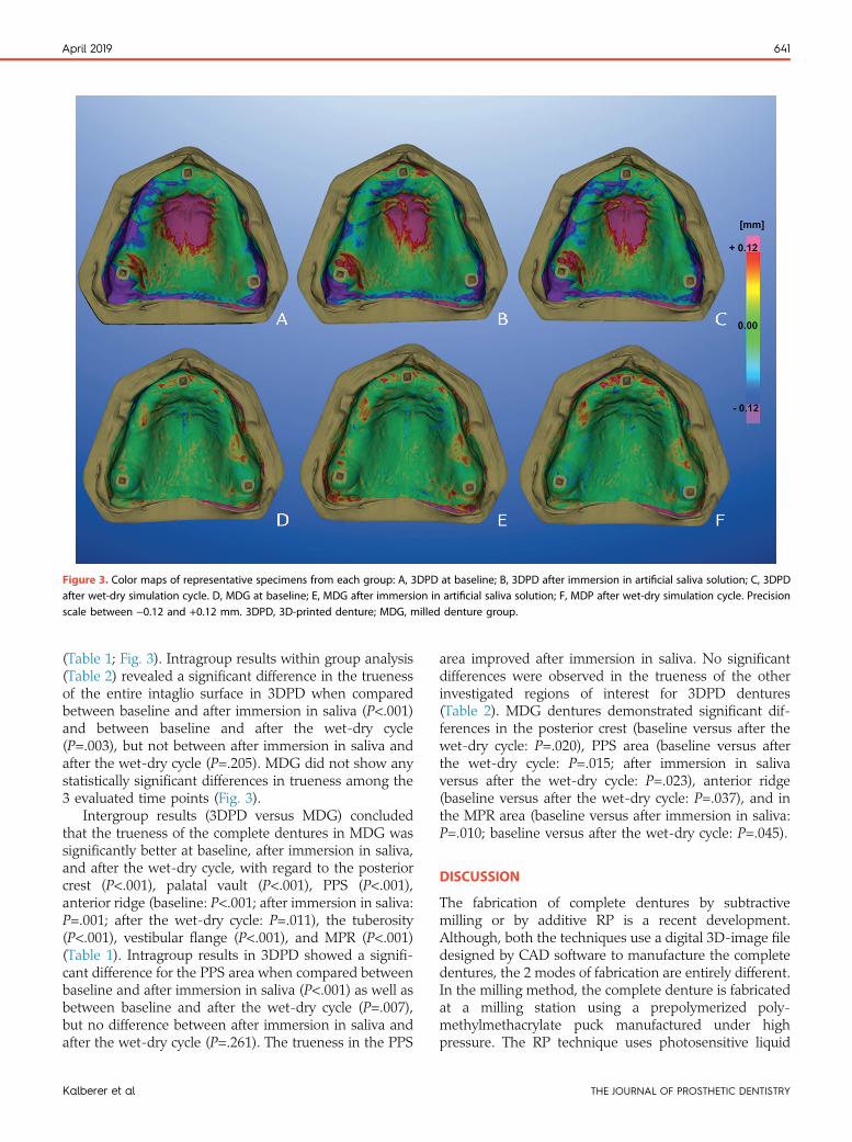
Figure 3. Color maps of representative specimens from each group: A, 3DPD at baseline; B, 3DPD after immersion in artificial saliva solution; C, 3DPDafter wet-dry simulation cycle. D, MDG at baseline; E, MDG after immersion in artificial saliva solution; F, MDP after wet-dry simulation cycle. Precisionscale between −0.12 and +0.12 mm. 3DPD, 3D-printed denture; MDG, milled denture group.
April 2019 641
(Table 1; Fig. 3). Intragroup results within group analysis(Table 2) revealed a significant difference in the truenessof the entire intaglio surface in 3DPD when comparedbetween baseline and after immersion in saliva (P<.001)and between baseline and after the wet-dry cycle(P=.003), but not between after immersion in saliva andafter the wet-dry cycle (P=.205). MDG did not show anystatistically significant differences in trueness among the3 evaluated time points (Fig. 3).
Intergroup results (3DPD versus MDG) concludedthat the trueness of the complete dentures in MDG wassignificantly better at baseline, after immersion in saliva,and after the wet-dry cycle, with regard to the posteriorcrest (P<.001), palatal vault (P<.001), PPS (P<.001),anterior ridge (baseline: P<.001; after immersion in saliva:P=.001; after the wet-dry cycle: P=.011), the tuberosity(P<.001), vestibular flange (P<.001), and MPR (P<.001)(Table 1). Intragroup results in 3DPD showed a signifi-cant difference for the PPS area when compared betweenbaseline and after immersion in saliva (P<.001) as well asbetween baseline and after the wet-dry cycle (P=.007),but no difference between after immersion in saliva andafter the wet-dry cycle (P=.261). The trueness in the PPS
Kalberer et al
area improved after immersion in saliva. No significantdifferences were observed in the trueness of the otherinvestigated regions of interest for 3DPD dentures(Table 2). MDG dentures demonstrated significant dif-ferences in the posterior crest (baseline versus after thewet-dry cycle: P=.020), PPS area (baseline versus afterthe wet-dry cycle: P=.015; after immersion in salivaversus after the wet-dry cycle: P=.023), anterior ridge(baseline versus after the wet-dry cycle: P=.037), and inthe MPR area (baseline versus after immersion in saliva:P=.010; baseline versus after the wet-dry cycle: P=.045).
DISCUSSION
The fabrication of complete dentures by subtractivemilling or by additive RP is a recent development.Although, both the techniques use a digital 3D-image filedesigned by CAD software to manufacture the completedentures, the 2 modes of fabrication are entirely different.In the milling method, the complete denture is fabricatedat a milling station using a prepolymerized poly-methylmethacrylate puck manufactured under highpressure. The RP technique uses photosensitive liquid
THE JOURNAL OF PROSTHETIC DENTISTRY
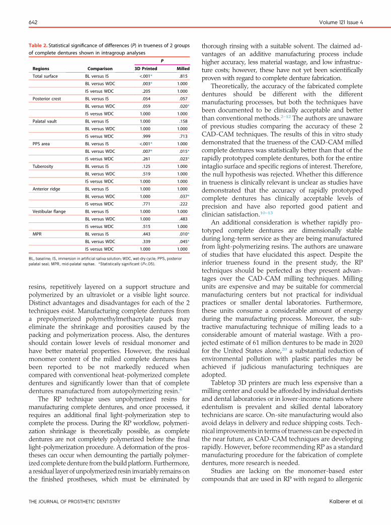
Table 2. Statistical significance of differences (P) in trueness of 2 groupsof complete dentures shown in intragroup analyses
Regions Comparison
P
3D Printed Milled
Total surface BL versus IS <.001* .815
BL versus WDC .003* 1.000
IS versus WDC .205 1.000
Posterior crest BL versus IS .054 .057
BL versus WDC .059 .020*
IS versus WDC 1.000 1.000
Palatal vault BL versus IS 1.000 .158
BL versus WDC 1.000 1.000
IS versus WDC .999 .713
PPS area BL versus IS <.001* 1.000
BL versus WDC .007* .015*
IS versus WDC .261 .023*
Tuberosity BL versus IS .125 1.000
BL versus WDC .519 1.000
IS versus WDC 1.000 1.000
Anterior ridge BL versus IS 1.000 1.000
BL versus WDC 1.000 .037*
IS versus WDC .771 .222
Vestibular flange BL versus IS 1.000 1.000
BL versus WDC 1.000 .483
IS versus WDC .515 1.000
MPR BL versus IS .443 .010*
BL versus WDC .339 .045*
IS versus WDC 1.000 1.000
BL, baseline; IS, immersion in artificial saliva solution; WDC, wet-dry cycle; PPS, posteriorpalatal seal; MPR, mid-palatal raphae. *Statistically significant (P<.05).
642 Volume 121 Issue 4
resins, repetitively layered on a support structure andpolymerized by an ultraviolet or a visible light source.Distinct advantages and disadvantages for each of the 2techniques exist. Manufacturing complete dentures froma prepolymerized polymethylmethacrylate puck mayeliminate the shrinkage and porosities caused by thepacking and polymerization process. Also, the denturesshould contain lower levels of residual monomer andhave better material properties. However, the residualmonomer content of the milled complete dentures hasbeen reported to be not markedly reduced whencompared with conventional heat-polymerized completedentures and significantly lower than that of completedentures manufactured from autopolymerizing resin.6
The RP technique uses unpolymerized resins formanufacturing complete dentures, and once processed, itrequires an additional final light-polymerization step tocomplete the process. During the RP workflow, polymeri-zation shrinkage is theoretically possible, as completedentures are not completely polymerized before the finallight-polymerization procedure. A deformation of the pros-theses can occur when demounting the partially polymer-ized completedenture from thebuildplatform. Furthermore,a residual layer of unpolymerized resin invariably remains onthe finished prostheses, which must be eliminated by
THE JOURNAL OF PROSTHETIC DENTISTRY
thorough rinsing with a suitable solvent. The claimed ad-vantages of an additive manufacturing process includehigher accuracy, less material wastage, and low infrastruc-ture costs; however, these have not yet been scientificallyproven with regard to complete denture fabrication.
Theoretically, the accuracy of the fabricated completedentures should be different with the differentmanufacturing processes, but both the techniques havebeen documented to be clinically acceptable and betterthan conventional methods.2e12 The authors are unawareof previous studies comparing the accuracy of these 2CAD-CAM techniques. The results of this in vitro studydemonstrated that the trueness of the CAD-CAM milledcomplete dentures was statistically better than that of therapidly prototyped complete dentures, both for the entireintaglio surface and specific regions of interest. Therefore,the null hypothesis was rejected. Whether this differencein trueness is clinically relevant is unclear as studies havedemonstrated that the accuracy of rapidly prototypedcomplete dentures has clinically acceptable levels ofprecision and have also reported good patient andclinician satisfaction.10e13
An additional consideration is whether rapidly pro-totyped complete dentures are dimensionally stableduring long-term service as they are being manufacturedfrom light-polymerizing resins. The authors are unawareof studies that have elucidated this aspect. Despite theinferior trueness found in the present study, the RPtechniques should be perfected as they present advan-tages over the CAD-CAM milling techniques. Millingunits are expensive and may be suitable for commercialmanufacturing centers but not practical for individualpractices or smaller dental laboratories. Furthermore,these units consume a considerable amount of energyduring the manufacturing process. Moreover, the sub-tractive manufacturing technique of milling leads to aconsiderable amount of material wastage. With a pro-jected estimate of 61 million dentures to be made in 2020for the United States alone,20 a substantial reduction ofenvironmental pollution with plastic particles may beachieved if judicious manufacturing techniques areadopted.
Tabletop 3D printers are much less expensive than amilling center and could be afforded by individual dentistsand dental laboratories or in lower-income nations whereedentulism is prevalent and skilled dental laboratorytechnicians are scarce. On-site manufacturing would alsoavoid delays in delivery and reduce shipping costs. Tech-nical improvements in terms of trueness can be expected inthe near future, as CAD-CAM techniques are developingrapidly. However, before recommending RP as a standardmanufacturing procedure for the fabrication of completedentures, more research is needed.
Studies are lacking on the monomer-based estercompounds that are used in RP with regard to allergenic
Kalberer et al
April 2019 643
potential, residual monomer levels, material and colorstability, material compatibility with conventional relines,mechanical properties, and biocompatibility. Theappearance of the 2 different denture types requiresevaluation as esthetics are of increasing importance. Inaddition, patient-centered outcome measures have to beconsidered before validating this evolving technique.
CONCLUSIONS
Based on the findings of this in vitro study, the followingconclusions were drawn;
1. CAD-CAM milled complete dentures were betterthan rapidly prototyped complete dentures in termsof trueness of the intaglio surfaces.
2. Further research is needed on the biomechanical,clinical, and patient-centered outcome measures todetermine the superiority of one technique over theother with regard to manufacturing complete den-tures by CAD-CAM techniques.
REFERENCES
1. Baba NZ, AlRumaih HS, Goodacre BJ, Goodacre CJ. Current techniques inCAD/CAM denture fabrication. Gen Dent 2016;64:23-8.
2. Goodacre BJ, Goodacre CJ, Baba NZ, Kattadiyil MT. Comparison of denturebase adaptation between CAD-CAM and conventional fabrication tech-niques. J Prosthet Dent 2016;116:249-56.
3. Srinivasan M, Cantin Y, Mehl A, Gjengedal H, Müller F, Schimmel M. CAD/CAM milled removable complete dentures: an in vitro evaluation of trueness.Clin Oral Investig 2017;21:2007-19.
4. Srinivasan M, Gjengedal H, Cattani-Lorente M, Moussa M, Durual S,Schimmel M, et al. CAD/CAM milled complete removable dental prostheses:an in vitro evaluation of biocompatibility, mechanical properties, and surfaceroughness. Dent Mater J 2018;37:526-33.
5. Steinmassl O, Dumfahrt H, Grunert I, Steinmassl PA. CAD/CAM producesdentures with improved fit. Clin Oral Investig 2018;22:2829-35.
6. Steinmassl PA, Wiedemair V, Huck C, Klaunzer F, Steinmassl O, Grunert I,et al. Do CAD/CAM dentures really release less monomer than conventionaldentures? Clin Oral Investig 2017;21:1697-705.
Kalberer et al
7. Steinmassl O, Dumfahrt H, Grunert I, Steinmassl PA. Influence of CAD/CAM fabrication on denture surface properties. J Oral Rehabil 2018;45:406-13.
8. Kattadiyil MT, Jekki R, Goodacre CJ, Baba NZ. Comparison of treatmentoutcomes in digital and conventional complete removable dental prosthesisfabrications in a predoctoral setting. J Prosthet Dent 2015;114:818-25.
9. Bidra AS, Farrell K, Burnham D, Dhingra A, Taylor TD, Kuo CL. Prospectivecohort pilot study of 2-visit CAD/CAM monolithic complete dentures andimplant-retained overdentures: Clinical and patient-centered outcomes.J Prosthet Dent 2016;115:578-86.
10. Pereyra NM, Marano J, Subramanian G, Quek S, Leff D. Comparison ofpatient satisfaction in the fabrication of conventional dentures vs. DENTCA(CAD/CAM) dentures: a case report. J N J Dent Assoc 2015;86:26-33.
11. Ucar Y, Akova T, Aysan I. Mechanical properties of polyamide versusdifferent PMMA denture base materials. J Prosthodont 2012;21:173-6.
12. Chen H, Wang H, Lv P, Wang Y, Sun Y. Quantitative evaluation of tissuesurface adaption of cad-designed and 3d printed wax pattern of maxillarycomplete denture. Biomed Res Int 2015;2015:453968.
13. Inokoshi M, Kanazawa M, Minakuchi S. Evaluation of a complete denturetrial method applying rapid prototyping. Dent Mater J 2012;31:40-6.
14. ISO 5725-1:1994 Accuracy (trueness and precision) of measurement methodsand results - Part 1: General principles and definitions. Available at: https://www.iso.org/standard/11833.html.
15. Faul F, Erdfelder E, Buchner A, Lang AG. Statistical power analyses usingG*Power 3.1: tests for correlation and regression analyses. Behav ResMethods 2009;41:1149-60.
16. Papaspyridakos P, Gallucci GO, Chen CJ, Hanssen S, Naert I,Vandenberghe B. Digital versus conventional implant impressions foredentulous patients: accuracy outcomes. Clin Oral Implants Res 2016;27:465-72.
17. Srinivasan M, Schimmel M, Badoud I, Ammann P, Herrmann FR, Müller F.Influence of implant angulation and cyclic dislodging on the retentive force oftwo different overdenture attachments - an in vitro study. Clin Oral ImplantsRes 2016;27:604-11.
18. Srinivasan M, Schimmel M, Kobayashi M, Badoud I, Ammann P,Herrmann FR, et al. Influence of different lubricants on the retentive force ofLOCATOR attachments - an in vitro pilot study. Clin Oral Implants Res2016;27:771-5.
19. Mehl A, Koch R, Zaruba M, Ender A. 3D monitoring and quality controlusing intraoral optical camera systems. Int J Comput Dent 2013;16:23-36.
20. Douglass CW, Shih A, Ostry L. Will there be a need for complete dentures inthe United States in 2020? J Prosthet Dent 2002;87:5-8.
Corresponding author:Dr Frauke MüllerDivision of Gerodontology and Removable ProsthodonticsUniversity Clinics of Dental Medicine, University of GenevaRue Michel-Servet 1, CH-1211 Geneva 4SWITZERLANDEmail: [email protected]
Copyright © 2018 by the Editorial Council for The Journal of Prosthetic Dentistry.https://doi.org/10.1016/j.prosdent.2018.09.001
THE JOURNAL OF PROSTHETIC DENTISTRY