Busuiphan lung - Thorax
17
Thorax (1969), 24, 639. Busuiphan lung W. A. LITTLER, J. M. KAY, P. S. HASLETON, AND DONALD HEATH From the Cardio-thoracic Unit, Broadgreen Hospital, Liverpool, and the Departmnent of Pathology, University of Liverpool A 61-year-old man with chronic myeloid leukaemia was treated with busulphan (Myleran). After receiving 1 g. of this drug over a period of 20 months he became dyspnoeic and developed crepitations in the lungs. Two months later radiographs of the chest revealed peri-hilar infiltrates and subsequently diffuse mottling throughout both lungs. Lung function tests showed a gross impairment of the transfer factor to a quarter of the predicted normal. At necropsy the lungs showed a striking proliferation of granular pneumocytes, many of which had disintegrated to produce intra-alveolar debris, some of which showed organization by fibrous tissue. There was associated interstitial pulmonary fibrosis. Electron microscopy confirmed the desquamated alveolar cells to be type II (granular) pneumocytes containing characteristic lamellar bodies. Many of these osmiophilic bodies, believed to be the source of pulmonary surfactant, had been liberated into the alveolar spaces, with the formation of phospholipid myelin figures and lattices. We think that the basic pathology of busulphan lung is a chemically induced alveolitis with pro- liferation of granular pneumocytes followed by fibrosis of alveolar walls and intra-alveolar contents. Here we describe in detail the clinical and patho- logical features of a patient who developed pul- monary complications following a course of busulphan (Myleran) for chronic myeloid leuk- aemia. Lung function studies were performed in addition to clinical examination. Electron micro- scopy of the lung was carried out as well as routine histopathology and has yielded new in- formation on the pathology of 'busulphan lung'. CASE REPORT The patient, a 61-year-old male clerk, was first seen in January 1967 because of blurred vision in the left eye and tiredness for three weeks. On examination the mucous membranes and nail beds were pale, the spleen was enlarged five fingerbreadths below the left costal margin, and the liver was also palpable. There were bilateral retinal haemorrhages and exu- dates. The chest, heart, and nervous system were normal on clinical examination, and the systemic blood pressure was moderately raised at 180/105 mm. Hg. The blood picture was typical of chronic myeloid leukaemia with a total white cell count of 256,000/ cu. mm. A differential count showed blast cells 8%, myelocytes 36%, metamyelocytes 5%, neutrophils 46%, lymphocytes 2%, and eosinophils 3 O/ . The platelet count was 129,000/cu. mm. and the haemo- globin level was 8-8 g./100 ml. (60%). Bone marrow examination was not carried out. Peripheral blood chromosome culture revealed the Philadelphia (Ph1) chromosome. The chest radiograph was normal (Fig. 1). A course of busulphan therapy was started on 13 January 1967. The initial dose of 4 mg. daily was reduced to a maintenance dose of 2 mg. four times a week on March 28, when he was in a satisfactory clinical and haematological remission with a total white cell count of 55,000/cu. mm., a platelet count of 465,000/cu. mm., and a haemoglobin level of 13-8 g./100 ml. (95%). This remission continued with doses of busulphan, 2 mg. four or five times a week, until 13 June 1968, when a total white cell count of 19,300/cu. mm. showed a differential count of blast cells 24%, myelocytes 7%, neutrophils 50%, lymphocytes 10%, monocytes 6%, eosinophils 1%, and basophils 2%. At this stage the platelet count was 150,000/cu. mm. and the haemoglobin level was 8-9 g./100 ml. (61%). The patient's only complaint was tiredness and the main physical finding was splenomegaly. Because of the myeloblastic blood picture the dose of busulphan was kept at 2 mg. four times a week, and in addi- tion a course of 6-mercaptopurine, 150 mg. daily, was started. On 9 July 1968 the haemoglobin level had fallen to 7-1 g./100 ml. (51%). He was trans- fused with four pints of blood and the dose of busulphan was tailed off and finally stopped on 26 July after 19 months' treatment with a total dose of 639 copyright. on October 6, 2021 by guest. Protected by http://thorax.bmj.com/ Thorax: first published as 10.1136/thx.24.6.639 on 1 November 1969. Downloaded from
Transcript of Busuiphan lung - Thorax

Thorax (1969), 24, 639.
Busuiphan lungW. A. LITTLER, J. M. KAY, P. S. HASLETON,
AND DONALD HEATH
From the Cardio-thoracic Unit, Broadgreen Hospital, Liverpool, and the Departmnent of Pathology,University of Liverpool
A 61-year-old man with chronic myeloid leukaemia was treated with busulphan (Myleran). Afterreceiving 1 g. of this drug over a period of 20 months he became dyspnoeic and developedcrepitations in the lungs. Two months later radiographs of the chest revealed peri-hilar infiltratesand subsequently diffuse mottling throughout both lungs. Lung function tests showed a grossimpairment of the transfer factor to a quarter of the predicted normal. At necropsy the lungsshowed a striking proliferation of granular pneumocytes, many of which had disintegrated toproduce intra-alveolar debris, some of which showed organization by fibrous tissue. There wasassociated interstitial pulmonary fibrosis. Electron microscopy confirmed the desquamatedalveolar cells to be type II (granular) pneumocytes containing characteristic lamellar bodies.Many of these osmiophilic bodies, believed to be the source of pulmonary surfactant, had beenliberated into the alveolar spaces, with the formation of phospholipid myelin figures and lattices.We think that the basic pathology of busulphan lung is a chemically induced alveolitis with pro-liferation of granular pneumocytes followed by fibrosis of alveolar walls and intra-alveolarcontents.
Here we describe in detail the clinical and patho-logical features of a patient who developed pul-monary complications following a course ofbusulphan (Myleran) for chronic myeloid leuk-aemia. Lung function studies were performed inaddition to clinical examination. Electron micro-scopy of the lung was carried out as well asroutine histopathology and has yielded new in-formation on the pathology of 'busulphan lung'.
CASE REPORT
The patient, a 61-year-old male clerk, was first seenin January 1967 because of blurred vision in the lefteye and tiredness for three weeks. On examinationthe mucous membranes and nail beds were pale, thespleen was enlarged five fingerbreadths below theleft costal margin, and the liver was also palpable.There were bilateral retinal haemorrhages and exu-dates. The chest, heart, and nervous system werenormal on clinical examination, and the systemicblood pressure was moderately raised at 180/105 mm.Hg.The blood picture was typical of chronic myeloid
leukaemia with a total white cell count of 256,000/cu. mm. A differential count showed blast cells 8%,myelocytes 36%, metamyelocytes 5%, neutrophils46%, lymphocytes 2%, and eosinophils 3 O/ . Theplatelet count was 129,000/cu. mm. and the haemo-
globin level was 8-8 g./100 ml. (60%). Bone marrowexamination was not carried out. Peripheral bloodchromosome culture revealed the Philadelphia(Ph1) chromosome. The chest radiograph wasnormal (Fig. 1).A course of busulphan therapy was started on 13
January 1967. The initial dose of 4 mg. daily wasreduced to a maintenance dose of 2 mg. four timesa week on March 28, when he was in a satisfactoryclinical and haematological remission with a totalwhite cell count of 55,000/cu. mm., a platelet countof 465,000/cu. mm., and a haemoglobin level of13-8 g./100 ml. (95%). This remission continuedwith doses of busulphan, 2 mg. four or five times aweek, until 13 June 1968, when a total white cellcount of 19,300/cu. mm. showed a differentialcount of blast cells 24%, myelocytes 7%, neutrophils50%, lymphocytes 10%, monocytes 6%, eosinophils1%, and basophils 2%. At this stage the plateletcount was 150,000/cu. mm. and the haemoglobin levelwas 8-9 g./100 ml. (61%).The patient's only complaint was tiredness and the
main physical finding was splenomegaly. Because ofthe myeloblastic blood picture the dose of busulphanwas kept at 2 mg. four times a week, and in addi-tion a course of 6-mercaptopurine, 150 mg. daily,was started. On 9 July 1968 the haemoglobin levelhad fallen to 7-1 g./100 ml. (51%). He was trans-fused with four pints of blood and the dose ofbusulphan was tailed off and finally stopped on 26July after 19 months' treatment with a total dose of
639
copyright. on O
ctober 6, 2021 by guest. Protected by
http://thorax.bmj.com
/T
horax: first published as 10.1136/thx.24.6.639 on 1 Novem
ber 1969. Dow
nloaded from

W. A. Littler, J. M. Kay, P. S. Hasleton, and Donald Heath
FIG. 1. Normal chest radiograph taken in January 1967 when chronic myeloidleukaemia had been diagnosed.
1,000 mg. A chest radiograph taken at this time wasnormal and unchanged.
In September 1968 the patient began to complainof dyspnoea on moderate activity such as climbingstairs or walking up an incline. On examination hehad slight sacral and ankle oedema but no jugularvenous engorgement. A soft apical pan-systolicmurmur was heard, but the heart sounds were normal.Bilateral basal crepitations were present. These find-ings were attributed to cardiac failure associated withanaemia, since the haemoglobin level was 7-3 g./100 ml. (50%). The blood film showed anisocytosisand polychromasia with 9 normoblasts per 100 whitecells. The reticulocyte count was 4%. The serumbilirubin was 11 mg./100 ml., but there was noexcess of urobilinogen in the urine and Schumm'stest was negative. The direct Coombs' test was nega-tive. The chest radiograph and the electrocardiogramwere normal. Four pints of blood were transfused andhe was given 40 mg. frusemide each day. The patient'ssymptoms improved and the systemic oedema went,but the basal crepitations in the lung persisted. Sincethere was haematological evidence of haemolysis,prednisolone was added to the treatment in an initialdose of 40 mg. daily, reducing over a five-day periodto 20 mg. daily.
During the next month his dyspnoea on effortincreased rapidly, so that he became breathless even
at rest and he also developed a non-productive cough.Examination on 4 October 1968 revealed tachypnoea,central cyanosis, bilateral basal crepitations, and afever of 1010 F. The blood film showed evidenceof reduced myeloblastic activity, 8% of the total whitecell count of 8,500/cu. mm. consisting of blast cells.The haemoglobin level was 10-8 g./100 ml. (74%).The patient was now considered to have a pulmonaryinfection and a week's course of ampicillin, 500 mg.six-hourly, was given. The temperature settled butthe dyspnoea, central cyanosis, and basal crepitationspersisted. On 6 November a chest radiograph showedperi-hilar infiltrates, particularly in the right sub-apical region, with an area of consolidation in theposterior segment of the left lower lobe (Fig. 2). Hewas treated with a transfusion of four pints of blood,tetracycline, 500 mg. six-hourly, digoxin, 0-25 mg.twice daily, frusemide, 40 mg. daily, and oxygen. Thehaemnoglobin level rose to 9-9 g./100 ml. (68%), butthe symptoms and signs persisted. Repeated blood,sputum, and urine cultures were negative and fungiwere not isolated from the sputum. There was furtherdeterioration of the appearance of the chest radio-graphs with the development of diffuse mottlingthroughout both lungs (Fig. 3).On 27 November prednisolone was increased to
60 mg. daily, reducing over four days to 30 mg. daily,and oxygen, 6 to 8 1./min., was given continuously
U640
copyright. on O
ctober 6, 2021 by guest. Protected by
http://thorax.bmj.com
/T
horax: first published as 10.1136/thx.24.6.639 on 1 Novem
ber 1969. Dow
nloaded from

Busulphan lung
FIG. 2. Chest radiograph takenon 6 November 1968 when thepatient had had chest symptomsfor two months. The radiographshows peri-hilar infiltrates, parti-cularly in the right sub-apicalregion, with an area of consolid-ation in the posterior segment ofthe left lower lobe.
FIG. 3. Chest radiograph takenon 6 December 1968, one weekbefore the patient's death. Thereis diffuise mottling throughoutboth lung fields.
641
copyright. on O
ctober 6, 2021 by guest. Protected by
http://thorax.bmj.com
/T
horax: first published as 10.1136/thx.24.6.639 on 1 Novem
ber 1969. Dow
nloaded from
W. A. Littler, J. M. Kay, P. S. Hasleton, and Donald Heath
by nasal catheter. Despite this treatment there wasno sign of clinical or radiological improvement.Myeloblastic activity continued in the blood (on 5December, out of a total white cell count of 16,700/cu. mm., 10% were myeloblasts, 6% myelocytes, and8% were metamyelocytes), although the haemoglobinlevel remained above 70% during the last three weeksof his life. The patient died on 12 December 1968from respiratory failure and pulmonary infection.
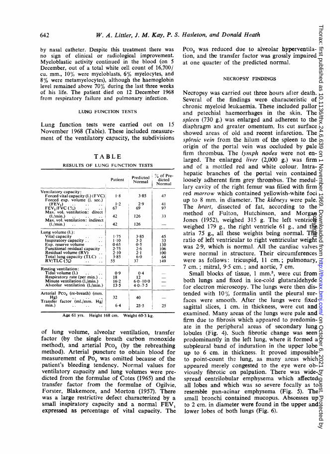
LUNG FUNCTION TESTS
Lung function tests were carried out on 15November 1968 (Table). These included measure-ment of the ventilatory capacity, the subdivisions
TABLERESULTS OF LUNG FUNCTION TESTS
P % of Pre-Patient Predictedidce
'NraINormalVentilatory capacity:
Forced vital capacity (I.) (FVC)Forced exp. volume (1. sec.)(FEVI)
FEV5/FVC(%)Max. vol. ventilation: direct
(I./min.)Max. vol. ventilation: indirect
(1./m mn.) .. . .
Lung volume (1.):Vital capacityInspiratory capacityExp. reserve volumeFunctional residual capacityResidual volume (RV)Total lung capacity (TLC)RV/TLC(%)
1 8 3-85
1-2 2967 69
42 126
42 126
1 75 3-851 10 3-30-65 0u5275 2-62-10 2-13-85 6-0
55 37
Resting ventilation:Tidal volume (I.) 0 9Respiratory rate (per min.) . 18Minute ventilation (I./min.) 16 2Alveolar ventilation (I./min.) 135
1-Arterial Pco2 (re-breath) (mm.
Hg) .. ..Transfer factor (mI/mm. Hg!
min.)
32
6-4
0 412
6-0-10 04 0-7 5
40
25-5
47
4197
33
453313010610064149
25
Age 61 yrs. Height 168 cm. Weight 60-3 kg.
of lung volume, alveolar ventilation, transferfactor (by the single breath carbon monoxidemethod), and arterial Pco2 (by the rebreathingmethod). Arterial puncture to obtain blood formeasurement of Po2 was omitted because of thepatient's bleeding tendency. Normal values forventilatory capacity and lung volumes were pre-
dicted from the formulae of Cotes (1965) and thetransfer factor from the formulae of Ogilvie,Forster, Blakemore, and Morton (1957). Therewas a large restrictive defect characterized by a
small inspiratory capacity and a normal FEV1expressed as percentage of vital capacity. The
Pco2 was reduced due to alveolar hyperventila-tion, and the transfer factor was grossly impairedat one quarter of the predicted normal.
NECROPSY FINDINGS
Necropsy was carried out three hours after death.Several of the findings were characteristic ofchronic myeloid leukaemia. These included pallorand petechial haemorrhages in the skin. Thespleen (730 g.) was enlarged and adherent to thediaphragm and greater omentum. Its cut surfaceshowed areas of old and recent infarction. Thesplenic vein from the hilum of the spleen to theorigin of the portal vein was occluded by palefirm thrombus. The lymph nodes were not en-larged. The enlarged liver (2,000 g.) was firmand of a mottled red and white colour. Intra-hepatic branches of the portal vein containedloosely adherent firm grey thrombus. The medul-lary cavity of the right femur was filied with firmred marrow which contained yellowish-white fociup to 8 mm. in diameter. The kidneys were pale.The heart, dissected of fat, according to themethod of Fulton, Hutchinson, and MorganJones (1952), weighed 315 g. The left ventricleweighed 179 g., the right ventricle 61 g., and theatria 75 g., all these weights being normal. Theratio of left ventricular to right ventricular weightwas 29, which is normal. All the cardiac valveswere normal in structure. Their circumferenceswere as follows: tricuspid, 11 cm.; pulmonary,7 cm.; mitral, 9 5 cm.; and aortic, 7 cm.
Small blocks of tissue, 1 mm.3, were cut fromboth lungs and fixed in ice-cold glutaraldehydefor electron microscopy. The lungs were then dis-tended with 10% formalin until the pleural sur-faces were smooth. After the lungs were fixedsagittal slices, 1 cm. in thickness, were cut andexamined. Many areas of the lungs were pale andfirm due to fibrosis which appeared to predomin-ate in the peripheral areas of secondary lunglobules (Fig. 4). Such fibrotic change was seenpredominantly in the left lung, where it formed asubpleural band of induration in the upper lobeup to 6 cm. in thickness. It proved impossibleto point-count the lung, as many areas whichappeared merely congested to the eye were ob-viously fibrotic on palpation. There was wide-spread centrilobular emphysema which affectedall lobes and which was so severe focally as toresemble pan-acinar emphysema (Fig. 5). Thesmall bronchi contained mucopus. Abscesses upto 2 cm. in diameter were found in the upper andlower lobes of both lungs (Fig. 6).
I 1-
642
copyright. on O
ctober 6, 2021 by guest. Protected by
http://thorax.bmj.com
/T
horax: first published as 10.1136/thx.24.6.639 on 1 Novem
ber 1969. Dow
nloaded from

Busulphan lung
FIG. 4. Cut surface of lung showing pale areas offibrosiswhich are situated mainly in the peripheral areas ofsecondary lung lobules ( x 2).
FIG. 5. Cut surface of lung showing confluent bronchiolaremphysema of such severity as to mimic pan-acinaremphysema. There is surrounding fibrosis ( x 2).
i:FIG. 6. Cut surface of lung showing one of theabscesses ( x 2).
HISTOPATHOLOGY
Characteristic cells of chronic myeloid leukaemiainfiltrated the spleen, the perivenous fat of thethrombosed splenic vein, the interstitial tissues ofthe pancreas, and the bone marrow.
LUNGS Sections of blocks of tissue taken fromareas of lung not obviously fibrotic showed pro-nounced dilatation of alveolar capillary bloodvessels with some diapedesis of red cells intoalveolar spaces. In addition many alveoli con-tained solid masses of amorphous granulareosinophilic material (Fig. 7). While some of thismay have been intra-alveolar exudate associatedwith the dilatation of the alveolar capillaries, muchof the eosinophilic debris appeared to have arisenfrom the break-up of the desquamated alveolar lin-ing cells described below (Fig. 8). In some areas ofthe debris there were ghost outlines of large intra-alveolar cells; in places some of these cells werepreserved relatively intact (Fig. 8). Even in theareas of lung which did not seem fibrosed to thenaked eye there was early organization of thisintra-alveolar debris. In the obviously fibrosedparts of the lung the organization of debris hadproceeded further, with the laying down of maturefibrous tissue (Fig. 9). In many places these intra-
643
copyright. on O
ctober 6, 2021 by guest. Protected by
http://thorax.bmj.com
/T
horax: first published as 10.1136/thx.24.6.639 on 1 Novem
ber 1969. Dow
nloaded from

W. A. Littler, J. M. Kay, P. S. Hasleton, and Donald Heath
FIG. 7
*4,:n§&DM. -... vX
4b .....
*i.
A:* e 1a*
alveolar masses had become adherent to and in-corporated into the alveolar walls, forming fibrousnodules, some of which had coalesced to produceextensive areas of fibrosis. Many intra-alveolarmasses of collagen had become covered by anextension of membranous pneumocytes from thealveolar walls. In addition to incorporation offibrous masses on to the alveolar walls, the wallsthemselves showed considerable thickening due tooedema and fibrosis (Fig. 10). Thus the largefibrous areas in the lung appear to have arisenfrom both true fibrous thickening of alveolarwalls and incorporation into them of organizedmasses of intra-alveolar fibrinous exudate.
4,-. A,si*,rs,4,A
4,.
'A~~A49.'i.is~ ~~~4 V..'AW
*' ;''~X\7_ S
>V%s*~~~~~~~~~~~FRP *1-*4\,t'/'4O ~ 4f
b, OF~*i
24 -~~~
FIG. 9
FIG. 7. Photomicrograph showing granular eosinophilicmaterial within the alveolar spaces (H. and E. x 125).FIG. 8. Photomicrograph showing an alveolar space whichcontains granular pneumocytes in various states of dis-integration to form the granular material illustrated inFig. 7. The surrounding lung tissue isfibrosed (H. and E.x 280).FIG. 9. Photomicrograph showing early organization ofthe intra-alveolar granular material. There is associatedearly interstitial pulmonary fibrosis (H. and E. x 145).
wt _
* tvb*b. .I ...
"
Wf-\
644
4.S
.00
copyright. on O
ctober 6, 2021 by guest. Protected by
http://thorax.bmj.com
/T
horax: first published as 10.1136/thx.24.6.639 on 1 Novem
ber 1969. Dow
nloaded from

Busulphan lung
their presence is clearly related to the intra-alveolar haemorrhages described above.
Nr;s*}y*.**gv The pulmonary vasculature showed no evidencerN£*,tV *of hypertensive pulmonary vascular disease. The
muscular pulmonary arteries (100 to 1,000 / inm~~~o diameter) showed some intimal elastosis and-! 4N,40,^ifta~ftfibrosis and there was atrophy of the underlying
media related to this. The pulmonary arteriolest >#a>4et%°>*6xk (less than 100 ,u in external diameter) had thin
walls composed of a single elastic lamina andshowed intimal fibrosis. A similar intimal changewas seen in the small pulmonary veins andvenules. Such fibrous changes in the small pul-
-R X w}**monary blood vessels were most pronounced in*," and about the fibrous areas of lung. They appear
to represent a co,mbination of normal age change(Brenner, 1935) and reaction to surroundingS
* tt { r * , t -;L' s ~~~~fibrosis.Histological examination confirmed the
- , _gt;B;6*<u,..WF presence of centrilobular emphysema with dilata-.-os;*,ttion of respiratory bronchioles and accumulation
5t ~ # S t < \' ' of carbon pigment in the surrounding lung sub-'9 8 ^ s v £i$5 stance. The acute abscesses noted in the lungf W were packed with neutrophil polymorphs and
>~~~~~~~Aiip¢.._,.............. X,.,.
FIG. 10. Interstitialpulmonaryfibrosis with intra-alveolar v " . frcollections of granular pneumocytes (H. and E. x 1l35).,
Large cells with large vesicular nuclei contain- - ,ing one or two prominent nucleoli were seen Wlining the alveolar walls (Fig. 11) or lying free $ I'within the alveolar spaces. Their cytoplasm was : , t.+ _ Ieosinophilic and contained numerous small clear -vacuoles which gave a positive periodic acid-Schiff staining reaction. In somne instances obvious AOmicrovilli were seen extending from their luminal itborders. These cells, having the cytologicalcharacteristics of granular pneumocytes, projected *;from the alveolar walls, forming bulbous promin- cences and demilunes. In the alveolar spaces theyeither remained solitary or formed small syn- A -
cytium-like masses. 8 '^ 4$g .The thickened alveolar walls were infiltrated by a*
lymphocytes, eosinophils, and plasma cells. Onlyscanty mast cells were found in this situation, butthis is not surprising since the lung had been \fixed in aqueous formalin, which is known to dis-solve out the metachromatic granules in humanmast cells (Riley, 1959). Groups of haemosiderin-laden macrophages were present in the lung in FIG. 11. Granular pneumocytes lining an alveolar wallalveolar spaces and within pleural lymphatics. thickened byfibrous tissue andshowing a chronic inflamma-These were found especially in fibrotic areas, and tory cellular exudate (H. and E. x 255).
3A
645
copyright. on O
ctober 6, 2021 by guest. Protected by
http://thorax.bmj.com
/T
horax: first published as 10.1136/thx.24.6.639 on 1 Novem
ber 1969. Dow
nloaded from

W. A. Littler, J. M. Kay, P. S. Hasleton, and Donald Heath
contained fungal hyphae which stained by theperiodic acid-Schiff reaction.The pleura showed fibrous thickening and con-
tained collateral vessels with fasciculi of longitud-inal muscle in their walls.
Cholesterol granulomas were also seen. Theseconsisted of a fibrous reaction around acicularclefts.
ELECTRON MICROSCOPY
After primary fixation with glutaraldehyde, theblocks of lung were post-fixed in osmium
tetroxide, stained with uranyl acetate, and em-bedded in araldite. Thin sections (1 u) were cutwith an LKB Ultratome III ultramicrotome,mounted on glass slides, and stained with tolui-dine blue for light microscopy and the selectionof suitable areas for electron microscopy. Theblocks were then trimmed and ultra-thin sections(900 A) were cut, mounted on copper grids,stained with lead citrate, and examined in an AETEM6B electron microscope.The enlarged alveolar epithelial cells which lined
the alveolar walls (Fig. 12) and occupied the
FIG. 12. Electron micrograph showing a granular pneumocyte attached to an alveolar wall (A). The cellcontains a nucleus (N) and numerous intracytoplasmic lamellar bodies (L.). Note the surface microvilli (arrow)( x 10,270).
646
copyright. on O
ctober 6, 2021 by guest. Protected by
http://thorax.bmj.com
/T
horax: first published as 10.1136/thx.24.6.639 on 1 Novem
ber 1969. Dow
nloaded from

Busulphan lung 647
4---
;s v ; , ... - -t-e 4. .e. 48
e: '> * , , -6 ;3i4PA;|; n ,4 t v , *.e X* . b i . .y X S M aCj. q wE;re X J X 5
... .e ]e 4.^ .,
jijSb,.. a
iI,i h :llk
s..
_S.. \ *W I... 04R
FIG. 13. Electron micrograph showing granular pneumocytes within an alveolar space. These cells contain charac-teristic osmiophilic lamellar bodies (L) and have numerous short microviil on the cell surface (arrow). Note thecytoplasmic streamer (X) of the granular pneumocyte to the left ( x 7,500).
copyright. on O
ctober 6, 2021 by guest. Protected by
http://thorax.bmj.com
/T
horax: first published as 10.1136/thx.24.6.639 on 1 Novem
ber 1969. Dow
nloaded from

W. A. Littler, J. M. Kay, P. S. Hasleton, and Donald Heath
iI
t....
0
y
FIG. 14. Detail of Fig. 13 to show the lamellar bodies(L) and microvilli (arrow) (x 23,000).
648
.iik
Adimm......1
W
.-AP
4W.
4,;:
copyright. on O
ctober 6, 2021 by guest. Protected by
http://thorax.bmj.com
/T
horax: first published as 10.1136/thx.24.6.639 on 1 Novem
ber 1969. Dow
nloaded from

Busulphan lung
FIG. 15. Electron micrograph showing the nature of the alveolar contents inbusulphan lung. The dark strands are fibrin (F). The rounded structures, consisting ofconcentric osmiophilic rings, are discharged lamellar bodies (L) ( x 18,750).
649
*W.Z.f .11
- A..%k-
1. liat pJ.,.-, :. :,&:""
copyright. on O
ctober 6, 2021 by guest. Protected by
http://thorax.bmj.com
/T
horax: first published as 10.1136/thx.24.6.639 on 1 Novem
ber 1969. Dow
nloaded from
650 W. A. Littler, J. M. Kay, P. S. Hasleton, and Donald Heath
pr
S
..A.
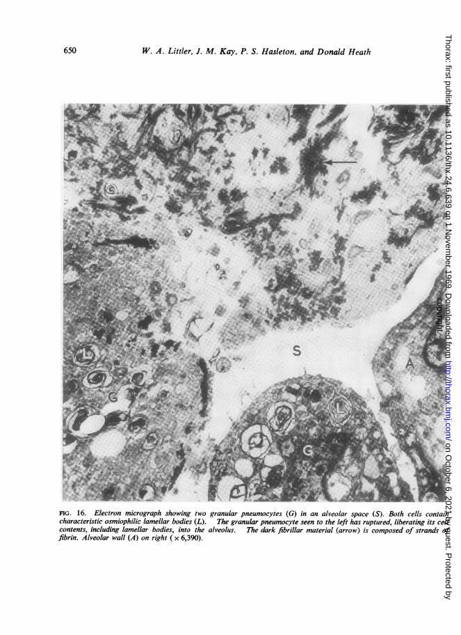
FIG. 16. Electron micrograph showing two granular pneumocytes (G) in an alveolar space (S). Both cells containcharacteristic osmiophilic lamellar bodies (L). The granular pneumocyte seen to the left has ruptured, liberating its cellcontents, including lamellar bodies, into the alveolus. The dark fibrillar material (arrow) is composed of strands offibrin. Alveolar wall (A) on right ( x 6,390).
C6f"
..
W.'
...I
copyright. on O
ctober 6, 2021 by guest. Protected by
http://thorax.bmj.com
/T
horax: first published as 10.1136/thx.24.6.639 on 1 Novem
ber 1969. Dow
nloaded from
Busulphan lung
4. +-
i
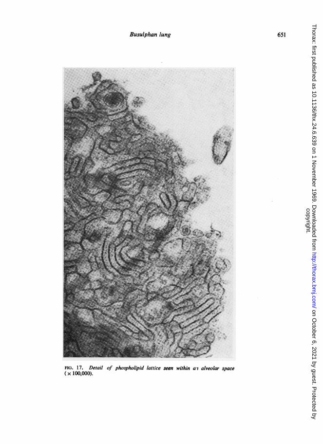
FIG. 17. Detail of phospholipid lattice seen within ai alveolar space(x 100,000).
651
copyright. on O
ctober 6, 2021 by guest. Protected by
http://thorax.bmj.com
/T
horax: first published as 10.1136/thx.24.6.639 on 1 Novem
ber 1969. Dow
nloaded from

W. A. Littler, J. M. Kay, P. S. Hasleton, and Donald Heath
#V
.t e 'i4 +)s-°Y.... 9, I
* 4> i 4;1>W B........
t.
A4'.........e
e ~ ~ ~ ~ ~ ~ ~ ~m 4+:
FIG. 18. Electron micrograph to show the nature of a non-cellular component of the intra-alveolar material. The dark granular material above is probably fibrin. Below are collagen fibresshowing characteristic periodicity (x 75,000).
..44
.
652
.:.X...
copyright. on O
ctober 6, 2021 by guest. Protected by
http://thorax.bmj.com
/T
horax: first published as 10.1136/thx.24.6.639 on 1 Novem
ber 1969. Dow
nloaded from

Busulphan lung
alveolar spaces (Fig. 13) were identified as granu-lar pneumocytes. They possessed prominent intra-cytoplasmic lamellar or spiral osmiophilic secre-tory inclusions which are usually called 'lamellarbodies' (Fig. 14). The cell surface was thrown upinto characteristic irregular, short microvilli.The intra-alveolar debris consisted of strands
of amorphous osmiophilic fibrillary material inter-spersed with roughly circular bodies composed ofconcentric osmiophilic membranes (Fig. 15). Thefibrillary material resembled fibrin and the circu-lar bodies appeared identical to the lamellarsecretory inclusion bodies of granular pneumo-cytes. In some instances it appeared that granularpneumocytes had disintegrated (Fig. 16) andliberated their lamellar bodies, so giving rise tothe intra-alveolar debris which characterized thehistological picture of this condition. Alsoscattered within the alveolar spaces were complexlattice structures (Fig. 17) composed of osmio-philic membranes which resembled the artificialphospholipid membranes prepared by Lucy andGlauert (1964). Evidence of organization of theintra-alveolar debris was seen in the form ofgroups of fibrils showing the periodic transversestriations characteristic of collagen (Fig. 18).
DISCUSSION
The development of pulmonary complications insome patients on 'busulphan therapy has beenrecognized since the original report of Oliner,Schwartz, Rubio, and Dameshek in 1961. Theydescribed two patients with chronic myeloidleukaemia receiving the drug who developzddyspnoea, fever, weakness, and weight loss; oneof these patients had a dry cough. The mainphysical findings noted, as in the present case,were bilateral basal crepitations.
Radiographs of the chest in their cases showeddiffuse, bilateral infiltrates which had the appear-ance of an inflammatory process. From ourpathological findings, and by analogy with theradiographic features of desquamative interstitialpneumonia (Liebow, Steer and Billingsley, 1965),in which there is a similar pathology of initialexudation of granular pneumocytes followed bythe development of fibrosing alveolitis, we thinkthat two radiographic pictures are to be expectedin busulphan lung. In the early stage, when thealveolar spaces are filled with large numbers ofgranular pneumocytes, the chest radiographs shouldshow a 'ground-glass' appearance, as in the casesof desquamative interstitial pneumonia illustratedby Liebow et al. (1965). In the later stage of
fibrous thickening of alveolar walls and organiza-tion of intra-alveolar exudate, we would anticipateseeing the well-known radiological appearancesof interstitial pulmonary fibrosis.The radiographic abnormalities of busulphan
lung were initially misdiagnosed as miliary tuber-culosis by Leake, Smith, and Woodliff (1963) inone of their two cases, and this patient was givenanti-tuberculosis therapy. The chest radiographswere said to be consistent with interstitial pul-monary fibrosis in the third case of Kyle,Schwartz, Oliner, and Dameshek (1961) and inthe case of Bates and Christie (1964). Our case isatypical in that, while in most previously reportedexamples of this disease chest radiographs haveusually shown changes from the onset of symp-toms, in our patient they showed no abnormalityin the early stages of the illness. When changesdid appear eventually they were not at firstcharacteristic, since they showed consolidation ofthe left lower lobe. This change appears to berelated to the abscess formation with surround-ing inflammatory change in this area that wenoted at necropsy.Pulmonary function tests were performed on
one of the patients of Oliner et al. (1961) andwere reported as being 'compatible with the syn-drome of alveolar-capillary block', although noactual figure for the transfer factor was given.Our patient showed evidence of 'restrictive lungdisease' from pulmonary function studies, with asevere reduction in transfer factor, so that it wasonly 25% of the normal (Table). We believe thatthe transfer factor may prove to be a useful in-dicator of the onset and severity of pulmonarycomplications of busulphan therapy.Pulmonary function tests were also said to
indicate 'alveolar capillary block' in one of fourcases (case 3) of an Addisonian-like syndromefollowing busulphan therapy reported by Kyleet al. (1961); once again no precise data fromthese tests were given by this author. Similar testscarried out on one of the cases of Leake et al.(1963) showed a slight defect in diffusion', but nofigures were given. Bates and Christie (1964)demonstrated a low transfier factor associatedwith a low haemoglobin during the course of ill-ness in a patient with busulphan lung. Correctionof the patient's anaemia restored the transferfactor to the normal range initially, but in theterminal stage of the illness the transfer factorwas low despite an adequate level of haemo-globin. The authors drew attention to the diffi-culties of interpreting changes in the value of thetransfer factor in the presence of anaemia. In
653
copyright. on O
ctober 6, 2021 by guest. Protected by
http://thorax.bmj.com
/T
horax: first published as 10.1136/thx.24.6.639 on 1 Novem
ber 1969. Dow
nloaded from

W. A. Littler, J. M. Kay, P. S. Hasleton, and Donald Heath
our patient the haemoglobin at the time that pul-monary function tests were carried out was 11 2g. /100 ml. (77 %). Anaemia of this degree wouldnot contribute significantly to the profound reduc-tion in transfer factor which we found.When pulmonary complications developed in
their patients, Oliner et al. (1961) discontinuedthe busulphan therapy and gave large doses ofprednisolone. Both patients responded to thistreatment with disappearance of their pulmonarysigns and symptoms; one of the patients alsoshowed radiological improvement. It seems likelyto us that improvement following the use ofsteroids will only occur when the disease is in itscellular phase with the exudation of granularpneumocytes. There seems little likelihood ofclinical or radiological improvement with suchtreatment when extensive pulmonary fibrosis hasoccurred. The patient designated case 3 by Kyleet al. (1961) responded to the stopping of busul-phan and the administration of prednisolone.Under this regime the pulmonary symptoms dis-appeared, although the patient subsequently diedfrom pulmonary oedema. One of the patients ofLeake et al. (1963) improved with cessation oftreatment by busulphan alone; in this instance nosteroids were given. Our patient was not givenprednisolone initially, and when it was started itwas given in relatively small doses because ofhaemolysis. When eventually bigger doses wereemployed the pulmonary syndrome was in anadvanced state. Where patients have respondedto prednisolone, big doses have been used (Olineret al., 1961 ; Kyle et al., 1961) whilst in thosecases where smaller doses were used (Smalley andWall, 1966) the patients have died with very littleresponse.
Death from pulmonary oedema has beenreported in patients with busulphan lung byOliner et al. (1961) and Kyle et al. (1961).Our patient shows a notable difference from
those reported previously in that busulphan hadbeen stopped for one month before the onset ofsymptoms. In the previous cases reported thepatients were taking the drug at the time whensymptoms appeared. Central cyanosis was prom-inent in our case, but it is mentioned as beingpresent in only one of the cases reported before(Smalley and Wall, 1966).
In the majority of cases of busulphan lung pre-viously reported the biopsy specimens of lunghave been described as showing interstitial pul-monary fibrosis (as in the cases of Oliner et al.(1961), the third case of Kyle et al. (1961), and
the second case of Leake et al. (1963) ). The con-cept of the pathology of this disease was broad-ened by the recognition of atypical intra-alveolarcells by Heard and Cooke (1968). We have beenable to confirm by means of electron microscopytheir suggestion made on the basis of light micro-scopy that these cells are the large alveolar (typeII) cells. The ultrastructure of these cells in thepresent case is characteristic of granular pneumo-cytes in possessing highly characteristic lamellarbodies (Figs 13 and 15) and in showing shortmicrovilli (Fig. 13).We have noted the same cells in conditions
characterized by chemical and physical irritationof the alveolar walls, with the production of firstthe cellular and later the fibrosing stages of alveo-litis. Thus we have seen this proliferation ofgranular pneumocytes in rats poisoned by Crota-laria spectabilis seeds to produce pulmonaryhypertension (Kay, Smith, and Heath, 1969). Wehave seen the same phenomenon in the lungs ofdogs subjected to irradiation, sent to us forexamination by Dr. H. W. C. Ward, of the QueenElizabeth Hospital, Birmingham. Finally, we havedemonstrated by electron microscopy (Brewer,Heath, and Asquith, 1969) the same outpouringof granular pneumocytes into the alveoli in a caseof the non-specific cellular alveolitis which hasbeen termed desquamative interstitial pneumonia(Liebow et al., 1965). It is apparent that the granu-lar pneumocyte is a common reactive cell in thealveolar wall in man, responding to all manner ofnoxious stimuli. The presence of such cells is inno way specific for busulphan lung.We suggest that the other features of the
pathology of busulphan lung follow this cellularalveolitis and we differ from Heard and Cooke(1968) in their interpretation of the histo-pathology. They are of the opinion that the intra-alveolar eosinophilic material is to be explainedon the basis of a persistent fibrinous oedema. Wethink it likely that much of this intra-alveolarsubstance represents the breakdown of previouslydesquamated granular pneumocytes. We havearrived at this conclusion by noting the intimateadmixture of intact or ghost outlines of pneumo-cytes with the intra-alveolar debris (Figs 8 and 16).Fibrinous oedema is associated in the productionof this alveolar material, but the disintegration ofgranular pneumocytes also plays an importantrole.We agree with Heard and Cooke (1968) that
much of this material undergoes subsequentorganization and incorporation into alveolar
654
copyright. on O
ctober 6, 2021 by guest. Protected by
http://thorax.bmj.com
/T
horax: first published as 10.1136/thx.24.6.639 on 1 Novem
ber 1969. Dow
nloaded from

Busulphan lung
walls, but we believe from our study of this case
that true innate fibrous thickening of alveolarwalls occurs. This appears to follow the naturalhistory of alveolitis progressing from the cellularto the fibrous stage, without in all cases an asso-
ciated thickening of alveolar walls brought aboutby organization and incorporation of intra-alveolar material.
It seems likely, too, that the disintegration ofthe granular pneumocytes (Fig. 16), with libera-tion of the phospholipid contained in theirlamellar bodies (Figs 15 and 16), may be respon-
sible for the lipid granulomas noted in this case.
Glancy, Frazier, and Roberts (1968) describecholesterol granulomas of identical histologicalstructure which they were able to show by x-ray
diffraction studies contained cholesterol palmitateand stearate. Although these authors associatedsuch granulomas with pulmonary hypertension, itshould be noted that identical granulomas in thiscase occurred without any morbid anatomicalevidence of a raised pulmonary arterial pressure.
It seems to us more likely that granulomas ofthis type arise as a reaction to phospholipidliberation by disintegrating granular pneumocytes.
REFERENCESBates, D. V., and Christie, R. V. (1964). Respiratory Function in
Disease. Case 48, p. 428. Saunders, Philadelphia.Brenner, 0. (1935). Pathology of the vessels of the pulmonary circula-
tion. Arch. intern. Med., 56, 211.Brewer, D. B., Heath, D., and Asquith, P. (1969). Electron microscopy
of desquamative interstitial pneumonia. J. Path., 97, 317.Cotes, J. E. (1965). Lung Function, p. 314. Blackwell, Oxford.Fulton, R. M., Hutchinson, E. C., and Morgan Jones, A. (1952).
Ventricular weight in cardiac hypertrophy. Brit. Heart J., 14,413.Glancy, D. L., Frazier, P. D., and Roberts, W. C. (1968). Pulmonary
parenchymal cholesterol-eater granulomas in patients withpulmonary hypertension. Amer. J. Med., 45, 198.
Heard, B. E., and Cooke, R. A. (1968). Busulphan lung. Thorax, 23,187.
Kay, J. M., Smith, P., and Heath, D. (1969). Electron microscopy ofcrotalaria pulmonary hypertension. Ibid., 24, 511.
Kyle, R. A., Schwartz, R. S., Oliner, H. L., and Dameshek, W. (1961).A syndrome resembling adrenal cortical insufficiency associatedwith long term busulfan (Myleran) therapy. Blood, 18, 497.
Leake, E., Smith, W. G., and Woodliff, H. J. (1963). Diffuse inter-stitial pulmonary fibrosis after busulphan therapy. Lancet, 2, 432.
Liebow, A. A., Steer, A., and Billingsley, J. G. (1965). Desquamativeinterstitial pneumonia. Amer. J. Med., 39, 369.
Lucy, J. A., and Glauert, A. M. (1964). Structure and assembly ofmacromolecular lipid complexes composed of globular micelles.J. molec. Biol., 8, 727.
Ogilvie, C. M., Forster, R. E., Blakemore, W. S., and Morton, J. W.(1957). A standardized breath holding technique for the clinicalmeasurement of the diffusing capacity of the lung for carbonmonoxide. J. clin. Invest., 36, 1.
Oliner, H., Schwartz, R., Rubio, F., Jr., and Dameshek, W. (1961).Interstitial pulmonary fibrosis following busulfan therapy. Amer.J.Med,,31,134.
Riley, J. F. (1959). The Mast Cells. Livingstone, Edinburgh.Smalley, R. V., and Wall, R. L. (1966). Two cases of busulfan toxicity.
Ann. intern. Med., 64, 154.
655
copyright. on O
ctober 6, 2021 by guest. Protected by
http://thorax.bmj.com
/T
horax: first published as 10.1136/thx.24.6.639 on 1 Novem
ber 1969. Dow
nloaded from