Burns and Wound healing Dr. Leonard Da Silva Molecular & Cellular Pathology.
44
Burns and Wound healing Dr. Leonard Da Silva Molecular & Cellular Pathology
-
Upload
ashlyn-hampton -
Category
Documents
-
view
216 -
download
0
Transcript of Burns and Wound healing Dr. Leonard Da Silva Molecular & Cellular Pathology.

Burns and Wound healing
Dr. Leonard Da Silva
Molecular & Cellular Pathology

Be sure to keep hot liquids out of reach of small children

U.S. Burn Statistics
• Approximately 2.4 million burn injuries are reported per year
• Medical professionals treat approximately 650,000 of the injuries;
• Between 8,000 and 12,000 of patients with burns die,
• Patients with major burns exceeding 60% of their total body surface area often do not survive

• Burns are one of the most expensive
catastrophic injuries to treat.
• “The cost of waiting for your own skin
to grow can be more painful than the burn itself!”
Hospital Costs

Occupational skin disease
- Physical – Heat, cold, radiation

Workers compensation• Evaluation
– Diagnosis– Causation– Impairment evaluation– Conclusions & recommendations– Physical examinations
Effective management requires an understanding of burns
pathophysiology
Different causes lead to different patterns of injury
Knowing the cause will help predict the pathological response and therefore the treatment

Anatomy of skin• Epidermis
– Outer layer contains the stratum corneum• The rate limiting step in dermal or percutaneous absorption is
diffusion through the epidermis
• Dermis– Much thicker than epidermis – True skin & is the main natural protection against trauma– Contains
• Sweat glands• Sebaceous glands• Blood vessels• Hair• Nails
• Subcutaneous Layer – Contains the fatty tissues which cushion & insulate

Review of skin functions
•Functions of the skin •Protection •Heat regulation •Sensory preception •Excretion •Vitamin D production
•Attempts to cover wounds and treat severe burns is cited as far back as 1500 B.C.
• Grafts from humans (allografts) or animals (xenografts)
Background Information
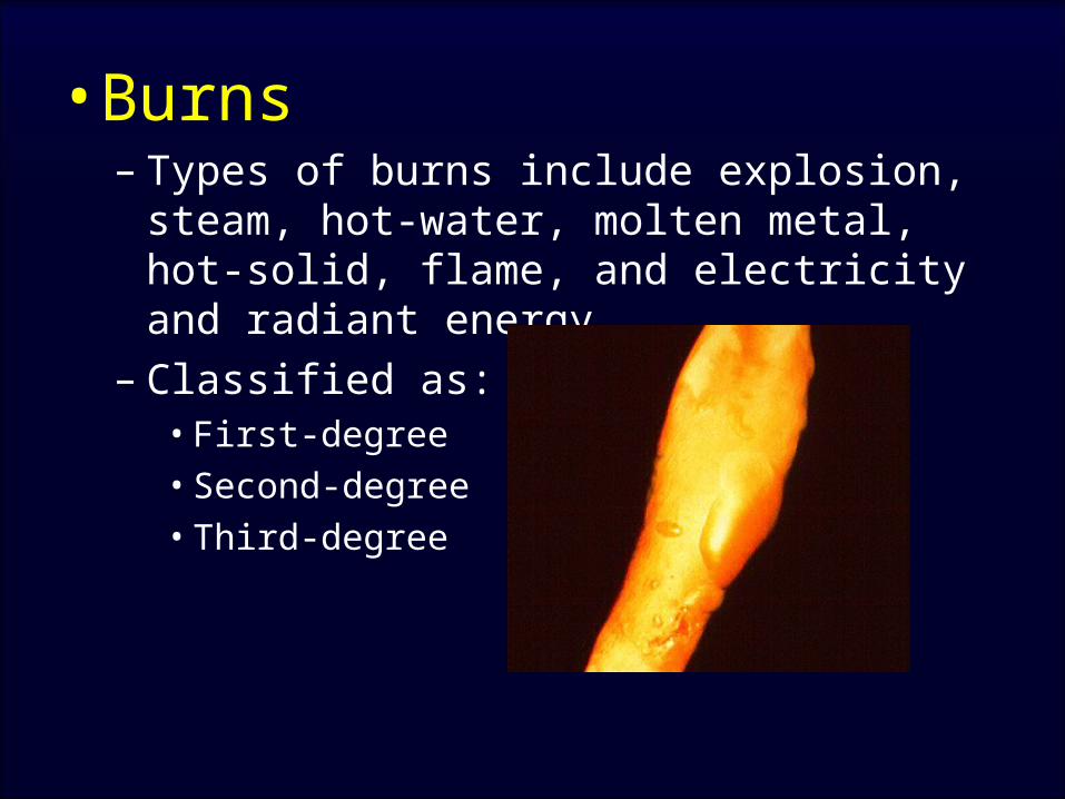
• Burns– Types of burns include explosion, steam, hot-
water, molten metal, hot-solid, flame, and electricity and radiant energy
– Classified as:• First-degree • Second-degree• Third-degree

Classification of burns
•Partial thickness - characterized by varying depth from epidermis to the
Superficial - includes only the epidermis Deep - involve entire epidermis and part of the dermis
•Full thickness - above + possible damage to the SC, muscle and bone

Mechanisms of burns injuries
Electrical burns Inhalation burns Chemical burns Radiation burns
Electrical burns
- high intensity heat
- brain or heart damage or musculoskeletal injuries associated with the electrical injuries.
- safely remove the person from the source of the electricity. Do not become a victim.

Electrical Burns
Voltage tissue damage 2 pathways of damage :
1. Electrical dysfunction of the cardiovascularconduction system and the nervous system
2. Conversion of electrical energy to heat energy when the current encounters the resistance of the tissues

Inhalation burns
• Occur in people trapped in burning buildings or vehicles
• Exposure to high temperature aerosolized flammable materials
• Damage to respiratory tract epithelium from the oral cavity to the alveoli

Chemical burns
• damage until it is completely removed
• Alkalis worse – penetrate deeper
• If cutaneous exposure remove clothes and wash area
• Usually seen in industrial setting or motor vehicle accident

CaOH

Radiation burns
• Treatment may result in burns
• Xray – medical imaging and radiotherapy

Radiation
• Ionizing radiation sources–Alpha radiation stopped by skin
»Ingestion
–Beta radiation can injure skin by contact »Localized at skin surface or outer
layers of skin
–Gamma radiation and x-rays are skin and systemic hazards
» Skin cancer may develop

Burn Healing

Phases of Wound Healing
1. Vascular Response
2. Blood coagulation
3. Inflammation
4. Formation of new tissue
5. Epithelialisation
6. Contraction & Remodeling

Early wound healing events
• Hemostasis– Platelet aggregation – Intrinsic and extrinsic coagulation cascade– Thrombin, fibrin– Vasoconstriction

Early wound healing events
• Inflammation
Vasodilatation
Increase in vascular permeability
Chemotaxis
Cellular response

Early wound healing events
• Homeostasis
• Neutrophils
• 48-72h- macrophages
• 5-7 days- few inflammatory cells.

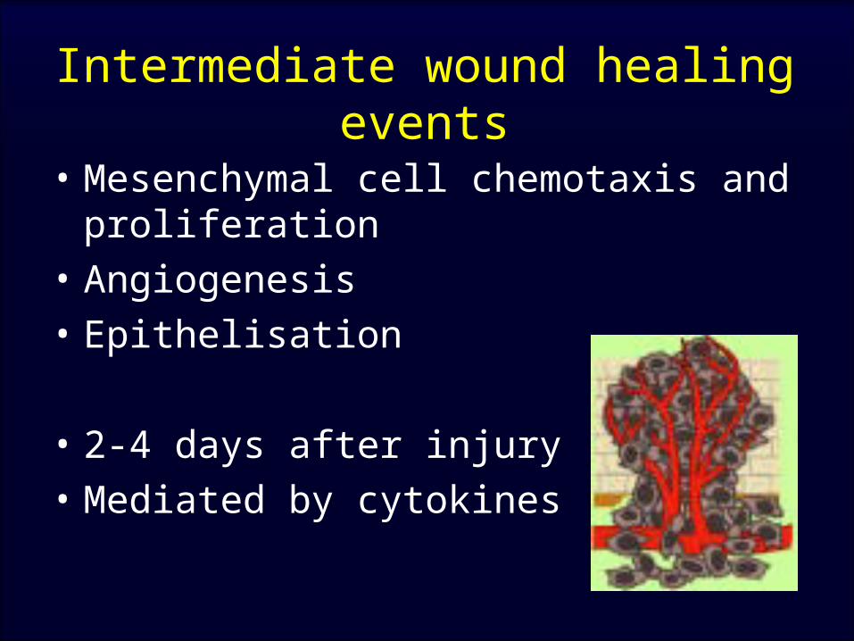
Intermediate wound healing events
• Mesenchymal cell chemotaxis and proliferation
• Angiogenesis
• Epithelisation
• 2-4 days after injury
• Mediated by cytokines

Intermediate wound healing events
Mesenchymal cell chemotaxis and proliferation
• Fibroblasts- migration and proliferation• Smooth muscle
Angiogenesis- reconstruction of vasculature
• Stimulate: High lactate, acidic Ph, low O2 tension• Endothelial cell migration and proliferation

Intermediate wound healing events
Epithelisation• Partial thickness- Cells derived from wound
edges and epithelial appendages.
• Incisional wound: cellular migration over less then 1 mm. Wound sealed in 24-48h.
• Cellular detachment
• Migration

Late wound healing events
Collagen synthesis
• 3 helical polypeptide chains
• Lysine and proline hydroxylation
Required for cross-linking
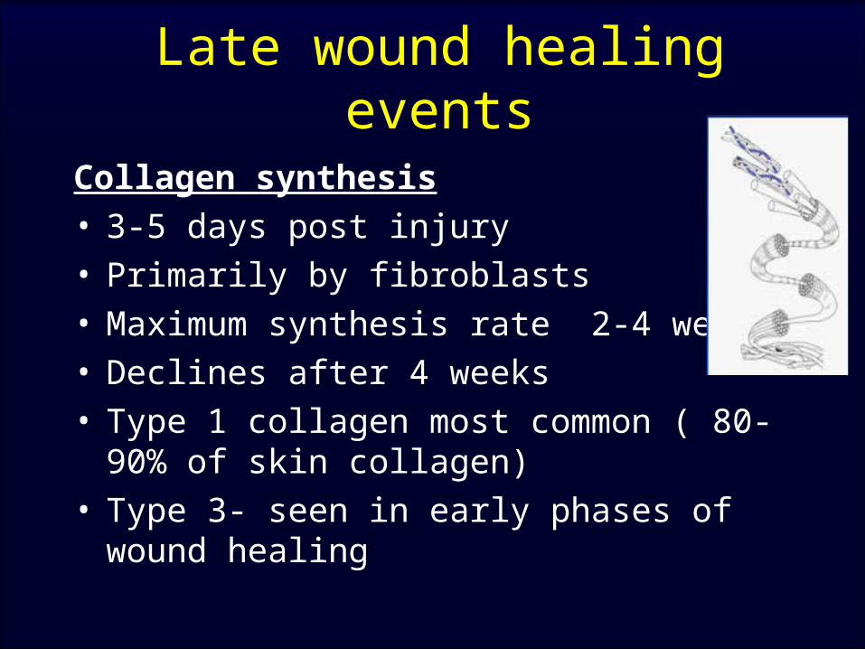
Late wound healing events
Collagen synthesis• 3-5 days post injury• Primarily by fibroblasts• Maximum synthesis rate 2-4 weeks• Declines after 4 weeks• Type 1 collagen most common ( 80-90% of
skin collagen)• Type 3- seen in early phases of wound
healing

Wound contraction
• Centripetal movement of the wound edges toward the center. ( 0.6-0.7 mm/day)
• Begins at 4-5 days• Maximal contraction 12-15 days• Trivial component in closed incisional wounds,
significant for closure of open wounds• Rate- depends on tissue• Circular wounds- slower closure but avoid
stenosis
Wound contraction• Mechanism- cell mediated processes,
not requiring collagen synthesis• Myofibroblasts- fibroblasts with
myofilaments in cytoplasm• Appear in wound day 3-21• Located in periphery- pull wound edges
together.• Contractures- contraction across joint
surface

Terminal wound healing events
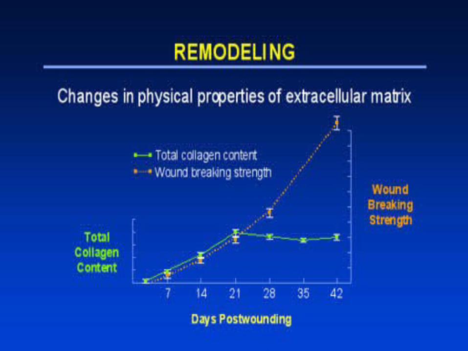
• Remodeling- turnover of collagen. Type 3 replaced by type 1
• Day 21- net accumulation of wound collagen becomes stable
• Wound bursting strength- 15% of normal.• Week 3-6- greatest rate of increase• 6 weeks- 80-90% of eventual strength.• 6 months maximum strength ( 90% ). Process
continues for 12 months
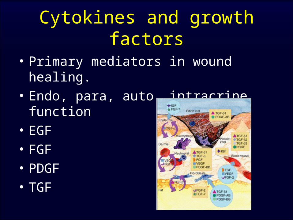
Cytokines and growth factors
• Primary mediators in wound healing.
• Endo, para, auto, intracrine function
• EGF
• FGF
• PDGF
• TGF

Growth factors in wound healing


Forensic pathology
A large percentage of the work done by forensic pathologists involves burns
All types of burn injuries are seen
Commonly incineration cases occur especially in motor vehicle accidents

Complications of burns
process of healing
immediate and delayed
Skin – fluid retention
Protection from infectious agents
Fluid loss ‐ haemoconcentration and poor vascular perfusion of the skin and viscera – shock and acutetubular necrosis

Delayed
Poor perfusion – infections and sepsis
Scar tissue – lack elastic properties – contractures

Are you one of those people that stays up to date on the latest sports scores and plays?

Wound care - grafting
Indications for grafting full thickness priority areas wound bed pink, firm, free of exudate bacterial count < 100,000/gram of tissue