Bronchogenic carcinoma in idiopathic pulmonary fibrosis ...
1
e-mail: [email protected] website: www.ildcare.eu Figure 1: HRCT patient A. Discussion After Ltx the incidence of bronchogenic carcinoma is increased. Risk factors are IPF per se, immunosuppressive drugs, single versus bilateral Ltx, smoking, increasing age and male gender. 3 In 6.9% of single Ltx a bronchogenic carcinoma arises in the native lung as we found in patient A. This is rarely accounted when a bilateral Ltx is performed. 3 Moreover, nowadays most bilateral Ltx are done in Leuven. In 2% of patients a bronchogenic carcinoma is unexpectedly found in the explanted lung, as we found in patient B and C. 1 Symptoms are usually aspecific or mimic an infection or rejection as in patient A. 4 Adenocarcinoma and squamous cell carcinoma represent the most frequent pathological types, followed by small cell carcinoma. 4 Although disease is often diagnosed in an early stage, the prognosis remains extremely poor. 3 Introduction Lung transplantation (Ltx) is an accepted therapy for patients with end-stage lung disease. The most important indications are chronic obstructive pulmonary disease (COPD) and idiopathic pulmonary fibrosis (IPF). 1 The incidence of lung cancer after Ltx is 20-25 times higher than in the general population, but diagnosis is often difficult. 2 We describe three Ltx patients suffering primarily from IPF. Cases Patient A presented 7 years after single Ltx with malaise. He was renovating his house. A HRCT of the chest showed an increasing opacity in the native lung replacing the fibrotic lesions (Figure 1). The differential diagnosis consisted of fungus infection, rejection and malignancy. Eventually he was diagnosed with a bronchogenic carcinoma, he died shortly after. Patient B underwent a bilateral Ltx. In the explanted right lung a T2N2 carcinoma was found. Two years after radical chemo-radiotherapy progression appeared, shortly after he died. Patient C complained of left pretibial pain before Ltx. 18 FDG-PET showed uptake in both lungs and the left tibia. The first was interpreted as compatible with her IPF, the latter was suggestive for Pierre Marie Bamberger. At the time of Ltx, however, she was diagnosed with an adenocarcinoma in both lungs. She died shortly after, see also Table 1. Table 1: Summary of the clinical data of the presented cases. References 1. Mathew J, et al. Lung cancer and lung transplantation: a review. J Thorac Oncol 2009;4:753-60 2. Bellil Y, et al. Bronchogenic carcinoma in solid organ transplant recipients. Curr Tr Opt Onc 2006:6:77-81 3. Dickson R, et al. High frequency of bronchogenic carcinoma after single-lung transplantation. J Heart Lung Transplant 2006;25:1297-301 4. Minai O, et al. Bronchogenic carcinoma after lung transplantation: characteristics and outcomes. JTO 2008;3:1404-9 Patient A, male Patient B, male Patient C, female Age diagnosis IPF / Ltx resp. 46 / 48 years 53 / 57 years 47 / 53 years Histology UIP UIP NSIP, fibrotic type Smoking status ex, 30 packyears ex, 26 packyears never Treatment IPF (all acetylcysteine/prednisone) cyclophosfamide azathioprine azathioprine Single or bilateral Ltx single (right) single (left), after rejection bilateral bilateral Histology explanted lung UIP, no malignancy left: UIP, right: squamous cell carcinoma massive bilateral adenocarcinoma Time Ltx - carcinoma 8 years in explanted lung in explanted lung Stage and treatment IV: none, poor performance T2N2M0 (IIIA): chemo- radiotherapy, progression: chemo cT4N2M1b (IV): palliative radiotherapy Time carcinoma - death 3 months 22 months 3 months Conclusions Transplanted IPF patients are at risk for developing primary bronchogenic carcinoma. Symptoms are often aspecific, diagnosis is difficult and prognosis is extremely poor. These cases stress the importance of actively searching for bronchogenic carcinoma before as well after lung transplantation in patients with IPF. HRCT of patient A showing an increasing opacity in the native lung replacing the fibrotic lesions, also ground glass areas in the transplant lung. Bronchogenic carcinoma in idiopathic pulmonary fibrosis patients after lung transplantation Hendriks LEL 1 , Drent M 2,3 , van Haren EHJ 1 , Verschakelen JA 3,4 , Verleden GM 5 1 Dept of Respiratory Medicine, Atrium Medical Centre, Heerlen, 2 Dept of Respiratory Medicine, 3 ild care team, Maastricht University Medical Centre+, Maastricht, The Netherlands, 4 Dept of Radiology, University Hospitals, 5 Dept of Lung Transplantation, University Hospital Gasthuisberg, Leuven, Belgium
Transcript of Bronchogenic carcinoma in idiopathic pulmonary fibrosis ...
e-mail: [email protected]: www.ildcare.eu
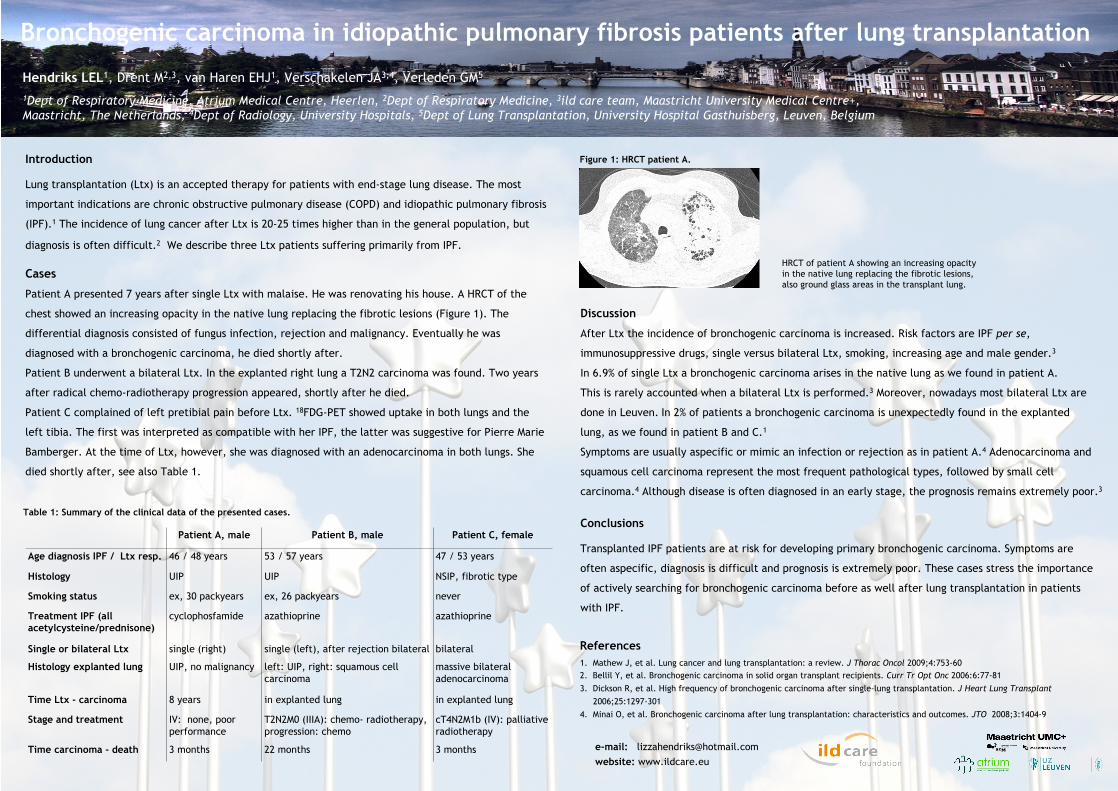
Figure 1: HRCT patient A.
Discussion
After Ltx the incidence of bronchogenic carcinoma is increased. Risk factors are IPF per se,
immunosuppressive drugs, single versus bilateral Ltx, smoking, increasing age and male gender.3
In 6.9% of single Ltx a bronchogenic carcinoma arises in the native lung as we found in patient A.
This is rarely accounted when a bilateral Ltx is performed.3 Moreover, nowadays most bilateral Ltx are
done in Leuven. In 2% of patients a bronchogenic carcinoma is unexpectedly found in the explanted
lung, as we found in patient B and C.1
Symptoms are usually aspecific or mimic an infection or rejection as in patient A.4 Adenocarcinoma and
squamous cell carcinoma represent the most frequent pathological types, followed by small cell
carcinoma.4 Although disease is often diagnosed in an early stage, the prognosis remains extremely poor.3
Introduction
Lung transplantation (Ltx) is an accepted therapy for patients with end-stage lung disease. The most
important indications are chronic obstructive pulmonary disease (COPD) and idiopathic pulmonary fibrosis
(IPF).1 The incidence of lung cancer after Ltx is 20-25 times higher than in the general population, but
diagnosis is often difficult.2 We describe three Ltx patients suffering primarily from IPF.
Cases
Patient A presented 7 years after single Ltx with malaise. He was renovating his house. A HRCT of the
chest showed an increasing opacity in the native lung replacing the fibrotic lesions (Figure 1). The
differential diagnosis consisted of fungus infection, rejection and malignancy. Eventually he was
diagnosed with a bronchogenic carcinoma, he died shortly after.
Patient B underwent a bilateral Ltx. In the explanted right lung a T2N2 carcinoma was found. Two years
after radical chemo-radiotherapy progression appeared, shortly after he died.
Patient C complained of left pretibial pain before Ltx. 18FDG-PET showed uptake in both lungs and the
left tibia. The first was interpreted as compatible with her IPF, the latter was suggestive for Pierre Marie
Bamberger. At the time of Ltx, however, she was diagnosed with an adenocarcinoma in both lungs. She
died shortly after, see also Table 1.
Table 1: Summary of the clinical data of the presented cases.
References1. Mathew J, et al. Lung cancer and lung transplantation: a review. J Thorac Oncol 2009;4:753-602. Bellil Y, et al. Bronchogenic carcinoma in solid organ transplant recipients. Curr Tr Opt Onc 2006:6:77-81
3. Dickson R, et al. High frequency of bronchogenic carcinoma after single-lung transplantation. J Heart Lung Transplant
2006;25:1297-3014. Minai O, et al. Bronchogenic carcinoma after lung transplantation: characteristics and outcomes. JTO 2008;3:1404-9
Patient A, male Patient B, male Patient C, female
Age diagnosis IPF / Ltx resp. 46 / 48 years 53 / 57 years 47 / 53 years
Histology UIP UIP NSIP, fibrotic type
Smoking status ex, 30 packyears ex, 26 packyears never
Treatment IPF (all acetylcysteine/prednisone)
cyclophosfamide azathioprine azathioprine
Single or bilateral Ltx single (right) single (left), after rejection bilateral bilateral
Histology explanted lung UIP, no malignancy left: UIP, right: squamous cell carcinoma
massive bilateral adenocarcinoma
Time Ltx - carcinoma 8 years in explanted lung in explanted lung
Stage and treatment IV: none, poor performance
T2N2M0 (IIIA): chemo- radiotherapy, progression: chemo
cT4N2M1b (IV): palliative radiotherapy
Time carcinoma - death 3 months 22 months 3 months
Conclusions
Transplanted IPF patients are at risk for developing primary bronchogenic carcinoma. Symptoms are
often aspecific, diagnosis is difficult and prognosis is extremely poor. These cases stress the importance
of actively searching for bronchogenic carcinoma before as well after lung transplantation in patients
with IPF.
HRCT of patient A showing an increasing opacity in the native lung replacing the fibrotic lesions, also ground glass areas in the transplant lung.
Bronchogenic carcinoma in idiopathic pulmonary fibrosis patients after lung transplantation
Hendriks LEL1, Drent M2,3, van Haren EHJ1, Verschakelen JA3,4, Verleden GM5
1Dept of Respiratory Medicine, Atrium Medical Centre, Heerlen, 2Dept of Respiratory Medicine, 3ild care team, Maastricht University Medical Centre+, Maastricht, The Netherlands, 4Dept of Radiology, University Hospitals, 5Dept of Lung Transplantation, University Hospital Gasthuisberg, Leuven, Belgium