British Society of Gastroenterology guidelines on the ... · PDF fileEmail alerting the box at...
38
doi: 10.1136/gutjnl-2013-305372 published online October 28, 2013 Gut Rebecca C Fitzgerald, Massimiliano di Pietro, Krish Ragunath, et al. management of Barrett's oesophagus guidelines on the diagnosis and British Society of Gastroenterology http://gut.bmj.com/content/early/2013/10/28/gutjnl-2013-305372.full.html Updated information and services can be found at: These include: Data Supplement http://gut.bmj.com/content/suppl/2013/10/26/gutjnl-2013-305372.DC1.html "Supplementary Data" References http://gut.bmj.com/content/early/2013/10/28/gutjnl-2013-305372.full.html#ref-list-1 This article cites 352 articles, 40 of which can be accessed free at: P<P Published online October 28, 2013 in advance of the print journal. service Email alerting the box at the top right corner of the online article. Receive free email alerts when new articles cite this article. Sign up in Collections Topic (298 articles) Oesophageal cancer Articles on similar topics can be found in the following collections (DOIs) and date of initial publication. publication. Citations to Advance online articles must include the digital object identifier citable and establish publication priority; they are indexed by PubMed from initial typeset, but have not not yet appeared in the paper journal. Advance online articles are Advance online articles have been peer reviewed, accepted for publication, edited and http://group.bmj.com/group/rights-licensing/permissions To request permissions go to: http://journals.bmj.com/cgi/reprintform To order reprints go to: http://group.bmj.com/subscribe/ To subscribe to BMJ go to: group.bmj.com on October 29, 2013 - Published by gut.bmj.com Downloaded from
Transcript of British Society of Gastroenterology guidelines on the ... · PDF fileEmail alerting the box at...

doi: 10.1136/gutjnl-2013-305372 published online October 28, 2013Gut
Rebecca C Fitzgerald, Massimiliano di Pietro, Krish Ragunath, et al. management of Barrett's oesophagusguidelines on the diagnosis and British Society of Gastroenterology
http://gut.bmj.com/content/early/2013/10/28/gutjnl-2013-305372.full.htmlUpdated information and services can be found at:
These include:
Data Supplement http://gut.bmj.com/content/suppl/2013/10/26/gutjnl-2013-305372.DC1.html
"Supplementary Data"
References http://gut.bmj.com/content/early/2013/10/28/gutjnl-2013-305372.full.html#ref-list-1
This article cites 352 articles, 40 of which can be accessed free at:
P<P Published online October 28, 2013 in advance of the print journal.
serviceEmail alerting
the box at the top right corner of the online article.Receive free email alerts when new articles cite this article. Sign up in
CollectionsTopic
(298 articles)Oesophageal cancer � Articles on similar topics can be found in the following collections
(DOIs) and date of initial publication. publication. Citations to Advance online articles must include the digital object identifier citable and establish publication priority; they are indexed by PubMed from initialtypeset, but have not not yet appeared in the paper journal. Advance online articles are Advance online articles have been peer reviewed, accepted for publication, edited and
http://group.bmj.com/group/rights-licensing/permissionsTo request permissions go to:
http://journals.bmj.com/cgi/reprintformTo order reprints go to:
http://group.bmj.com/subscribe/To subscribe to BMJ go to:
group.bmj.com on October 29, 2013 - Published by gut.bmj.comDownloaded from

Notes
(DOIs) and date of initial publication. publication. Citations to Advance online articles must include the digital object identifier citable and establish publication priority; they are indexed by PubMed from initialtypeset, but have not not yet appeared in the paper journal. Advance online articles are Advance online articles have been peer reviewed, accepted for publication, edited and
http://group.bmj.com/group/rights-licensing/permissionsTo request permissions go to:
http://journals.bmj.com/cgi/reprintformTo order reprints go to:
http://group.bmj.com/subscribe/To subscribe to BMJ go to:
group.bmj.com on October 29, 2013 - Published by gut.bmj.comDownloaded from

British Society of Gastroenterology guidelines on thediagnosis and management of Barrett’s oesophagusRebecca C Fitzgerald,1 Massimiliano di Pietro,1 Krish Ragunath,2 Yeng Ang,3
Jin-Yong Kang,4 Peter Watson,5 Nigel Trudgill,6 Praful Patel,7 Philip V Kaye,8
Scott Sanders,9 Maria O’Donovan,10 Elizabeth Bird-Lieberman,11 Pradeep Bhandari,12
Janusz A Jankowski,13 Stephen Attwood,14 Simon L Parsons,15 Duncan Loft,16
Jesper Lagergren,17 Paul Moayyedi,18 Georgios Lyratzopoulos,19 John de Caestecker20
▸ Additional material ispublished online only. To viewplease visit the journal online(http://dx.doi.org/10.1136/gutjnl-2013-305372).
For numbered affiliations seeend of article
Correspondence toProfessor Rebecca C Fitzgerald,MRC Cancer Unit, University ofCambridge, Box 197,Cambridge BiomedicalCampus, Cambridge,CB2 0XZ, UK;[email protected]
Received 31 May 2013Revised 14 August 2013Accepted 1 September 2013
To cite: Fitzgerald RC, diPietro M, Ragunath K, et al.Gut Published Online First:[please include Day MonthYear] doi:10.1136/gutjnl-2013-305372
ABSTRACTThese guidelines provide a practical and evidence-basedresource for the management of patients with Barrett’soesophagus and related early neoplasia. The Appraisalof Guidelines for Research and Evaluation (AGREE II)instrument was followed to provide a methodologicalstrategy for the guideline development. A systematicreview of the literature was performed for Englishlanguage articles published up until December 2012 inorder to address controversial issues in Barrett’soesophagus including definition, screening anddiagnosis, surveillance, pathological grading fordysplasia, management of dysplasia, and early cancerincluding training requirements. The rigour and quality ofthe studies was evaluated using the SIGN checklistsystem. Recommendations on each topic were scored byeach author using a five-tier system (A+, strongagreement, to D+, strongly disagree). Statements thatfailed to reach substantial agreement among authors,defined as >80% agreement (A or A+), were revisitedand modified until substantial agreement (>80%) wasreached. In formulating these guidelines, we took intoconsideration benefits and risks for the population andnational health system, as well as patient perspectives.For the first time, we have suggested stratification ofpatients according to their estimated cancer risk basedon clinical and histopathological criteria. In order toimprove communication between clinicians, werecommend the use of minimum datasets for reportingendoscopic and pathological findings. We advocateendoscopic therapy for high-grade dysplasia and earlycancer, which should be performed in high-volumecentres. We hope that these guidelines will standardiseand improve management for patients with Barrett’soesophagus and related neoplasia.
PURPOSE AND METHODS
The purpose of this guideline is to provide a prac-tical and evidence-based resource for the manage-ment of patients with Barrett’s oesophagus andrelated early neoplasia. This document is thereforeaimed at gastroenterologists, physicians and nursepractitioners, as well as members of multidisciplin-ary teams (MDTs; surgeons, radiologists, patholo-gists), who take decisions on the management ofsuch patients. The population covered by theseguidelines includes: patients with gastro-oesophageal reflux disease or other risk factors for
Barrett’s (obesity, family history for Barrett’s andoesophageal adenocarcinoma (OAC)); every patientwith incident or prevalent Barrett’s oesophagusregardless of their age, sex or comorbidities; patientswith early OAC and patients with intestinal metapla-sia (IM) at the gastro-oesophageal junction (GOJ)with no endoscopic evidence of Barrett’soesophagus. The previous British Society ofGastroenterology (BSG) guidelines were publishedin 2005 and since then there have been advances inthe diagnostic and management tools available.Within these guidelines, we have systematicallyreviewed the literature in order to address contro-versial issues in Barrett’s oesophagus and to formu-late practical recommendations to guide patientmanagement. In particular, we have covered the fol-lowing key questions.1. How should Barrett’s oesophagus be defined
and which patients should undergo regularsurveillance?
2. Are there clinical features associated withincreased cancer risk in Barrett’s oesophagus,which should influence the frequency of endo-scopic surveillance?
3. Are there diagnostic tools that should be uti-lised to screen the population at risk forBarrett’s oesophagus?
4. Which imaging modality should be used forthe endoscopic diagnosis and surveillance ofBarrett’s oesophagus?
5. How should we best manage dysplasia inBarrett’s oesophagus?
6. Which staging modality is preferred forBarrett’s-related early OAC?
7. What are the indications for endoscopic and/or surgical therapy in Barrett’s-relatedadenocarcinoma?
8. Are there minimum standards for training andmaintenance of skills in the field of endoscopictherapy?
9. How should patients be followed-up afterendoscopic therapy?
10. Are there chemopreventive interventionsrecommended to reduce the likelihood of theprogression of Barrett’s oesophagus?
11. What are the priorities for research and devel-opment in the field of Barrett’s carcinogenesis?
The Appraisal of Guidelines for Research andEvaluation (AGREE II) instrument1 was used toprovide a methodological strategy for the
Fitzgerald RC, et al. Gut 2013;0:1–36. doi:10.1136/gutjnl-2013-305372 1
Guidelines Gut Online First, published on October 28, 2013 as 10.1136/gutjnl-2013-305372
Copyright Article author (or their employer) 2013. Produced by BMJ Publishing Group Ltd (& BSG) under licence.
group.bmj.com on October 29, 2013 - Published by gut.bmj.comDownloaded from group.bmj.com on October 29, 2013 - Published by gut.bmj.comDownloaded from group.bmj.com on October 29, 2013 - Published by gut.bmj.comDownloaded from group.bmj.com on October 29, 2013 - Published by gut.bmj.comDownloaded from group.bmj.com on October 29, 2013 - Published by gut.bmj.comDownloaded from group.bmj.com on October 29, 2013 - Published by gut.bmj.comDownloaded from group.bmj.com on October 29, 2013 - Published by gut.bmj.comDownloaded from group.bmj.com on October 29, 2013 - Published by gut.bmj.comDownloaded from group.bmj.com on October 29, 2013 - Published by gut.bmj.comDownloaded from group.bmj.com on October 29, 2013 - Published by gut.bmj.comDownloaded from group.bmj.com on October 29, 2013 - Published by gut.bmj.comDownloaded from group.bmj.com on October 29, 2013 - Published by gut.bmj.comDownloaded from group.bmj.com on October 29, 2013 - Published by gut.bmj.comDownloaded from group.bmj.com on October 29, 2013 - Published by gut.bmj.comDownloaded from group.bmj.com on October 29, 2013 - Published by gut.bmj.comDownloaded from group.bmj.com on October 29, 2013 - Published by gut.bmj.comDownloaded from group.bmj.com on October 29, 2013 - Published by gut.bmj.comDownloaded from group.bmj.com on October 29, 2013 - Published by gut.bmj.comDownloaded from group.bmj.com on October 29, 2013 - Published by gut.bmj.comDownloaded from group.bmj.com on October 29, 2013 - Published by gut.bmj.comDownloaded from group.bmj.com on October 29, 2013 - Published by gut.bmj.comDownloaded from group.bmj.com on October 29, 2013 - Published by gut.bmj.comDownloaded from group.bmj.com on October 29, 2013 - Published by gut.bmj.comDownloaded from group.bmj.com on October 29, 2013 - Published by gut.bmj.comDownloaded from group.bmj.com on October 29, 2013 - Published by gut.bmj.comDownloaded from group.bmj.com on October 29, 2013 - Published by gut.bmj.comDownloaded from group.bmj.com on October 29, 2013 - Published by gut.bmj.comDownloaded from group.bmj.com on October 29, 2013 - Published by gut.bmj.comDownloaded from group.bmj.com on October 29, 2013 - Published by gut.bmj.comDownloaded from group.bmj.com on October 29, 2013 - Published by gut.bmj.comDownloaded from group.bmj.com on October 29, 2013 - Published by gut.bmj.comDownloaded from group.bmj.com on October 29, 2013 - Published by gut.bmj.comDownloaded from group.bmj.com on October 29, 2013 - Published by gut.bmj.comDownloaded from group.bmj.com on October 29, 2013 - Published by gut.bmj.comDownloaded from group.bmj.com on October 29, 2013 - Published by gut.bmj.comDownloaded from group.bmj.com on October 29, 2013 - Published by gut.bmj.comDownloaded from

development of the guidelines and to aid assessment of thequality of the guidelines. Three appraisers in the author listassessed the compliance of the guidelines to the AGREE IIdomains. As part of the AGREE II criteria, external review ofthis manuscript was also performed by two internationallyrenowned experts in the field (Dr L Lovat and Professor JBergman). The authors comprised gastroenterologists, endosco-pists, surgeons, pathologists, economists, public health physiciansand patient representatives. Individuals were selected on the basisof their current membership of the relevant BSG committees ortheir expertise in the field in order to ensure representationacross all the relevant disciplines. A working group was formedfor each topic (working groups listed under Contributors) andthe authors of that group were then responsible for conducting acomprehensive literature search to identify references relevant toindividual topics. Studies were divided according to their meth-odologies (systematic reviews and meta-analyses, randomisedcontrolled trials (RCTs), cohort studies, diagnostic studies andeconomic studies), and the rigour and quality of the study wasevaluated using the SIGN checklist system (http://www.sign.ac.uk/methodology/checklists.html). The authors included as manystudies as possible to support the evidence; however, studies withsuboptimal quality were excluded, or included if they repre-sented the only evidence to address particular clinical questions.Cohort studies with very small patient groups, feasibility studies,systematic reviews without meta-analysis and biomarker pilot dis-covery studies were excluded from evidence-generating literature,as well as studies with methodological flaws that were consideredunacceptable after careful review. Evidence was finally scoredusing the North of England evidence-based guidelines2 asfollows.▸ Ia: Evidence obtained from meta-analysis of RCTs.▸ Ib: Evidence obtained from at least one RCT.▸ IIa: Evidence obtained from at least one well-designed con-
trolled study without randomisation.▸ IIb: Evidence obtained from at least one other type of well-
designed quasi-experimental study.▸ III: Evidence obtained from well-designed descriptive studies
such as comparative studies, correlative studies and casestudies.
▸ IV: Evidence obtained from expert committee reports, oropinions or clinical experience of respected authorities.The literature search was performed for Nursing and Allied
Health Literature (CINAHL) for English language articles pub-lished up until December 2012. We performed additionalsearches of Medline using the Ovid database, including OvidMedline 1948 to the present and Ovid Medline (R) in-processand other non-indexed citations. The principal search termswere ‘Barrett’s (o)esophagus’, ‘dysplasia’, ‘screening’, ‘surveil-lance’, ‘high-grade dysplasia’ (‘HGD’), ‘intramucosal carcinoma’,‘radiofrequency ablation’, ‘endoscopic mucosal resection’,‘photodynamic therapy’ (‘PDT’), ‘argon plasma coagulation’,‘(o)esophagectomy’, ‘biomarkers’, ‘p53’, ‘model’, ‘economic’and ‘Markov’. The panel graded each of the recommendationson the basis of the strength of the evidence, taking into consid-eration limitations of the studies and weighing the differencebetween the estimated benefits and risks of the intervention.Therefore recommendations were graded as follows.▸ Grade A requires at least one RCTof good quality addressing
the topic of recommendation.▸ Grade B requires the availability of clinical studies without
randomisation on the topic of recommendation.▸ Grade C requires evidence from category IV in the absence
of directly applicable clinical studies.
Recommendations were scored by each individual author onthe basis of a five-tier system comprising the following agree-ment categories: A+, strong agreement; A, agree with reserva-tion; U, undecided; D, disagree; D+, strongly disagree.Statements that failed to reach substantial agreement amongauthors, defined as >80% agreement (A or A+), on the firstround of voting were revisited and modified according toauthors’ comments. Further rounds of voting were then contin-ued until substantial agreement (>80%) was reached. Onlinesupplementary appendix 1 shows the percentage of authors’agreement on individual statements and the number votingrequired to meet the minimum threshold of 80%.
Detailed attention has been paid to other published guide-lines, in particular the American Gastroenterology Association(AGA) Medical position Statement,3 a recent systematic reviewwith consensus statements (BADCAT)4 and National Institute ofHealth and Care Exellence (NICE) guidelines for managementof dysplastic Barrett’s,5 6 in order to try to align internationalpractices and to aid useful comparisons of clinical outcomes foraudit and research.
In formulating these guidelines, we took into considerationbenefits and risks for the population and national health systemas well as side effects. For example, we considered the benefitsto the population derived from the reduction of the incidenceand mortality for OAC achievable through screening, endo-scopic surveillance for Barrett’s and endoscopic therapy for dys-plasia. We considered risks inherent in invasive interventions,such as endoscopic surveillance and therapy. We also took intoaccount implications for the healthcare system, which can arisefrom expensive interventions, such as endoscopic screening orsurveillance, and economic considerations using existing data inthe field. We considered psychological morbidity and reductionof quality of life (QOL) resulting from repeated interventions(surveillance and endotherapy for dysplasia as a preventivemeasure for cancer development). Patient perspectives weretaken into consideration by consulting with two patient repre-sentatives. These lay members were consulted from the outsetto ensure that patient perspectives were taken into accountduring the literature review process and in deciding whichtopics should be addressed before the literature review process.Draft guidelines were then resubmitted to the lay members, andmodifications made in accordance with their comments.
After completion, the guidelines underwent appraisal andexternal review in accordance with the AGREE II instrument, asdiscussed above. The recommendations were then posted on theBSG website for open consultation and reviewed by BSG andAssociation of Upper GI Surgeons (AUGIS) Clinical ServicesCommittee reviewers before publication. It is anticipated that athorough review of these guidelines will be required in about5 years, and specific sections may need reviewing in the interimas new data emerge when results from the ongoing trials, suchas Aspirin Esomeprazole Chemoprevention Trial (AspECT)(UKCRN ID 1339), BEST (UKCRN ID 9461), BOSS (UKCRNID 4943) and SURF (NTR1198), are available.
DISSEMINATION AND IMPLEMENTATIONOF THE GUIDELINESThese guidelines have been written to be as practical as possibleand it is intended that this will be supplemented by endoscopicand histopathological images for educational purposes.Dissemination will be achieved through publication in the peer-reviewed journal Gut and through presentations at national BSGconferences as well as at relevant training courses. Some of thestatements in these guidelines, particularly those concerning
2 Fitzgerald RC, et al. Gut 2013;0:1–36. doi:10.1136/gutjnl-2013-305372
Guidelines

endoscopic therapy, are in line with NICE recommendations,6 7
which represent an additional source of guidance for the man-agement of this disease. In this article, we have provided tablesthat should help guide practitioners to acquire the minimumdataset of clinical information in order to optimise patient man-agement (endoscopy and pathology proforma) and ensure con-sistency among hospitals. There is also a patient informationsheet explaining the diagnosis of Barrett’s oesophagus(Appendix 4) and the latest surveillance recommendations.These can be easily adapted to individual clinical settings. Auditand monitoring of these guidelines will be carried out throughusers’ feedback on the BSG website forum (http://www.bsg.org.uk/forum). This is a list of elements in clinical practice that canbe subjected to monitoring and auditing activity.▸ Adherence of endoscopists to the Seattle protocol▸ Use of a minimum dataset for endoscopy reporting▸ Use of a minimum dataset for pathology reporting▸ Revision of diagnoses of dysplasia by second GI pathologist▸ Adherence to recommendations for endoscopic surveillance▸ Volume of cases of endoscopic therapy to assess fitness of
service provision▸ Safety and efficacy of endoscopic therapy for Barrett’s dys-
plasia and early neoplasia▸ MDT discussion of cases with HGD and Barrett’s early
cancer
EXECUTIVE SUMMARY OF KEY RECOMMENDATIONSDiagnosis▸ Barrett’s oesophagus is defined as an oesophagus in which
any portion of the normal distal squamous epithelial lininghas been replaced by metaplastic columnar epithelium, whichis clearly visible endoscopically (≥1 cm) above the GOJ andconfirmed histopathologically from oesophageal biopsies(Recommendation grade C).
▸ The proximal limit of the longitudinal gastric folds withminimal air insufflation is the easiest landmark to delineatethe GOJ and is the suggested minimum requirement(Recommendation grade B).
▸ Endoscopic reporting should be performed using a minimumdataset including a record of the length using the Prague cri-teria (circumferential extent (C), maximum extent (M) ofendoscopically visible columnar-lined oesophagus in centi-metres and any separate islands above the main columnar-lined segment noted) (Recommendation grade B).
▸ In order to improve the standard of care and to ease discus-sion between experts, the use of a minimum dataset isrecommended to report histopathological findings(Recommendation grade C).
Screening for Barrett’s oesophagus▸ Screening with endoscopy is not feasible or justified for an
unselected population with gastro-oesophageal reflux symp-toms (Recommendation grade B).
▸ Endoscopic screening can be considered in patients withchronic GORD symptoms and multiple risk factors (at leastthree of age 50 years or older, white race, male sex, obesity).However, the threshold of multiple risk factors should belowered in the presence of family history including at leastone first-degree relative with Barrett’s or OAC(Recommendation grade C).
Surveillance▸ Although RCT data are lacking, given the evidence from the
published studies that surveillance correlates with earlierstage and improved survival from cancer, surveillance is gen-erally recommended (Recommendation grade B).
▸ Endoscopic monitoring with histopathological assessment ofdysplasia is the only current method of surveillance withsufficient evidence to be recommended (Recommendationgrade B).
▸ Surveillance regimens should take into account the presenceof IM and length of the Barrett’s segment (Recommendationgrade B).
▸ Dysplasia confirmed by two GI pathologists is currently thebest tissue biomarker for the assessment of cancer risk(Recommendation grade B).
▸ Until randomised controlled evidence is available, biomarkerpanels cannot yet be recommended as routine of care(Recommendation grade C).
Practicalities of endoscopic surveillance▸ Patients should have early access to an outpatient clinic to be
informed about a new diagnosis of Barrett’s oesophagus and tohave an initial discussion about the pros and cons of surveil-lance with written information provided (Recommendationgrade C).
▸ For a given patient, whether or not surveillance is indicatedshould be determined on the basis of an estimate of the like-lihood of cancer progression and patient fitness for repeatendoscopies, as well as patient preference (Recommendationgrade C).
▸ High-resolution endoscopy should be used in Barrett’soesophagus surveillance (Recommendation grade C).
▸ There is insufficient evidence to recommend transnasalendoscopy as a replacement for transoral endoscopy(Recommendation grade C).
▸ Advanced imaging modalities, such as chromoendoscopy or‘virtual chromoendoscopy’, are not superior to standardwhite light endoscopy in Barrett’s oesophagus surveillanceand are therefore not recommended for routine use(Recommendation grade A).
▸ Adherence to a quadrantic, 2 cm biopsy protocol in additionto sampling any visible lesions is recommended for allpatients undergoing surveillance. This should also apply tolong segments (Recommendation grade B).
▸ Surveillance is generally not recommended in patients with IMat the cardia or in those with an irregular Z-line regardless ofthe presence of IM (Recommendation grade C).
▸ For patients with Barrett’s oesophagus shorter than 3 cm,without IM or dysplasia, a repeat endoscopy with quadranticbiopsies is recommended to confirm the diagnosis. Ifrepeat endoscopy confirms the absence of IM, dischargefrom surveillance is encouraged as the risks for endoscopyprobably outweigh the benefits (Recommendation grade C).
▸ Patients with Barrett’s oesophagus shorter than 3 cm, withIM, should receive endoscopic surveillance every 3–5 years(Recommendation grade C).
▸ Patients with segments of 3 cm or longer should receivesurveillance every 2–3 years (Recommendation grade C).
Histopathological diagnosis of dysplasia▸ Given the important management implications for a diagno-
sis of dysplasia, we recommend that all cases of suspecteddysplasia are reviewed by a second GI pathologist, withreview in a cancer centre if intervention is being considered(Recommendation grade C).
▸ Given the difficulties associated with the management of the‘indefinite for dysplasia’ category, all such cases should alsobe reviewed by a second GI pathologist, and the reasons foruse of the ‘indefinite for dysplasia’ category should be givenin the histology report in order to aid patient management(Recommendation grade C).
Fitzgerald RC, et al. Gut 2013;0:1–36. doi:10.1136/gutjnl-2013-305372 3
Guidelines

▸ The addition of a p53 immunostain to the histopathologicalassessment may improve the diagnostic reproducibility of adiagnosis of dysplasia in Barrett’s oesophagus and should beconsidered as an adjunct to routine clinical diagnosis(Recommendation grade B).
Management of dysplasia and early cancer▸ Patients with a diagnosis of indefinite for dysplasia should be
managed with optimisation of antireflux medication andrepeat endoscopy in 6 months. If no definite dysplasia isfound on subsequent biopsies, then the surveillance strategyshould follow the recommendation for non-dysplasticBarrett’s oesophagus (Recommendation grade C).
▸ Management of low-grade dysplasia (LGD) is unclear in viewof limited data about the natural history. It is essential that thediagnosis is confirmed by two pathologists, and patientsshould be surveyed endoscopically at 6 monthly intervals.Currently, ablation therapy cannot be recommended routinelyuntil more data are available (Recommendation grade C).
▸ Expert high-resolution endoscopy (HRE) should be carriedout in all Barrett’s patients with biopsy-detected HGD inorder to detect visible abnormalities suitable for endoscopicresection (ER) (Recommendation grade B).
▸ Visible lesions should be considered malignant until provenotherwise (Recommendation grade C).
▸ Description of lesion morphology using the Paris classifica-tion gives an indication of the likelihood of invasive cancerand aids communication between clinicians. This shouldtherefore be used for all visible lesions but cannot at presentbe used to predict prognosis (Recommendation grade C).
▸ All patients with dysplasia or early cancer, for whom therapyis considered, should be discussed at the specialist MDT foroesophago-gastric cancer. This team should include an inter-ventional endoscopist, upper GI cancer surgeon, radiologistand a GI pathologist (minimum standard) (Recommendationgrade C).
▸ Patients with dysplasia or early cancer should be informed oftreatment options and have access to consultation with allspecialists as required (Recommendation grade C).
Endoscopic therapy for Barrett’s-related neoplasia▸ For HGD and Barrett’s-related adenocarcinoma confined to
the mucosa, endoscopic therapy is preferred over oesophagect-omy or endoscopic surveillance (Recommendation grade B).
▸ Endoscopic therapy of Barrett’s neoplasia should be per-formed at centres where endoscopic and surgical options canbe offered to patients (Recommendation grade C).
▸ A minimum of 30 supervised cases of ER and 30 cases ofendoscopic ablation should be performed to acquire compe-tence in technical skills, management pathways and compli-cations (Recommendation grade C).
▸ ER should be performed in high-volume tertiary referralcentres. Radiofrequency ablation (RFA) should be performedin centres equipped with ER facilities and expertise(Recommendation grade C).
ER for Barrett’s-related neoplasia associated with visible lesions▸ Endoscopic assessment will usually identify the area with the
most advanced neoplasia. ER should aim to resect all visibleabnormalities (Recommendation grade C).
▸ ER is recommended as the most accurate staging interventionfor Barrett’s early neoplasia (Recommendation grade B).
▸ ER should be considered the therapy of choice for dysplasiaassociated with visible lesions and T1a adenocarcinoma(Recommendation grade B).
▸ For patients at high surgical risk, endoscopic therapy can beoffered as an alternative to surgery for treatment of good
prognosis T1b adenocarcinomas (T1b sm1, well differen-tiated and without lymph vascular invasion)(Recommendation grade C).
▸ For T1b adenocarcinomas with involvement of the secondsubmucosal layer or beyond (T1b sm2-sm3), endoscopictherapy should not be considered curative (Recommendationgrade B).
▸ The cap and snare technique with submucosal injection andthe band ligation technique without submucosal injection areconsidered to be equally effective (Recommendation grade A).
Pathology reporting of ER▸ Use of a minimum dataset for the reporting of ER specimens
is recommended to ensure that all prognostic information isincluded in reports (Recommendation grade C).
▸ The presence of tumour cells at the deep margin indicatesincomplete resection and warrants further treatment(Recommendation grade C).
Imaging for HGD and T1 carcinoma: role of CT–positron emis-sion tomography (PET) and endoscopic ultrasound (EUS)▸ Before ER, neither CT nor PET–CT have a clear role in the
staging of patients with Barrett’s HGD or suspected T1cancer and neither is routinely required (Recommendationgrade B).
▸ Since EUS can both overstage and understage T1 lesions, itsroutine use cannot be recommended for staging before ERfor suspected early lesions (Recommendation grade B).
▸ In selected cases where the endoscopist cannot excludeadvanced stage on the basis of the endoscopic appearance ofnodular lesions, EUS with or without fine needle aspiration(FNA) is recommended to inform the therapeutic decision(Recommendation grade C).
▸ EUS with or without FNA of visible lymph nodes is recom-mended in selected cases with T1b (sm1) disease on stagingER for which endoscopic therapy is selected, because of thesignificant risk of lymph nodal involvement(Recommendation grade C).
Ablative therapy for flat HGD and residual Barrett’s after ER▸ In the presence of HGD or intramucosal cancer without
visible lesions (flat HGD/intramucosal cancer), these shouldbe managed with an endoscopic ablative technique(Recommendation grade A).
▸ There are few comparative data among ablative techniques,but RFA currently has a better safety and side-effect profileand comparable efficacy (Recommendation grade C).
▸ Eradication of residual Barrett’s oesophagus after focal ERreduces the risk of metachronous neoplasia and is recom-mended (Recommendation grade B).
▸ Endoscopic follow-up is recommended after endoscopictherapy of Barrett’s neoplasia, with biopsies taken from theGOJ and within the extent of the previous Barrett’s oesopha-gus (Recommendation grade B).
Surgical management of early Barrett’s neoplasia▸ Surgical therapy is considered the treatment of choice for
early adenocarcinoma that has extended into submucosabecause of the significant risk of lymph node metastasis(Recommendation grade B).
▸ Oesophagectomy should be performed in high-volumecentres, as these are associated with lower in-hospital mortal-ity than low-volume centres (Recommendation grade B).
▸ There is currently no evidence to support one techniqueof oesophagogastrectomy over another. It is recommendedthat the procedure is tailored to the particular case andthe expertise available in that centre (Recommendationgrade C).
4 Fitzgerald RC, et al. Gut 2013;0:1–36. doi:10.1136/gutjnl-2013-305372
Guidelines

▸ There are not sufficient data to recommend endoscopic sur-veillance after oesophagectomy for HGD or T1 adenocarcin-oma provided that surgery has removed all the Barrett’smucosa. Until further evidence is available, endoscopyshould be performed on a symptomatic basis(Recommendation grade C).
Documentation and audit of treatment for HGD and early cancer▸ Findings and management decisions for HGD and early
cancer should be entered into the National Audit(Recommendation grade C).
Economic considerations▸ There are insufficient data to indicate that endoscopic screen-
ing and surveillance for Barrett’s oesophagus are cost-effective. Further studies on non-endoscopic diagnosticmethods are awaited (Recommendation grade C).
▸ Endoscopic therapy for dysplastic Barrett’s oesophagus andearly OAC is cost-effective compared with oesophagectomy(Recommendation grade B).
Strategies for chemoprevention and symptom control▸ There is not yet sufficient evidence to advocate acid-
suppression drugs as chemopreventive agents(Recommendation grade C).
▸ Use of medication to suppress gastric acid production is recom-mended for symptom control (Recommendation grade A).
▸ Proton pump inhibitors (PPIs) have the best clinical profilefor symptomatic management (Recommendation grade A).
▸ Antireflux surgery is not superior to pharmacological acidsuppression for the prevention of neoplastic progression ofBarrett’s oesophagus (Recommendation grade C).
▸ Antireflux surgery should be considered in patients withpoor or partial symptomatic response to PPIs(Recommendation grade A).
▸ There is currently insufficient evidence to support the use ofaspirin, non-steroidal anti-inflammatory drugs (NSAIDs) orother chemopreventive agents in patients with Barrett’soesophagus (Recommendation grade C).
Patient perspective▸ All patients should be offered an appointment to discuss
management decisions. When intervention is considered,therapeutic options should be discussed with an endoscopistas well as a surgeon (Recommendation grade C).
Future developmentsThe following developments would revolutionise the care of
individuals with Barrett’s oesophagus and should be prioritiesfor policy makers and funders.▸ A non-endoscopic test(s) for diagnosis and surveillance▸ Studies to determine whether surveillance actually reduces
mortality▸ Better understanding of the impact of screening and surveil-
lance on QOL▸ More research into the use of advanced imaging modalities
to improve dysplasia detection and cost-effectiveness ofsurveillance
▸ Better risk stratification biomarkers to augment or replacethe reliance on a histopathological assessment of dysplasiaand better inform the indication for endoscopic ablativetherapy
▸ More studies on the natural history of Barrett’s oesophagus,especially in the context of very short segments of columnarlined epithelium, LGD and cases with particular molecularprofiles
▸ Research is required to inform the debate surroundingwhether patients with LGD or no dysplasia should receiveablation therapy
▸ Evidence that endoscopic therapies are durable and do notrequire long-term endoscopic monitoring or that long-termsurveillance can be replaced with a cost-effectivenon-endoscopic technique
▸ Studies to further delineate the role of chemoprevention▸ Health-economic studies should be performed in parallel
with trials to evaluate new management algorithms▸ Effects of current and future care pathways on patient QOL
should be formally evaluated.
INTRODUCTION AND HISTORICAL PERSPECTIVESince the original eponymous description in 1950, there havebeen numerous definitions of the condition, Barrett’s oesopha-gus, which have led to difficulties in diagnosis and managementas well as hampering comparison between research studies.Between 1950 and 1970, it was established that Barrett’soesophagus is an acquired condition occurring in response togastro-oesophageal reflux leading to a columnar lined distaloesophagus.8–10 It then became apparent that this entityembraced a spectrum of at least three different cellular types,which commonly occur as a mosaic. These are principally agastric fundic-type (oxyntocardiac) epithelium comprisingmucus-secreting, parietal and chief cells, a cardiac-type (transi-tional) mucosa comprising almost entirely mucus-secreting cells,and an intestinal type characterised by goblet cells.11 A multi-layered columnar epithelium is also described, possibly specificfor an early phase in the development of Barrett’s oesophagus.12
The association with adenocarcinoma was established in the1970s, and, as a result of this endoscopic surveillance, protocolshave been introduced. However, there has been significantdebate surrounding which features of Barrett’s oesophagus pre-dispose to malignant conversion and hence which patientsshould be classified as having Barrett’s oesophagus and the fre-quency of follow-up advised. For example, the length of theBarrett’s segment (ultra-short, short and long) and the differentcellular subtypes (gastric or intestinal) have been subclassifiedover the years with different recommendations emerging overtime and between different countries and specialist societies.More recently, there has been interest in whether the relativecontribution of individual lifestyle, inherited factors andmolecular alterations of the tissue might also alter the potentialfor malignant conversion.
DIAGNOSISDefinition summaryIn these guidelines, we have taken the view that the basic defin-ition should be descriptive of the acquired metaplastic state andclearly separated from the question of malignant potential. Theestimated likelihood of cancer development is an evolving area,which the working group felt should be assessed on the basis ofa synthesis of the endoscopic, histopathological and molecularfeatures according to the current evidence in order to informthe precise follow-up or surveillance recommendations.
Barrett’s oesophagus is defined as an oesophagus in which anyportion of the normal distal squamous epithelial lining has beenreplaced by metaplastic columnar epithelium, which is clearly visibleendoscopically (≥1 cm) above the GOJ and confirmed histopatho-logically from oesophageal biopsies (Recommendation grade C).
Endoscopic diagnosis of Barrett’s oesophagusand irregular Z-lineDefining the GOJAt the present time, the gold standard diagnostic tool forBarrett’s oesophagus is endoscopy. The term endoscopy here
Fitzgerald RC, et al. Gut 2013;0:1–36. doi:10.1136/gutjnl-2013-305372 5
Guidelines

refers to standard transoral endoscopy; however, transnasalendoscopy has also been investigated and recently been provento be an accurate and well-tolerated alternative.13 14 Transnasalendoscopy has been shown to have a sensitivity and specificityof 98% and 100%, respectively, for the endoscopic diagnosis ofBarrett’s oesophagus when compared with standard endoscopyin the study of Shariff and coworkers13 (Evidence grade Ib).The role of transnasal endoscopy in Barrett’s oesophagus sur-veillance is a different question and will be discussed below.
At endoscopy, in order to ascertain whether there is acolumnar-lined segment in the lower oesophagus, it is essentialto accurately delineate the GOJ. This can be achieved by visua-lising the distal end of the palisade vessels, which lie in theoesophageal mucosa but penetrate the submucosal layer atthe level of the GOJ,15 or by delineating the proximal end ofthe gastric folds16 17 (Evidence grade III). Theoretically, the twolandmarks should coincide at the GOJ; however, the presenceof oesophagitis, the degree of insufflation, vascular anatomicalvariants of the oesophageal vessels, as well as respiration andperistalsis can make the correspondence between these twolandmarks inconsistent.3 In a study comparing these two diag-nostic methods, the palisading criteria resulted in an overallpoor diagnostic reproducibility with a κ value of 0.14; endo-scopic experience had no impact on the level of agreement.18
After an explanation of the Prague C&M Criteria (see below)using the gastric folds, there was a statistically significantimprovement in diagnostic agreement (Evidence grade III).
Barrett’s oesophagus should be endoscopically distinguishedfrom an irregular Z-line, whereby the squamocolumnar junctionappears with tongues of columnar epithelium shorter than 1 cmand with no confluent columnar-lined segment. In a case–control study, an irregular Z-line has been found with higherfrequency in patients with reflux disease19 (Evidence grade IIa).Although one study found that about 40% of cases of irregularZ-line harboured IM on biopsy samples, the significance of thisendoscopic finding is still unclear20 (Evidence grade III). Onlinesupplementary appendix 2 shows examples of normal GOJ andirregular Z-lines in contrast with clearly visible Barrett’s.
The proximal limit of the longitudinal gastric folds withminimal air insufflation is the easiest landmark to delineate theGOJ and is the suggested minimum requirement (Recommendationgrade B).
Documentation of endoscopic findings (proformaof minimum dataset)It is important to measure the length and shape of thecolumnar-lined segment using a standardised methodology inorder to aid communication between clinicians and to helpdetermine the level of diagnostic confidence and the perceivedrisk of adenocarcinoma development, which can alter withsegment length as discussed below (table 1). It is appreciatedthat distinguishing between an irregular Z-line within physiolo-gically normal limits and a short tongue of columnar-linedmucosa can be very difficult. Endoscopists need to ensure thatthey have carefully delineated the GOJ as discussed above and,if uncertain about whether the appearance of an irregular Z-lineis sufficient to support a confident endoscopic diagnosis ofBarrett’s oesophagus, then an endoscopic diagnosis of Barrett’soesophagus should not be made. As stated in the definition ‘col-umnar epithelium should be clearly visible endoscopically abovethe gastro-oesophageal junction’. Since the diagnosis of anirregular Z-line is subjective and there is no accepted lengthcut-off to distinguish between an irregular Z-line and Barrett’soesophagus, we would suggest that 1 cm (M of Prague criteria)
should be the minimum length for an endoscopic diagnosis ofBarrett’s (Evidence grade IV). Biopsies are generally not recom-mended if there is an irregular Z-line. However, according tothe degree of suspicion, biopsies may be performed to aid thediagnosis. If the biopsy specimens are taken within an irregularZ-line, with no clear endoscopic evidence of Barrett’s, theyshould be then labelled as GOJ and not oesophageal biopsysamples. Since the presence of pure fundic/oxyntic mucosa is avery rare finding in Barrett’s oesophagus, this pathologicalfinding would suggest sampling of the GOJ (see section on‘Minimum dataset for histopathology diagnosis and clinico-pathological correlation’).
The Prague C&M classification for Barrett’s length is basedon validated, explicit, consensus-driven criteria.21 TheInternational Working Group for Classification of Oesophagitis(IWGCO) developed criteria including assessment of the cir-cumferential (C) and maximal (M) extent of the endoscopicallyvisualised Barrett’s segment, as well as endoscopic landmarkssuch as the diaphragmatic hiatal pinch and the proximal extentof the gastric folds. Video recordings were scored by an inter-national panel of 29 endoscopists, and the overall reliabilitycoefficients for endoscopic recognition of Barrett’s ≥1 cm was0.72, whereas for Barrett’s <1 cm, it was 0.22. The reliabilitycoefficients for recognising the location of the GOJ and the dia-phragmatic pinch were 0.88 and 0.85, respectively (Evidencegrade III). These findings have been reproduced in differentpatient populations22 23 and have recently been validated in amulticentre study24 (Evidence grade III). The Prague classifica-tion includes recording as subtext the presence of Barrett’sislands, which are increasingly prevalent after endoscopictherapy. In future, a modification of the Prague classificationmay provide an easier system for recording columnar-lined epi-thelium that is not continuous with the squamocolumnar junc-tion. The presence and location of visible lesions should also berecorded according to the Paris classification25 in order toimprove lesion recognition at the time of endoscopic therapy.Information on the number of biopsy samples taken is necessaryto assess the quality of a surveillance endoscopy.
Table 1 Minimum endoscopic dataset required when reportingthe finding of Barrett’s oesophagus
Finding Reporting system Nomenclature
Barrett’s oesophaguslength
Prague classification CnMn (where n islength in cm)
Barrett’s islands Describe distance from theincisors and length in cm
Descriptive in the text
Hiatus hernia Distance betweendiaphragmatic pinch and GOJ
yes/no; cm
Visible lesions Number and distance fromincisors
yes/no; cm
Classification ofvisible lesions
Paris classification 0-Ip, protrudedpedunculated0-Is, protruded sessile0-IIa, superficialelevated0-IIb, flat0-IIc, superficialdepressed0-III, excavated
Biopsies Location and number ofsamples taken
n cm (distance fromincisors) Xn
GOJ, gastro-oesophageal junction.
6 Fitzgerald RC, et al. Gut 2013;0:1–36. doi:10.1136/gutjnl-2013-305372
Guidelines

Endoscopic reporting should be performed using a minimumdataset including a record of the length using the Prague criteria(circumferential extent (C), maximum extent (M) of endoscopic-ally visible columnar-lined oesophagus in centimetres and anyseparate islands above the main columnar-lined segment noted)(Recommendation grade B).
Biopsy protocol and site mappingThe Seattle biopsy protocol, which entails four-quadrantrandom biopsies every 2 cm in addition to targeted biopsies onmacroscopically visible lesions, is recommended at the time ofdiagnosis and at subsequent surveillance 26 (Evidence grade III).If a patient is unable to tolerate this procedure at the initial diag-nostic evaluation, often performed under local anaesthetic spray,then it is recommended that the patient is brought back at theearliest opportunity for further evaluation including the fullbiopsy protocol in order to inform further management.
Targeted biopsy samples from visible lesions should be takenbefore random biopsies. Distal areas should be biopsied firststarting 1–2 cm above the GOJ and advancing proximally tominimise obscured view from bleeding.
Histopathological diagnosisHistological features indicative of an oesophageal originof the biopsy specimensFrom a histopathological perspective, it has been proposed that:‘the true GOJ is distal to the end of the tubular oesophagus andproximal to rugal folds as shown by the presence of submucosaloesophageal glands in this region’. Hence, the distinctionbetween columnar-lined oesophagus and IM at the gastriccardia (CIM) can only be made definitively histologically whencolumnar mucosa with or without IM is seen juxtaposed withnative anatomical oesophageal structures such as submucosalglands and/or gland ducts.27–29 Reports also suggest that multi-layered epithelium or squamous islands are helpful, as theformer is reported as pathognomonic of Barrett’s, and the latterare almost always seen in continuity with the superficial portionof gland ducts.12 28 30 In large studies, however, native struc-tures are seen in only 10–15% of biopsy samples and thereforeare present in less than one in six diagnostic procedures; adefinitive oesophageal or gastric origin can only therefore bedetermined in the minority of biopsy samples.27 31 32 The greatmajority of samples may include columnar mucosa of cardiac,oxyntic or intestinal type, often juxtaposed with squamousmucosa, but lacking native structures. The presence of IM inthese is highly corroborative but not specific for a diagnosis ofBarrett’s oesophagus, as CIM cannot be confidently ruled out(see below). Owing to the relative paucity of native structures,it is no longer considered helpful to classify these patientsseparately as in the previous guidelines. However, this infor-mation should be recorded, and the diagnosis of Barrett’soesophagus should take into account the degree of confidencebased on a combined analysis of endoscopic and histopatho-logical criteria.
The relevance of IMIM in Barrett’s is most commonly of an incomplete (type II orIII) subtype comprising mucous cells and goblet cells, althougha complete type (type I with absorptive cells) may also beseen.33 34
There is a body of evidence to suggest that, of the types ofmetaplastic columnar epithelium in the oesophagus, intestinal isthe most biologically unstable with the greatest risk of neoplasticprogression through dysplasia to adenocarcinoma. This comes
from early pathological studies35 36 and more recent population-based studies37 (Evidence grade III). It is this evidence that hasled the AGA to conclude in their most recent guidelines that:‘for the purposes of this statement the definition of Barrett’sesophagus is the condition in which any extent of metaplasticcolumnar epithelium that predisposes to cancer developmentreplaces the stratified squamous epithelium that normally linesthe distal esophagus. Presently intestinal metaplasia is requiredfor the diagnosis of Barrett’s metaplasia because intestinal meta-plasia is the only one of the three types of oesophageal colum-nar epithelium that clearly predisposes to malignancy.’…‘therefore we suggest that the term ‘Barrett’s oesophagus’ pres-ently should be used only for patients who have intestinal meta-plasia in the esophagus’.
This AGA definition of Barrett’s oesophagus is at odds withthe definition in previous BSG guidelines38 (BSG 2005) becauseof concern that confirmation of the presence of IM can belimited by sampling error in mucosal biopsy samples. In a studyby Harrison et al39 of 1646 biopsy samples from 125 patientswith long-segment Barrett’s oesophagus, the optimum numberof samples needed to demonstrate goblet cells in 67.9% ofendoscopies was eight, but, in contrast, if only four wereobtained, only 34.7% of endoscopies yielded a positive resultfor identification of goblet cells. Thus there are some data toshow that the chance of detecting goblet cells is maximised bytaking a minimum of eight samples throughout the Barrett’ssegment (Evidence grade III). In addition, Gatenby et al40 foundthat, although the rate of development of dysplasia and cancerin patients without IM at index biopsies (n=322) was equal tothat of patients with IM (n=612), they also found that >50%of the patients without IM had evidence of IM at the 5-yearfollow-up and >90% were diagnosed with IM at 10 years(Evidence grade III). These two studies indicate that a singleendoscopy with a low number of biopsy samples is not sufficientto exclude IM, particularly in a short segment of Barrett’soesophagus.
Two additional studies challenged the notion that IM is themost biologically unstable type of columnar metaplasia in theoesophagus. Takubo et al41 carefully analysed the columnarmucosa adjacent to 141 early OACs resected endoscopically andfound that fewer than half of them showed evidence of IM,concluding that cancer may also arise in a non-intestinalised col-umnar epithelium (Evidence grade III). This study, however,does not indicate whether these patients had evidence of IM inthe remainder of their Barrett’s segment and therefore onecannot exclude the possibility that cancer may be associatedwith loss of intestinal differentiation. In a retrospective study,Kelty and colleagues found that the cancer risk in a historicalcohort of 379 patients with oesophageal IM was similar to agroup of 319 patients with columnar-lined oesophagus withoutIM (Evidence grade III).42 This study, however, lacks informa-tion about endoscopic findings and whether patients withoutIM did go on to develop IM during later surveillance. Inkeeping with data from these studies, there is also evidence thatthe non-goblet columnar epithelium may harbour similarmolecular abnormalities to goblet cell epithelium.43–46
On the other hand, the recent population-based study fromthe Northern Ireland register found that the annual incidence ofHGD and cancer in patients with IM is significantly higher thanin those without IM (0.38% vs 0.07%).37 Even though thisstudy has some of the same limitations as the study of Keltyet al, it is a population study with over 8000 patients, of which40% had documented endoscopic evidence of Barrett’soesophagus, and 20% had information on the length of
Fitzgerald RC, et al. Gut 2013;0:1–36. doi:10.1136/gutjnl-2013-305372 7
Guidelines

Barrett’s (Evidence grade III). In addition, there was no signifi-cant difference in the cancer incidence between patients withand without endoscopic correlation, suggesting that the absenceof endoscopy data in 60% of the cohort is unlikely to affect theoverall results.
For these reasons, even though the insistence of the identifica-tion of IM to define or confirm a diagnosis of Barrett’s oesopha-gus is problematic, it is recognised that the inclusion ofgastric-type mucosa in short tongues of columnar-linedoesophagus is of less clinical importance in terms of the likeli-hood of malignant transformation and has the potential togreatly influence the frequency of diagnosis of Barrett’soesophagus at index endoscopy and the number of patientsentering into follow-up and surveillance programmes. This mayin turn profoundly influence our understanding of the naturalhistory and biology of the condition. However, whether or notIM is present can be taken into consideration when determiningthe frequency and necessity of follow-up of patients. Hence, wesuggest that the presence of IM is not a prerequisite for the def-inition of Barrett’s oesophagus, but should be taken intoaccount when deciding on the clinical management, as discussedin the surveillance section.
Distinguishing between true Barrett’s oesophagus and IM of thecardiaIt is not recommended that biopsy specimens from the cardiaare taken routinely. However, if there is concern about theappearance at that site or if specimens are taken in patientshaving ablation therapy, then the following considerations needto be taken into account. Differentiation of oesophageal IMfrom IM of the proximal stomach (‘cardia’) in a mucosal biopsysample from the GOJ region on morphological grounds is diffi-cult in most circumstances, apart from when oesophageal nativestructures are seen. The different forms of IM may occur atboth sites, and, similarly, studies suggesting a distinctive type ofcytokeratin 7 and 20 immunocytochemical staining in Barrett’shave not been sufficiently reproducible to apply in routine set-tings.27 47–50 In view of the lack of reliable markers to distin-guish between IM of the cardia and oesophagus, this distinctionneeds to be made endoscopically, and the endoscopist is there-fore required to carefully label the site from which biopsysamples were taken in reference to the endoscopic landmarks, inorder to inform the clinico–pathological correlation.
Minimum dataset for histopathology diagnosis andclinicopathological correlationThe histopathological information needs to be integrated withthe endoscopic findings in order to reach an accurate clinicaldiagnosis and determine the ramifications for follow-up. Thepathologist should record the following elements in the histo-pathological report:▸ number of biopsy samples analysed at each level;▸ the type of mucosa present (squamous or columnar);▸ the presence of any native oesophageal structures;▸ the presence of gastric- (cardiac/fundic) or intestinal-type
metaplasia;▸ the presence and grade of dysplasia.
This minimum dataset is recommended to standardise thehistopathological reporting for Barrett’s oesophagus and toensure that all the information required for the assessment ofdisease is included. This dataset can be incorporated into a pro-forma to facilitate the interpretation of the report, which is par-ticularly encouraged in the presence of dysplasia. Examples of ashort proforma (figure 1) and a more comprehensive proforma
(figure 2) are given, which may be adapted to suit particularclinical settings and practice.
We have taken the decision to abandon the previous nomen-clature from the 2005 guidelines, since, although academicallyappealing, it was cumbersome and the distinction between ‘diag-nostic’, ‘corroborative of ’ and ‘in keeping with’ are difficult toremember. In particular, as discussed above, although nativeoesophageal structures do identify the oesophageal origin of thebiopsy samples, these only occur in a minority and hencecannot be relied upon to help reach a diagnosis.
In the context of biopsy specimens confidently labelled by theendoscopist as being taken within the tubular oesophagus and inthe presence of endoscopically visible Barrett's oesophagus, thefollowing diagnostic terms are advocated:1. ‘Barrett’s oesophagus with gastric metaplasia only’ (glandu-
lar epithelium with cardiac/fundic metaplasia)2. ‘Barrett’s oesophagus with IM’ (glandular epithelium with
IM)3. ‘No evidence of Barrett’s oesophagus’ (squamous mucosa
without glandular tissue).Online supplementary appendix 3 shows histological exam-
ples of Barrett’s with gastric metaplasia and IM.Particular attention to exclude sampling from the hiatus
hernia or cardia should be given when fundic/oxyntic mucosaonly is found, since pure fundic metaplasia is a rare finding inBarrett’s oesophagus51 (Evidence grade III). This can be usefulwhen trying to distinguish between an irregular Z-line and trueBarrett’s oesophagus.
The endoscopist should record whether the biopsy samples aretaken at the GOJ (irregular Z-line, without convincing endo-scopic evidence of Barrett’s oesophagus), as this will lead to thedistinct histopathological diagnosis of ‘Junctional mucosa withcardiac or oxyntic epithelium with/without intestinal metaplasia’.
In order to improve the standard of care and to ease discussionbetween experts, the use of a minimum dataset is recommendedto report histopathological findings (Recommendation grade C).
SCREENING FOR BARRETT’S OESOPHAGUSIn order to determine the usefulness and potential feasibility ofscreening, it is necessary to consider: the population prevalence;
Figure 1 Example of a short proforma for reporting histopathologydiagnosis and surveillance biopsy findings. This could be adapted tosuit your locality.
8 Fitzgerald RC, et al. Gut 2013;0:1–36. doi:10.1136/gutjnl-2013-305372
Guidelines

the identifiable risk factors that might help focus screening onsubgroups at higher risk; and the diagnostic tests available.52
Prevalence of Barrett’s oesophagusThe prevalence of Barrett’s oesophagus in the population atlarge remains uncertain, which is due to the need for endoscopyto define this condition. Two studies have attempted to assessthe prevalence via endoscopy screening of the unselected adultpopulation. An Italian study conducted endoscopies in 1033individuals, showing a prevalence of Barrett’s oesophagus of1.3%.53 A Swedish population study of 1000 people revealed aprevalence of 1.6%.54 However, the limited participation rateremained a concern in both these studies, since it introduced arisk of selection bias resulting in a possible overestimate of theprevalence.
Risk factors for Barrett’s oesophagusMale gender,55–57 older age56 58 and history of reflux symp-toms56–61 are the main established predictors of increased riskof Barrett’s oesophagus (Evidence grade IIa). There is also anassociation with obesity, at least when assessed as waist to hipratio56 62 and abdominal circumference63 (Evidence grade IIa),while studies of body mass index only have shown more contra-dictory results.62–65 A history of cigarette smoking is associatedwith Barrett’s oesophagus in some studies,56 59 60 but not all.65
Familial clustering for Barrett’s oesophagus is reported in about7% of individuals with Barrett’s oesophagus or OAC.66 A posi-tive family history of Barrett’s oesophagus or OAC is associatedwith an increased risk of Barrett’s oesophagus,66 67 and up to28% of first-degree relatives of patients with OAC or Barrett’s
HGD also have Barrett’s oesophagus.68 (Evidence grade IIa).Studies on familial aggregation have implicated genetic factorsin the development of Barrett’s,67 and a recent genome-wideassociation study has identified the first two loci associated withthe disease.69 Studies on this topic are summarised in table 2.
Diagnostic technologiesThe diagnostic technologies used for screening also affect thefeasibility and cost-effectiveness of such a programme. Forexample, ultrathin transnasal endoscopy may have advantagesover standard endoscopy, and non-endoscopic cytology devicesmay also be much more suitable for population-based screening.The data on the sensitivity of these devices and associated assaysare summarised in table 3. The use of an immuno-based assaysignificantly enhances the sensitivity and specificity of a cytologycollection device (Cytosponge), and this is promising, butresults of further trials, such as the ongoing BEST2 trial, arerequired before such technologies can be recommended forscreening outside of research.
Since the literature search was conducted, a study has beenpublished demonstrating that patients belonging to practiceswith the lowest rates of gastroscopy are at greater risk of pooroutcome when oesophagogastric cancer is diagnosed.68 Thishighlights the importance of referring patients appropriately forendoscopy when risk factors are present.
Screening with endoscopy is not feasible or justified for anunselected population with gastro-oesophageal reflux symptoms(Recommendation grade B).
Endoscopic screening can be considered in patients withchronic GORD symptoms and multiple risk factors (at least
Figure 2 Example of acomprehensive proforma for reportinghistopathology diagnosis andsurveillance biopsy findings.
Fitzgerald RC, et al. Gut 2013;0:1–36. doi:10.1136/gutjnl-2013-305372 9
Guidelines

three of age 50 years or older, white race, male sex, obesity).However, the threshold of multiple risk factors should belowered in the presence of a family history including at least onefirst-degree relative with Barrett’s or OAC (Recommendationgrade C).
SURVEILLANCERationale for endoscopic surveillanceSurvival rate for invasive OAC is very poor with <13% overallsurvival at 5 years71 (also available at http://info.cancerresearchuk.org/cancerstats/). The aim of endoscopic sur-veillance is to detect cancer or precancer at a stage when inter-vention may be curative. Specifically, surveillance should detectcancer before invasion of the submucosa when the risk oflymph node metastases significantly increases and variesbetween 9% and 50% depending on the depth of invasionwithin the submucosa.72 The practice of surveillance is wide-spread among European and North American gastroenterolo-gists despite the lack of RCT evidence to demonstrate itsefficacy. The BOSS Trial, which is a RCT for systematic Barrett’ssurveillance compared with endoscopy ‘at the time of need’, isnow in the follow-up phase, and it is hoped that this willprovide clear evidence one way or the other. In the meantime,the current evidence base is from comparative studies and epi-demiological retrospective cohort studies73–80 (Evidence grade
III). A study has been published since the literature review thatis worthy of mention. Corley et al81 conducted a retrospectivecase–control study during the years 1995–2009, which com-pared surveillance histories in 38 cases of OAC in patients witha prior diagnosis of Barrett’s oesophagus with 101 livingpatients under surveillance for Barrett’s oesophagus, matchedfor age, sex and duration of follow-up who had not died fromOAC. The data demonstrated that surveillance within 3 yearswas not associated with a decrease in mortality from OAC.However, it can also be seen that patients were more likely tohave had dysplasia during surveillance and ∼50% had advanceddisease at diagnosis, suggesting that there is a problem with thequality of surveillance. Hence, we have paid particular attentionto providing guidance for how surveillance should be con-ducted, including the management algorithms when dysplasia isidentified (see following sections).
The first consideration with regard to the justification forBarrett’s surveillance is the annual cancer conversion rate.Historically, this has been quoted as 0.5% per annum based ona number of case series.82–88 These have tended to be small andsubject to publication bias.89 However, two new population-based studies have suggested that the true rate may be lowerthan this. In a Northern Ireland population-based study, theincidence of cancer and HGD was determined in 8522 patientswith an endoscopic diagnosis of Barrett’s with or without IM
Table 2 Summary of risk factors for development of Barrett’s oesophagus
Study Year No of patients Design Risk factorsGrade ofevidence
Gerson et al57 2001 517 GORD (99 withBarrett’s)
Prospective questionnaire Male genderHeartburnNocturnal painOdynophagia
III
Eloubeidi et al58 2001 104 GORD107 Barrett’s
Prospective comparative study Questionnaireto patients with GORD vs Barrett’s
Age>40Heartburn or regurgitationHeartburn >once a week
III
Avidan et al59 2002 256 Barrett’s229 non-erosiveGORD
Prospective case–control study NERD vsBarrett’s oesophagus
No of reflux episodesHiatus herniaExcess of smoking and alcohol
IIa
El-Serag et al64 2005 36 with Barrett’s93 without Barrett’s
Retrospective case–control studyPatients with endoscopy + CT
BMIBMI>30: OR for Barrett’s 4.0 (95% CI 1.4 to 11.1)
IIa
Smith et al60 2005 167 with Barrett’s261 controls
Population-based case–control Weekly acid reflux (OR 29.7)Smoking (OR 3.1)Positive interaction between reflux and obesity or smoking
IIa
Cook et al55 2005 Meta-analysis male vs female Male/female ratio 1.96:1 IaEdelstein et al62 2007 193 with Barrett’s
211 controlsCase–control study Obesity: waist-to-hip ratio (OR 2.4)
Association with BMI weakerIIa
Corley et al63 2007 320 with Barrett’s316 with GORD317 controls
Case–control study Obesity: association with abdominal circumference >80No association with BMI
IIa
Anderson et al65 2007 224 with Barrett’s227 with OAC260 controls
Population-based case–control study GORD symptoms, BMI and smoking associated with OA butnot Barrett’s
IIa
Edelstein et al56 2009 197 with Barrett’s418 controls
Case–control study Older ageMale genderObesity (waist-to-hip ratio)Smoking
IIa
Taylor et al61 2010 Meta-analysis of 26 studies GORD symptoms associated with long-segment Barrett’s(heterogeneous association with short-segment Barrett’s)
Ia
Chak et al67 2002 58 with Barrett’s106 controls
Case–control study In individuals with family history, OR for Barrett’s 12.2 (95%CI 3.3 to 44.8)
IIa
Su et al69 2012 Discovery cohort1852 with Barrett’s5172 controls
GWAS 6p21 locus OR 1.2116q24 OR 1.14
IIa
BMI, body mass index; GWAS, genome-wide association study; NERD, non-erosive reflux disease; OAC, oesophageal adenocarcinoma; OR, odds ratio.
10 Fitzgerald RC, et al. Gut 2013;0:1–36. doi:10.1136/gutjnl-2013-305372
Guidelines

with a mean follow-up of 7.0 years (59 784 patient years). Theoverall risk of HGD and OAC was 0.22% per year (or 0.16%per year for OAC only), which increased to 0.38% per yearwhen the analysis was restricted to those with IM.37 In a Danishstudy, the ascertainment was through histopathology recordsonly on the basis of a diagnosis of IM. A total of 11 028patients were identified with a median follow-up of 5.2 years(58 547 patient years).90 Here the annual risk for HGD andOAC was 0.26% per year (or 0.12% for OAC only). The risk inthis Danish cohort is similar to that in individuals with shortsegments (0.11% per annum for <3 cm in Northern Irelandcohort and 0.19% in a recent meta-analysis91), which is a grouplikely to be over-represented when ascertainment is based onhistopathological criteria.92 Geographical differences in inci-dence between different countries should also be borne in mind,as there is evidence of a higher incidence of OAC in the UKcompared with other areas, including the USA and NorthernEurope.93 94 Meta-analyses are a useful calibrator, and, in themost recent published meta-analysis,91 57 studies comprising11 434 patients and 58 547 years of follow-up were selected asmeeting the required criteria. Here the incidence of OAC innon-dysplastic Barrett’s was 0.33% (95% CI 0.28% to 0.38%)with no evidence of publication bias.
When comparing the cancer risk in patients with Barrett’soesophagus with other conditions, even taking the most
conservative study, the standardised incidence ratio of OACwas 11.3,90 which is 4.7-fold and 3.9-fold higher than thecolon cancer risk in ulcerative colitis95 and primary sclerosingcholangitis96, respectively, 4.5-fold higher than the risk of anylymphoproliferative disorder/malignancy in coeliac disease,97
and roughly equal to the risk of breast cancer in first degreerelatives of BRCA1/2 mutation carriers with breast cancer.98
Therefore, methods to detect individuals at increased risk meritcareful consideration.
If surveillance is worthwhile, then it should detect earlier-stage cancers and hence should be a reasonable predictor oflonger survival. The published literature suggests that cancersdetected during surveillance are generally earlier stage and asso-ciated with improved survival (table 4) (Evidence grade III).However, although improved survival rates are the most desir-able indicators of the effectiveness of any surveillance pro-gramme, these data are often not available and, when they are,are confounded by inherent lead-time bias and length bias.
Although RCT data are lacking, given the evidence from thepublished studies that surveillance correlates with earlier stagingand improved survival from cancer, surveillance is generallyrecommended (Recommendation grade B).
Endoscopic monitoring with histopathological assessment ofdysplasia is the only current method of surveillance with suffi-cient evidence to be recommended (Recommendation grade B).
Table 4 Summary of studies examining impact of surveillance on OAC outcomes
Study YearTotal patients withOAC and GOJAC
No of patients with cancerdetected during surveillance
Association withearlier cancer stage
Association withimproved survival
Grade ofevidence
Streitz et al73 1993 77 11 p=0.006 p=0.007 IIIPeters et al74 1994 52 17 p=0.01 p=0.05 IIIVan Sandick et al75 1998 70 16 p=0.0001 p=0.0029 IIICorley et al76 2002 23 15 p=0.02 p=0.001 IIICooper et al77 2002 1633 9.70% p<0.001* p<0.01 IIIFountoulakis et al78 2004 91 17 p=0.001 p=0.008 IIIRubenstain et al79 2008 155 25 p=0.02 HR 0.82 (95% CI 0.52 to 1.29) IIICooper et al137 2009 2754 8.10% p=0.001 p=0.001 III
*0.06 for cardia cancer.GOJAC, gastro-oesophageal junction adenocarcinoma; HR, hazard ratio; OAC, oesophageal adenocarcinoma.
Table 3 Technologies investigated for screening in Barrett’s oesophagus and OAC
Study Year Technique No of patients Design Findings Grade
Gerson et al361 2009 Standard OGD 126 asymptomatic women Prospective single-centrescreening
6% Barrett’s prevalence (all SSBO) III
Rex et al362 2003 Standard OGD 961 patients undergoingcolonoscopy
Prospective multicentrescreening
6.8% Barrett’s prevalence (8.3% in symptomaticindividuals)
III
Jobe et al363 2006 Standard OGD vs TNE 121 with GORD or knownBarrett’s
Randomised crossover Similar prevalence of Barrett’s with the twotechniques
Ib
Shariff et al13 2012 Standard OGD vs TNE 82 (49 with known Barrett’s+33controls)
Randomised crossover TNE had 98% sens and 100% spec for diagnosisof Barrett’s
Ib
Lin et al364 2007 CE followed by OGD 90 with GORD or knownBarrett’s
Prospective blindedcomparative
CE had 67% sens and 84% spec for diagnosis ofBarrett’s
III
Galmicheet al365
2008 CE followed by OGD 77 referred for OGD Prospective blindedcomparative
CE had 60% sens and 100% spec for diagnosisof Barrett’s
III
Ramirezet al366
2008 String CE followed byOGD
100 with GORD Prospective blindedcomparative
CE had 73% sens and 84% spec for diagnosis ofBarrett’s
III
Kadri et al367 2010 Cytosponge followed byOGD
501 with GORD Prospective blindedcomparative
CE had 73% sens and 94% spec for diagnosis ofBarrett’s (≥1 cm)
III
Qin et al368 1993 Occult blood bead 233 825 individuals Prospective cohort Sensitivity for upper GI cancer: 3.4% III
CE, capsule endoscopy; OAC, oesophageal adenocarcinoma; sens, sensitivity; spec, specificity; SSBO, short segment of Barrett’s; TNE, transnasal endoscopy.
Fitzgerald RC, et al. Gut 2013;0:1–36. doi:10.1136/gutjnl-2013-305372 11
Guidelines

Clinical and demographic risk factors associatedwith malignant progressionAs discussed above, there is evidence that the presence of IMcorrelates with greater biological instability. This has been con-firmed in the population study on the Northern Irish cohort,where the cancer risk in patients with IM was almost threetimes as high as that in patients without IM.37
There have been multiple studies published over the last20 years demonstrating that men are at increased risk of devel-oping OAC compared with women, and the median age peaksin the 6th decade. In the largest population dataset available,the overall risk (with and without IM for all segment lengths)was 0.28% per year in men and 0.13% per year in women.37
However, there is a paucity of data and inconsistency across thestudies concerning the association of male sex and the progres-sion to cancer (table 5) and hence different management formen is not currently indicated.
The same group has examined the effect of lifestyle factorsand has shown that current tobacco smoking was significantlyassociated with an increased risk of progression (HR=2.03;95% CI 1.29 to 3.17) compared with never smokers, and acrossall strata of smoking intensity99 (Evidence grade III). Alcoholconsumption was not related to risk of progression. Measures ofbody size were rarely reported in studies, and body size was notassociated with risk of progression.
The majority of the recent studies (three meta-analyses, 11cohort studies and two case–control studies) reported a positivecorrelation between the length of Barrett’s segment and the riskfor adenocarcinoma, although this did not reach statisticalsignificance in all of them37 57 84 100–113 (Evidence grade III)(table 5). Traditionally, 3 cm has been used as a cut-off to distin-guish between long and short segments, and this has beenreflected in the majority of the studies. While this is arbitrary,data suggest that interobserver agreement is reduced for veryshort segments, especially once they are <1 cm21. These studiesare summarised in table 5 (see recommendation below). Besidessegment length, the presence of ulcers, strictures and nodulesare indicative of prevalent malignancy and should be reassessedwithout delay, including multiple targeted biopsies or diagnosticER if appropriate.86 114
In the future, surveillance intervals should take into accountall the socio-demographic risk factors and characteristics of theBarrett’s segment; however, such risk algorithms have not yetbeen developed and validated sufficiently. In the meantime, thesegment length seems the most striking discriminator, and thelow rate of progression in segments <3 cm is sufficient towarrant differences in surveillance frequency (figure 3).
Surveillance regimens should take into account the presence ofIM and length of the Barrett’s segment (Recommendation grade B).
IM at the cardia and GOJThe presence of IM in the gastric cardia or at the GOJ is acommon pathological finding at endoscopy and can occur in 5–18% of the normal population.110 115 116 This appears to havea distinct epidemiological and clinical profile compared withBarrett’s oesophagus. IM at the cardia or GOJ has a higherprevalence in female subjects and non-white races, and, accord-ing to some, but not all, of the studies can be more often asso-ciated with Helicobacter pylori infection110 117 118 (Evidencegrade III). More importantly, there is evidence that individualswith IM at the cardia or GOJ have a significantly lower cancerrisk than patients with Barrett’s.110 119 120 In particular, onerecent population study that followed-up 86 patients with IM at
the GOJ for a median interval of 8 years has found no incidentcases of cancer118 (Evidence grade III).
Surveillance is generally not recommended in patients with IMat the cardia or in those with an irregular Z-line regardless of thepresence of IM (Recommendation grade C).
Correlation of histopathological grade of dysplasia andtissue molecular markers with risk of malignant progressionThe risk of cancer in Barrett’s has been shown repeatedly to behigher in glandular mucosa harbouring IM, as discussed above.The current biomarker is dysplasia, which is based on morpho-logical criteria and reflects the underlying complex array ofmolecular alterations leading to abnormal cell kinetics, differen-tiation status and epithelial polarity. There is robust evidencethat dysplasia is a risk factor for cancer progression, but thereare important drawbacks related to the pathological diagnosis ofdysplasia.
During surveillance, patients with non-dysplastic Barrett’smay be at least 10 times more likely to die from an unrelatedcause than to develop OAC.91 121–123 Furthermore, the riskappears to decrease over time since the initial diagnosis in non-dysplastic Barrett’s.124
In the Danish population study, the risk of LGD was fivetimes higher than that of non-dysplastic Barrett’s,90 and, in theNorthern Ireland population, the HR for development of HGDand OAC combined was 5.67 for LGD, with no dysplasia as1.00 as the referent37 (Evidence grade III). In a Dutch study inwhich all cases of Barrett’s oesophagus with LGD were reviewedby expert histopathologists, the progression rate was 13.4% perannum for those that were confirmed compared with 0.49% perannum for the 85% of cases that were down-staged to non-dysplastic Barrett’s125 (Evidence grade III). The impact of theconsensus diagnosis on the progression rate was confirmed in aUK study.126 On the other hand, in a US study with a similardesign, the review by expert pathologists did not make any dif-ference; however, the κ value for agreement for LGD was0.18,103 highlighting the extreme practical limitations of thisdiagnosis. The extent of LGD—that is, the number of biopsysamples with LGD, has also been suggested to correlate withrisk of progression.127 However, a more recent study has notconfirmed this finding.103
Overall, the natural history of LGD is still unclear and islikely to be heavily influenced by the histopathological strin-gency of the diagnosis.
Dysplasia confirmed by two GI pathologists is currently thebest tissue biomarker for the assessment of cancer risk(Recommendation grade B).
A number of molecular abnormalities have been characterisedduring the progression to adenocarcinoma, and several of thesehave been suggested as suitable biomarkers to supplement orreplace the current problematic assessment of dysplasia128 (sum-marised in online supplementary table S1). Most of these havenot been validated sufficiently to justify clinical use, and techno-logical considerations have also hampered application in routinehistopathology laboratories. However, molecular methodologiesare being increasingly introduced into routine clinical laborator-ies, and more robust validation studies suggest that progress isbeing made.129 There is evidence that immunohistochemistryfor p53 can improve interobserver agreement for dysplasia andimprove patient stratification126 130 131 (Evidence grade III)(table 6). This is discussed in more detail in the section on thehistopathological diagnosis of dysplasia.
12 Fitzgerald RC, et al. Gut 2013;0:1–36. doi:10.1136/gutjnl-2013-305372
Guidelines

Table 5 Studies reporting association with Barrett’s oesophagus length and sex with cancer progression
Study Year No of patients/studies Study design Length of Barrett’s oesophagus Gender (male (M) vs female (F))
Desai et al91 2011 967 patients with SSBO (16studies) vs a pool of 11 434with NDBO (57 studies)
Meta-analysis The annual incidence of OAC was 0.19% (95%CI 0.08 to 0.34) in SSBO as opposed to 0.33%(95% CI 0.28 to 0.38) overall
Not reported
Yousef et al102 2008 6 studies with informationon SSBO vs 26 studies withinformation on LSBO
Meta-analysis The annual incidence of OAC was 0.61% (95%CI 3.1 to 12.2) in SSBO as opposed to 0.67%in LSBO (95% CI 5.2 to 8.6)
OAC incidence: 1.02% per year(95% CI 0.63% to 1.64%) in Mand 0.45% (95% CI 0.22% to0.92%) in F
Thomas et al101 2007 258 patients with SSBO vs960 with LSBO (6 studies)
Meta-analysis Non-significant trend towards reduction in riskof developing cancer in SSBO (RR 0.55, 95% CI0.19 to 1.5). Patients who developed cancerhad significantly longer Barrett’s segments(p<0.002)
Not reported
Hvid-Jensenet al90
2011 11 028 patients withBarrett’s
Cohort study Not available OAC incidence: 0.15% per year(95% CI 0.11% to 0.19%) in Mand 0.05% (95% CI 0.03% to0.1%) in F
Bhat et al37 2011 681 patients with SSBO vs947 with LSBO
Cohort study (8522patients withBarrett’s)
Data on Barrett’s length only available for<20% of patients. Incidence of OAC was0.07% (95% CI 0.02% to 0.20%) in SSBO and0.22% (95% CI 0.13% to 0.37%) in LSBO.Significant in the univariate analysis
OAC incidence: 0.17% per year(95% CI 0.13% to 0.22%) in Mand 0.08% (95% CI 0.05% to0.12%) in F
Wani et al105 2011 1000 patients with Barrett’s<6 cm vs 362 with Barrett’s>6 cm
Cohort study (1204patients with Barrett’soesophagus)
The length of the Barrett’s oesophagus wasassociated significantly with progression.Barrett’s oesophagus <6 cm, 0.09% (95% CI0.03% to 0.24%) vs Barrett’s oesophagus≥6 cm, 0.65% (95% CI 0.33% to 1.25%);p=0.001)
HGD/OAC incidence: 0.66% peryear (95% CI 0.48% to 0.91%) inM and 0.44% (95% CI 0.14% to1.36%) in F
Sikkema et al104 2011 713 patients with Barrett’soesophagus >2 cm
Prospective cohortstudy
A longer length of Barrett’s was independentpredictor of progression to HGD or OAC (RR1.11 per cm increase; 95% CI 1.01 to 1.2)
M had a RR of 1.7 (95% CI 0.6 to4.5) compared with F
Wani et al103 2011 210 patients with Barrett’sand LGD
Multicentre cohortstudy
No significant difference (p=0 .39) in theincidence of cancer in patients with SSBO(0.29% (95% CI 0.07% to 1.16%)) comparedwith LSBO (0.6% (95% CI 0.22% to 1.62%))
Not reported
Wong et al106 2010 155 patients with SSBO and93 with LSBO
Retrospective cohortstudy
Length >3 cm was found to be associated withdysplasia (OR 1.2; 95% CI 1.07 to 1.34;p=0.004)
Not reported (predominantly Mpopulation)
Weston et al107 2004 550 patient with Barrett’s(309 with SSBO and 241with LSBO)
Prospective cohortstudy
Length was associated with progression toHGD/OAC (HR 1.15, 95% CI 1.03 to 1.29).Log-rank tests showed significant differencesonly between group <3 cm vs group 6 cm(p<0.001)
Not reported (predominantly Mpopulation)
Hage et al84 2004 104 patients with LSBO Retrospective cohortstudy
A longer length of Barrett’s was associatedwith an increased risk of progression to HGD orcancer (p<0.02)
Not reported
Gopal et al108 2003 309 patients Retrospective cohortstudy
Greater prevalence of dysplasia in LSBO vsSSBO (23% vs 9%, p=0.0001). Lengthindependently associated with dysplasia inmultivariate analysis
Gender not associated withdysplasia (predominantly Mpopulation)
Avidan et al113 2002 131 cases of Barrett’s withHGD/OAC and 1189 controlswith benign Barrett’soesophagus
Retrospective case–controlstudy
OR for cancer 1.17 (95% CI 1.07 to 1.27) forany increase in the Barrett’s length by 1 cm
M gender had a 1.2 OR for HGD/OAC (95% CI 0.12 to 12.16)
Rudolph et al109 2000 309 patients followed-upprospectively (83 with SSBOand 226 with LSBO)
Prospective cohortstudy
A 5 cm difference in segment length wasassociated with a 1.7-fold (95% CI 0.8 to 3.8)increase in cancer risk
Not reported
Hirota et al110 1999 13 patients with LSBO and50 with SSBO
Retrospective cohortstudy (833 patientsreferred for OGD)
Higher prevalence of OAC in LSBO comparedwith SSBO (p=0.043)
Not reported
O’Connor et al111 1999 136 patients with Barrett’s(30 with SSBO and 106 withLSBO)
Prospective cohortstudy
The relative risk of LGD, HGD or cancerincreased by 1.42 (95% CI 1.07 to 1.89) foreach 3 cm increase in length of Barrett’sepithelium (p=0.02)
Not reported
Menke-Pluymerset al112
1993 96 patients with benignBarrett’s and 62 with cancerin Barrett’s oesophagus
Retrospective case–control
Greater length associated with increased risk ofcancer: a doubling of any given length involveda 1.7 increase in risk (p<0.05).
M gender had a borderlineassociation with OAC (p=0.06)
HGD, high-grade dysplasia; LGD, low-grade dysplasia; LSBO, long segment of Barrett’s; NDBO, non-dysplastic Barrett’s; OAC, oesophageal adenocarcinoma; OGD,oesophagogastroduodenoscopy; SSBO, short segment of Barrett’s.
Fitzgerald RC, et al. Gut 2013;0:1–36. doi:10.1136/gutjnl-2013-305372 13
Guidelines
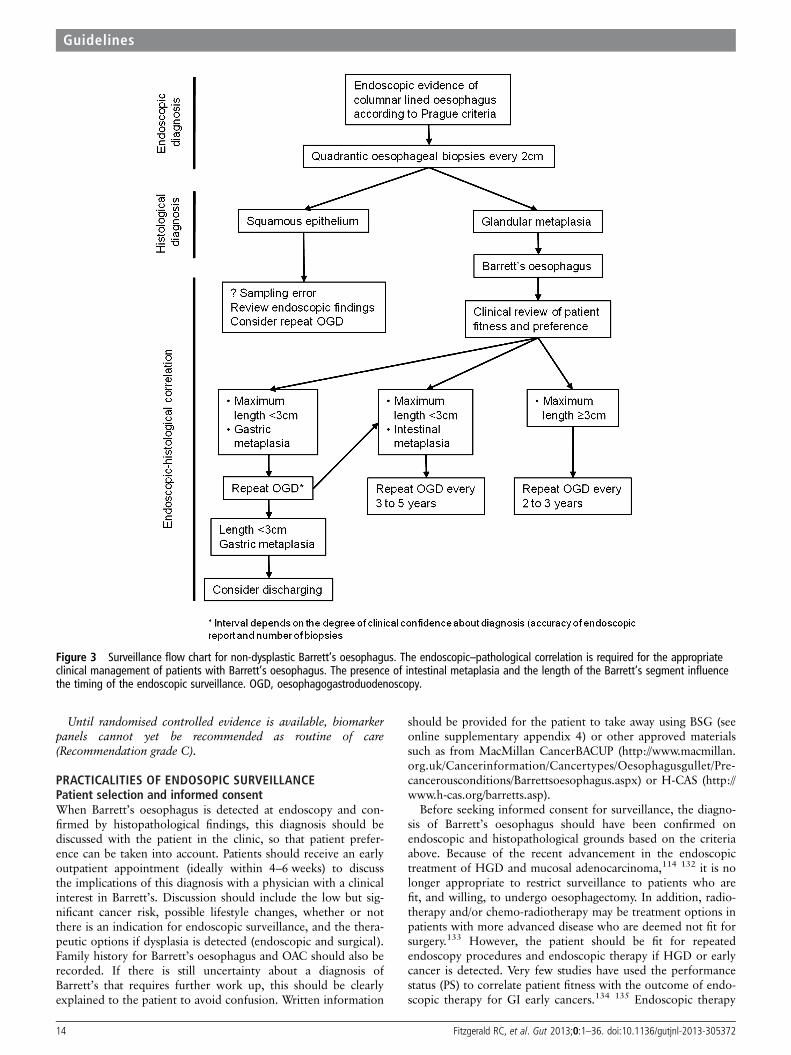
Until randomised controlled evidence is available, biomarkerpanels cannot yet be recommended as routine of care(Recommendation grade C).
PRACTICALITIES OF ENDOSOPIC SURVEILLANCEPatient selection and informed consentWhen Barrett’s oesophagus is detected at endoscopy and con-firmed by histopathological findings, this diagnosis should bediscussed with the patient in the clinic, so that patient prefer-ence can be taken into account. Patients should receive an earlyoutpatient appointment (ideally within 4–6 weeks) to discussthe implications of this diagnosis with a physician with a clinicalinterest in Barrett’s. Discussion should include the low but sig-nificant cancer risk, possible lifestyle changes, whether or notthere is an indication for endoscopic surveillance, and the thera-peutic options if dysplasia is detected (endoscopic and surgical).Family history for Barrett’s oesophagus and OAC should also berecorded. If there is still uncertainty about a diagnosis ofBarrett’s that requires further work up, this should be clearlyexplained to the patient to avoid confusion. Written information
should be provided for the patient to take away using BSG (seeonline supplementary appendix 4) or other approved materialssuch as from MacMillan CancerBACUP (http://www.macmillan.org.uk/Cancerinformation/Cancertypes/Oesophagusgullet/Pre-cancerousconditions/Barrettsoesophagus.aspx) or H-CAS (http://www.h-cas.org/barretts.asp).
Before seeking informed consent for surveillance, the diagno-sis of Barrett’s oesophagus should have been confirmed onendoscopic and histopathological grounds based on the criteriaabove. Because of the recent advancement in the endoscopictreatment of HGD and mucosal adenocarcinoma,114 132 it is nolonger appropriate to restrict surveillance to patients who arefit, and willing, to undergo oesophagectomy. In addition, radio-therapy and/or chemo-radiotherapy may be treatment options inpatients with more advanced disease who are deemed not fit forsurgery.133 However, the patient should be fit for repeatedendoscopy procedures and endoscopic therapy if HGD or earlycancer is detected. Very few studies have used the performancestatus (PS) to correlate patient fitness with the outcome of endo-scopic therapy for GI early cancers.134 135 Endoscopic therapy
Figure 3 Surveillance flow chart for non-dysplastic Barrett’s oesophagus. The endoscopic–pathological correlation is required for the appropriateclinical management of patients with Barrett’s oesophagus. The presence of intestinal metaplasia and the length of the Barrett’s segment influencethe timing of the endoscopic surveillance. OGD, oesophagogastroduodenoscopy.
14 Fitzgerald RC, et al. Gut 2013;0:1–36. doi:10.1136/gutjnl-2013-305372
Guidelines

can be safely performed in patients with Eastern CooperativeOncology Group PS 0–2.136 Therefore it is reasonable to con-sider endoscopic surveillance in patients with PS 0–2, providedthat the estimated patient life expectancy is sufficiently long forthe individual to benefit from surveillance if dysplasia or earlycancer were detected.
If surveillance is thought to be clinically indicated, then theclinician should discuss with the patient the possible benefits ofsurveillance in detecting early-stage tumours and improvingcancer survival. However, this discussion should also mentionthe lack of randomised controlled data to prove the benefits ofsurveillance, and clinicians must also emphasise to the patientthat the actual risk of death from oesophageal cancer is small.Furthermore, the disadvantages of endoscopy surveillanceshould also be discussed, including the small risks of the proced-ure26 and the associated psychological morbidity.137 Forexample, in an American study conducted in a population ofVeterans with a diagnosis of Barrett’s, more than half of thepatients missed their follow-up endoscopy, suggesting that notall patients are willing to adhere to surveillance programmes.138
Clinicians should also emphasise that, as with any monitoringprogramme, there is a failure rate, in that surveillance cannotguarantee to detect every tumour that may develop. There areno clear data to support how best to impart this complex infor-mation, and more work in this area is warranted.
Patients should have early access to an outpatient clinic to beinformed about a new diagnosis of Barrett’s oesophagus and tohave an initial discussion about the pros and cons of surveillancewith written information provided (Recommendation grade C).
For a given patient, whether or not surveillance is indicatedshould be determined on the basis of an estimate of the likeli-hood of cancer progression and patient fitness for repeat endosco-pies, as well as patient preference (Recommendation grade C).
Endoscopic assessmentTechnological advancement with new-generation charge coupleddevices has allowed the routine use of high-resolution endos-copy (HRE), which produces images with resolutions rangingfrom 850 000 to more than one million pixels. HRE allows finedefinition of the mucosal layer for the recognition of subtlesuperficial abnormalities, with theoretical advantage in the rec-ognition of dysplasia and Barrett’s oesophagus-related early
neoplasia. It is the opinion of the experts that HRE, in conjunc-tion with careful cleaning of the mucosal surface of mucus,saliva and food debris, is the minimum standard for the evalu-ation of patients with known Barrett’s oesophagus4; however, todate, there is no randomised trial comparing conventionalendoscopy with HRE in Barrett’s oesophagus dysplasia detec-tion (Evidence grade IV). In an RCT, HRE performed equallycompared with chromoendoscopy and narrow band imaging(NBI) in the overall diagnosis of dysplasia139 (Evidence gradeIb). Mucolytic agents (eg, 4–10% N-acetylcysteine) or antifoam-ing agents (eg, simethicone) can be used to disperse excessmucus and bubbles. There is also evidence that longer inspec-tion times during assessment with white light endoscopy is asso-ciated with an increased detection rate for HGD and earlycancer140 (Evidence grade III). This should be taken intoaccount when planning how much time to allocate for endo-scopic surveillance of very long segments of Barrett’s, particu-larly those longer than 10 cm.
Although transnasal endoscopy has been shown to be accuratein the diagnosis of Barrett’s oesophagus (Evidence grade Ib), therandomised studies performed so far either included a smallnumber of patients,14 or were performed in a low-risk popula-tion.13 Furthermore, it should be noted that the biopsy speci-mens taken with these endoscopes are significantly smaller,13
and this may increase sampling bias and hamper the interpret-ation of dysplasia. Therefore there is currently a lack of robustdata to recommend transnasal endoscopy in routine Barrett’soesophagus surveillance.
HRE should be used in Barrett’s oesophagus surveillance(Recommendation grade C).
There is insufficient evidence to recommend transnasal endos-copy as a replacement for transoral endoscopy (Recommendationgrade C).
Use of chromoendoscopy and advanced endoscopic imagingAdvanced endoscopic imaging has been investigated to increasethe detection of both IM and dysplasia in Barrett’s oesophaguswith the aim to help target biopsies (table 7).
Chromoendoscopy uses dyes to enhance endoscopic detec-tion. Methylene blue (MB) is a vital dye actively absorbed bycolumnar intestinal-type cells141 and has been used to improvethe yield of IM in Barrett’s oesophagus142–144 (Evidence grade
Table 6 Studies investigating correlation of abnormal p53 expression by immunohistochemistry and cancer risk in Barrett’s oesophagus
Study Finding Sample size EDRN stageGrade ofevidence
Weston et al 2001359 Kaplan–Meier curves differed significantly between p53-positive and-negative patients for outcome defined as progression of LGD
Progressors n=5,non-progressors n=43
Prospectivephase 4
IIa
Murray et al 2006196 OAC/HGD end point: OR 8.42 (95% CI 2.37 to 30.0) Progressors n=35, controlsn=175
Phase 3:retrospective
IIa
SIkkema et al 2009198 HR 6.5 (95% CI 2.5 to 17.1) Remained a risk factor on multivariableanalysis
Progressors n=27,non-progressors n=27
Prospectivephase 4
IIa
Younes et al 1997370 Progression from LGD to HGD/OAC, p=0.0108. p53 accumulation has asensitivity of 100%, specificity of 93%, and a predictive value of a positivetest of 0.56
Progressors n=5,non-progressors n=25
Phase 3:retrospective
IIa
Skacel et al 2002130 Progression from LGD to HGD/OAC. A correlation with clinical progressionwas seen, p=0.017 (88% sensitivity and 75% specificity for progression)
Progressors n=8,non-progressors n=8
Phase 3:retrospective
IIa
Bani-Hani 2000197 OR=2.99 (95% CI=0.57 to 15.76; p=0.197). Nested case–control(unmatched), n=12 cases
Phase 3:retrospective
IIa
Kastelein 2012131 RR=6.2 (95% CI=3.6 to 10.9) Progressors n=49,non-progressors n=586
Phase 3:retrospective
IIa
HGD, high-grade dysplasia; HR, hazard ratio; LGD, low-grade dysplasia; OAC, oesophageal adenocarcinoma; OR, odds ratio.
Fitzgerald RC, et al. Gut 2013;0:1–36. doi:10.1136/gutjnl-2013-305372 15
Guidelines

III). In a historical cohort, Sharma and coworkers found signifi-cant enrichment of IM in MB-targeted biopsy samples com-pared with random samples145 (Evidence grade III). Thedetection rate of IM and dysplasia during MB chromoendo-scopy has been investigated in a number of randomised andcohort studies with conflicting data14–151 (table 7). A recentmeta-analysis has found no incremental yield of both IM anddysplasia with MB chromoendoscopy compared with standardendoscopy with random samples152 (Evidence grade Ia). Itshould also be noted that MB may damage DNA, which,coupled with the lack of evidence for efficacy, suggests that itsuse cannot be recommended153 (Evidence grade III).
Indigo carmine (IC) is a contrast agent that allows detailedinspection of the mucosal pattern in combination with magnifi-cation endoscopy.154 A prospective multicentre study found thatthe ridged/villous pattern had a 71% sensitivity for IM, whilethe irregular/distorted pattern had an 83% sensitivity and an88% specificity for HGD/early cancer155 (Evidence grade III).The limitation of IC chromoendoscopy is the need for highmagnification with consequent narrow field of view. Only onerandomised trial has evaluated IC chromoendoscopy for detec-tion of dysplasia in Barrett’s, but failed to find an increased rateof dysplasia compared with high-resolution white light endos-copy139 (Evidence grade Ib).
The value of acetic acid (AA) to improve the diagnostic yieldof surveillance endoscopy has also been studied. AA inducesintracellular protein denaturation, with swelling of the mucosalsurface and enhancement of the architecture. Randomised cross-over studies have produced contradictory results on the diagnos-tic yield of AA-enhanced magnification endoscopy for IM156 157
(Evidence grade Ib). AA-enhanced magnification endoscopy hasbeen shown to have a higher dysplasia yield in Barrett’soesophagus surveillance, with 24% of patients having histo-logical upgrade compared with a previous standard endoscopywith random biopsies performed in a non-specialist centre158
(Evidence grade III). In a large single-centre prospective study,Pohl et al159 found that AA-targeted biopsies had a sensitivity of96.7% and a specificity of 66.5% for a diagnosis of HGD/earlycancer. A single-centre retrospective cohort study has showed
significantly increased dysplasia yield (p=0.001) compared withstandard endoscopy with random biopsies160 (Evidence gradeIII). The same group showed that histology on AA-targetedbiopsies was more cost-effective than the Seattle protocol in ahigh-risk population.161 More data are needed to decide on theusefulness of this technique.
With recent technological advancements, ‘virtual chromoendo-scopy’ has become available, which allows chromoendoscopywithout the use of dyes. This is based on light filters (NBI,Olympus) or post-image acquisition processing (i-scan, Pentaxand FICE, Fujinon). The most extensively studied ‘virtual chro-moendoscopy’ technique in Barrett’s oesophagus is NBI, whichhighlights the mucosal pattern and the superficial vasculature.A number of different classifications have been proposed todescribe mucosal pits in non-dysplastic and dysplastic Barrett’s,which yielded high diagnostic accuracy162–164 (Evidence gradeIII). When NBI was compared with standard imaging techniques,one prospective tandem study showed an incremental diagnosticyield for dysplasia in the per-patient analysis165 (Evidence gradeIIa), and two additional studies reported an increased dysplasiadetection only in the per-biopsy analysis139 166 (Evidence gradeIb). A meta-analysis of eight studies has found that NBI has a sen-sitivity and specificity of 96% and 94%, respectively, for the diag-nosis of HGD, and 95% and 65%, respectively, for the diagnosisof IM.167 However, the interobserver agreement for the inter-pretation of the NBI images is only moderate.168 Overall, despitethe finding that NBI performed by an expert endoscopist mayincrease the targeted yield of dysplasia, it also transpires thathigh-resolution endoscopy alone is sufficient to maximise dyspla-sia detection on a per-patient basis.
Autofluorescence imaging (AFI), which exploits endogenousfluorophores excited by short wavelengths, has been studied inthe context of Barrett’s oesophagus.169 170 Initial single-centrecohort studies showed that AFI can improve the diagnostic yieldof dysplasia compared with standard endoscopy, but with afalse-positive rate as high as 80%171 172 (Evidence grade III). Toovercome this, AFI has been incorporated into an HRE-NBIscope with magnification, also known as endoscopic trimodalimaging (ETMI). Although an initial multicentre non-
Table 7 Comparative studies between standard and advanced imaging techniques for the diagnosis of IM and dysplasia in Barrett’soesophagus
Study Year Technique Study designNo ofpatients
Increased detectionof IM
Increased detectionof dysplasia
Grade ofevidence
Sharma et al145 2001 MB Cohort 158 p=0.024 N/A IIbCanto et al147 2000 MB Randomised MB vs WLE 43 p=0.0001 p=0.03 IbRagunath et al150 2003 MB Randomised cross-over 57 p=0.032 p=ns IbWo et al151 2001 MB Randomised cross-over 47 p=ns p=ns IbHorwhat et al149 2008 MB Randomised cross-over 48 p=ns p=ns IbGossner et al146 2006 MB Cohort cross-over 86 N/A p=0.053 IIaLim et al148 2006 MB Randomised cross-over 30 N/A p=0.02* IbNgamruengphong et al152 2009 MB Meta-analysis 450 p=ns p=ns IaKara et al139 2005 IC/NBI Randomised cross-over 28 N/A p=ns IbHoffman et al156 2006 AA Randomised cross-over 31 p<0.001 N/A IbFerguson et al157 2006 AA Randomised 137 p=ns N/A IbLongcroft-Wheaton et al160 2010 AA Retrospective cohort 190 N/A p=0.001 IIIWolfsen et al165 2008 NBI Cohort tandem 65 N/A p<0.001 IIaSharma et al166 2013 NBI Randomised cross-over 123 N/A p=ns IbCurvers et al174 2010 ETMI Randomised cross-over 87 N/A p=ns IbCurvers et al175 2011 ETMI Randomised cross-over 99 N/A p=ns Ib
*Higher yield of dysplasia in WLE with random biopsies compared with MB chromoendoscopy.AA, acetic acid; ETMI, endoscopic trimodal imaging; IC, indigo carmine; IM, intestinal metaplasia; MB, methylene blue; NBI, narrow band imaging; WLE, white light endoscopy.
16 Fitzgerald RC, et al. Gut 2013;0:1–36. doi:10.1136/gutjnl-2013-305372
Guidelines

randomised feasibility study showed that ETMI increased thediagnostic yield for dysplasia from 63% to 90% compared withstandard endoscopy with random biopsies173 (Evidence gradeIII), this was not confirmed in two subsequent multicentre ran-domised studies, where ETMI only improved the diagnosticyield of dysplasia in the per-biopsy analysis174 175 (Evidencegrade Ib). Overall, these studies showed that, in selected high-risk cohorts of patients, ETMI does not allow the requirementfor random biopsies to be abandoned. Further studies inlow-risk patients will inform whether AFI can have a role inreducing the number of biopsies without loss of diagnosticaccuracy.
Other imaging techniques that have showed some value inBarrett’s oesophagus include confocal laser endomicroscopy,spectroscopy and optical coherence tomography170 176–178;however, further studies are needed to clarify whether they canimprove diagnostic accuracy during Barrett’s oesophagussurveillance.
In the future, molecular imaging may improve our imagingarmamentarium to increase dysplasia detection. Molecularimaging exploits fluorescently labelled molecules that bind withdifferent affinity to dysplastic compared with non-dysplasticcells. Two types of compound have been studied so far. In aproof-of-principle study, Li and colleagues identified a 7-aminoacid peptide that binds an OAC cell line more avidly than a non-dysplastic Barrett’s oesophagus cell line, and they confirmed thedifferential binding in surgical specimens of OAC ex vivo.179
Similarly, Bird-Lieberman and coworkers identified a naturallectin (wheat germ agglutinin) that differentially binds surfaceglycoproteins of dysplastic and non-dysplastic cells and used anautofluorescence endoscope in surgically resected oesophagi tovalidate the ex vivo findings.180 In vivo studies are needed tovalidate these techniques.
Advanced imaging modalities, such as chromoendoscopy or‘virtual chromoendoscopy’, are not superior to standard white lightendoscopy in Barrett’s oesophagus surveillance and are thereforenot recommended for routine use (Recommendation grade A).
Biopsy protocolTo find dysplasia, endoscopists have generally relied on thedirected sampling of any visible lesions, which may be aided byenhanced endoscopic visualisation tools as discussed above,together with systematic, four-quadrant biopsies every 2 cmaccording to the so-called ‘Seattle protocol’.26 A prospectivestudy has demonstrated a significant increase in the detection ofearly lesions through the introduction of such a protocol181
(Evidence grade III). However, adherence to this protocol islimited and ranges from 10% to 79%, with poorer adherencefor longer segments,182–184 and failure to adhere to the protocolhas been shown to result in a significantly lower rate of dysplasiadetection.185 Overall, although intense and time-consuming, themultiple biopsies involved in the Seattle protocol have beendemonstrated to be safe when performed by experienced endos-copists.26 Another limitation of this technique is the high costgenerated by processing multiple biopsies, but this still seemsjustified at the current time in the absence of an alternative.Future RCTs will need to compare cost-effectiveness of thestandard practice with alternative techniques such as histologyon targeted biopsy samples guided by conventional or virtualchromoendoscopy.
Adherence to a quadrantic, 2 cm biopsy protocol in additionto sampling any visible lesions is recommended for all patientsundergoing surveillance. This should also apply to long segments(Recommendation grade B).
Frequency of surveillance for non-dysplasticBarrett’s oesophagusIn the previous BSG guidelines published in 2005, the recom-mended surveillance interval for non-dysplastic Barrett’s wasevery 2 years. However, given the recent data suggesting thatthe overall risk of malignant conversion is lower than previouslythought, we recommend that the interval should be lengthenedin line with other guidelines.3 We therefore advocate a new sur-veillance strategy whereby the managing clinician synthesises theendoscopic and histopathological findings to tailor the surveil-lance interval on a more individual basis (figure 3). A degree ofvariation in this interval is permitted, which may be influencedby the presence of risk factors for the development of cancer.
To summarise, in practical terms, short segments of columnarepithelium with no IM have an extremely low risk of malignantconversion37 91 (∼0.05% per annum) (Evidence grade III). Forthese patients, it is recommended to repeat the endoscopy oncein 3–5 years time to confirm the findings and account for sam-pling and measurement error. If there is doubt, the endoscopycould be repeated sooner. If two good-quality endoscopies, eachwith a minimum number of four oesophageal biopsies wherepossible, confirm a short segment (<3 cm) with gastric metapla-sia only, then discharge is encouraged, as the risks of endoscopyprobably outweigh the benefits. In selected cases with a strongpersonal risk profile for OAC (see recommendation on screen-ing), continued endoscopic surveillance can be considered.
For patients with Barrett’s oesophagus shorter than 3 cm,without IM or dysplasia, a repeat endoscopy with quadranticbiopsies is recommended to confirm the diagnosis. If repeatendoscopy confirms the absence of IM, discharge from surveil-lance is encouraged, as the risks of endoscopy probably outweighthe benefits (Recommendation grade C).
There is evidence that the risk of cancer progression corre-lates significantly with the length of the Barrett’s segment, suchthat segments shorter than 3 cm have a lower cancer incidence(table 5). Therefore, in view of the recent evidence supporting alower cancer risk in non-dysplastic Barrett’s oesophagus thanpreviously thought, it is reasonable for patients with short seg-ments containing IM to have a longer endoscopic surveillanceinterval than patients with long segments. We propose a rangeof 3–5 years to allow the clinician to tailor surveillance on theperceived individual cancer risk.
Patients with Barrett’s oesophagus shorter than 3 cm, with IM,should receive endoscopic surveillance every 3–5 years(Recommendation grade C).
For longer segments (>3 cm), a shorter surveillance interval ismore appropriate. This is regardless of the presence of IM, sinceit is noted, that in long segments, IM is almost always present,but can be missed due to sampling error. We propose a range(between 2 and 3 years), which may be informed by the individ-ual risk factors and patient and physician preference. Because ofthe poor adherence to the surveillance biopsy protocol for longsegments of Barrett’s oesophagus, consideration should be givento refer patients with a very long segment (>10 cm) to tertiaryreferral centres for endoscopic surveillance, as suggested also inthe new Dutch guidelines (personal communication).
Patients with segments of 3 cm or longer should receive sur-veillance every 2–3 years (Recommendation grade C).
Histopathological diagnosis of dysplasiaPathological features and reporting of dysplasiaOnline supplementary appendix 3 shows histological examplesof Barrett’s with different degrees of dysplasia.
Fitzgerald RC, et al. Gut 2013;0:1–36. doi:10.1136/gutjnl-2013-305372 17
Guidelines
There are very few studies that investigated reporting of dys-plasia in Barrett’s oesophagus. Two studies examined the Viennaclassification and found a degree of agreement among patholo-gists that was at best moderate for HGD, fair for LGD, andpoor for indefinite for dysplasia.126 186 The approach to report-ing upper GI tract neoplasia differs significantly in certain partsof the world and this has led to inconsistency in the termin-ology used and hence inconsistent data on incidence and clinicalprogression outcomes. The most recent recommendations bythe WHO state that dysplasia should be graded as either low orhigh grade.187 The revised Vienna classification for GI mucosalneoplasia attempts to standardise diagnostic terminology intobiologically similar groupings with scores of 1–5188 dependingon the presence or absence of dysplasia or malignancy.
Revised Vienna classification and dysplasia subtypes
1. Negative for dysplasiaThis includes normal epithelium, metaplastic epithelium
showing reactive or regenerative changes, and mucosa showingreactive/regenerative changes including nuclear enlargement,nuclear hyperchromasia and prominent nucleoli.
2. Indefinite for dysplasiaThis category is used for cases where the morphological fea-
tures between true dysplasia and regenerative/inflammatoryatypia are blurred.189 It is important to appreciate that this diag-nosis may in fact mean that the patient has features suspiciousof HGD, but not enough certainty is present to warrant thiscall. This may be due to technical factors, such as poor staining,poor orientation, cross cutting or denuded surface epithelium,or to severe active inflammation or ulceration leading to markedatypia, precluding a confident diagnosis of dysplasia. In othercases, the epithelium appears abnormal, but the features are notsufficiently well developed to justify a definite diagnosis of dys-plasia. Features favouring dysplasia are the presence of anabrupt transition from normal to atypical epithelium, togetherwith nuclear pleomorphism, atypical mitoses and loss of nuclearpolarity.190 Evidence of ‘surface maturation’—that is, loss of thecytological atypia seen in the deeper glands as the mucosamatures into the surface epithelium—is often taken as the bestmarker to favour regeneration rather than dysplasia, althoughthis is also not invariably true (eg, crypt dysplasia describedbelow). Explicit mention in the pathology report of the reasonjustifying this diagnosis can be useful to aid patientmanagement.
3. Low-grade dysplasiaIn LGD, glandular architecture is relatively preserved and the
diagnosis is made on the basis of cytological atypia.
Morphological patterns of LGD▸ LGD generally shows an ‘adenomatous’ cytological appear-
ance (resembling the dysplastic changes associated with aden-omatous polyps of the colon) in which nuclei are elongated(pencil shaped), slightly enlarged and hyperchromatic withinconspicuous nucleoli. There may be mild pleomorphism,mucin depletion, mild loss of polarity, nuclear crowding, andstratification of nuclei up to three-quarters of the height ofthe cell, but not touching the luminal surface. Mitoses andapoptotic debris may be seen on the surface or in the upperportions of the glands. Evidence of loss of ‘surface matur-ation’—that is, presence of cytological atypia seen in thedeeper glands—into the surface epithelium is often taken asthe best marker to distinguish true dysplasia from
regenerative atypia; however, in the presence of ulceration,regenerative surface epithelium may also closely mimic LGD.
▸ A ‘non-adenomatous’ (foveolar) type composed of smallround cells with abundant cytoplasm may occasionally beseen.191 Although this is less well characterised, cells withnuclear/cytoplasmic ratios <50% are probably best put intothis low-grade category.
4a. HGD (incorporating carcinoma in situ)The distinction between HGD and LGD is largely based on thepresence of architectural changes in conjunction with moremarked nuclear atypia. These changes may be accompanied bycomplex architectural changes including a papillary or villoussurface (although villiform change may also be seen in reactiveepithelium), in conjunction with branching, complex buddingor back-to-back ‘crowding’ arrangements. Intraluminal papillae,bridges or cribriform patterns are also seen. There are increasednumbers of atypical mitoses on upper levels of crypts, togetherwith mucin depletion and a loss of nuclear polarity. Of note,HGD can be accompanied by acute inflammation and shouldnot be downgraded in its presence.190
Morphological patterns of HGD▸ ‘Adenomatous’ cytological appearance: nuclei are elongated,
pencil shaped, enlarged, hyperchromatic and show crowdingand stratification up to the luminal surface of the cells. Thedistinction between the upper end of ‘low grade’ and ‘highgrade’ dysplasia can be subjective.
▸ ‘Non adenomatous’, which includes the term foveolar dys-plasia: cells have a cytological appearance characterised byrounded nuclei showing marked nuclear enlargement andmarked atypia with increased nuclear/cytoplasmic ratios,irregular nuclear membranes (including angular edges),coarse chromatin, and prominent or irregular nucleoli. Thefoveolar type may have a more bland appearance, comprisingsmall round nuclei with conspicuous nucleoli. The gradingof this variant is less well characterised; however, thenuclear/cytoplasmic ratio appears to be more important forgrading, with high ratios (nuclei involving >50% of cell)being put into a high-grade category.
Crypt dysplasiaSignificant cytological atypia in the crypt bases with surface mat-uration has been reported in up to 7.3% of cases of Barrett’s.192
Previously, this would have been regarded as either ‘negative fordysplasia’ or ‘indefinite for dysplasia’, as the atypia does notreach the surface epithelium. It seems likely, however, that cryptdysplasia represents an early stage in the development of dyspla-sia, and the atypia is highly likely to progress up to the surfaceover time and so warrants recognition.193 Crypt dysplasia standsout as a focus that is distinctly different from the surroundingcrypts and can appear as low-grade or high-grade cytologicalatypia. Although the dysplasia can be of the ‘adenomatous’ or‘non-adenomatous’ round cell type, the most common featuresare nuclear enlargement, loss of polarity, marked pleomorphismwith irregular shapes and sizes, nuclear crowding, increasedmitotic activity and goblet cell dystrophy. Crypt dysplasiashould not be diagnosed purely on the basis of stratification andhyperchromasia in the absence of significant nuclear pleomorph-ism, as these changes may often be seen in regenerative cryptalepithelium. p53 immunohistochemistry may be a helpfuladjunct for the assessment of crypt dysplasia. Crypt dysplasiashould be reported according to the degree of dysplasia present.If there is uncertainty, then the ‘indefinite’ category may be
18 Fitzgerald RC, et al. Gut 2013;0:1–36. doi:10.1136/gutjnl-2013-305372
Guidelines

appropriate. Some pathologists favour putting cases with high-grade features into an ‘at least low grade’ category in view ofthe likely early nature of the lesion and the implications of ahigh-grade diagnosis; however, cases of isolated crypt dysplasiaare probably best managed as low grade until further databecome available.193
4b. Intramucosal carcinoma (including suspicious for invasivecarcinoma)
Intramucosal carcinoma is a lesion in which neoplastic cellshave penetrated the basement membrane and invaded thelamina propria or muscularis mucosae, but without invasioninto the submucosa. However, histological recognition oflamina propria invasion may be difficult because of the absenceof objective criteria. Patterns of lamina propria invasion that areused by gastrointestinal pathologists include sheets of neoplasticcells, abortive angulated glands, a never-ending/anastomosinggland pattern, a highly complex cribriform arrangement ofglands, tightly packed small tubular glandular arrays, and single-cell infiltration. Recognition of each of these patterns is some-what subjective, with κ statistics varying between 0.21 and 0.47,suggesting poor or, at best, moderate agreement.126 186 If defini-tive submucosal invasion is in question, the term ‘suspicious ofinvasive carcinoma’ can be used.
5. Submucosal invasion by adenocarcinomaUnequivocal invasion of submucosa or deeper structures
often accompanied and characterised by a desmoplastic responsein the tissue stroma to invasive tumour cords/acini.
Given the important management implications for a diagnosis ofdysplasia, we recommend that all cases of suspected dysplasia arereviewed by a second GI pathologist, with review in a cancer centreif intervention is being considered (Recommendation grade C).
Given the difficulties associated with the management of the‘indefinite for dysplasia’ category, all such cases should alsobe reviewed by a second GI pathologist, and the reasons for useof the ‘indefinite for dysplasia’ category should be given in thehistology report in order to aid patient management(Recommendation grade C).
Dysplasia reporting and reproducibilityIt has long been recognised that there is inter- andintra-observer variability in the diagnosis of GI tract dysplasia.This relates to differentiating between HGD and intramucosaladenocarcinoma, HGD and LGD and also distinguishingbetween regenerative changes and LGD. In the case of definitedysplasia, this is because these divisions involve unnaturalcut-offs along a biological/histological continuum.126 Studieshave shown that the prediction of progression of oesophagealdysplasia is improved if at least two expert pathologists agree onthe diagnosis and increases further when a greater number ofpathologists concur with the diagnosis.125 130 For practicalreasons, and in day-to-day diagnostic practice, a diagnosis ofdysplasia in the setting of Barrett’s should be corroborated by asecond pathologist with a specialist GI interest. The RoyalCollege of Pathology recommends that ‘double’ reporting of adiagnosis of HGD in the upper GI tract should be mandatoryand this has been confirmed by consensus statements agreed byBarrett’s international experts.4 We have extended this consen-sus reporting to all grades of dysplasia.
Aids to histological diagnosis of dysplasia and p53immunostainingOf all the putative experimental molecular markers, the onewith the greatest body of evidence and which can also be
applied in the routine clinical setting is immunohistochemistryfor nuclear p53. Although the p53 positivity rate in Barrett’soesophagus dysplasia is variably reported in the literature,ranging from 50% to 89%,194 195 when positive it can improveinterobserver agreement for reporting dysplasia126 and can be apowerful predictor of progression, with an OR between threeand eight in different studies129 131 196–198 (table 6). In a studyfrom Skacel et al130, who analysed factors predictive of progres-sion in patient with LGD, p53 immunostaining positivity and100% agreement among three GI pathologists on LGD diagno-sis correlated with the risk of progression, suggesting that p53might improve interobserver agreement. This was replicated in alater study.126 Interpretation of p53 immunostaining can beproblematic and poorly reproducible subject to variation inmethodology and interobserver variation. Notwithstanding this,some pathologists find staining for p53 of use, especially in dis-tinguishing between atypical reactive proliferation (indefinite fordysplasia) and true LGD. Low background wild-type p53expression is often seen in nuclei of normal columnar and basallayers of squamous mucosa, which is a useful baseline to iden-tify the overexpression pattern typical of dysplasia.Overexpression is generally a consequence of mutations that sta-bilise the inactivated protein.199 However, not all p53 mutationslead to stabilisation of a mutated inactive p53 protein. A studyperformed in non-small cell lung cancer showed that, asopposed to missense mutations, the majority of null mutationsdid not lead to p53 overexpression.200 In such cases, mutationis expected to lead to failed translation of the protein. In fact,an absent pattern of p53 immunostaining, when compared withnormal wild-type background, is now recognised as an abnormalpattern which also occurs in dysplasia as a result of silencingmutations of the p53 gene.194 Online supplementary appendix3 shows immunohistochemical examples of Barrett’s with over-expression and loss of p53.
The addition of p53 immunostaining to the histopathologicalassessment may improve the diagnostic reproducibility of adiagnosis of dysplasia in Barrett’s oesophagus and should beconsidered as an adjunct to routine clinical diagnosis(Recommendation grade C).
MANAGEMENT OF DYSPLASIA AND EARLY CANCERIndefinite for dysplasia, a controversial entityThere is very little published literature on the management ofpatients with this histopathological condition. A study lookingat interobserver variability showed that the degree of agreementamong pathologists for a diagnosis of indefinite for dysplasia islower than that for LGD, with κ values of 0.18 and 0.35,respectively186 (Evidence grade III). Younes and coworkersshowed that the rate of cancer progression in patients withindefinite for dysplasia was similar to non-dysplastic patients;however, if the indefinite for dysplasia was multifocal, the rateof progression was as high as in patients with LGD201 (Evidencegrade III). An excess of inflammation is linked to cellular atypia,and this could be resolved by improved medical control of thegastro-oesophageal reflux, although scientific evidence for this islacking (figure 4).
Patients with a diagnosis of indefinite for dysplasia should bemanaged with an optimisation of the antireflux medical therapyand re-endoscoped in 6 months. If no definite dysplasia is found onsubsequent biopsies, then the surveillance strategy should followthe recommendation for non-dysplastic Barrett’s oesophagus(Recommendation grade C).
Fitzgerald RC, et al. Gut 2013;0:1–36. doi:10.1136/gutjnl-2013-305372 19
Guidelines
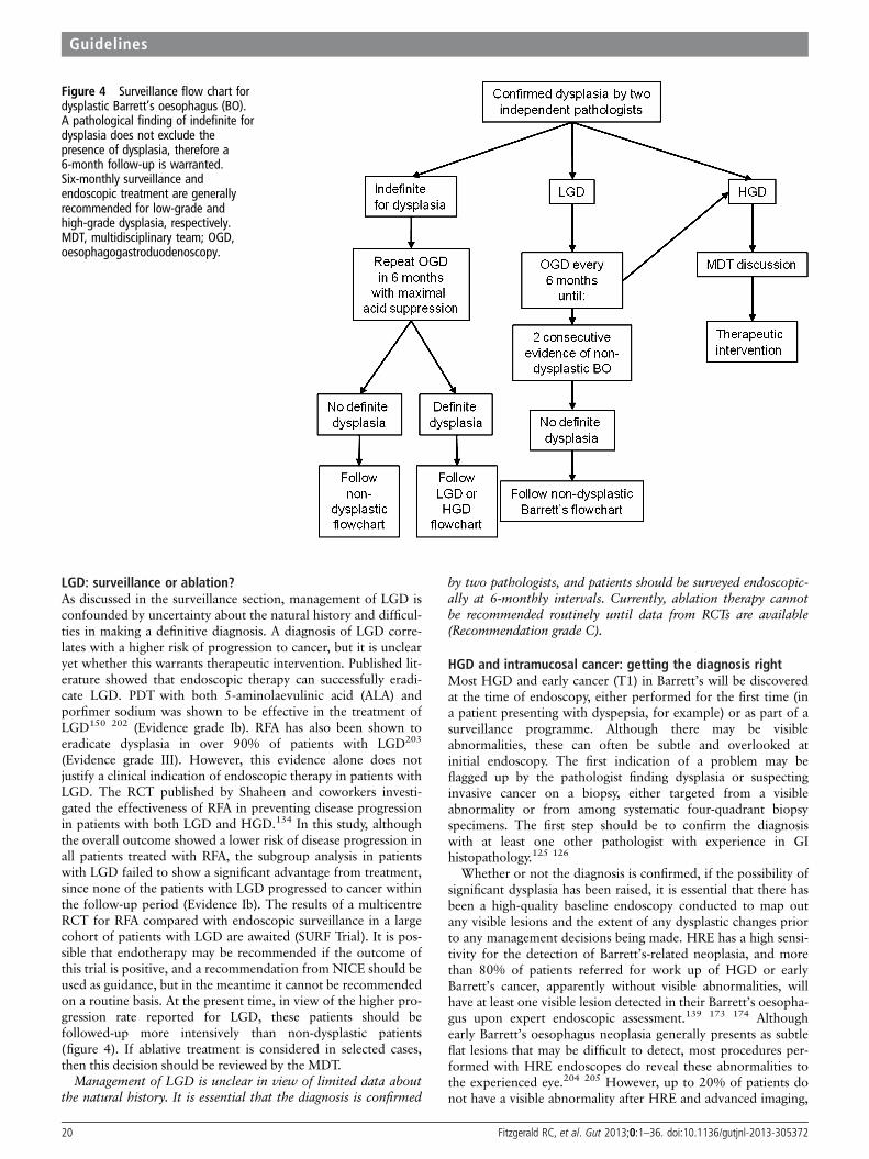
LGD: surveillance or ablation?As discussed in the surveillance section, management of LGD isconfounded by uncertainty about the natural history and difficul-ties in making a definitive diagnosis. A diagnosis of LGD corre-lates with a higher risk of progression to cancer, but it is unclearyet whether this warrants therapeutic intervention. Published lit-erature showed that endoscopic therapy can successfully eradi-cate LGD. PDT with both 5-aminolaevulinic acid (ALA) andporfimer sodium was shown to be effective in the treatment ofLGD150 202 (Evidence grade Ib). RFA has also been shown toeradicate dysplasia in over 90% of patients with LGD203
(Evidence grade III). However, this evidence alone does notjustify a clinical indication of endoscopic therapy in patients withLGD. The RCT published by Shaheen and coworkers investi-gated the effectiveness of RFA in preventing disease progressionin patients with both LGD and HGD.134 In this study, althoughthe overall outcome showed a lower risk of disease progression inall patients treated with RFA, the subgroup analysis in patientswith LGD failed to show a significant advantage from treatment,since none of the patients with LGD progressed to cancer withinthe follow-up period (Evidence Ib). The results of a multicentreRCT for RFA compared with endoscopic surveillance in a largecohort of patients with LGD are awaited (SURF Trial). It is pos-sible that endotherapy may be recommended if the outcome ofthis trial is positive, and a recommendation from NICE should beused as guidance, but in the meantime it cannot be recommendedon a routine basis. At the present time, in view of the higher pro-gression rate reported for LGD, these patients should befollowed-up more intensively than non-dysplastic patients(figure 4). If ablative treatment is considered in selected cases,then this decision should be reviewed by the MDT.
Management of LGD is unclear in view of limited data aboutthe natural history. It is essential that the diagnosis is confirmed
by two pathologists, and patients should be surveyed endoscopic-ally at 6-monthly intervals. Currently, ablation therapy cannotbe recommended routinely until data from RCTs are available(Recommendation grade C).
HGD and intramucosal cancer: getting the diagnosis rightMost HGD and early cancer (T1) in Barrett’s will be discoveredat the time of endoscopy, either performed for the first time (ina patient presenting with dyspepsia, for example) or as part of asurveillance programme. Although there may be visibleabnormalities, these can often be subtle and overlooked atinitial endoscopy. The first indication of a problem may beflagged up by the pathologist finding dysplasia or suspectinginvasive cancer on a biopsy, either targeted from a visibleabnormality or from among systematic four-quadrant biopsyspecimens. The first step should be to confirm the diagnosiswith at least one other pathologist with experience in GIhistopathology.125 126
Whether or not the diagnosis is confirmed, if the possibility ofsignificant dysplasia has been raised, it is essential that there hasbeen a high-quality baseline endoscopy conducted to map outany visible lesions and the extent of any dysplastic changes priorto any management decisions being made. HRE has a high sensi-tivity for the detection of Barrett’s-related neoplasia, and morethan 80% of patients referred for work up of HGD or earlyBarrett’s cancer, apparently without visible abnormalities, willhave at least one visible lesion detected in their Barrett’s oesopha-gus upon expert endoscopic assessment.139 173 174 Althoughearly Barrett’s oesophagus neoplasia generally presents as subtleflat lesions that may be difficult to detect, most procedures per-formed with HRE endoscopes do reveal these abnormalities tothe experienced eye.204 205 However, up to 20% of patients donot have a visible abnormality after HRE and advanced imaging,
Figure 4 Surveillance flow chart fordysplastic Barrett’s oesophagus (BO).A pathological finding of indefinite fordysplasia does not exclude thepresence of dysplasia, therefore a6-month follow-up is warranted.Six-monthly surveillance andendoscopic treatment are generallyrecommended for low-grade andhigh-grade dysplasia, respectively.MDT, multidisciplinary team; OGD,oesophagogastroduodenoscopy.
20 Fitzgerald RC, et al. Gut 2013;0:1–36. doi:10.1136/gutjnl-2013-305372
Guidelines

and, in such cases, four-quadrant biopsies are required to detectHGD.173 174 There is some evidence that magnification and elec-tronic/optical manipulation of the image can increase detectionof dysplasia in Barrett’s oesophagus when compared with HRE,as discussed in the endoscopic surveillance section; however,advanced imaging modalities rarely change the overall diagnosticoutcome.166 175 These imaging techniques can, however, aid def-inition and detailed mapping of the edges of visible lesions inorder to guide complete ER.206 207
Expert HRE should be carried out in all Barrett’s patientswith biopsy-detected HGD in order to detect visible abnormal-ities suitable for ER (Recommendation grade B).
Visible lesions should be considered malignant until provenotherwise (Recommendation grade C).
Any endoscopic abnormalities should be documented usingthe Paris classification, which is based on the Japanese systemused to classify early gastric cancer. It has been used used toclassify mucosal irregularities in patients with Barrett’s oesopha-gus,25 although, unlike the Japanese classification for earlygastric cancer, it has not been validated as a prognostic tool inthis context. Superficial Paris 0-IIa and 0-IIb lesions are unlikelyto contain invasive cancer, while type 0-Is sessile lesions andtype 0-IIc ‘depressed’ lesions are more likely to contain invasivecancer, with up to 25% of cases subsequently being shown tohave submucosal (sm) invasion.204 205 207
Description of lesion morphology using the Paris classificationgives an indication of the likelihood of invasive cancer and aidscommunication between clinicians. This should therefore be usedfor all visible lesions but cannot at present be used to predictprognosis (Recommendation grade C).
Role of specialist teams and decision makingTreatment for HGD and early cancer involves endoscopic orsurgical management. The success of both modalities should bemeasured in terms of morbidity and mortality related to theprocedure (or series of procedures), long-term survival andQOL. These recommendations are entirely consistent with theguidelines for the management of oesophageal and gastriccancer recently published by Allum et al,133 which, however,focus specifically on HGD and early cancer.
Recommendations for treatment of patients withBarrett’s-related neoplasia should be taken in the context ofan upper GI specialist MDT taking into account patientcomorbidities, nutritional status, patient preferences and staging(figure 5).6 Before treatment decisions are made, the patientshould have the opportunity to discuss the options in an out-patient clinic setting (not in endoscopy after sedation), and thismay comprise a joint discussion with an endoscopist and asurgeon. The patient should have access to all the supportingevidence being used to make the recommendation according totheir wishes. Patients should be copied into any clinical corres-pondence, as recommended by NICE guidelines for the man-agement of colorectal cancer.209
All patients with dysplasia or early cancer, for which therapyis considered, should be discussed at the specialist MDT for oeso-phagogastric cancer. This team should include an interventionalendoscopist, upper GI cancer surgeon, radiologist and a GI path-ologist (minimum standard) (Recommendation grade C).
Patients with dysplasia or early cancer should be informed oftreatment options and have access to all specialists if required(Recommendation grade C).
Endoscopic therapy for Barrett’s-related neoplasiaAlthough, surgery remains an effective treatment for Barrett’sneoplasia, it is associated with significant morbidity and mortal-ity compared with endoscopic therapy. Cohort studies suggestthat endoscopic therapy for mucosal OAC has similar long-termdisease-specific survival to surgery, but lower death rates.210–212
A number of retrospective case series comparing endoscopicand surgical therapy have demonstrated high survival outcomesin both groups, but higher short-term mortality in the surgicalgroup than in the endoscopic group, but patients were not wellmatched.212–215 A systematic review showed a mortality of1.2% in the surgical group compared with 0.04% in the endo-scopic group.216 A more recent study showed lower morbidityin the endoscopic group214 (Evidence grade III). A cost-effectiveness study demonstrated that RFA was likely to be morecost-effective than surgery.217 Recent consensus statementsissued by a large group of international experts have indicatedthat endoscopic therapy should be preferred over surgery forthe management of early Barrett’s neoplasia.4
For HGD and Barrett’s-related adenocarcinoma confined tothe mucosa, endoscopic therapy is preferred over oesophagect-omy or endoscopic surveillance (Recommendation grade B).
Endoscopic therapy in the oesophagus carries a low but sig-nificant risk of complication.218 219 It is important that therapyis carried out in centres that have the specialist expertise to offerand the necessary back-up if required. For example, endoscopictherapy should be carried out in centres that can also offersurgery if a complication occurs.
Endoscopic therapy of Barrett’s neoplasia should be performedat centres where endoscopic and surgical options can be offeredto patients (Recommendation grade C).
There are now good observational data to support the per-formance of oesophageal surgery in specialist centres for treat-ment of adenocarcinoma.220 Results for individual surgeonsimprove with experience, and patient outcomes have consist-ently been shown to be better in high-volume centres220 221
(Evidence grade III). ER series reported from expert high-volume centres have shown a low rate of significant complica-tions (<3%),114 212 218 but the complication rate is significantlyhigher in the hands of less experienced endoscopists duringtheir first 20 ER procedures performed.222 Therefore it seemslogical that, similar to the recommendations for oesophagect-omy, endoscopic therapy should also be performed in centreswith the experience, facilities and high throughput of cases tooptimise outcomes. Endoscopic training should start withknowledge acquisition, followed by resection and ablation intissue animal models, before training in human subjects. The GIpathologist should also be trained in the report of ER speci-mens. In order to achieve a good level of training, it is crucialthat professional societies guarantee wide availability of trainingcourses and fellowships for Barrett’s endoscopic therapy.
A minimum of 30 supervised cases of ER and 30 cases ofendoscopic ablation should be performed to acquire competencein technical skills, management pathways and complications(Recommendation grade C).
ER should be performed in tertiary referral cancer centres foroesophageal cancer disease, which must be equipped with high-resolution endoscopes and all relevant ER facilities. These spe-cialist cancer centres should have an adequate volume of cases,which in the absence of published evidence we have set as 15ERs per annum for HGD or early cancer in a given centre basedon the opinion of experts. Centres should preferably have twoendoscopists performing endoscopic therapy, and low-volume
Fitzgerald RC, et al. Gut 2013;0:1–36. doi:10.1136/gutjnl-2013-305372 21
Guidelines

centres should consider developing joint services. The endosco-pist performing endoscopic therapy must be fully trained inlesion recognition and ER techniques, with expertise in at leastone enhanced endoscopy imaging modality (electronic imagingor AA chromoendoscopy). Progression of disease is describedduring treatment with RFA,132 223 therefore an ER may berequired during the ablation pathway.
ER should be performed in high-volume tertiary referralcentres. RFA should be performed in centres equipped with ERfacilities and expertise (Recommendation grade C).
Endoscopic resectionER should always be performed with therapeutic intent;however, histopathological assessment of the ER specimen isalso the most accurate staging technique for Barrett’soesophagus-related early neoplasia.224 225 ER is preferred oversurveillance biopsies because of the difficulty in differentiatingHGD from mucosal cancer and deep invasive cancer on biopsymaterial.4 226 Visible lesions contain the most advanced histo-logical staging in surgical resection specimens, and this is con-firmed by experience with step-wise radical ER of the entire
Barrett’s segment.227 228 This supports a policy of removal ofall visible abnormalities by ER.
Endoscopic assessment will usually identify the area with themost advanced neoplasia. ER should aim to resect all visibleabnormalities (Recommendation grade C).
ER is recommended as the most accurate staging interventionfor Barrett’s early neoplasia (Recommendation grade B).
Tumours confined to the mucosa (T1a) have been shown tohave significantly better 5-year recurrence-free and overall survivalrates (100% and 91%, respectively) than those showing involve-ment of the submucosa (60% and 58%).229 Endoscopic therapyfor Barrett’s neoplasia has been developed on the evidence thatHGD and T1a OAC is associated with a low rate of lymph nodemetastasis: endoscopic and surgical series indicate a 0–10% risk inT1a cancer, while submucosal invasion carries a higher risk (up to46%).215 229–236 A recent systematic review reported no evidenceof nodal metastasis in patients with a final stage of HGD.72
Further stratification of these superficial lesions as in table 8 yieldsadditional prognostic information in that the risk of nodal metasta-sis correlates with the depth of penetration into the layers,229 234
but not all the studies have confirmed this.230 236
Figure 5 Recommended flow chartfor the management of high-gradedysplasia (HGD) and early oesophagealadenocarcinoma (OAC). A diagnosis ofHGD and early OAC should bediscussed in a multidisciplinary team(MDT) setting, and treatment optionsshould be explained in the clinic to thepatient. Endoscopic treatment andsurgery are generally recommended formucosal disease and submucosalcancer, respectively. Good prognosiscancer with involvement of superficialsubmucosal layers (sm1) can betreated endoscopically in patients athigh surgical risk. EC, early cancer;HRE, high-resolution endoscopy; OGD,oesophagogastroduodenoscopy; RFA,radiofrequency ablation.
22 Fitzgerald RC, et al. Gut 2013;0:1–36. doi:10.1136/gutjnl-2013-305372
Guidelines

It is still unclear whether T1sm1 cancer (invading the superfi-cial submucosa within 500 mm) can be confidently treated by ER,and although series from Amsterdam and Wiesbaden suggest thiscarries a low risk of lymph node metastases,230 234 237 not all theliterature supports this.72 236 This debate is further complicatedby the technical challenges faced by the pathologist to accuratelyidentify the submucosal invasion depth on the ER specimen. Theimportance of experienced pathology in deciding whetherendotherapy is likely to be curative or not is critical, since notonly the depth of invasion but features of the tumour (lymphaticor vascular invasion, degree of differentiation) are thought to beimportant in the decision-making process.238 Free deep resectionmargins on the ER specimen (R0), together with an absence oflymphovascular invasion (LVI−) and good differentiation (G1)are all indicative of a good prognosis for early cancer.230
Lymphovascular space invasion and lymph node metastases havebeen reported in 17% and 10% of patients, respectively, withinvasion into the duplicated muscularis mucosae, although theseare technically intramucosal.235
ER should be considered the therapy of choice for dysplasiaassociated with visible lesions and T1a adenocarcinoma(Recommendation grade B).
For patients at high surgical risk, endoscopic therapy can beoffered as an alternative to surgery for treatment of good progno-sis T1b adenocarcinomas (T1b sm1, well differentiated andwithout lymph vascular invasion) (Recommendation grade C).
For T1b adenocarcinomas with involvement of the secondsubmucosal layer or beyond (T1b sm2–sm3), endoscopic therapyshould not be considered curative (Recommendation grade B)
ER techniquesThe basic principle of ER involves identification of the lesionmargins by marking with a diathermy device followed by a sys-tematic resection of the marked area. Common resection techni-ques are cap and snare and band ligation.
The band ligation technique involves suction of the markedarea into a distal attachment (preloaded with multiple rubberbands) and deployment of the band on to the tissue to create apseudo-polyp, which is then resected with a snare. The cap andsnare technique also involves a dedicated transparent cap at thedistal end of the scope, which is preloaded with a crescentsnare. Differently from the band ligation, the area of interest isalways injected (submucosal space) and lifted before its suctioninto the cap. Once the mucosa is placed into the cap, then thesnare can be closed around and the tissue resected.
Both cap and snare and band ligation techniques appear tohave similar success rates of the order of 85–98%.224 239–242
There are two RCTs that compared the two techniques.219 243
These studies did not show significant differences either interms of the depth of resection or complication rates, leading tothe conclusion that both techniques were similarly safe andeffective. Some endoscopists believe that submucosal injectionimproves safety of the ER when an attempt is made to resectnodular lesions, which carry a high risk of invasive cancers. Insuch cases, the presence of the ‘non-lifting sign’ will inform theendoscopist about the possibility of deep invasive cancer and itsassociated risks.
The cap and snare technique with submucosal injection andthe band ligation technique without submucosal injection areconsidered to be equally effective (Recommendation grade A).
Pathology reporting of ER specimensER specimens should be pinned out on a rigid support (eg,cork) and formalin fixed for at least 12 h. Photographs of the
fixed specimens can act as a useful record. They should be seri-ally sectioned at 2 mm intervals along the axis of the closestmargin and completely embedded. Both of the end piecesshould be embedded en face.
The status of the tissue margins is the most significant prog-nostic factor; however, if the sample has been received in mul-tiple pieces, it will not be possible to identify the true lateralmargins, and only deep margins will be relevant.244 Tumoursshould be graded and T staged. T1a and T1b intramucosal car-cinomas can be subclassified as shown in table 8.245 A commentshould be made regarding the presence or absence of lympho-vascular space invasion, background IM and dysplasia. Seeminimum reporting dataset (table 9).
Artefacts such as haemorrhage, heat/cutting artefact, surfaceloss, fixation contraction with rolling and folding of edgesleading to poor orientation of tissue at embedding may allhinder the histological assessment244 (Evidence grade III). Thesuction technique can cause artefactual lifting and/or disruptionof the surface epithelium, with intramucosal haemorrhage,oedema and fibrin deposition on the surface. This should not beconfused with true ulceration. Biopsy samples taken 1–16 daysafter gastric ER can show signet ring cell change and clear celldegeneration in areas of ischaemia.246 Although there are norelevant studies that looked at mucosal changes after ER, tissueregeneration in the immediate post-ER period can hamperproper histological assessment, and therefore it is recommendedto delay post-ER biopsies to 6–8 weeks (Evidence grade IV).
It can be difficult to accurately stage these tumours on ERspecimens, when there is duplication of the muscularis mucosae,a characteristic finding in Barrett’s. This may lead to overstagingby misinterpreting the space between duplicated muscularis assubmucosa, and in some instances it may not even be possibleto determine if true submucosa is present (see online supple-mentary appendix 4). The largest and most recent studiessuggest that invasion into the space between the duplicated mus-cularis mucosae has a similar low risk of lymph node metastasesto lamina propria invasion if other features (lymphovascularinvasion and poor differentiation) are absent230 247 (Evidencegrade III). In addition, it is important to be aware of entrappedglands and submucosal glands that may mimic submucosaladenocarcinoma.
Further, the use of the muscularis propria as a reference pointfor determining depth of submucosal invasion is problematic inERs, as it is generally not represented. In the latter instance, itmay be more appropriate to state that submucosal invasion ispresent and then measure the depth of invasion beyond themuscularis mucosae. In the colon, depth of invasion beyond thesubmucosa is widely used as a marker of likely lymph node
Table 8 Subclassification of T1a and T1b oesophagealadenocarcinoma
Class Description
T1am1 Carcinoma in situ or with questionable invasion beyond
the basement membranem2 Invasion into the lamina propriam3 Invasion into the muscularis mucosa
T1bsm1 Invasion into the upper third of the submucosa within 500 mmsm2 Invasion into the middle third of the submucosasm3 Invasion into the lower third of the submucosa
Fitzgerald RC, et al. Gut 2013;0:1–36. doi:10.1136/gutjnl-2013-305372 23
Guidelines

metastases, but studies in OAC have been conflicting, with dif-ferences between surgical and ER series.229 230 Measurement ofthe distance from the tumour to the nearest deep margin isrecommended to document the adequacy of resection. The pres-ence of tumour cells at the deep margin indicates incompleteresection requiring further treatment.
Use of a minimum dataset for the reporting of ER specimensis recommended to ensure that all prognostic information isincluded in reports (Recommendation grade C).
The presence of tumour cells at the deep margin indicatesincomplete resection and warrants further treatment(Recommendation grade C).
Imaging for HGD and T1 carcinoma: role of CT–PET and EUSThere is little published evidence to support a role for CT (orCT–PET) with regard to early cancer (HGD, mucosal lesions),with most of the evidence being for advanced cancer (seesection on surgical treatment). Where biopsy specimens showinvasive cancer, CT scanning is often advocated in order todetect distant metastases. However, it has poor accuracy in localT staging and it is less accurate than EUS in detecting locallymph node metastases.248 249 PET–CT has a higher accuracythan CT for distant and locoregional metastases, but is inferiorto EUS.250–252
Before ER, neither CT nor PET–CT have a clear role in thestaging of patients with Barrett’s HGD or suspected T1 cancerand neither is routinely required (Recommendation grade B).
Initial evidence that endoscopic resectability (differentiation ofT 1/2) could be predicted by EUS has been tempered by most ofthe recent series, which indicated that 15–25% of cases are under-staged compared with ER, while about 4–12% are overstaged.253–256 One study compared endoscopic assessment with EUS andfound that accuracy of both techniques in predicting depth of inva-sion was similar.257 High-frequency EUS miniprobes are moreaccurate than conventional EUS, but still fail to correctly stage T1cancers in one-third of cases (Evidence grade Ib).258 Overall, thesestudies show that EUS misclassifies the T stage in approximatelyone-quarter of patients with early oesophageal cancer, and this haslimited clinical impact over expert high-resolution imaging plusER. However, in routine clinical practice, the endoscopist may notalways be able to confidently exclude an advanced stage (T>1a)on the basis of the endoscopic appearance of nodular lesions, and,in this circumstance, EUS should be performed to guide the thera-peutic choice.
EUS is the most accurate tool available for detecting regionallymph node involvement and can add staging information inpatients with T1b disease after ER.231 249 259 Endosonographiccriteria that are suggestive of malignant involvement of visiblelymph nodes include a width greater than 10 mm, round shape,smooth border, and echo-poor pattern.260 When all four
suspicious features are present, there is an 80–100% chance ofmetastatic involvement; however, only 25% of malignant nodeswill have all of these features. Therefore, with the use of echocriteria alone, sensitivity and specificity for regional lymph nodemetastases were 80% (95% CI 75% to 84%) and 70% (95% CI65% to 75%), respectively, in this study. FNA can determinemore precisely whether or not suspicious lymph nodes are infil-trated, and it has high sensitivity in coeliac, mediastinal andperigastric lymph nodes.261 Sensitivity, specificity and accuracyof EUS FNA for locoregional lymph nodes are currently allaround 85%262 (Evidence grade III). One cohort study on 25patients with baseline HGD or intramucosal adenocarcinoma onbiopsies has showed that EUS FNA of suspicious lymph nodeschanged management decision in 20% of patients referred forendoscopic therapy of cases263 (Evidence grade III).
Since EUS can both overstage and understage T1 lesions, itsroutine use cannot be recommended for staging before ER forsuspected early lesions (Recommendation grade B).
In selected cases where the endoscopist cannot excludeadvanced stage on the basis of endoscopic appearance of nodularlesions, EUS with or without FNA is recommended to informthe therapeutic decision (Recommendation grade C).
EUS with or without FNA of visible lymph nodes is recom-mended in selected cases with T1b (sm1) disease on staging ERfor which endoscopic therapy is selected, because of the significantrisk of lymph nodal involvement (Recommendation grade C).
Ablative therapy for flat HGD/intramucosal cancerand residual Barrett’s after ERIf HGD or intramucosal cancer are confirmed and there are novisible lesions after expert HRE review, then ablative therapy isthe treatment of choice. After ER of any visible lesions, thestage and extent of disease will have been clarified. If disease isconfined to the mucosa, then endoscopic therapy is preferredfor eliminating the remaining Barrett’s segment. Numerous caseseries on different endoscopic ablative techniques (PDT, argonplasma coagulation (APC), cryotherapy, RFA and step-wiseradical ER) reported eradication rates of HGD ranging from36% to 100%.228 264–270 Some were combined with ER forvisible lesions. Randomised controlled trial data in patients withHGD are available for some of these techniques: PDT vsPPI,271 RFA vs Sham,132 APC vs PDT,272 RFA vs step-wiseradical ER,273 PDT with ALA vs PDT with photofrin (porfimersodium).274 All ablation modalities improve eradication com-pared with surveillance for HGD (Evidence grade Ib), but theyshould only be used as a primary treatment modality in the caseof flat dysplasia.275 The studies with the best outcomes relate toPDT and RFA. ALA PDT has been showed to have a bettersafety and efficacy profile than photofrin PDT, but only inBarrett’s shorter than 6 cm.274 Three-year follow-up data ofRFA for HGD showed a reduced risk of progression to cancercompared with surveillance.132 276 RFA was associated withimprovement in disease-specific health-related QOL,277 and thisimprovement appears secondary to a perceived decrease in therisk of cancer. Endoscopic treatment is associated with a higherrate of recurrence during follow-up; however, recurrent lesionscan be effectively treated by further endoscopic therapy.114 212
RFA has been compared with step-wise ER for eradication ofresidual Barrett’s epithelium after ER for HGD or mucosalOAC with equivalent efficacy, but with a higher stricture rate inthe ER arm273 (Evidence grade Ib). Safety and tolerability weresuperior, with a decreased number of therapeutic sessions in theRFA group (Evidence grade Ib). RFA has not been comparedwith PDT, but patients with failed PDT have been successfully
Table 9 Minimum dataset for reporting endoscopic resectionspecimens
Intestinal metaplasia Yes/noDysplasia grade Indefinite/LGD/HGD/adenocarcinomaDifferentiation Well, moderate or poorly differentiatedT1 subclassification T1a m1–3/T1b sm1–3Lymphovascular space invasion Yes/noDeep margin Positive/negativeDistance to deep margins mmLateral margins (en block resection) Positive/negative
HGD, high-grade dysplasia; LGD, low-grade dysplasia.
24 Fitzgerald RC, et al. Gut 2013;0:1–36. doi:10.1136/gutjnl-2013-305372
Guidelines

treated with RFA.278 PDT has a significant photosensitivity andstricture rate, therefore RFA appears to have a better safetyprofile and is easier to administer. In addition, RFA also hasbeen associated with a lower incidence of buried glands thanPDT and APC.279 280 Effectiveness in dysplasia eradication,safety profile and general aspects of all the techniques inpatients with HGD are summarised in table 10. It transpiresthat the overall efficacy and safety profile support RFA as thebest ablation modality currently available, as long as it is usedfor treatment of flat dysplasia only.
In the presence of HGD or intramucosal cancer withoutvisible lesions (flat HGD/intramucosal cancer), these should bemanaged with an endoscopic ablative technique. There is littlecomparative data among ablative techniques, but RFA currentlyhas a better safety and side-effect profile and comparable efficacy(Recommendation grade C).
More than 20% of patients treated with ER of visible lesionsdevelop metachronous lesions in the Barrett’s segment within2 years.114 Recurrence of neoplasia after ER can be significantlyreduced if the residual Barrett’s is completely ablated114 132 273
(Evidence grade III). After ER for early focal neoplasia, >80%of patients will have HGD or LGD detected in the remainingBarrett’s epithelium.281 Eradication of the remaining Barrett’sepithelium by RFA is a safe and effective treatment for anyremaining flat dysplasia281 282 (Evidence grade III). Ablationmay also be useful to avoid strictures, which are associated withstep-wise radical ER of circumferential lesions, particularly withlonger Barrett’s segments.228 Combining ER with RFA reducesthis risk and is effective in eradicating the remaining Barrett’sepithelium273 (Evidence grade Ib).
Eradication of residual Barrett’s oesophagus after focal ERreduces the risk of metachronous neoplasia and is recommended(Recommendation grade B).
Follow-up after endoscopic therapyThe goal of endoscopic mucosal resection and ablation is to elim-inate the subsequent risk of cancer. This requires long-termfollow-up data, which are not currently available for ablative tech-niques. From the limited studies available after ablation, new squa-mous (neosquamous) epithelium reveals no molecularabnormalities, and seems biologically stable.283 Five-year and3-year follow-up data from the RCTs of PDT and RFA,
respectively, in dysplastic Barrett’s oesophagus showed a durableresponse to treatment.271 276 However, buried metaplasia hasbeen reported within neosquamous epithelium biopsy specimensand this can predispose to the development of so-called ‘buried’cancer.284 Current data suggest that the occurrence of buriedglands is higher in patients treated with PDT (14.2%) comparedwith RFA (0.9%).279 Recurrence of Barrett’s at the GOJ has beenreported and seems important.285 Currently, follow-up is manda-tory not only to detect recurrence but also to allow furthertherapy to be applied as required.4 Empirically, in patients treatedfor HGD, endoscopic follow-up is recommended 3-monthly for1 year and yearly thereafter. This should include biopsies at theGOJ and within the previous extent of the Barrett’s epithelium.
Pathological reporting of biopsies after ablation therapyAblation therapy is associated with an early acute and chronicinflammatory response, together with reactive epithelial changesfollowed by lamina propria fibrosis and duplication and splittingof the muscularis mucosae after a few months.244 Neosquamousepithelium is associated with ablation therapy, and interestinglycan also be seen with PPI treatment without ablation therapy,especially after extensive biopsies.286 287 This neosquamous epi-thelium can replace the Barrett’s segment, forming islands orsheets of ‘normal’ squamous epithelium. However, this mayoverlie buried glands or buried carcinoma.279 A systematicreview has shown an overall incidence of buried glands in14.2% of patients who received PDT and 0.9% of patients whoreceived RFA.279 However one post-RFA study on the neosqua-mous epithelium found that only 37% of biopsy specimens con-tained lamina propria and deeper tissue suitable for assessmentof buried metaplasia,283 whereas the other studies do notmention the presence of lamina propria in neosquamous biopsysamples.290 Buried dysplasia can be difficult to evaluate, particu-larly as the atypia does not reach the surface.244 Again p53immunohistochemistry may be a helpful adjunct, as dysplasticglands may demonstrate a significant staining pattern.126 194 289
If there is uncertainty about the significance of the atypiapresent, then it is best to use the ‘indefinite for dysplasia’ report-ing category. In summary, the importance of buried metaplasiaafter ablation remains unclear, as well as the adequacy of biop-sies to detect it, leaving some uncertainties about the usefulnessof biopsy specimens taken from the neosquamous epithelium.
Table 10 Summary of clinical profiles of different ablative techniques for HGD
Technique Rate of dysplasia eradication Advantages Disadvantages
Step-wise radical ER 97–100% ▸ Accurate diagnosis▸ Low costs
▸ High risk of stenosis▸ Feasible only for Barrett’s oesophagus <5 cm
APC 67–86% ▸ Large availability▸ Low costs
▸ Buried glands▸ Feasible for short segments only
PDT 40–77% ▸ RCT available▸ Treatment of nodular dysplasia
▸ High risk of stricture▸ Buried glands▸ Photosensitivity
RFA 80–98% ▸ RCT available▸ High response rate▸ Low complication rate
▸ High costs▸ No long-term follow-up data
Cryotherapy 68–88% ▸ Good safety profile ▸ Small studies (no RCTs)▸ No long-term follow-up data
APC, argon plasma coagulation; ER,endoscopic resection; PDT, photodynamic therapy; RCT, randomised controlled trial; RFA, radiofrequency ablation.
Fitzgerald RC, et al. Gut 2013;0:1–36. doi:10.1136/gutjnl-2013-305372 25
Guidelines

However, until further evidence becomes available, neosqua-mous epithelium biopsies are warranted.
Endoscopic follow-up is recommended after endoscopictherapy of Barrett’s neoplasia with biopsy samples taken from theGOJ and within the extent of the previous Barrett’s oesophagus(Recommendation grade B).
Surgical management of early Barrett’s neoplasiaEfficacy and safetyFor HGD and T1 OAC, case series suggest that the 5-year sur-vival rates range between 80% and 90%, and the 3-year survivalexceeds 90%.211 214 290 Tumour recurrence after resection ofearly cancer is rare (1%). There are no reports of long-termtumour recurrence in patients who have had complete surgicalresection of HGD alone, and all-cause mortality is equivalent tonon-surgical treatments211 290 291 (Evidence grade III).
Mortality for patients undergoing oesophagectomy for HGDor early adenocarcinoma is difficult to estimate because data arefrom retrospective studies conducted in self-selected high-volume centres, thus the quality of data was assessed as low. Anumber of case series21 292–301 evaluating over 500 patientswith HGD or early adenocarcinoma show an operative mortal-ity for oesophagectomy for HGD and early adenocarcinoma ofless than 5%, with an overall operative 30-day mortality ofapproximately 2%. These results are comparable to the recentUK national audit results, where the mortality for 2200 patientsundergoing oesophagectomy for any operable cancer (HGDexcluded) was 3.8% for 30-day mortality and 4.5% forin-hospital mortality301 (Evidence grade III).
Surgical resection is associated with significant short-termmorbidity (6–37%).290 303 A recent retrospective comparison ofoesophagectomy (61 patients) versus endotherapy (40 patients)for HGD or T1 cancer in one centre revealed much lower mor-bidity in the endotherapy group with a similar survival,although follow-up was relatively short in the endotherapygroup.214
Surgical therapy is considered the treatment of choice for earlyadenocarcinoma that has extended into submucosa because ofthe significant risk of lymph node metastasis (Recommendationgrade B).
Volume effect in specialist surgical centresGood observational data indicate that oesophageal surgery fortreatment of adenocarcinoma should be performed in specialistcentres, since patient outcomes have been shown to be better inhigh-volume centres and individual surgeons’ results improvewith experience220 221 (Evidence grade III). The recent AUGISguidelines on minimum surgeon volumes for oesophagogastricunits recommend that each unit should consist of at least foursurgeons carrying out a minimum of 15–20 resections peryear.221
Oesophagectomy should be performed in high-volume centres,as these are associated with lower in-hospital mortality thanlow-volume centres (Recommendation grade B).
Type of surgerySurgical resection for Barrett’s dysplasia and early cancer mustinclude complete resection of the Barrett’s segment (longitu-dinal margins). Lymphadenectomy should be performed for T1sm tumours because of the significant risk of lymph nodeinvolvement304–306 (Evidence grade III). There is insufficientevidence to support the use of one technique above the others.Attempts have been made to tailor a less radical, less morbidsurgical procedure for these patients with early lesions who do
not require extensive lymphadenectomy. These are not asso-ciated with a detrimental effect on long-term survival. However,the majority of surgical data come from retrospective observa-tional studies, often comparing outcomes with outdated histor-ical controls.
Data directly comparing the various techniques, or fromseries restricted to HGD and intramucosal cancer, are extremelylimited. A series of 36 patients including left thoracoabdominaloesophagectomy (60%), transhiatal oesophagectomy (20%) andIvor Lewis oesophagectomy (20%) reported no operative mor-tality and major complications in only 11% of patients.296 At amean follow-up of 5 years, QOL outcomes were comparable toage- and sex-matched controls. A study of a further series ofpatients with HGD and T1 oesophageal cancer undergoingopen surgery by either the transhiatal or transthoracic routesreports operative mortality of 2.5% and 5-year survival of 77%.This series included patients with tumour-involved lymphnodes, for whom survival is significantly impaired comparedwith patients with HGD307 (Evidence grade III).
Transhiatal oesophagectomyTranshiatal oesophagectomy avoids the need for thoracotomy,with low in-hospital mortality (3%) and length of stay (50% dis-charges in 1 week) in one high-volume centre.308 Concernsabout the adequacy of lymphadenectomy with this proceduremay not apply in the context of HGD and intramucosal cancer.In an RCT, the transhiatal procedure was associated with lessperioperative morbidity than a transthoracic procedure, andthere was no significant difference in perioperative mortality(2% vs 4%, p=0.45).309 Long-term follow-up showed no differ-ence in overall survival when all patients were considered,although a subgroup of patients with between one and eightlymph nodes involved had a better survival in the transthoracicprocedure.310 This trial included patients with operable diseaseof all stages, but does suggest transthoracic oesophagectomy is apreferable procedure where there is a significant risk of lymph-adenopathy such as in T1 sm disease (Evidence grade Ib).
Vagal-sparing oesophagectomy for HGDThe vagal-sparing technique aims to reduce the postoperativedumping and diarrhoea associated with oesophagectomy. Astudy of patients with HGD or intramucosal cancer found thatthe 49 patients who underwent vagal sparing oesophagectomyhad shorter hospital stays and fewer major complications than39 patients who underwent transhiatal procedures and 21 whounderwent en bloc resections.297 Postoperative dumping syn-drome and diarrhoea were decreased in the vagal-sparing group,and there was no detrimental effect on long-term outcomes.The vagal-sparing procedure does not involve a lymphadenect-omy, and meticulous preoperative staging is required to excludethe presence of submucosal invasive disease, where the risk oflymph node involvement would make this procedure inadequate(Evidence grade III).
Merendino segmental oesophagectomyA further option for HGD arising within a short (<3 cm)segment of Barrett’s is a Merendino limited resection withjejunal interposition. In a series of 24 patients, this was asso-ciated with no operative mortality, significantly less periopera-tive morbidity than standard oesophagectomy, and a normalQOL at 1 year311 (Evidence grade III).
26 Fitzgerald RC, et al. Gut 2013;0:1–36. doi:10.1136/gutjnl-2013-305372
Guidelines

Minimally invasive laparoscopic or thoracoscopic oesophagectomyThe term ‘minimally invasive oesophagectomy (MIO)’ incorpo-rates a very heterogeneous group of procedures. There are noRCTs reported comparing MIO with open surgery, althoughsuch a trial is currently underway in France312. A series of 222patients undergoing MIO, including 47 with HGD, has shownthat this procedure is safe, with a 30-day mortality of 1.4%.The median hospital stay was shorter than that usually seen inopen surgery, at only 7 days, but the procedure remains asso-ciated with major complications including a leak rate of11.7%313 (Evidence grade III).
A UK series has attempted to compare consecutive series ofopen Ivor Lewis and minimally invasive procedures. This groupfound that overall morbidity and mortality were similar for thetwo groups, but there were fewer pulmonary complications inthe minimally invasive group (8% vs 23%)314 (Evidence gradeIII). A similar reduction in pulmonary complications was foundin a recent study of early oesophageal cancer,315 with improvedearly QOL data in the first few months after surgery. In therecent UK national audit, there were 1541 open oesophagec-tomies performed for oesophageal cancer compared with 659minimally invasive procedures. The postoperative morbidity andmortality were comparable in the two groups except that therewere statistically significantly more anastomotic leaks in the min-imally invasive group (10.8%)302 (Evidence grade III).Long-term outcome data are scarce in the patients having min-imally invasive procedures.
Overall, there is insufficient evidence to support the use ofone technique above the others. In summary, MIO is evolvingand becoming more commonly used. As it develops, it is likelyto offer some advantages over open oesophagectomy, althoughfurther data are required.
There is currently no evidence to support one technique ofoesophagectomy over another. It is recommended that the pro-cedure is tailored to the particular case and the expertise avail-able in that centre (Recommendation grade C).
Quality of lifePatients having oesphagectomy report worse functional,symptom and global health-related QOL scores than beforesurgery. Oesophagectomy has a negative impact on QOL; theadverse effects lessen in patients who survive for 2 or moreyears.316 There is some evidence that QOL is improved withMIO compared with open surgery, at least in the short term.315
The evidence of QOL levels after MIO depend more on thecurative effect than the mode of resection294 296 317–321
(Evidence grade III).
Follow-up after treatment of HGD or early carcinoma by surgeryThere are three322–324 surgical follow-up series after oesopha-gectomy involving 102 patients with Barrett’s oesophagus.These studies report that new Barrett’s oesophagus can occurafter curative subtotal oesophagectomy, with gastric conduitreconstruction for adenocarcinoma, squamous cell carcinoma orHGD. The development of Barrett’s oesophagus occurs in halfof patients studied and may recur from 6 months or less aftersurgery to 10 years after surgery. Even though a BADCAT con-sensus statement suggested an endoscopy at 2, 5 and 10 yearsafter surgery,4 it is still unclear whether surveillance strategiesafter oesophagectomy are justified.
There is not sufficient data to recommend endoscopic surveil-lance after oesophagectomy for HGD or T1 adenocarcinomaprovided that surgery has removed all Barrett’s mucosa. Until
further evidence is available, endoscopy should be performed ona symptomatic basis (Recommendation grade C).
Documentation and audit of treatment for HGDand early cancerFrom 1 April 2012, the National Oesophago-Gastric Canceraudit has included patients with oesophageal HGD in Barrett’soesophagus as well as an ongoing audit of the management ofinvasive carcinoma.325 The initial estimate of the incidence ofHGD in England and Wales is 1350 per year, which corre-sponds to around 45 patients per Cancer Network per year. Incomparison, there are around 17 000 patients diagnosed withoesophagogastric cancer annually.
The National Audit collects data on staging, treatment andoutcomes. The questions about the management of HGDpatients have been kept simple, partly because their manage-ment is a complex and varied process within hospitals inEngland and Wales. The audit questions related to patients withoesophageal HGD are:1. Has oesophageal HGD been confirmed by two GI
pathologists?2. What are the characteristics of the HGD at diagnosis (endo-
scopic appearance, length of Barrett’s segment, characteris-tics of the lesion, focal or multifocal)?
3. Has the patient been discussed in a specialist MDT meeting?4. What treatments were planned for the patient? (EMR, abla-
tion or surgery)5. What were the post-treatment pathology results?6. What are the short-term outcomes of oesophagectomy in
patients diagnosed with HGD?Alongside the national oesophago-gastric audit, individual
cancer centres are required, in accordance with the NICE guid-ance,5 to audit efficacy and safety of endoscopic therapy forBarrett’s oesophagus. Other topics that should be considered foraudit include the adherence to the Seattle protocol for Barrett’ssurveillance, the complication rate in patients undergoingBarrett’s surveillance, and the compliance with the endoscopicminimum dataset for reporting Barrett’s oesophagus. Auditingresults will need to be used to implement changes in practice.
Findings and management decisions for HGD and earlycancer should be entered into the National Audit(Recommendation grade C).
ECONOMIC CONSIDERATIONSCost-effectiveness of endoscopic screening of patients withGORD with the aim to offer endoscopic surveillance to thosewith Barrett’s oesophagus is highly controversial (see online sup-plementary table S2). There was wide divergence in cost-effectiveness estimates from US$12 336/quality-adjusted lifeyears (QALY) gained to US$95 559/QALY gained, leaving hugeuncertainty about the cost-effectiveness of this approach.326–329
Non-endoscopic devices could prove to be more cost-effective.A cost utility study using microsimulation modelling comparedthe Cytosponge and endoscopy as screening intervention inpatients with GORD and found that they led to a similar QALYgained, but the Cytosponge was more cost-effective330
(Evidence grade III). Future studies in larger cohorts, such as theongoing BEST2 study, are required to confirm whether theCytosponge has sufficient diagnostic accuracy to be used as ascreening intervention.
When comparing cost-effectiveness of surveillance of Barrett’soesophagus versus no surveillance, only one study found thatendoscopic surveillance is cost-effective, while others found thatsurveillance was dominated by ‘do nothing’ or had an
Fitzgerald RC, et al. Gut 2013;0:1–36. doi:10.1136/gutjnl-2013-305372 27
Guidelines
incremental cost-effectiveness ratio of about US$90 000/QALYgained, which is above conventional thresholds of what is con-sidered to be cost-effective.331–334 When surveillance was ana-lysed in the context of offering ablative therapy for dysplasticBarrett’s oesophagus, endoscopic ablation proved to dominateand be cost-effective in patients with HGD in comparison withsurgery, especially when RFA was modelled.217 335–337 However,none of these studies considered the cost of identifying patientswith dysplasia in the first place—that is, surveillance of patientswith non-dysplastic Barrett’s oesophagus. In addition, moststudies assumed ∼0.5% annual conversion rate to malignancy,whereas recent data suggests 0.3% may be more appropriate.This would make Barrett’s surveillance more expensive percancer identified and therefore less cost-effective. On the otherhand, some models, reported before the advent of ablative ther-apies, provided more conservative estimates of cost-effectivenessthan in the present era, due to the fact that ablative therapiesare more cost-effective than surgery in patients with HGD.Some studies evaluated uncertainty in the assumptions made inthe models using one-way sensitivity analyses. These do notcapture all the uncertainty in the model, and probabilistic sensi-tivity analyses (PSA) should be conducted. Those that usedextensive PSA approaches commented on substantial uncertaintyin model estimates.333 Most papers evaluated cost-effectivenessfrom a third-party payer perspective (eg, healthcare system), andnot from a societal perspective.
Overall, it is very unclear whether surveillance is cost-effectivefor non-dysplastic Barrett’s oesophagus. More evidence isneeded on natural disease progression, such as the ongoingBOSS trial. Furthermore, future models should: use more con-servative estimates for conversion to malignancy; encompassnon-endoscopic testing methods; evaluate the impact of ablativetherapy in the context of surveillance; assess model uncertaintyusing PSA; and evaluate the cost-effectiveness from a societalperspective.
Endoscopic therapy, and in particular RFA and the combin-ation of ER and RFA, has been proven to be more cost-effectivethen surgery. Despite the finding from the NICE cost analysisthat surgery was the most cost-effective intervention,339 this hasnot taken into account the most recent data on more updatedmodels, which showed that endoscopic therapy should be pre-ferred to surgery from an economic perspective.217 330 340
However, some techniques such as RFA still carry significantcosts, and other techniques such as ER require appropriatetraining to optimise the competence of the operator and ensurethe best long-term outcome for the patient. Therefore, therecommendations regarding the minimum number of proce-dures required for training and for the maintenance of skills(section on Endoscopic therapy for Barrett’s-related neoplasia)also have economic implications. In other words, the cost-effectiveness of endoscopic therapy is related to the proportionof patients that is successfully shifted from a surgical to an endo-scopic pathway. In order to accomplish this, centralisation ofendoscopic services in cancer centres is indicated to reduce costsas well as to improve patient management.
Despite the uncertainties of the cost-effectiveness of screeningand surveillance, in these guidelines we have given a weak rec-ommendation (grade C) for screening in selected cases and amoderate recommendation for surveillance (grade B). There areimportant clinical justifications for these choices. The recom-mendation of screening in selected cases is one of the very fewmeasures currently available to modify the worrying epidemio-logical increase in OAC incidence over the last 30 years. Theincreasing incidence and poor outcomes for this disease have
led to ‘a great pathological concern’, as highlighted in the 2008Annual Report from the Chief Medical Officer.341 The futureadvent of cheaper and less invasive techniques for diagnosis willmake screening more feasible and cost-effective. The recommen-dation for surveillance in Barrett’s patients also emanates from asimilar standpoint. However, we think we have made a stepforward compared with the previous guidelines, in that we haveidentified subcategories of patients who could be discharged,because of very low cancer risk, and other subcategories ofpatients with intermediate risk where surveillance at less fre-quent intervals is justified, with potential positive impact on thecost-effectiveness of surveillance. Focus will therefore be shiftedtowards the higher-risk groups. Overall, until definite data willresolve the uncertainties about cost-effectiveness of surveillance,a conservative position on surveillance is justified.
There are insufficient data to indicate that endoscopic screen-ing and surveillance for Barrett’s oesophagus are cost-effective.Further studies on non-endoscopic diagnostic methods areawaited (Recommendation grade C).
Endoscopic therapy for dysplastic Barrett’s oesophagus andearly OAC is cost-effective compared with oesophagectomy(Recommendation grade B).
STRATEGIES FOR CHEMOPREVENTION AND SYMPTOMCONTROLChemoprevention is defined as the use of pharmacologicalagents or surgical strategies to prevent the development ofcancer. Most of the currently available evidence revolves aroundstudies of PPIs and NSAIDs.
Acid suppression drugsOne prospective longitudinal cohort study found a significantlylower OR for developing dysplasia in patients treated with PPIs(0.25 (95% CI 0.13 to 0.47)) compared with patients receivingno therapy or histamine-2 receptor antagonists (H2RA)
342
(Evidence grade III). A retrospective observational study foundan OR of 0.43 (95% CI 0.21 to 0.83) in patients receiving PPIscompared with no medical therapy87 (Evidence grade III).When considering the balance of risks and benefits, these drugsare very safe, although long-term side effects including osteo-porosis, GI infections and pneumonia need to be taken intoaccount. On the other hand, the benefits, while not well under-stood, are potentially significant, and, while no placebo-controlled trial data are likely to be forthcoming, the data fromthe AspECT trial comparing low- versus high-dose PPIs areawaited. However, the vast majority of patients with Barrett’soesophagus have reflux symptoms and are recommended to takemedical therapy for symptomatic control.7 An RCT comparingomeprazole 40 mg twice daily with ranitidine 150 mg twicedaily found that the better acid suppression in the omeprazolearm correlated with a small but significant reduction in thelength of the Barrett’s oesophagus.343 These data must be inter-preted carefully because of the subjectivity of endoscopic meas-urement of Barrett’s oesophagus length, and a Cochrane reviewthat pooled together data from two other RCTs did not confirmthis finding.344 Another Cochrane review found that PPIs aremore effective than H2RA for symptom control in patients withreflux disease345 (Evidence grade Ia).
There is not yet sufficient evidence to advocate acid suppressiondrugs as chemopreventive agents (Recommendation grade C).
Use of medications to suppress gastric acid production isrecommended for symptom control (Recommendation grade A).
PPIs have the best clinical profile for symptomatic manage-ment (Recommendation grade A).
28 Fitzgerald RC, et al. Gut 2013;0:1–36. doi:10.1136/gutjnl-2013-305372
Guidelines

Antireflux surgerySome surgical series have suggested that Nissen fundoplicationcan promote regression of Barrett’s oesophagus and preventprogression to dysplasia;346–348 these results must be interpretedcarefully, since these are small retrospective studies, which madelittle reference to pathological sampling error in very short seg-ments of Barrett’s oesophagus or to the subjectivity of a diagno-sis of LGD (Evidence grade III). One series did find a significantprogression rate to HGD of 0.8%/year in patients who under-went antireflux surgery.349 One RCT compared surgery withmedical therapy and found no difference in progression ratesbetween the two groups82 (Evidence grade Ib). There are noRCTs that compared surgery versus no therapy. Two RCTs com-paring surgery versus PPIs found that they are both effective forsymptom control, with a slightly better outcome in the surgicalgroup350 351 (Evidence grade Ib).
Antireflux surgery is not superior to pharmacological acid sup-pression for the prevention of neoplastic progression of Barrett’soesophagus (Recommendation grade C).
Antireflux surgery should be considered in patients with poor orpartial symptomatic response to PPIs (Recommendation grade A).
Non-steroidal anti-inflammatory drugsIn a recent meta-analysis of RCTs, low-dose aspirin at a dose ofat least 75 mg reduced cancer mortality particularly after5 years, with an OR of 0.63 (95% CI 0.49 to 0.83)352
(Evidence grade Ia). Both aspirin and NSAIDs have been asso-ciated in another recent meta-analysis with a 40% reduction inthe risk of OAC.353 The crux of the debate lies in the risk/benefit ratio of aspirin, and this is currently unclear.354
While some have argued that the risk/benefit ratio is favour-able, the true risks of GI bleeding and haemorrhagic stroke are,however, unclear. In this regard, it has been argued that thepatients who suffer from aspirin-related GI bleeds tend topresent early.355 356 However, recent evidence has indicated thatthe risk of bleeding in aspirin users seems more strongly asso-ciated with the dose than with the duration of aspirin.357 TheAspECT is a randomised trial using a 2×2 factorial design inwhich patients are assigned to low- or high-dose PPI therapywith or without 300 mg aspirin. A total of 2513 patients havebeen enrolled, and this trial is specifically powered to answerquestions about the role of aspirin in cancer prevention forBarrett’s oesophagus. The results of this trial are awaited. Otheragents such as statins, curcumin, multivitamin and antioxidantshave been suggested to potentially prevent cancer progression inBarrett’s oesophagus; however, evidence is not yet sufficient todraw conclusions on this subject.358–360
There is currently insufficient evidence to support the use ofaspirin, NSAIDs or other chemopreventive agents in patientswith Barrett’s oesophagus (Recommendation grade C).
PATIENT PERSPECTIVEPatients should be fully informed about their diagnosis and havean initial discussion about the pros and cons of surveillance. Inthe case of dysplasia (including indefinite, low grade and highgrade), the diagnosis should be confirmed by an independent,expert histopathological review, and the patient should beinformed of the diagnosis and the implications therein. Whenintervention is considered, the case should be reviewed by a spe-cialist MDT for upper GI cancer, and the patient should havethe opportunity to discuss the options in detail with expertsfrom endoscopic and surgical disciplines.
All patients should be offered an appointment to discuss man-agement decisions. When intervention is considered, therapeuticoptions should be discussed with an endoscopist as well as asurgeon (Recommendation grade C).
FUTURE DEVELOPMENTSThe following developments would revolutionise the care ofindividuals with Barrett’s oesophagus and should be prioritiesfor policy makers and funders.▸ A non-endoscopic test(s) for diagnosis and surveillance▸ Studies to determine whether surveillance actually reduces
mortality▸ Better understanding of the impact of screening and surveil-
lance on QOL▸ More research into the use of advanced imaging modalities
to improve dysplasia detection and cost-effectiveness ofsurveillance
▸ Better risk stratification biomarkers to augment or replacethe reliance on a histopathological assessment of dysplasiaand better inform the indication for endoscopic ablativetherapy
▸ More studies on the natural history of Barrett’s oesophagus,especially in the context of very short segments of columnar-lined epithelium, LGD and cases with particular molecularprofiles
▸ Research is required to inform the debate surroundingwhether patients with LGD or no dysplasia should receiveablation therapy
▸ Evidence that endoscopic therapies are durable and do notrequire long-term endoscopic monitoring or replacement oflong-term surveillance with a cost-effective non-endoscopictechnique
▸ Studies to further delineate the role of chemoprevention▸ Health economic studies should be performed in parallel
with trials to evaluate new management algorithms▸ Effects of current and future care pathways on patient QOL
should be formally evaluatedIt is difficult to forecast how long it will take to address these
important questions, although it is likely that a time period of5–7 years will be required before the majority of these questionswill be answered. Nevertheless, we suggest that these guidelinesshould be reviewed in 4–6 years’ time or earlier to take intoaccount emerging evidence. In addition, NICE guidelines pro-duced in the interim will be complementary and essential tohelp implement these recommendations.
Author affiliations1MRC Cancer Unit, University of Cambridge, Cambridge, UK2Nottingham Digestive Diseases Centre, NIHR Biomedical Research Unit, QueensMedical Centre, Nottingham University Hospitals NHS Trust3GI Science Centre, University of Manchester, Salford Royal NHS Foundation Trust,Salford, UK4Department of Gastroenterology, St George’s Hospital, London, UK5Department of Gastroenterology, Royal Victoria Hospital, Belfast, UK6Department of Gastroenterology, Sandwell and West Birmingham Hospitals,Birmingham, UK7Department of Gastroenterology, Southampton University Hospitals, Southampton, UK8Department of Histopathology, University Hospitals NHS Trust, Nottingham, UK9Department of Histopathology, South Warwickshire NHS Foundation Trust, Warwick,UK10Department of Histopathology, Cambridge University Hospitals, Cambridge, UK11Department of Gastroenterology, Oxford University Hospitals, Oxford, UK12Department of Gastroenterology, Queen Alexandra Hospital, Portsmouth HospitalsNHS Trust, Portsmouth, UK13Plymouth University Peninsula, Schools of Medicine and Dentistry, Plymouth, UK14Department of Surgery, Northumbria Healthcare NHS Foundation Trust, NorthTyneside Hospital, North Shields, UK
Fitzgerald RC, et al. Gut 2013;0:1–36. doi:10.1136/gutjnl-2013-305372 29
Guidelines

15Department of Surgery, Nottingham University Hospitals, Nottingham, UK16Department of Gastroenterology, University Hospital, Coventry, UK17Department of Research Oncology, King’s College, London, UK18McMaster University, Hamilton, Ontario, Canada19Department of Public Health and Primary Care, University of Cambridge, Cambridge,UK20Digestive Diseases Centre, Leicester University Hospitals, Leicester, UK
Acknowledgements We are grateful to the BADCAT team for generating anevidence-based consensus review on management of Barrett’s neoplasia, which hasbeen used as an aid to the evidence base and recommendations for the sections onthe management of dysplasia. We are grateful to Dr Nick Carroll (Department ofRadiology, Cambridge University Hospitals) for his critical review of the section onradiological staging of Barrett’s-related neoplasia. We would like to thank Dr LaurenceLovat and Professor Jacques Bergman, who are experts in the field, who provided anindependent critical review of the guidelines. We are also grateful to Mimi McCordand Peter Benest, who provided valuable input from the patient perspective.
Contributors All the authors contributed to the literature search and manuscriptwriting of individual sections. All the authors approved the final version of themanuscript. Authors were divided in working groups as follows: definition ofBarrett’s, RCF, SS, KR; pathology section, PVK, SS, MO’D; risk factors and screening,PW, JL, J-YK, JAJ, RCF; endoscopic surveillance, KR, SS, YA, DL, MDP and NT; riskstratification and biomarkers, RCF, SS and EB-L; management of dysplasia and earlycancer, KR, JAJ, PB, SA, SP, PP, JDC; economic considerations, PM, GL;chemoprevention, JAJ, MDP and RCF; manuscript editing, RCF, MDP and KR.
Competing interests RCF developed the Cytosponge technology which has beenlicensed by MRC-Technology to Covidien. RCF has no direct financial arrangementwith Covidien. KR received educational and research grants, speaker and consultancyhonoraria from Olympus/Keymed, and research support and speaker honorariafrom Barrx/Covidien. MDP received educational grants form Olympus/Keymed,Pentax and Barrx/Covidien. JDC discloses occasional remunerated statements on OTCZantac utility. PP receives financial support from Synmed Limited for the WessexBarrett’s charity. PB received educational grants from Olympus/Keymed. PM receivedspeaker honoraria from AstraZeneca. JAJ has consultancies with AstraZeneca andDr Falk.
Provenance and peer review Not commissioned; externally peer reviewed.
REFERENCES1 Brouwers MC, Kho ME, Browman GP, et al. AGREE II: advancing guideline
development, reporting and evaluation in health care. CMAJ 2010;182:E839–42.2 Eccles M, Clapp Z, Grimshaw J, et al. North of England evidence based guidelines
development project: methods of guideline development. BMJ 1996;312:760–2.3 Spechler SJ, Sharma P, Souza RF, et al. American Gastroenterological Association
technical review on the management of Barrett’s esophagus. Gastroenterology2011;140:e18–52; quiz e13.
4 Bennett C, Vakil N, Bergman J, et al. Consensus statements for management ofBarrett’s dysplasia and early-stage esophageal adenocarcinoma, based on a Delphiprocess. Gastroenterology 2012;143:336–46.
5 NICE. Epithelial radiofrequency ablation for Barrett’s oesophagus. 2010. http://www.nice.org.uk/nicemedia/live/11359/48933/48933.pdf
6 NICE and N.I.f.H.a.C. Excellence. Barrett’s Oesophagus—Ablative Therapy for theTreatment of Barrett’s Oesophagus CG106. 2010:1–96. http://www.nice.org.uk
7 NICE and N.I.f.H.a.C. Excellence. Management of dyspepsia in adults in primarycare. 2004:1–47. http://www.nice.org.uk
8 Moersch RN, Ellis FH Jr, Mc DJ. Pathologic changes occurring in severe refluxesophagitis. Surg Gynecol Obstet 1959;108:476–84.
9 Hayward J. The lower end of the oesophagus. Thorax 1961;16:36–41.10 Bremner CG, Lynch VP, Ellis FH Jr. Barrett’s esophagus: congenital or acquired?
An experimental study of esophageal mucosal regeneration in the dog. Surgery1970;68:209–16.
11 Paull A, Trier JS, Dalton MD, et al. The histologic spectrum of Barrett’s esophagus.N Engl J Med 1976;295:476–80.
12 Glickman JN, Spechler SJ, Souza RF, et al. Multilayered epithelium in mucosalbiopsy specimens from the gastroesophageal junction region is a histologic markerof gastroesophageal reflux disease. Am J Surg Pathol 2009;33:818–25.
13 Shariff MK, Bird-Lieberman EL, O’Donovan M, et al. Randomized crossover studycomparing efficacy of transnasal endoscopy with that of standard endoscopy todetect Barrett’s esophagus. Gastrointest Endosc 2012;75:954–61.
14 Saeian K, Staff DM, Vasilopoulos S, et al. Unsedated transnasal endoscopyaccurately detects Barrett’s metaplasia and dysplasia. Gastrointest Endosc2002;56:472–8.
15 Hoshihara Y. [Complications of gastroesophageal reflux disease. 2. Endoscopicdiagnosis of Barrett esophagus—can Barrett esophagus be diagnosed byendoscopic observation alone?]. Nihon Naika Gakkai Zasshi 2000;89:85–90.
16 McClave SA, Boyce HW Jr, Gottfried MR. Early diagnosis of columnar-linedesophagus: a new endoscopic diagnostic criterion. Gastrointest Endosc1987;33:413–16.
17 Sharma P, Morales TG, Sampliner RE. Short segment Barrett’s esophagus—theneed for standardization of the definition and of endoscopic criteria. Am JGastroenterol 1998;93:1033–6.
18 Amano Y, Ishimura N, Furuta K, et al. Which landmark results in a moreconsistent diagnosis of Barrett’s esophagus, the gastric folds or the palisadevessels? Gastrointest Endosc 2006;64:206–11.
19 Kim JB, Shin SR, Shin WG, et al. Prevalence of minimal change lesions in patientswith non-erosive reflux disease: a case-control study. Digestion 2012;85:288–94.
20 Dickman R, Levi Z, Vilkin A, et al. Predictors of specialized intestinal metaplasia inpatients with an incidental irregular Z line. Eur J Gastroenterol Hepatol2010;22:135–8.
21 Sharma P, Dent J, Armstrong D, et al. The development and validation of anendoscopic grading system for Barrett’s esophagus: the Prague C & M criteria.Gastroenterology 2006;131:1392–9.
22 Lee YC, Cook MB, Bhatia S, et al. Interobserver reliability in the endoscopicdiagnosis and grading of Barrett’s esophagus: an Asian multinational study.Endoscopy 2010;42:699–704.
23 Chang CY, Lee YC, Lee CT, et al. The application of Prague C and M criteria inthe diagnosis of Barrett’s esophagus in an ethnic Chinese population. Am JGastroenterol 2009;104:13–20.
24 Vahabzadeh B, Seetharam AB, Cook MB, et al. Validation of the Prague C & Mcriteria for the endoscopic grading of Barrett’s esophagus by gastroenterologytrainees: a multicenter study. Gastrointest Endosc 2012;75:236–41.
25 Endoscopic Classification Review Group. Update on the paris classification ofsuperficial neoplastic lesions in the digestive tract. Endoscopy 2005;37:570–8.
26 Levine DS, Blount PL, Rudolph RE, et al. Safety of a systematic endoscopic biopsyprotocol in patients with Barrett’s esophagus. Am J Gastroenterol2000;95:1152–7.
27 Srivastava A, Odze RD, Lauwers GY, et al. Morphologic features are useful indistinguishing Barrett esophagus from carditis with intestinal metaplasia. Am JSurg Pathol 2007;31:1733–41.
28 Shi L, Der R, Ma Y, et al. Gland ducts and multilayered epithelium in mucosalbiopsies from gastroesophageal-junction region are useful in characterizingesophageal location. Dis Esophagus 2005;18:87–92.
29 Coad RA, Woodman AC, Warner PJ, et al. On the histogenesis of Barrett’soesophagus and its associated squamous islands: a three-dimensional study oftheir morphological relationship with native oesophageal gland ducts. J Pathol2005;206:388–94.
30 Glickman JN, Chen YY, Wang HH, et al. Phenotypic characteristics of a distinctivemultilayered epithelium suggests that it is a precursor in the development ofBarrett’s esophagus. Am J Surg Pathol 2001;25:569–78.
31 Takubo K, Tsuchiya S, Fukushi K, et al. Dysplasia and reserve cell hyperplasia-likechange in human esophagus. Acta Pathol Jpn 1981;31:999–1013.
32 Takubo K, Nixon JM, Jass JR. Ducts of esophageal glands proper and Paneth cells inBarrett’s esophagus: frequency in biopsy specimens. Pathology 1995;27:315–17.
33 Gottfried MR, McClave SA, Boyce HW. Incomplete intestinal metaplasia in thediagnosis of columnar lined esophagus (Barrett’s esophagus). Am J Clin Pathol1989;92:741–6.
34 Rothery GA, Patterson JE, Stoddard CJ, et al. Histological and histochemicalchanges in the columnar lined (Barrett’s) oesophagus. Gut 1986;27:1062–8.
35 Smith RR, Hamilton SR, Boitnott JK, et al. The spectrum of carcinoma arising inBarrett’s esophagus. A clinicopathologic study of 26 patients. Am J Surg Pathol1984;8:563–73.
36 Skinner DB, Walther BC, Riddell RH, et al. Barrett’s esophagus. Comparison ofbenign and malignant cases. Ann Surg 1983;198:554–65.
37 Bhat S, Coleman HG, Yousef F, et al. Risk of malignant progression in Barrett’sesophagus patients: results from a large population-based study. J Natl Cancer Inst2011;103:1049–57.
38 Watson A, Heading RC, Shepherd NA, et al. Guidelines for the diagnosis andmanagement of Barrett’s columnar-lined oesophagus. 2005. http://www.bsg.org.uk/images/stories/docs/clinical/guidelines/oesophageal/Barretts_Oes.pdf
39 Harrison R, Perry I, Haddadin W, et al. Detection of intestinal metaplasia inBarrett’s esophagus: an observational comparator study suggests the need for aminimum of eight biopsies. Am J Gastroenterol 2007;102:1154–61.
40 Gatenby PA, Ramus JR, Caygill CP, et al. Relevance of the detection of intestinalmetaplasia in non-dysplastic columnar-lined oesophagus. Scand J Gastroenterol2008;43:524–30.
41 Takubo K, Aida J, Naomoto Y, et al. Cardiac rather than intestinal-typebackground in endoscopic resection specimens of minute Barrett adenocarcinoma.Hum Pathol 2009;40:65–74.
42 Kelty CJ, Gough MD, Van Wyk Q, et al. Barrett’s oesophagus: intestinalmetaplasia is not essential for cancer risk. Scand J Gastroenterol 2007;42:1271–4.
43 Liu W, Hahn H, Odze RD, et al. Metaplastic esophageal columnar epitheliumwithout goblet cells shows DNA content abnormalities similar to gobletcell-containing epithelium. Am J Gastroenterol 2009;104:816–24.
30 Fitzgerald RC, et al. Gut 2013;0:1–36. doi:10.1136/gutjnl-2013-305372
Guidelines

44 Chaves P, Crespo M, Ribeiro C, et al. Chromosomal analysis of Barrett’s cells:demonstration of instability and detection of the metaplastic lineage involved. ModPathol 2007;20:788–96.
45 DeMeester SR, Wickramasinghe KS, Lord RV, et al. Cytokeratin and DAS-1immunostaining reveal similarities among cardiac mucosa, CIM, and Barrett’sesophagus. Am J Gastroenterol 2002;97:2514–23.
46 Riddell RH, Odze RD. Definition of Barrett’s esophagus: time for a rethink—isintestinal metaplasia dead? Am J Gastroenterol 2009;104:2588–94.
47 Oh DS, DeMeester SR, Tanaka K, et al. The gene expression profile of cardiaintestinal metaplasia is similar to that of Barrett’s esophagus, not gastric intestinalmetaplasia. Dis Esophagus 2011;24:516–22.
48 Ormsby AH, Vaezi MF, Richter JE, et al. Cytokeratin immunoreactivity patterns inthe diagnosis of short-segment Barrett’s esophagus. Gastroenterology2000;119:683–90.
49 El-Zimaity HM, Graham DY. Cytokeratin subsets for distinguishing Barrett’sesophagus from intestinal metaplasia in the cardia using endoscopic biopsyspecimens. Am J Gastroenterol 2001;96:1378–82.
50 Glickman JN, Wang H, Das KM, et al. Phenotype of Barrett’s esophagus andintestinal metaplasia of the distal esophagus and gastroesophageal junction: animmunohistochemical study of cytokeratins 7 and 20, Das-1 and 45 MI. Am J SurgPathol 2001;25:87–94.
51 Chandrasoma P, Wijetunge S, Demeester SR, et al. The histologic squamo-oxynticgap: an accurate and reproducible diagnostic marker of gastroesophageal refluxdisease. Am J Surg Pathol 2010;34:1574–81.
52 Lao-Sirieix P, Fitzgerald RC. Screening for oesophageal cancer. Nat Rev Clin Oncol2012;9:278–87.
53 Zagari RM, Fuccio L, Wallander MA, et al. Gastro-oesophageal reflux symptoms,oesophagitis and Barrett’s oesophagus in the general population: theLoiano-Monghidoro study. Gut 2008;57:1354–9.
54 Ronkainen J, Aro P, Storskrubb T, et al. Prevalence of Barrett’s esophagus in thegeneral population: an endoscopic study. Gastroenterology 2005;129:1825–31.
55 Cook MB, Wild CP, Forman D. A systematic review and meta-analysis of the sexratio for Barrett's esophagus, erosive reflux disease, and nonerosive reflux disease.Am J Epidemiol 2005;162:1050–61.
56 Edelstein ZR, Bronner MP, Rosen SN, et al. Risk factors for Barrett’s esophagusamong patients with gastroesophageal reflux disease: a community clinic-basedcase-control study. Am J Gastroenterol 2009;104:834–42.
57 Gerson LB, Edson R, Lavori PW, et al. Use of a simple symptom questionnaire topredict Barrett’s esophagus in patients with symptoms of gastroesophageal reflux.Am J Gastroenterol 2001;96:2005–12.
58 Eloubeidi MA, Provenzale D. Clinical and demographic predictors of Barrett’sesophagus among patients with gastroesophageal reflux disease: a multivariableanalysis in veterans. J Clin Gastroenterol 2001;33:306–9.
59 Avidan B, Sonnenberg A, Schnell TG, et al. Hiatal hernia and acid reflux frequencypredict presence and length of Barrett’s esophagus. Dig Dis Sci 2002;47:256–64.
60 Smith KJ, O’Brien SM, Smithers BM, et al. Interactions among smoking, obesity,and symptoms of acid reflux in Barrett’s esophagus. Cancer Epidemiol BiomarkersPrev 2005;14(11 Pt 1):2481–6.
61 Taylor JB, Rubenstein JH. Meta-analyses of the effect of symptoms ofgastroesophageal reflux on the risk of Barrett’s esophagus. Am J Gastroenterol2010;105:1729, 1730–7; quiz 1738.
62 Edelstein ZR, Farrow DC, Bronner MP, et al. Central adiposity and risk of Barrett’sesophagus. Gastroenterology 2007;133:403–11.
63 Corley DA, Kubo A, Levin TR, et al. Abdominal obesity and body mass index asrisk factors for Barrett’s esophagus. Gastroenterology 2007;133:34–41; quiz 311.
64 El-Serag HB, Kvapil P, Hacken-Bitar J, et al. Abdominal obesity and the risk ofBarrett’s esophagus. Am J Gastroenterol 2005;100:2151–6.
65 Anderson LA, Watson RG, Murphy SJ, et al. Risk factors for Barrett’s oesophagusand oesophageal adenocarcinoma: results from the FINBAR study. World JGastroenterol 2007;13:1585–94.
66 Chak A, Ochs-Balcom H, Falk G, et al. Familiality in Barrett’s esophagus,adenocarcinoma of the esophagus, and adenocarcinoma of the gastroesophagealjunction. Cancer Epidemiol Biomarkers Prev 2006;15:1668–73.
67 Chak A, Lee T, Kinnard MF, et al. Familial aggregation of Barrett’s oesophagus,oesophageal adenocarcinoma, and oesophagogastric junctional adenocarcinoma inCaucasian adults. Gut 2002;51:323–8.
68 Juhasz A, Mittal SK, Lee TH, et al. Prevalence of Barrett esophagus in first-degreerelatives of patients with esophageal adenocarcinoma. J Clin Gastroenterol2011;45:867–71.
69 Su Z, Gay LJ, Strange A, et al. Common variants at the MHC locus and at chromosome16q24.1 predispose to Barrett’s esophagus. Nat Genet 2012;44:1131–6.
70 Shawihdi M, Thompson E, Kapoor N, et al. Variation in gastroscopy rate in Englishgeneral practice and outcome for oesophagogastric cancer: retrospective analysisof Hospital Episode Statistics. Gut Published Online First: 20 Feb 2013.doi:10.1136/gutjnl-2012-304202
71 Eloubeidi MA, Mason AC, Desmond RA, et al. Temporal trends (1973–1997) insurvival of patients with esophageal adenocarcinoma in the United States: aglimmer of hope? Am J Gastroenterol 2003;98:1627–33.
72 Dunbar KB, Spechler SJ. The risk of lymph-node metastases in patients withhigh-grade dysplasia or intramucosal carcinoma in Barrett’s esophagus: asystematic review. Am J Gastroenterol 2012;107:850–62; quiz 863.
73 Streitz JM Jr, Andrews CW Jr, Ellis FH Jr. Endoscopic surveillance of Barrett’s esophagus.Does it help? J Thorac Cardiovasc Surg 1993;105:383–7; discussion 387–8.
74 Peters JH, Clark GW, Ireland AP, et al. Outcome of adenocarcinoma arising inBarrett’s esophagus in endoscopically surveyed and nonsurveyed patients. J ThoracCardiovasc Surg 1994;108:813–21; discussion 821–2.
75 van Sandick JW, van Lanschot JJ, Kuiken BW, et al. Impact of endoscopic biopsysurveillance of Barrett’s oesophagus on pathological stage and clinical outcome ofBarrett’s carcinoma. Gut 1998;43:216–22.
76 Corley DA, Levin TR, Habel LA, et al. Surveillance and survival in Barrett’sadenocarcinomas: a population-based study. Gastroenterology 2002;122:633–40.
77 Cooper GS, Yuan Z, Chak A, et al. Association of prediagnosis endoscopy withstage and survival in adenocarcinoma of the esophagus and gastric cardia. Cancer2002;95:32–8.
78 Fountoulakis A, Zafirellis KD, Dolan K, et al. Effect of surveillance of Barrett’soesophagus on the clinical outcome of oesophageal cancer. Br J Surg2004;91:997–1003.
79 Rubenstein JH, Sonnenberg A, Davis J, et al. Effect of a prior endoscopy onoutcomes of esophageal adenocarcinoma among United States veterans.Gastrointest Endosc 2008;68:849–55.
80 Cooper GS, Kou TD, Chak A. Receipt of previous diagnoses and endoscopy andoutcome from esophageal adenocarcinoma: a population-based study withtemporal trends. Am J Gastroenterol 2009;104:1356–62.
81 Corley DA, Mehtani K, Quesenberry C, et al. Impact of endoscopic surveillance onmortality from Barrett’s Esophagus-associated esophageal adenocarcinomas.Gastroenterology 2013;145:312–19 e1.
82 Parrilla P, Martinez de Haro LF, Ortiz A, et al. Long-term results of a randomizedprospective study comparing medical and surgical treatment of Barrett’sesophagus. Ann Surg 2003;237:291–8.
83 Ferraris R, Bonelli L, Conio M, et al. Incidence of Barrett’s adenocarcinoma in anItalian population: an endoscopic surveillance programme. Gruppo Operativo perlo Studio delle Precancerosi Esofagee (GOSPE). Eur J Gastroenterol Hepatol1997;9:881–5.
84 Hage M, Siersema PD, van Dekken H, et al. Oesophageal cancer incidence andmortality in patients with long-segment Barrett’s oesophagus after a meanfollow-up of 12.7 years. Scand J Gastroenterol 2004;39:1175–9.
85 Gladman L, Chapman W, Iqbal TH, et al. Barrett’s oesophagus: an audit ofsurveillance over a 17-year period. Eur J Gastroenterol Hepatol 2006;18:271–6.
86 Switzer-Taylor V, Schlup M, Lubcke R, et al. Barrett’s esophagus: a retrospectiveanalysis of 13 years surveillance. J Gastroenterol Hepatol 2008;23:1362–7.
87 Nguyen DM, El-Serag HB, Henderson L, et al. Medication usage and the risk ofneoplasia in patients with Barrett’s esophagus. Clin Gastroenterol Hepatol2009;7:1299–304.
88 Gatenby PA, Ramus JR, Caygill CP, et al. Treatment modality and risk ofdevelopment of dysplasia and adenocarcinoma in columnar-lined esophagus. DisEsophagus 2009;22:133–42.
89 Easterbrook PJ, Berlin JA, Gopalan R, et al. Publication bias in clinical research.Lancet 1991;337:867–72.
90 Hvid-Jensen F, Pedersen L, Drewes AM, et al. Incidence of adenocarcinoma amongpatients with Barrett’s esophagus. N Engl J Med 2011;365:1375–83.
91 Desai TK, Krishnan K, Samala N, et al. The incidence of oesophagealadenocarcinoma in non-dysplastic Barrett’s oesophagus: a meta-analysis. Gut2012;61:970–6.
92 di Pietro M, O’Donovan M, Fitzgerald RC. Where is the truth when it comes tocancer risk in Barrett’s esophagus? Gastroenterology 2012;142:1245–7.
93 Botterweck AA, Schouten LJ, Volovics A, et al. Trends in incidence ofadenocarcinoma of the oesophagus and gastric cardia in ten European countries.Int J Epidemiol 2000;29:645–54.
94 Jankowski JA, Provenzale D, Moayyedi P. Esophageal adenocarcinoma arising fromBarrett’s metaplasia has regional variations in the west. Gastroenterology2002;122:588–90.
95 Jess T, Rungoe C, Peyrin-Biroulet L. Risk of colorectal cancer in patients withulcerative colitis: a meta-analysis of population-based cohort studies. ClinGastroenterol Hepatol 2012;10:639–45.
96 de Valle MB, Bjornsson E, Lindkvist B. Mortality and cancer risk related to primarysclerosing cholangitis in a Swedish population-based cohort. Liver Int2012;32:441–8.
97 Tio M, Cox MR, Eslick GD. Meta-analysis: coeliac disease and the risk of all-causemortality, any malignancy and lymphoid malignancy. Aliment Pharmacol Ther2012;35:540–51.
98 Rebbeck TR, Domchek SM. Variation in breast cancer risk in BRCA1 and BRCA2mutation carriers. Breast Cancer Res 2008;10:108–1.
99 Coleman HG, Bhat S, Johnston BT, et al. Tobacco smoking increases the risk ofhigh-grade dysplasia and cancer among patients with Barrett’s esophagus.Gastroenterology 2012;142:233–40.
Fitzgerald RC, et al. Gut 2013;0:1–36. doi:10.1136/gutjnl-2013-305372 31
Guidelines

100 Desai TK, Singh J, Samala N, et al. The incidence of esophageal adenocarcinomain Barrett’s esophagus has been overestimated. Am J Gastroenterol2011;106:1364–5; author reply 1365–6.
101 Thomas T, Abrams KR, De Caestecker JS, et al. Meta analysis: cancer risk inBarrett’s oesophagus. Aliment Pharmacol Ther 2007;26:1465–77.
102 Yousef F, Cardwell C, Cantwell MM, et al. The incidence of esophageal cancerand high-grade dysplasia in Barrett’s esophagus: a systematic review andmeta-analysis. Am J Epidemiol 2008;168:237–49.
103 Wani S, Falk GW, Post J, et al. Risk factors for progression of low-grade dysplasia inpatients with Barrett’s esophagus. Gastroenterology 2011;141:1179–86, 1186 e1.
104 Sikkema M, Looman CW, Steyerberg EW, et al. Predictors for neoplasticprogression in patients with Barrett’s Esophagus: a prospective cohort study. Am JGastroenterol 2011;106:1231–8.
105 Wani S, Falk G, Hall M, et al. Patients with nondysplastic Barrett’s esophagushave low risks for developing dysplasia or esophageal adenocarcinoma. ClinGastroenterol Hepatol 2011;9:220–7; quiz e26.
106 Wong T, Tian J, Nagar AB. Barrett’s surveillance identifies patients with earlyesophageal adenocarcinoma. Am J Med 2010;123:462–7.
107 Weston AP, Sharma P, Mathur S, et al. Risk stratification of Barrett’s esophagus:updated prospective multivariate analysis. Am J Gastroenterol 2004;99:1657–66.
108 Gopal DV, Lieberman DA, Magaret N, et al. Risk factors for dysplasia in patientswith Barrett’s esophagus (BE): results from a multicenter consortium. Dig Dis Sci2003;48:1537–41.
109 Rudolph RE, Vaughan TL, Storer BE, et al. Effect of segment length on risk forneoplastic progression in patients with Barrett esophagus. Ann Intern Med2000;132:612–20.
110 Hirota WK, Loughney TM, Lazas DJ, et al. Specialized intestinal metaplasia,dysplasia, and cancer of the esophagus and esophagogastric junction: prevalenceand clinical data. Gastroenterology 1999;116:277–85.
111 O’Connor JB, Falk GW, Richter JE. The incidence of adenocarcinoma and dysplasiain Barrett’s esophagus: report on the Cleveland Clinic Barrett’s Esophagus Registry.Am J Gastroenterol 1999;94:2037–42.
112 Menke-Pluymers MB, Hop WC, Dees J, et al. Risk factors for the development ofan adenocarcinoma in columnar-lined (Barrett) esophagus. The RotterdamEsophageal Tumor Study Group. Cancer 1993;72:1155–8.
113 Avidan B, Sonnenberg A, Schnell TG, et al. Hiatal hernia size, Barrett’s length, andseverity of acid reflux are all risk factors for esophageal adenocarcinoma. Am JGastroenterol 2002;97:1930–6.
114 Pech O, Behrens A, May A, et al. Long-term results and risk factor analysis forrecurrence after curative endoscopic therapy in 349 patients with high-gradeintraepithelial neoplasia and mucosal adenocarcinoma in Barrett’s oesophagus.Gut 2008;57:1200–6.
115 Spechler SJ, Zeroogian JM, Antonioli DA, et al. Prevalence of metaplasia at thegastro-oesophageal junction. Lancet 1994;344:1533–6.
116 Wallner B, Sylvan A, Stenling R, et al. The Z-line appearance and prevalence ofintestinal metaplasia among patients without symptoms or endoscopical signsindicating gastroesophageal reflux. Surg Endosc 2001;15:886–9.
117 Chang Y, Liu B, Liu GS, et al. Short-segment Barrett’s esophagus and cardiaintestinal metaplasia: a comparative analysis. World J Gastroenterol2010;16:6151–4.
118 Jung KW, Talley NJ, Romero Y, et al. Epidemiology and natural history of intestinalmetaplasia of the gastroesophageal junction and Barrett’s esophagus: apopulation-based study. Am J Gastroenterol 2011;106:1447–55; quiz 1456.
119 Horwhat JD, Baroni D, Maydonovitch C, et al. Normalization of intestinalmetaplasia in the esophagus and esophagogastric junction: incidence and clinicaldata. Am J Gastroenterol 2007;102:497–506.
120 Sharma P, Weston AP, Morales T, et al. Relative risk of dysplasia for patients withintestinal metaplasia in the distal oesophagus and in the gastric cardia. Gut2000;46:9–13.
121 Moayyedi P, Burch N, Akhtar-Danesh N, et al. Mortality rates in patients withBarrett’s oesophagus. Aliment Pharmacol Ther 2008;27:316–20.
122 Cook MB, Wild CP, Everett SM, et al. Risk of mortality and cancer incidence inBarrett’s esophagus. Cancer Epidemiol Biomarkers Prev 2007;16:2090–6.
123 Solaymani-Dodaran M, Logan RF, West J, et al. Mortality associated with Barrett’sesophagus and gastroesophageal reflux disease diagnoses-a population-basedcohort study. Am J Gastroenterol 2005;100:2616–21.
124 Gaddam S, Singh M, Balasubramanian G, et al. Decreasing Rates of EsophagealAdenocarcinoma (EAC) in Patients With Persistent Non Dysplastic Barrett’sEsophagus (NDBE): Results From a Large Multicenter Cohort, in Gastroenterology.2012. p. S-166.
125 Curvers WL, ten Kate FJ, Krishnadath KK, et al. Low-grade dysplasia in Barrett’sesophagus: overdiagnosed and underestimated. Am J Gastroenterol2010;105:1523–30.
126 Kaye PV, Haider SA, Ilyas M, et al. Barrett’s dysplasia and the Viennaclassification: reproducibility, prediction of progression and impact ofconsensus reporting and p53 immunohistochemistry. Histopathology2009;54:699–712.
127 Srivastava A, Hornick JL, Li X, et al. Extent of low-grade dysplasia is a risk factorfor the development of esophageal adenocarcinoma in Barrett’s esophagus. Am JGastroenterol 2007;102:483–93; quiz 694.
128 Varghese S, Lao-Sirieix P, Fitzgerald RC. Identification and clinical implementationof biomarkers for Barrett’s esophagus. Gastroenterology 2012;142:435–41 e2.
129 Bird-Lieberman EL, Dunn JM, Coleman HG, et al. Population-based study reveals newrisk-stratification biomarker panel for Barrett’s esophagus. Gastroenterology2012;143:927–35.
130 Skacel M, Petras RE, Rybicki LA, et al. p53 expression in low grade dysplasia inBarrett’s esophagus: correlation with interobserver agreement and diseaseprogression. Am J Gastroenterol 2002;97:2508–13.
131 Kastelein F, Biermann K, Steyerberg EW, et al. Aberrant p53 protein expression isassociated with an increased risk of neoplastic progression in patients withBarrett’s oesophagus. Gut Published Online First: 20 December 2012.doi:10.1136/gutjnl-2012-303594
132 Shaheen NJ, Sharma P, Overholt BF, et al. Radiofrequency ablation in Barrett’sesophagus with dysplasia. N Engl J Med 2009;360:2277–88.
133 Allum WH, Blazeby JM, Griffin SM, et al. Guidelines for the management ofoesophageal and gastric cancer. Gut 2011;60:1449–72.
134 Takeshita K, Tani M, Inoue H, et al. Endoscopic treatment of early oesophageal orgastric cancer. Gut 1997;40:123–7.
135 Yoshida N, Naito Y, Sakai K, et al. Outcome of endoscopic submucosaldissection for colorectal tumors in elderly people. Int J Colorectal Dis2010;25:455–61.
136 Oken MM, Creech RH, Tormey DC, et al. Toxicity and response criteria of theEastern Cooperative Oncology Group. Am J Clin Oncol 1982;5:649–55.
137 Cooper SC, El-agib A, Dar S, et al. Endoscopic surveillance for Barrett’s oesophagus:the patients’ perspective. Eur J Gastroenterol Hepatol 2009;21:850–4.
138 Ajumobi A, Bahjri K, Jackson C, et al. Surveillance in Barrett’s esophagus:an audit of practice. Dig Dis Sci 2010;55:1615–21.
139 Kara MA, Peters FP, Rosmolen WD, et al. High-resolution endoscopy pluschromoendoscopy or narrow-band imaging in Barrett’s esophagus: a prospectiverandomized crossover study. Endoscopy 2005;37:929–36.
140 Gupta N, Gaddam S, Wani SB, et al. Longer inspection time is associated withincreased detection of high-grade dysplasia and esophageal adenocarcinoma inBarrett’s esophagus. Gastrointest Endosc 2012;76:531–8.
141 Canto MI. Vital staining and Barrett’s esophagus. Gastrointest Endosc 1999;49(3Pt 2):S12–16.
142 Canto MI, Setrakian S, Willis JE, et al. Methylene blue staining of dysplastic andnondysplastic Barrett’s esophagus: an in vivo and ex vivo study. Endoscopy2001;33:391–400.
143 Kiesslich R, Hahn M, Herrmann G, et al. Screening for specialized columnarepithelium with methylene blue: chromoendoscopy in patients with Barrett’sesophagus and a normal control group. Gastrointest Endosc 2001;53:47–52.
144 Breyer HP, Silva De Barros SG, Maguilnik I, et al. Does methylene blue detectintestinal metaplasia in Barrett’s esophagus? Gastrointest Endosc 2003;57:505–9.
145 Sharma P, Topalovski M, Mayo MS, et al. Methylene blue chromoendoscopy fordetection of short-segment Barrett’s esophagus. Gastrointest Endosc2001;54:289–93.
146 Gossner L, Pech O, May A, et al. Comparison of methylene blue-directed biopsiesand four-quadrant biopsies in the detection of high-grade intraepithelial neoplasiaand early cancer in Barrett’s oesophagus. Dig Liver Dis 2006;38:724–9.
147 Canto MI, Setrakian S, Willis J, et al. Methylene blue-directed biopsies improvedetection of intestinal metaplasia and dysplasia in Barrett’s esophagus.Gastrointest Endosc 2000;51:560–8.
148 Lim CH, Rotimi O, Dexter SP, et al. Randomized crossover study that usedmethylene blue or random 4-quadrant biopsy for the diagnosis of dysplasia inBarrett’s esophagus. Gastrointest Endosc 2006;64:195–9.
149 Horwhat JD, Maydonovitch CL, Ramos F, et al. A randomized comparison ofmethylene blue-directed biopsy versus conventional four-quadrant biopsy for thedetection of intestinal metaplasia and dysplasia in patients with long-segmentBarrett’s esophagus. Am J Gastroenterol 2008;103:546–54.
150 Ragunath K, Krasner N, Raman VS, et al. A randomized, prospective cross-overtrial comparing methylene blue-directed biopsy and conventional random biopsyfor detecting intestinal metaplasia and dysplasia in Barrett’s esophagus. Endoscopy2003;35:998–1003.
151 Wo JM, Ray MB, Mayfield-Stokes S, et al. Comparison of methylene blue-directedbiopsies and conventional biopsies in the detection of intestinal metaplasia anddysplasia in Barrett’s esophagus: a preliminary study. Gastrointest Endosc2001;54:294–301.
152 Ngamruengphong S, Sharma VK, Das A. Diagnostic yield of methylene bluechromoendoscopy for detecting specialized intestinal metaplasia and dysplasia inBarrett’s esophagus: a meta-analysis. Gastrointest Endosc 2009;69:1021–8.
153 Davies J, Burke D, Olliver JR, et al. Methylene blue but not indigo carmine causesDNA damage to colonocytes in vitro and in vivo at concentrations used in clinicalchromoendoscopy. Gut 2007;56:155–6.
32 Fitzgerald RC, et al. Gut 2013;0:1–36. doi:10.1136/gutjnl-2013-305372
Guidelines

154 Sharma P, Weston AP, Topalovski M, et al. Magnification chromoendoscopy forthe detection of intestinal metaplasia and dysplasia in Barrett’s oesophagus. Gut2003;52:24–7.
155 Sharma P, Marcon N, Wani S, et al. Non-biopsy detection of intestinal metaplasiaand dysplasia in Barrett’s esophagus: a prospective multicenter study. Endoscopy2006;38:1206–12.
156 Hoffman A, Kiesslich R, Bender A, et al. Acetic acid-guided biopsies aftermagnifying endoscopy compared with random biopsies in the detection ofBarrett’s esophagus: a prospective randomized trial with crossover design.Gastrointest Endosc 2006;64:1–8.
157 Ferguson DD, DeVault KR, Krishna M, et al. Enhanced magnification-directedbiopsies do not increase the detection of intestinal metaplasia in patients withGERD. Am J Gastroenterol 2006;101:1611–16.
158 Fortun PJ, Anagnostopoulos GK, Kaye P, et al. Acetic acid-enhanced magnificationendoscopy in the diagnosis of specialized intestinal metaplasia, dysplasia and earlycancer in Barrett’s oesophagus. Aliment Pharmacol Ther 2006;23:735–42.
159 Pohl J, Pech O, May A, et al. Incidence of macroscopically occult neoplasias inBarrett’s esophagus: are random biopsies dispensable in the era of advancedendoscopic imaging? Am J Gastroenterol 2010;105:2350–6.
160 Longcroft-Wheaton G, Duku M, Mead R, et al. Acetic acid spray is an effectivetool for the endoscopic detection of neoplasia in patients with Barrett’sesophagus. Clin Gastroenterol Hepatol 2010;8:843–7.
161 Bhandari P, Kandaswamy P, Cowlishaw D, et al. Acetic acid-enhancedchromoendoscopy is more cost-effective than protocol-guided biopsies in ahigh-risk Barrett’s population. Dis Esophagus 2012;25:386–92.
162 Kara MA, Ennahachi M, Fockens P, et al. Detection and classification of themucosal and vascular patterns (mucosal morphology) in Barrett’s esophagus byusing narrow band imaging. Gastrointest Endosc 2006;64:155–66.
163 Sharma P, Bansal A, Mathur S, et al. The utility of a novel narrow band imagingendoscopy system in patients with Barrett’s esophagus. Gastrointest Endosc2006;64:167–75.
164 Anagnostopoulos GK, Yao K, Kaye P, et al. Novel endoscopic observation inBarrett’s oesophagus using high resolution magnification endoscopy and narrowband imaging. Aliment Pharmacol Ther 2007;26:501–7.
165 Wolfsen HC, Crook JE, Krishna M, et al. Prospective, controlled tandem endoscopystudy of narrow band imaging for dysplasia detection in Barrett’s Esophagus.Gastroenterology 2008;135:24–31.
166 Sharma P, Hawes RH, Bansal A, et al. Standard endoscopy with random biopsiesversus narrow band imaging targeted biopsies in Barrett’s oesophagus:a prospective, international, randomised controlled trial. Gut 2012;62:15–21.
167 Mannath J, Subramanian V, Hawkey CJ, et al. Narrow band imaging forcharacterization of high grade dysplasia and specialized intestinal metaplasia inBarrett’s esophagus: a meta-analysis. Endoscopy 2010;42:351–9.
168 Curvers WL, Bohmer CJ, Mallant-Hent RC, et al. Mucosal morphology in Barrett’sesophagus: interobserver agreement and role of narrow band imaging. Endoscopy2008;40:799–805.
169 Panjehpour M, Overholt BF, Vo-Dinh T, et al. Endoscopic fluorescence detection ofhigh-grade dysplasia in Barrett’s esophagus. Gastroenterology 1996;111:93–101.
170 Georgakoudi I, Jacobson BC, Van Dam J, et al. Fluorescence, reflectance, andlight-scattering spectroscopy for evaluating dysplasia in patients with Barrett’sesophagus. Gastroenterology 2001;120:1620–9.
171 Kara MA, Peters FP, Ten Kate FJ, et al. Endoscopic video autofluorescence imagingmay improve the detection of early neoplasia in patients with Barrett’s esophagus.Gastrointest Endosc 2005;61:679–85.
172 Kara MA, Peters FP, Fockens P, et al. Endoscopic video-autofluorescence imagingfollowed by narrow band imaging for detecting early neoplasia in Barrett’sesophagus. Gastrointest Endosc 2006;64:176–85.
173 Curvers WL, Singh R, Song LM, et al. Endoscopic tri-modal imaging for detectionof early neoplasia in Barrett’s oesophagus: a multi-centre feasibility study usinghigh-resolution endoscopy, autofluorescence imaging and narrow band imagingincorporated in one endoscopy system. Gut 2008;57:167–72.
174 Curvers WL, Herrero LA, Wallace MB, et al. Endoscopic tri-modal imaging is moreeffective than standard endoscopy in identifying early-stage neoplasia in Barrett’sesophagus. Gastroenterology 2010;139:1106–14.
175 Curvers WL, van Vilsteren FG, Baak LC, et al. Endoscopic trimodal imaging versusstandard video endoscopy for detection of early Barrett’s neoplasia: a multicenter,randomized, crossover study in general practice. Gastrointest Endosc2011;73:195–203.
176 Sharma P, Meining AR, Coron E, et al. Real-time increased detection of neoplastictissue in Barrett’s esophagus with probe-based confocal laser endomicroscopy:final results of an international multicenter, prospective, randomized, controlledtrial. Gastrointest Endosc 2011;74:465–72.
177 Wallace MB, Perelman LT, Backman V, et al. Endoscopic detection of dysplasia inpatients with Barrett’s esophagus using light-scattering spectroscopy.Gastroenterology 2000;119:677–82.
178 Evans JA, Poneros JM, Bouma BE, et al. Optical coherence tomography to identifyintramucosal carcinoma and high-grade dysplasia in Barrett’s esophagus. ClinGastroenterol Hepatol 2006;4:38–43.
179 Li M, Anastassiades CP, Joshi B, et al. Affinity peptide for targeted detection ofdysplasia in Barrett’s esophagus. Gastroenterology 2010;139:1472–80.
180 Bird-Lieberman EL, Neves AA, Lao-Sirieix P, et al. Molecular imaging usingfluorescent lectins permits rapid endoscopic identification of dysplasia in Barrett’sesophagus. Nat Med 2012;18:315–21.
181 Fitzgerald RC, Saeed IT, Khoo D, et al. Rigorous surveillance protocol increasesdetection of curable cancers associated with Barrett’s esophagus. Dig Dis Sci2001;46:1892–8.
182 Curvers WL, Peters FP, Elzer B, et al. Quality of Barrett’s surveillance in TheNetherlands: a standardized review of endoscopy and pathology reports. Eur JGastroenterol Hepatol 2008;20:601–7.
183 Ramus JR, Caygill CP, Gatenby PA, et al. Current United Kingdom practice in thediagnosis and management of columnar-lined oesophagus: results of the UnitedKingdom National Barrett’s Oesophagus Registry endoscopist questionnaire. Eur JCancer Prev 2008;17:422–5.
184 Das D, Ishaq S, Harrison R, et al. Management of Barrett’s esophagus in the UK:overtreated and underbiopsied but improved by the introduction of a nationalrandomized trial. Am J Gastroenterol 2008;103:1079–89.
185 Abrams JA, Kapel RC, Lindberg GM, et al. Adherence to biopsy guidelines forBarrett’s esophagus surveillance in the community setting in the United States.Clin Gastroenterol Hepatol 2009;7:736–42.
186 Sonwalkar SA, Rotimi O, Scott N, et al. A study of indefinite for dysplasia inBarrett’s oesophagus: reproducibility of diagnosis, clinical outcomes and predictingprogression with AMACR (alpha-methylacyl-CoA-racemase). Histopathology2010;56:900–7.
187 Reid BJ, Haggitt RC, Rubin CE, et al. Observer variation in the diagnosis ofdysplasia in Barrett’s esophagus. Hum Pathol 1988;19:166–78.
188 Schlemper RJ, Riddell RH, Kato Y, et al. The Vienna classification ofgastrointestinal epithelial neoplasia. Gut 2000;47:251–5.
189 Odze RD. Barrett esophagus: histology and pathology for the clinician. Nat RevGastroenterol Hepatol 2009;6:478–90.
190 Odze RD. Diagnosis and grading of dysplasia in Barrett’s oesophagus. J ClinPathol 2006;59:1029–38.
191 Mahajan D, Bennett AE, Liu X, et al. Grading of gastric foveolar-type dysplasia inBarrett’s esophagus. Mod Pathol 2010;23:1–11.
192 Lomo LC, Blount PL, Sanchez CA, et al. Crypt dysplasia with surface maturation:a clinical, pathologic, and molecular study of a Barrett’s esophagus cohort. Am JSurg Pathol 2006;30:423–35.
193 Coco DP, Goldblum JR, Hornick JL, et al. Interobserver variability in the diagnosisof crypt dysplasia in Barrett esophagus. Am J Surg Pathol 2011;35:45–54.
194 Kaye PV, Haider SA, James PD, et al. Novel staining pattern of p53 in Barrett’sdysplasia—the absent pattern. Histopathology 2010;57:933–5.
195 Khan S, Do KA, Kuhnert P, et al. Diagnostic value of p53 immunohistochemistry inBarrett’s esophagus: an endoscopic study. Pathology 1998;30:136–40.
196 Murray L, Sedo A, Scott M, et al. TP53 and progression from Barrett’s metaplasiato oesophageal adenocarcinoma in a UK population cohort. Gut 2006;55:1390–7.
197 Bani-Hani K, Martin IG, Hardie LJ, et al. Prospective study of cyclin D1 overexpressionin Barrett’s esophagus: association with increased risk of adenocarcinoma. J NatlCancer Inst 2000;92:1316–21.
198 Sikkema M, Kerkhof M, Steyerberg EW, et al. Aneuploidy and overexpression ofKi67 and p53 as markers for neoplastic progression in Barrett’s esophagus: acase-control study. Am J Gastroenterol 2009;104:2673–80.
199 Greenblatt MS, Bennett WP, Hollstein M, et al. Mutations in the p53 tumorsuppressor gene: clues to cancer etiology and molecular pathogenesis. Cancer Res1994;54:4855–78.
200 Hashimoto T, Tokuchi Y, Hayashi M, et al. p53 null mutations undetected byimmunohistochemical staining predict a poor outcome with early-stage non-smallcell lung carcinomas. Cancer Res 1999;59:5572–7.
201 Younes M, Lauwers GY, Ertan A, et al. The significance of “indefinite fordysplasia” grading in Barrett metaplasia. Arch Pathol Lab Med 2011;135:430–2.
202 Ackroyd R, Kelty CJ, Brown NJ, et al. Eradication of dysplastic Barrett’soesophagus using photodynamic therapy: long-term follow-up. Endoscopy2003;35:496–501.
203 Sharma VK, Jae Kim H, Das A, et al. Circumferential and focal ablation of Barrett’sesophagus containing dysplasia. Am J Gastroenterol 2009;104:310–17.
204 Pech O, Gossner L, Manner H, et al. Prospective evaluation of the macroscopictypes and location of early Barrett’s neoplasia in 380 lesions. Endoscopy2007;39:588–93.
205 Thomas T, Gilbert D, Kaye PV, et al. High-resolution endoscopy and endoscopicultrasound for evaluation of early neoplasia in Barrett’s esophagus. Surg Endosc2010;24:1110–16.
206 Thomas T, Singh R, Ragunath K. Trimodal imaging-assisted endoscopic mucosalresection of early Barrett’s neoplasia. Surg Endosc 2009;23:1609–13.
207 Curvers WL, Kiesslich R, Bergman JJ. Novel imaging modalities in the detection ofoesophageal neoplasia. Best Pract Res Clin Gastroenterol 2008;22:687–720.
208 Peters FP, Brakenhoff KP, Curvers WL, et al. Histologic evaluation of resectionspecimens obtained at 293 endoscopic resections in Barrett’s esophagus.Gastrointest Endosc 2008;67:604–9.
Fitzgerald RC, et al. Gut 2013;0:1–36. doi:10.1136/gutjnl-2013-305372 33
Guidelines

209 NICE and N.I.f.H.a.C. Excellence. Guidance on Cancer Services—ImprovingOutcomes in Colorectal Cancers. 2004:1–136. http://www.nice.org.uk
210 Pech O, Bollschweiler E, Manner H, et al. Comparison between endoscopic andsurgical resection of mucosal esophageal adenocarcinoma in Barrett’s esophagusat two high-volume centers. Ann Surg 2011;254:67–72.
211 Prasad GA, Wang KK, Buttar NS, et al. Long-term survival following endoscopicand surgical treatment of high-grade dysplasia in Barrett’s esophagus.Gastroenterology 2007;132:1226–33.
212 Ell C, May A, Pech O, et al. Curative endoscopic resection of early esophagealadenocarcinomas (Barrett’s cancer). Gastrointest Endosc 2007;65:3–10.
213 Wang VS, Hornick JL, Sepulveda JA, et al. Low prevalence of submucosal invasivecarcinoma at esophagectomy for high-grade dysplasia or intramucosaladenocarcinoma in Barrett’s esophagus: a 20-year experience. Gastrointest Endosc2009;69:777–83.
214 Zehetner J, DeMeester SR, Hagen JA, et al. Endoscopic resection and ablationversus esophagectomy for high-grade dysplasia and intramucosal adenocarcinoma.J Thorac Cardiovasc Surg 2011;141:39–47.
215 Prasad GA, Wu TT, Wigle DA, et al. Endoscopic and surgical treatment of mucosal(T1a) esophageal adenocarcinoma in Barrett’s esophagus. Gastroenterology2009;137:815–23.
216 Menon D, Stafinski T, Wu H, et al. Endoscopic treatments for Barrett’s esophagus:a systematic review of safety and effectiveness compared to esophagectomy. BMCGastroenterol 2010;10:111.
217 Boger PC, Turner D, Roderick P, et al. A UK-based cost-utility analysis ofradiofrequency ablation or oesophagectomy for the management of high-gradedysplasia in Barrett’s oesophagus. Aliment Pharmacol Ther 2010;32:1332–42.
218 Peters FP, Brakenhoff KP, Curvers WL, et al. Endoscopic cap resection fortreatment of early Barrett’s neoplasia is safe: a prospective analysis of acute andearly complications in 216 procedures. Dis Esophagus 2007;20:510–15.
219 Pouw RE, van Vilsteren FG, Peters FP, et al. Randomized trial on endoscopicresection-cap versus multiband mucosectomy for piecemeal endoscopic resectionof early Barrett’s neoplasia. Gastrointest Endosc 2011;74:35–43.
220 Sutton DN, Wayman J, Griffin SM. Learning curve for oesophageal cancer surgery.Br J Surg 1998;85:1399–402.
221 AUGIS. Guidance on minimum surgeon volumes. 2010. http://www.augis.org/pdf/reports/AUGIS_recommendations_on_Minimum_Volumes.pdf
222 van Vilsteren FG, Pouw RE, Herrero LA, et al. Learning to perform endoscopicresection of esophageal neoplasia is associated with significant complications evenwithin a structured training program. Endoscopy 2012;44:4–12.
223 Titi M, Overhiser A, Ulusarac O, et al. Development of subsquamous high-gradedysplasia and adenocarcinoma after successful radiofrequency ablation of Barrett’sesophagus. Gastroenterology 2012;143:564–6 e1.
224 Conio M, Repici A, Cestari R, et al. Endoscopic mucosal resection for high-gradedysplasia and intramucosal carcinoma in Barrett’s esophagus: an Italianexperience. World J Gastroenterol 2005;11:6650–5.
225 Mino-Kenudson M, Brugge WR, Puricelli WP, et al. Management of superficialBarrett’s epithelium-related neoplasms by endoscopic mucosal resection:clinicopathologic analysis of 27 cases. Am J Surg Pathol 2005;29:680–6.
226 Ormsby AH, Petras RE, Henricks WH, et al. Observer variation in the diagnosis ofsuperficial oesophageal adenocarcinoma. Gut 2002;51:671–6.
227 Konda VJ, Ross AS, Ferguson MK, et al. Is the risk of concomitant invasiveesophageal cancer in high-grade dysplasia in Barrett’s esophagus overestimated?Clin Gastroenterol Hepatol 2008;6:159–64.
228 Pouw RE, Seewald S, Gondrie JJ, et al. Stepwise radical endoscopic resection foreradication of Barrett’s oesophagus with early neoplasia in a cohort of 169patients. Gut 2010;59:1169–77.
229 Liu L, Hofstetter WL, Rashid A, et al. Significance of the depth of tumor invasionand lymph node metastasis in superficially invasive (T1) esophagealadenocarcinoma. Am J Surg Pathol 2005;29:1079–85.
230 Alvarez Herrero L, Pouw RE, van Vilsteren FG, et al. Risk of lymph node metastasisassociated with deeper invasion by early adenocarcinoma of the esophagus andcardia: study based on endoscopic resection specimens. Endoscopy2010;42:1030–6.
231 Buskens CJ, Westerterp M, Lagarde SM, et al. Prediction of appropriateness oflocal endoscopic treatment for high-grade dysplasia and early adenocarcinoma byEUS and histopathologic features. Gastrointest Endosc 2004;60:703–10.
232 Sepesi B, Watson TJ, Zhou D, et al. Are endoscopic therapies appropriate forsuperficial submucosal esophageal adenocarcinoma? An analysis ofesophagectomy specimens. J Am Coll Surg 2010;210:418–27.
233 Stein HJ, Feith M, Bruecher BL, et al. Early esophageal cancer: pattern oflymphatic spread and prognostic factors for long-term survival after surgicalresection. Ann Surg 2005;242:566–73; discussion 573–5.
234 Westerterp M, Koppert LB, Buskens CJ, et al. Outcome of surgical treatment forearly adenocarcinoma of the esophagus or gastro-esophageal junction. VirchowsArch 2005;446:497–504.
235 Abraham SC, Krasinskas AM, Correa AM, et al. Duplication of the muscularismucosae in Barrett esophagus: an underrecognized feature and its implication forstaging of adenocarcinoma. Am J Surg Pathol 2007;31:1719–25.
236 Barbour AP, Jones M, Brown I, et al. Risk stratification for early esophagealadenocarcinoma: analysis of lymphatic spread and prognostic factors. Ann SurgOncol 2010;17:2494–502.
237 Manner H, May A, Pech O, et al. Early Barrett’s carcinoma with “low-risk”submucosal invasion: long-term results of endoscopic resection with a curativeintent. Am J Gastroenterol 2008;103:2589–97.
238 Vieth M, Ell C, Gossner L, et al. Histological analysis of endoscopic resectionspecimens from 326 patients with Barrett’s esophagus and early neoplasia.Endoscopy 2004;36:776–81.
239 Lopes CV, Hela M, Pesenti C, et al. Circumferential endoscopic resection ofBarrett’s esophagus with high-grade dysplasia or early adenocarcinoma. SurgEndosc 2007;21:820–4.
240 May A, Gossner L, Pech O, et al. Intraepithelial high-grade neoplasia and earlyadenocarcinoma in short-segment Barrett’s esophagus (SSBE): curative treatmentusing local endoscopic treatment techniques. Endoscopy 2002;34:604–10.
241 Nijhawan PK, Wang KK. Endoscopic mucosal resection for lesions with endoscopicfeatures suggestive of malignancy and high-grade dysplasia within Barrett’sesophagus. Gastrointest Endosc 2000;52:328–32.
242 Soehendra N, Seewald S, Groth S, et al. Use of modified multiband ligatorfacilitates circumferential EMR in Barrett’s esophagus (with video). GastrointestEndosc 2006;63:847–52.
243 May A, Gossner L, Behrens A, et al. A prospective randomized trial of twodifferent endoscopic resection techniques for early stage cancer of the esophagus.Gastrointest Endosc 2003;58:167–75.
244 Odze RD, Lauwers GY. Histopathology of Barrett’s esophagus after ablation andendoscopic mucosal resection therapy. Endoscopy 2008;40:1008–15.
245 Soetikno RM, Gotoda T, Nakanishi Y, et al. Endoscopic mucosal resection.Gastrointest Endosc 2003;57:567–79.
246 Mitsuhashi T, Lauwers GY, Ban S, et al. Post-gastric endoscopic mucosal resectionsurveillance biopsies: evaluation of mucosal changes and recognition of potentialmimics of residual adenocarcinoma. Am J Surg Pathol 2006;30:650–6.
247 Estrella JS, Hofstetter WL, Correa AM, et al. Duplicated muscularis mucosaeinvasion has similar risk of lymph node metastasis and recurrence-free survival asintramucosal esophageal adenocarcinoma. Am J Surg Pathol 2011;35:1045–53.
248 Kelly S, Harris KM, Berry E, et al. A systematic review of the staging performanceof endoscopic ultrasound in gastro-oesophageal carcinoma. Gut 2001;49:534–9.
249 Pech O, May A, Gunter E, et al. The impact of endoscopic ultrasound andcomputed tomography on the TNM staging of early cancer in Barrett’s esophagus.Am J Gastroenterol 2006;101:2223–9.
250 van Westreenen HL, Westerterp M, Bossuyt PM, et al. Systematic review of thestaging performance of 18F-fluorodeoxyglucose positron emission tomography inesophageal cancer. J Clin Oncol 2004;22:3805–12.
251 Westerterp M, Van Westreenen HL, Sloof GW, et al. Role of positron emissiontomography in the (re-)staging of oesophageal cancer. Scand J Gastroenterol Suppl2006:116–22.
252 Crabtree TD, Yacoub WN, Puri V, et al. Endoscopic ultrasound for early stageesophageal adenocarcinoma: implications for staging and survival. Ann ThoracSurg 2011;91:1509–15; discussion 1515–6.
253 Larghi A, Lightdale CJ, Memeo L, et al. EUS followed by EMR for staging ofhigh-grade dysplasia and early cancer in Barrett’s esophagus. Gastrointest Endosc2005;62:16–23.
254 Young PE, Gentry AB, Acosta RD, et al. Endoscopic ultrasound does not accuratelystage early adenocarcinoma or high-grade dysplasia of the esophagus. ClinGastroenterol Hepatol 2010;8:1037–41.
255 Pouw RE, Heldoorn N, Herrero LA, et al. Do we still need EUS in the workup ofpatients with early esophageal neoplasia? A retrospective analysis of 131 cases.Gastrointest Endosc 2011;73:662–8.
256 Pech O, Gunter E, Dusemund F, et al. Accuracy of endoscopic ultrasound inpreoperative staging of esophageal cancer: results from a referral center for earlyesophageal cancer. Endoscopy 2010;42:456–61.
257 May A, Gunter E, Roth F, et al. Accuracy of staging in early oesophageal cancerusing high resolution endoscopy and high resolution endosonography: acomparative, prospective, and blinded trial. Gut 2004;53:634–40.
258 Pech O, Gunter E, Dusemund F, et al. Value of high-frequency miniprobes andconventional radial endoscopic ultrasound in the staging of early Barrett’scarcinoma. Endoscopy 2010;42:98–103.
259 Keswani RN, Early DS, Edmundowicz SA, et al. Routine positron emissiontomography does not alter nodal staging in patients undergoing EUS-guided FNAfor esophageal cancer. Gastrointest Endosc 2009;69:1210–17.
260 Catalano MF, Sivak MV Jr, Rice T, et al. Endosonographic features predictive oflymph node metastasis. Gastrointest Endosc 1994;40:442–6.
261 Vazquez-Sequeiros E, Wiersema MJ, Clain JE, et al. Impact of lymph node stagingon therapy of esophageal carcinoma. Gastroenterology 2003;125:1626–35.
262 Wiersema MJ, Vilmann P, Giovannini M, et al. Endosonography-guided fine-needleaspiration biopsy: diagnostic accuracy and complication assessment.Gastroenterology 1997;112:1087–95.
263 Shami VM, Villaverde A, Stearns L, et al. Clinical impact of conventionalendosonography and endoscopic ultrasound-guided fine-needle aspiration in the
34 Fitzgerald RC, et al. Gut 2013;0:1–36. doi:10.1136/gutjnl-2013-305372
Guidelines

assessment of patients with Barrett’s esophagus and high-grade dysplasia orintramucosal carcinoma who have been referred for endoscopic ablation therapy.Endoscopy 2006;38:157–61.
264 Gossner L, Stolte M, Sroka R, et al. Photodynamic ablation of high-grade dysplasiaand early cancer in Barrett’s esophagus by means of 5-aminolevulinic acid.Gastroenterology 1998;114:448–55.
265 Wolfsen HC, Hemminger LL, Wallace MB, et al. Clinical experience of patientsundergoing photodynamic therapy for Barrett’s dysplasia or cancer. AlimentPharmacol Ther 2004;20:1125–31.
266 Keeley SB, Pennathur A, Gooding W, et al. Photodynamic therapy with curativeintent for Barrett’s esophagus with high grade dysplasia and superficialesophageal cancer. Ann Surg Oncol 2007;14:2406–10.
267 Van Laethem JL, Jagodzinski R, Peny MO, et al. Argon plasma coagulation in thetreatment of Barrett’s high-grade dysplasia and in situ adenocarcinoma. Endoscopy2001;33:257–61.
268 Dumot JA, Vargo JJ 2nd, Falk GW, et al. An open-label, prospective trial ofcryospray ablation for Barrett’s esophagus high-grade dysplasia and earlyesophageal cancer in high-risk patients. Gastrointest Endosc 2009;70:635–44.
269 Shaheen NJ, Greenwald BD, Peery AF, et al. Safety and efficacy of endoscopicspray cryotherapy for Barrett’s esophagus with high-grade dysplasia. GastrointestEndosc 2010;71:680–5.
270 Ganz RA, Overholt BF, Sharma VK, et al. Circumferential ablation of Barrett’sesophagus that contains high-grade dysplasia: a U.S. Multicenter Registry.Gastrointest Endosc 2008;68:35–40.
271 Overholt BF, Wang KK, Burdick JS, et al. Five-year efficacy and safety ofphotodynamic therapy with Photofrin in Barrett’s high-grade dysplasia.Gastrointest Endosc 2007;66:460–8.
272 Ragunath K, Krasner N, Raman VS, et al. Endoscopic ablation of dysplasticBarrett’s oesophagus comparing argon plasma coagulation and photodynamictherapy: a randomized prospective trial assessing efficacy and cost-effectiveness.Scand J Gastroenterol 2005;40:750–8.
273 van Vilsteren FG, Pouw RE, Seewald S, et al. Stepwise radical endoscopic resectionversus radiofrequency ablation for Barrett’s oesophagus with high-grade dysplasiaor early cancer: a multicentre randomised trial. Gut 2011;60:765–73.
274 Dunn JM, Mackenzie GD, Banks MR, et al. A randomised controlled trial of ALAvs. Photofrin photodynamic therapy for high-grade dysplasia arising in Barrett’soesophagus. Lasers Med Sci 2012;28:707–15.
275 Pouw RE, Sharma VK, Bergman JJ, et al. Radiofrequency ablation for totalBarrett’s eradication: a description of the endoscopic technique, its clinical resultsand future prospects. Endoscopy 2008;40:1033–40.
276 Shaheen NJ, Overholt BF, Sampliner RE, et al. Durability of radiofrequency ablationin Barrett’s esophagus with dysplasia. Gastroenterology 2011;141:460–8.
277 Shaheen NJ, Peery AF, Hawes RH, et al. Quality of life following radiofrequencyablation of dysplastic Barrett’s esophagus. Endoscopy 2010;42:790–9.
278 Dunn JM, Banks MR, Oukrif D, et al. Radiofrequency ablation is effective for thetreatment of high-grade dysplasia in Barrett’s esophagus after failed photodynamictherapy. Endoscopy 2011;43:627–30.
279 Gray NA, Odze RD, Spechler SJ. Buried metaplasia after endoscopic ablation ofBarrett’s esophagus: a systematic review. Am J Gastroenterol2011;106:1899–908; quiz 1909.
280 Kelty CJ, Ackroyd R, Brown NJ, et al. Endoscopic ablation of Barrett’s oesophagus:a randomized-controlled trial of photodynamic therapy vs. argon plasmacoagulation. Aliment Pharmacol Ther 2004;20:1289–96.
281 Pouw RE, Wirths K, Eisendrath P, et al. Efficacy of radiofrequency ablationcombined with endoscopic resection for barrett’s esophagus with early neoplasia.Clin Gastroenterol Hepatol 2010;8:23–9.
282 Herrero LA, van Vilsteren FG, Pouw RE, et al. Endoscopic radiofrequency ablationcombined with endoscopic resection for early neoplasia in Barrett’s esophaguslonger than 10 cm. Gastrointest Endosc 2011;73:682–90.
283 Pouw RE, Gondrie JJ, Rygiel AM, et al. Properties of the neosquamous epitheliumafter radiofrequency ablation of Barrett’s esophagus containing neoplasia. Am JGastroenterol 2009;104:1366–73.
284 Titi M, Overhiser A, Ulusarac O, et al. Development of subsquamous high-gradedysplasia and adenocarcinoma after successful radiofrequency ablation of Barrett’sesophagus. Gastroenterology 2012;143:564–6.
285 Vaccaro BJ, Gonzalez S, Poneros JM, et al. Detection of intestinal metaplasia aftersuccessful eradication of Barrett’s Esophagus with radiofrequency ablation. Dig DisSci 2011;56:1996–2000.
286 Bronner MP, Overholt BF, Taylor SL, et al. Squamous overgrowth is not a safetyconcern for photodynamic therapy for Barrett’s esophagus with high-gradedysplasia. Gastroenterology 2009;136:56–64; quiz 351–2.
287 Biddlestone LR, Barham CP, Wilkinson SP, et al. The histopathology of treatedBarrett’s esophagus: squamous reepithelialization after acid suppression and laserand photodynamic therapy. Am J Surg Pathol 1998;22:239–45.
288 Shaheen NJ, Peery AF, Overholt BF, et al. Biopsy depth after radiofrequencyablation of dysplastic Barrett’s esophagus. Gastrointest Endosc 2010;72:490–6 e1.
289 Hage M, Siersema PD, Vissers KJ, et al. Molecular evaluation of ablative therapyof Barrett’s oesophagus. J Pathol 2005;205:57–64.
290 Williams VA, Watson TJ, Herbella FA, et al. Esophagectomy for high gradedysplasia is safe, curative, and results in good alimentary outcome. J GastrointestSurg 2007;11:1589–97.
291 Green S, Tawil A, Barr H, et al. Surgery versus radical endotherapies for earlycancer and high grade dysplasia in Barrett’s oesophagus. Cochrane Database SystRev 2009;(2):CD007334.
292 DeMeester SR. Evaluation and treatment of superficial esophageal cancer.J Gastrointest Surg 2010;14(Suppl 1):S94–100.
293 Reed MF, Tolis G Jr, Edil BH, et al. Surgical treatment of esophageal high-gradedysplasia. Ann Thorac Surg 2005;79:1110–15; discussion 1110–5.
294 Chang LC, Oelschlager BK, Quiroga E, et al. Long-term outcome ofesophagectomy for high-grade dysplasia or cancer found during surveillance forBarrett’s esophagus. J Gastrointest Surg 2006;10:341–6.
295 Rice TW. Pro: esophagectomy is the treatment of choice for high-grade dysplasiain Barrett’s esophagus. Am J Gastroenterol 2006;101:2177–9.
296 Moraca RJ, Low DE. Outcomes and health-related quality of life afteresophagectomy for high-grade dysplasia and intramucosal cancer. Arch Surg2006;141:545–9; discussion 549–51.
297 Peyre CG, DeMeester SR, Rizzetto C, et al. Vagal-sparing esophagectomy: theideal operation for intramucosal adenocarcinoma and barrett with high-gradedysplasia. Ann Surg 2007;246:665–71; discussion 671–4.
298 Tseng EE, Wu TT, Yeo CJ, et al. Barrett’s esophagus with high grade dysplasia:surgical results and long-term outcome—an update. J Gastrointest Surg2003;7:164–70; discussion 170–1.
299 Altorki NK, Lee PC, Liss Y, et al. Multifocal neoplasia and nodal metastases in T1esophageal carcinoma: implications for endoscopic treatment. Ann Surg2008;247:434–9.
300 Lerut T, Moons J, Coosemans W, et al. Postoperative complications after transthoracicesophagectomy for cancer of the esophagus and gastroesophageal junction arecorrelated with early cancer recurrence: role of systematic grading of complicationsusing the modified Clavien classification. Ann Surg 2009;250:798–807.
301 Griffin SM, Shaw IH, Dresner SM. Early complications after Ivor Lewis subtotalesophagectomy with two-field lymphadenectomy: risk factors and management.J Am Coll Surg 2002;194:285–97.
302 Hardwick R. National Oesophago-Gastric Cancer Audit 2010. 2010. http://www.augis.org/clinical_audits/clinical_audits_og_cancer.htm
303 Sujendran V, Sica G, Warren B, et al. Oesophagectomy remains the gold standardfor treatment of high-grade dysplasia in Barrett’s oesophagus. Eur J CardiothoracSurg 2005;28:763–6.
304 Pennathur A, Farkas A, Krasinskas AM, et al. Esophagectomy for T1 esophagealcancer: outcomes in 100 patients and implications for endoscopic therapy. AnnThorac Surg 2009;87:1048–54; discussion 1054–5.
305 Portale G, Hagen JA, Peters JH, et al. Modern 5-year survival of resectableesophageal adenocarcinoma: single institution experience with 263 patients. J AmColl Surg 2006;202:588–96; discussion 596–8.
306 Stein HJ, Hutter J, Feith M, et al. Limited surgical resection and jejunalinterposition for early adenocarcinoma of the distal esophagus. Semin ThoracCardiovasc Surg 2007;19:72–8.
307 Rice TW, Blackstone EH, Goldblum JR, et al. Superficial adenocarcinoma of theesophagus. J Thorac Cardiovasc Surg 2001;122:1077–90.
308 Orringer MB, Marshall B, Chang AC, et al. Two thousand transhiatalesophagectomies: changing trends, lessons learned. Ann Surg 2007;246:363–72;discussion 372–4.
309 Hulscher JB, van Sandick JW, de Boer AG, et al. Extended transthoracic resectioncompared with limited transhiatal resection for adenocarcinoma of the esophagus.N Engl J Med 2002;347:1662–9.
310 Omloo JM, Lagarde SM, Hulscher JB, et al. Extended transthoracic resectioncompared with limited transhiatal resection for adenocarcinoma of the mid/distalesophagus: five-year survival of a randomized clinical trial. Ann Surg2007;246:992–1000; discussion 1000–1.
311 Stein HJ, Feith M, Mueller J, et al. Limited resection for early adenocarcinoma inBarrett’s esophagus. Ann Surg 2000;232:733–42.
312 Briez N, Piessen G, Bonnetain F, et al. Open versus laparoscopically-assistedoesophagectomy for cancer: a multicentre randomised controlled phase III trial—the MIRO trial. BMC Cancer 2011;11:310.
313 Luketich JD, Alvelo-Rivera M, Buenaventura PO, et al. Minimally invasiveesophagectomy: outcomes in 222 patients. Ann Surg 2003;238:486–94;discussion 494–5.
314 Parameswaran R, Veeramootoo D, Krishnadas R, et al. Comparative experience ofopen and minimally invasive esophagogastric resection. World J Surg2009;33:1868–75.
315 Nafteux P, Moons J, Coosemans W, et al. Minimally invasive oesophagectomy:a valuable alternative to open oesophagectomy for the treatment of earlyoesophageal and gastro-oesophageal junction carcinoma. Eur J Cardiothorac Surg2011;40:1455–63; discussion 1463–4.
316 Whistance RN, Blazeby JM. Systematic review: quality of life aftertreatment for upper gastrointestinal cancer. Curr Opin Support Palliat Care2011;5:37–46.
Fitzgerald RC, et al. Gut 2013;0:1–36. doi:10.1136/gutjnl-2013-305372 35
Guidelines

317 Blazeby JM, Alderson D, Farndon JR. Quality of life in patients with oesophagealcancer. Recent Results Cancer Res 2000;155:193–204.
318 Deschamps C, Nichols FC 3rd, Cassivi SD, et al. Long-term function and quality oflife after esophageal resection for cancer and Barrett’s. Surg Clin North Am2005;85:649–56, xi.
319 Headrick JR, Nichols FC 3rd, Miller DL, et al. High-grade esophageal dysplasia:long-term survival and quality of life after esophagectomy. Ann Thorac Surg2002;73:1697–702; discussion 1702–3.
320 Lagergren P, Avery KN, Hughes R, et al. Health-related quality of life amongpatients cured by surgery for esophageal cancer. Cancer 2007;110:686–93.
321 Crockett SD, Lippmann QK, Dellon ES, et al. Health-related quality of life inpatients with Barrett’s esophagus: a systematic review. Clin Gastroenterol Hepatol2009;7:613–23.
322 Wolfsen HC, Hemminger LL, DeVault KR. Recurrent Barrett’s esophagus andadenocarcinoma after esophagectomy. BMC Gastroenterol 2004;4:18.
323 Dresner SM, Griffin SM, Wayman J, et al. Human model ofduodenogastro-oesophageal reflux in the development of Barrett’s metaplasia. Br JSurg 2003;90:1120–8.
324 Hamilton SR, Yardley JH. Regnerative of cardiac type mucosa and acquisition of Barrettmucosa after esophagogastrostomy. Gastroenterology 1977;72(4 Pt 1):669–75.
325 National Oesophago-Gastric Cancer Audit 2012. 2012:1–66. http://www.ic.nhs.uk/webfiles/Services/NCASP/audits%20and%20reports/NOGCA_2012_Interactive.pdf
326 Gerson LB, Groeneveld PW, Triadafilopoulos G. Cost-effectiveness model ofendoscopic screening and surveillance in patients with gastroesophageal refluxdisease. Clin Gastroenterol Hepatol 2004;2:868–79.
327 Gupta N, Bansal A, Wani SB, et al. Endoscopy for upper GI cancer screening inthe general population: a cost-utility analysis. Gastrointest Endosc2011;74:610–24 e2.
328 Inadomi JM, Sampliner R, Lagergren J, et al. Screening and surveillance for Barrettesophagus in high-risk groups: a cost-utility analysis. Ann Intern Med2003;138:176–86.
329 Nietert PJ, Silverstein MD, Mokhashi MS, et al. Cost-effectiveness of screening apopulation with chronic gastroesophageal reflux. Gastrointest Endosc2003;57:311–18.
330 Benaglia T, Sharples LD, Fitzgerald RC, et al. Health Benefits andCost-Effectiveness of Endoscopic and Non-Endoscopic Cytosponge Screening forBarrett’s Esophagus. Gastroenterology 2012;144:62–73.
331 Das A, Wells C, Kim HJ, et al. An economic analysis of endoscopic ablative therapyfor management of nondysplastic Barrett’s esophagus. Endoscopy 2009;41:400–8.
332 Provenzale D, Schmitt C, Wong JB. Barrett’s esophagus: a new look at surveillancebased on emerging estimates of cancer risk. Am J Gastroenterol1999;94:2043–53.
333 Somerville M, Garside R, Pitt M, et al. Surveillance of Barrett’s oesophagus: is itworthwhile? Eur J Cancer 2008;44:588–99.
334 Sonnenberg A, Soni A, Sampliner RE. Medical decision analysis of endoscopicsurveillance of Barrett’s oesophagus to prevent oesophageal adenocarcinoma.Aliment Pharmacol Ther 2002;16:41–50.
335 Comay D, Blackhouse G, Goeree R, et al. Photodynamic therapy for Barrett’sesophagus with high-grade dysplasia: a cost-effectiveness analysis. Can JGastroenterol 2007;21:217–22.
336 Hur C, Nishioka NS, Gazelle GS. Cost-effectiveness of photodynamic therapy fortreatment of Barrett’s esophagus with high grade dysplasia. Dig Dis Sci2003;48:1273–83.
337 Inadomi JM, Somsouk M, Madanick RD, et al. A cost-utility analysis of ablativetherapy for Barrett’s esophagus. Gastroenterology 2009;136:2101–14 e1–6.
338 Vij R, Triadafilopoulos G, Owens DK, et al. Cost-effectiveness of photodynamictherapy for high-grade dysplasia in Barrett’s esophagus. Gastrointest Endosc2004;60:739–56.
339 NICE. Cost effectiveness analysis for Barrett’s oesophagus. http://www.nice.org.uk/nicemedia/live/13096/50250/50250.pdf
340 Hur C, Choi SE, Rubenstein JH, et al. The cost effectiveness of radiofrequencyablation for Barrett’s esophagus. Gastroenterology 2012;143:567–75.
341 Donaldson L. A pathological concern: understanding the rise in oesophagealcancer. http://www.dh.gov.uk/prod_consum_dh/groups/dh_digitalassets/@dh/@en/documents/digitalasset/dh_086182.pdf. CMO Annual Report, 2008:44–51.
342 El-Serag HB, Aguirre TV, Davis S, et al. Proton pump inhibitors are associated withreduced incidence of dysplasia in Barrett’s esophagus. Am J Gastroenterol2004;99:1877–83.
343 Peters FT, Ganesh S, Kuipers EJ, et al. Endoscopic regression of Barrett’s oesophagusduring omeprazole treatment; a randomised double blind study. Gut 1999;45:489–94.
344 Rees JR, Lao-Sirieix P, Wong A, et al. Treatment for Barrett’s oesophagus.Cochrane Database Syst Rev 2010;(1):CD004060.
345 van Pinxteren B, Sigterman KE, Bonis P, et al. Short-term treatment with protonpump inhibitors, H2-receptor antagonists and prokinetics for gastro-oesophagealreflux disease-like symptoms and endoscopy negative reflux disease. CochraneDatabase Syst Rev 2010;(11):CD002095.
346 O’Riordan JM, Byrne PJ, Ravi N, et al. Long-term clinical and pathologic responseof Barrett’s esophagus after antireflux surgery. Am J Surg 2004;188:27–33.
347 Hofstetter WL, Peters JH, DeMeester TR, et al. Long-term outcome of antirefluxsurgery in patients with Barrett’s esophagus. Ann Surg 2001;234:532–8;discussion 538–9.
348 Simonka Z, Paszt A, Abraham S, et al. The effects of laparoscopic Nissenfundoplication on Barrett’s esophagus: long-term results. Scand J Gastroenterol2012;47:13–21.
349 Zehetner J, DeMeester SR, Ayazi S, et al. Long-term follow-up after anti-refluxsurgery in patients with Barrett’s esophagus. J Gastrointest Surg2010;14:1483–91.
350 Anvari M, Allen C, Marshall J, et al. A randomized controlled trial of laparoscopicNissen fundoplication versus proton pump inhibitors for the treatment of patientswith chronic gastroesophageal reflux disease (GERD): 3-year outcomes. SurgEndosc 2011;25:2547–54.
351 Mehta S, Bennett J, Mahon D, et al. Prospective trial of laparoscopic nissenfundoplication versus proton pump inhibitor therapy for gastroesophageal refluxdisease: Seven-year follow-up. J Gastrointest Surg 2006;10:1312–16; discussion1316–7.
352 Rothwell PM, Price JF, Fowkes FG, et al. Short-term effects of daily aspirin oncancer incidence, mortality, and non-vascular death: analysis of the time course ofrisks and benefits in 51 randomised controlled trials. Lancet 2012;379:1602–12.
353 Liao LM, Vaughan TL, Corley DA, et al. Nonsteroidal anti-inflammatory drug usereduces risk of adenocarcinomas of the esophagus and esophagogastric junction ina pooled analysis. Gastroenterology 2012;142:442–52 e5; quiz e22–3.
354 Moayyedi P, Jankowski JA. Does long term aspirin prevent cancer? BMJ 2010;341:c7326.
355 Slattery J, Warlow CP, Shorrock CJ, et al. Risks of gastrointestinal bleeding duringsecondary prevention of vascular events with aspirin—analysis of gastrointestinalbleeding during the UK-TIA trial. Gut 1995;37:509–11.
356 de Abajo FJ, Garcia Rodriguez LA. Risk of upper gastrointestinal bleeding andperforation associated with low-dose aspirin as plain and enteric-coatedformulations. BMC Clin Pharmacol 2001;1:1.
357 Huang ES, Strate LL, Ho WW, et al. Long-term use of aspirin and the risk ofgastrointestinal bleeding. Am J Med 2011;124:426–33.
358 Rawat N, Alhamdani A, McAdam E, et al. Curcumin abrogates bile-inducedNF-kappaB activity and DNA damage in vitro and suppresses NF-kappaB activitywhilst promoting apoptosis in vivo, suggesting chemopreventative potential inBarrett’s oesophagus. Clin Transl Oncol 2012;14:302–11.
359 Alexandre L, Clark AB, Cheong E, et al. Systematic review: potential preventiveeffects of statins against oesophageal adenocarcinoma. Aliment Pharmacol Ther2012;36:301–11.
360 Dong LM, Kristal AR, Peters U, et al. Dietary supplement use and risk ofneoplastic progression in esophageal adenocarcinoma: a prospective study. NutrCancer 2008;60:39–48.
361 Gerson LB, Banerjee S. Screening for Barrett’s esophagus in asymptomatic women.Gastrointest Endosc 2009;70:867–73.
362 Rex DK, Cummings OW, Shaw M, et al. Screening for Barrett’s esophagus incolonoscopy patients with and without heartburn. Gastroenterology2003;125:1670–7.
363 Jobe BA, Hunter JG, Chang EY, et al. Office-based unsedated small-caliberendoscopy is equivalent to conventional sedated endoscopy in screening andsurveillance for Barrett’s esophagus: a randomized and blinded comparison. Am JGastroenterol 2006;101:2693–703.
364 Lin OS, Schembre DB, Mergener K, et al. Blinded comparison of esophagealcapsule endoscopy versus conventional endoscopy for a diagnosis of Barrett’sesophagus in patients with chronic gastroesophageal reflux. Gastrointest Endosc2007;65:577–83.
365 Galmiche JP, Sacher-Huvelin S, Coron E, et al. Screening for esophagitis andBarrett’s esophagus with wireless esophageal capsule endoscopy: a multicenterprospective trial in patients with reflux symptoms. Am J Gastroenterol2008;103:538–45.
366 Ramirez FC, Akins R, Shaukat M. Screening of Barrett’s esophagus with string-capsule endoscopy: a prospective blinded study of 100 consecutive patients usinghistology as the criterion standard. Gastrointest Endosc 2008;68:25–31.
367 Kadri SR, Lao-Sirieix P, O’Donovan M, et al. Acceptability and accuracy of a non-endoscopic screening test for Barrett’s oesophagus in primary care: cohort study.BMJ 2010;341:c4372.
368 Qin DX, Wang GQ, Zuo JH, et al. Screening of esophageal and gastric cancer byoccult blood bead detector. Cancer 1993;71:216–8.
369 Weston AP, Banerjee SK, Sharma P, et al. p53 protein overexpression in low gradedysplasia (LGD) in Barrett’s esophagus: immunohistochemical marker predictive ofprogression. Am J Gastroenterol 2001;96:1355–62.
370 Younes M, Ertan A, Lechago LV, et al. p53 Protein accumulation is a specificmarker of malignant potential in Barrett’s metaplasia. Dig Dis Sci1997;42:697–701.
36 Fitzgerald RC, et al. Gut 2013;0:1–36. doi:10.1136/gutjnl-2013-305372
Guidelines