
BREAST PATHOLOGY Emad Raddaoui, MD, FCAP, FASC 1.
105
BREAST PATHOLOGY Emad Raddaoui, MD, FCAP, FASC 1
-
Upload
dorcas-brown -
Category
Documents
-
view
218 -
download
1
Transcript of BREAST PATHOLOGY Emad Raddaoui, MD, FCAP, FASC 1.

BREAST PATHOLOGY
Emad Raddaoui, MD, FCAP, FASC
1

Normal Breast
• Specialized epithelium and stroma that
gives rise to both benign and malignant lesions• Six to ten major ductal systems originate at the
nipple.• Branching of the large ducts leads to the terminal
duct lobular units.• The TDU branches into grapelike clusters of small
acini to form the lobule.
2

Figure 23-1 Normal breast anatomy and anatomical location of common breast lesions.
Downloaded from: Robbins & Cotran Pathologic Basis of Disease (on 28 April 2008 09:47 AM)
© 2007 Elsevier
3
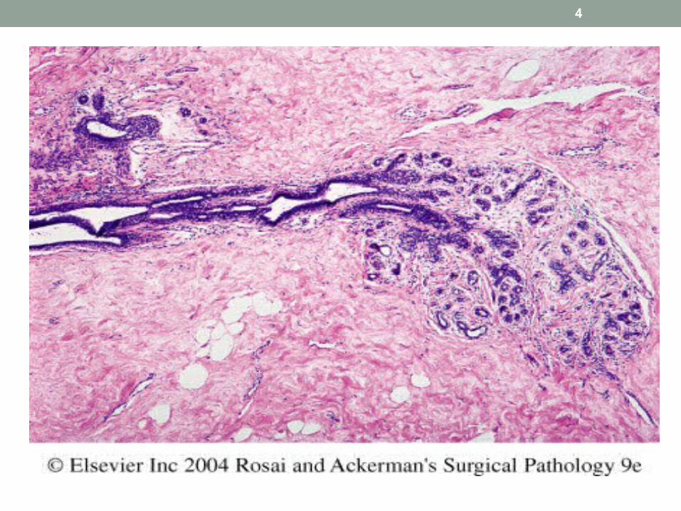
4

Breast, Clinical Presentation
1) Pain (mastalgia): is the most common breast symptom and may be cyclical with menses or noncyclical.
- Diffuse cyclical pain has no pathologic significance.- Noncyclical pain is usually associated with a focal site in
the breast. - Causes include ruptured cysts or areas of prior injury or
infections,or sometime no specific cause.
- Although the great majority of painful masses are benign, about 10% of breast cancers present with pain, and all masses need to be investigated.
5

Clinical Presentation
2)Palpable mass
3)Nipple discharge: Milky discharge has not been associated with
malignancy. Bloody or serous discharges are most commonly
associated with benign lesions but, rarely, can be due to a malignancy.
6
Characteristics of Breast Carcinomas by Clinical Presentation
• Palpable mass
• Mammographic density
• Mammographic calcifications
7

• Mammographic screening was introduced in the 1980s as a means to detect small, nonpalpable breast carcinomas not associated with breast symptoms.
• Screening is generally recommended to start at age 40.
• Younger women usually undergo mammography only if they are at high risk for developing carcinoma.
• The principal mammographic signs of breast carcinoma are densities and calcifications.
8

Breast ,Benign Epithelial Lesions
1- Non proliferative breast changes
2- Proliferative breast disease
3- Atypical hyperplasia
9

Fibrocystic change • Often produce palpable lumps • Characterized by various combinations of cysts, fibrous
overgrowth, and epithelial proliferation. • The cause of fibrocystic change is not known. • It is the single most common disorder of the breast.• The condition is diagnosed frequently between the ages of 20
and 55 and decreases progressively after the menopause.• Fibrocystic change presents with asymptomatic masses in the
breast, which are discovered by palpation. The masses vary from diffuse small irregularities (lumpy bumpy breast) to a discrete mass or masses.
• It may also present with pain, which may be cyclical with midcycle or premenstrual discomfort.
• Pain may be focal or diffuse and may or may not be associated with the lumps.
10

Fibrocystic changes
• Three patterns of morphologic changes :
1- Cyst formation
2- Fibrosis
3- Adenosis
11

• Histologically, cysts may be lined by flattened epithelium, columnar epithelium with features of apocrine cells or may completely lack an epithelial lining.
12

Fibrocystic changes
• Cysts :small to big in size ,lined by benign epithelium with apocrine metaplasia
• Semi-translucent or turbid fluid• Fibrosis : contribute to the palpable firmness of the
breast• Adenosis : Increase in the number of acini per lobule.• Adenosis can be seen in pregnancy.
13

Downloaded from: Robbins & Cotran Pathologic Basis of Disease (on 11 November 2007 08:05 PM)
© 2007 Elsevier
14

Benign Epithelial Lesionsproliferative Disease without Atypia
• Rarely form palpable masses• Detected as mammographic densities.• Incidental finding • e.g.Large duct papilloma present in 80% as nipple
discharge.• Risk for cancer is 1.5 – 2 times normal
15

Benign Epithelial Lesionsproliferative Disease without Atypia
• Proliferation of ductal epithelium and/or stroma without cellular abnormalities that are suggestive of cancer
• Many entities included here :
1- Epithelial hyperplasia
2- Sclerosing adenosis
3- complex sclerosing lesions/radial scar
3- Papillomas
16

Benign Epithelial Lesionsproliferative Disease without Atypia
Epithelial Hyperplasia.• In the normal breast, only myoepithelial cells and a
single layer of luminal cells. Epithelial or ductal hyperplasia is a proliferative condition in which there is an increase in the cellularity of the epithelium of the TDLU.
• Epithelial hyperplasia is defined by the presence of more than two cell layers. Hyperplasia ranges from mild, moderate to florid and from typical (i.e. without atypia) to atypical short of malignancy.
17

• The proliferating epithelium distends the ducts and ductules. It shows two distinct cell populations, epithelial and myoepithelial cells.
• It is a microscopic finding, which cannot be predicted clinically or by mammographic examination.
• The lesion may coexist with other features of fibrocystic change, but in some cases may form the predominant pattern.
18

19

Benign Epithelial Lesionsproliferative Disease without Atypia
Sclerosing Adenosis. • This condition most often occurs as an incidental
microscopic finding but may manifest as a palpable mass that may be mistaken clinically for cancer.
• It is almost always associated with other forms of fibrocystic change.
• Diffuse microcalcifications are commonly seen in the lesion, which may mimic carcinoma on mammography.
20

Sclerosing Adenosis
- Microscopically, sclerosing adenosis consists of proliferation of ductular structures and stroma with distortion of the TDLU.
21

)
22

Benign Epithelial Lesionsproliferative Disease without Atypia
Complex Sclerosing Lesion (Radial Scar). • Radial scars are stellate lesions characterized by a
central nidus of entrapped glands in a hyalinized stroma
• can resemble irregular invasive carcinomas mammographically or on gross examination..
23

Benign Epithelial Lesionsproliferative Disease without Atypia
Complex Sclerosing Lesion (Radial Scar). • "scar" refers to the morphologic appearance, as these
lesions are not associated with prior trauma or surgery.
24

25

Benign Epithelial Lesionsproliferative Disease without Atypia
Papillomas • This is a papillary tumor that arises from the duct
epithelium including large ducts.• It arises more often in the central part of the breast
from the lactiferous ducts (75%) but can occur in any quadrant.
• It is more commonly solitary, consisting of a single tumor in one duct, but multiple discrete tumors, usually in contiguous branches of the ductal system may occur.
26

Benign Epithelial Lesionsproliferative Disease without Atypia
• Large duct papillomas are usually solitary and situated in the lactiferous sinuses of the nipple.
• Small duct papillomas are commonly multiple and located deeper within the ductal system.
• Small duct papillomas have been shown to increase the risk of subsequent carcinoma.
27

• Nipple discharge, which may be bloody, is the most common presentation for central papillomas and less commonly of peripheral tumors.
• A subareolar mass may be palpable. • Age range is from 30 to 50 years.
28

29

Proliferative Breast Disease with Atpyia
• Risk for cancer is 4-5 times normal• Atypical hyperplasia is a cellular proliferation
resembling ductal carcinoma in situ (DCIS) or lobular carcinoma in situ (LCIS) but lacking sufficient qualitative or quantitative features for a diagnosis of carcinoma in situ.
30

Proliferative Breast Disease with Atpyia
• Include two entities
1 –Atypical ductal hyperplasia
2 –Atypical lobular hyperplasia
31

• Atypical hyperplasia has some of the architectural and cytologic features of carcinoma in situ but lack the complete criteria for that diagnosis and is categorized as ductal or lobular in type
32

© 2
33

STROMAL TUMORS
34

STROMAL TUMORS
• Two basic stromal tumors are
- fibroadenoma
- Phylloids tumor
35

36
Fibroadenoma
• The most common benign tumor of the female breast • Composed of both epithelial and stromal tissue
derived from the TDLU. • Any age ,most common before age 30• Usually present with a palpable mass • Regression usually occurs after menopause
Fibroadenoma
• The tumor presents as a spherical, rubbery nodule, which is sharply circumscribed from the surrounding breast tissue and so is freely movable and can be shelled out.
• It may increase in size during pregnancy and cease to grow after menopause. The tumor is usually solitary but may be multiple and involve both breasts. The cut surface is pearl-white
37

38
Fibroadenoma
• Spherical nodules• Sharply demarcated• Freely movable• Size vary • Proliferation in both glands and stroma• Treatment: lumpectomy (only the lump is
removed)

Fibroadenoma
• Histologically, the tumor is composed of a mixture of ducts and fibrous connective tissue
• The tumor is completely benign. • Rarely, carcinoma may arise within a fibroadenoma.
The predominant type has been lobular carcinoma
39

40

41
fibroadenoma

42

Phylloides tumor
• Phyllodes tumors, like fibroadenomas, arise from intralobular stroma. Although they can occur at any age, most present in the sixth decade, 10 to 20 years later than the average presentation of a fibroadenoma
• Most present as palpable masses
43

Phylloides tumor
• Phyllodes tumors must be excised with wide margins to avoid the high risk of local recurrences.
• The majority are low-grade tumors that may recur locally but only rarely metastasize. Rare high-grade lesions behave aggressively, with frequent local recurrences and distant hematogenous metastases in about one third of cases.
44

45
Breast cancer

Breast Carcinoma
• The most common malignancy of breast is carcinoma• Carcinoma of the breast is the most common cancer
in women• Women who lives to age 90 has a one in eight chance
to have breast cancer
46

Breast Cancer• Mammographic screening increased dramatically the
detection of small invasive cancers• DCIS by itself is almost exclusively detected by
mammography ,so the incidence of DCIS is increased with the use of mammography.
• The number of women with an advanced cancer is markedly decreased
47

Breast Cancer
• In 1994,the mortality rate started to decline• Currently only 20% of the women with breast cancer
are expected to die of the disease
48

49

Breast CancerRisk Factors• Age : steady increase in the incidence of breast
cancer, as women grow older. The age-specific incidence rate is highest in postmenopausal women. Breast cancer is rare before 25 yrs, except familial forms ,77% of cases occur in women >50 yrs of age. The average age at diagnosis is 64 years.
• Age at Menarche: The younger a woman’s age at menarche, the higher her risk of breast cancer. For each 2 years delay in onset of menstrual activity, the risk is reduced by about 10%. Menarche younger than age 11 have a 20% increased risk to that who have their menarch at 14yrs.
50

Breast Cancer, Risk Factors
• First Live birth: The earlier a woman has her first birth, the lower her lifetime risk for breast cancer. This is independent of parity. A woman who has her first birth after 30 years has an increased risk. A nulliparous woman has increased risk. Full term pregnancy before age 20 years has half the risk of nulliparous ,or women who have first birth after age 35.
• First Degree relative with Breast Cancer . The relative risk of breast cancer in a woman with breast cancer in first-degree relative (mother, sister, or daughter) ranges from 1.5 to 2.5.The risk increases with the number of affected first degree relatives. The majority of cancers occur in women without such history
51

Breast Cancer, Risk Factors
• Breast Biopsy :Atypical hyperplasia increases the risk for breast cancer
• Race :Overall incidence of breast cancer is lower in African American women
• Estrogen Exposure: The later a woman’s age at menopause, the higher her risk of breast cancer. Women who had their menopause after 55 years have 2 times the risk of those who had their menopause before 45 years postmenopausal hormone replacement slightly increase the risk
52

Breast Cancer, Risk Factors
• Radiation exposure: Higher rate of breast cancer• Women who have had a breast cancer have a 10-fold
increased risk of developing a second primary breast cancer.
• Geographic influence : Breast cancer is more common in Western industrialized countries than in developing countries. Four to seven times in USA and Europe higher than those in other countries.
• Diet: Fat might increase the risk• Obesity : may play a role
53

Breast Cancer, Risk Factors
• Exercise :some studies showed degreased risk• Breast–Feeding :The longer the women breast –
feed ,the lower the risk• Environmental toxins: pesticides .• Tobacco :Not associated with breast cancer,but
associated with the development of peri-ductal mastitis,or sub-areolar abscess .
54

Hereditary Breast Cancer
• A family history of breast cancer in a first-degree relative is reported in 13% of women with the disease
• About 25% of familial cancers (or around 3% of all breast cancers) can be attributed to two highly penetrant autosomal-dominant genes: BRCA1 and BRCA2
55

Sporadic Breast Cancer
• The major risk factors for sporadic breast cancer are related to hormone exposure: gender, age at menarche and menopause, reproductive history, breast-feeding, and exogenous estrogens. The majority of these cancers occur in postmenopausal women and overexpress estrogen
56

Breast CarcinomaClassification
• Almost all are Adenocarcinoma• Divided into In situ Carcinoma( non-invasive) and
Invasive carcinoma• Noninvasive carcinoma (Carcinoma in situ): This is
epithelial proliferation that is still confined to the TDLU, has not invaded beyond the basement membrane and is therefore incapable of metastasis.
57

Breast CarcinomaClassification ,Carcinoma in situ
There are two subtypes:
1) Ductal carcinoma in situ (DCIS) or intraductal carcinoma 80%
2) Lobular carcinoma in situ. The incidence in autopsy studies is about 20%.
58

DCIS
• DCIS comprises a heterogeneous group of noninvasive neoplastic proliferation with risk of development of subsequent invasive carcinoma.
• The tumor distends and distorts the ducts in the TDLU so that the terminal ducts enlarge and resemble large ducts.
59

DCIS• DCIS occurs throughout the age range of breast
carcinoma with mean age at diagnosis between 50 and 59 years, similar to the mean age of women with invasive ductal carcinoma.
• Mammography is a very sensitive diagnostic procedure for detecting DCIS, as a substantial proportion is not palpable. Mammographically detected microcalcifications are found in 72 to 98% of DCIS.
60

DCIS( Ductal Carcinoma In Situ)
• Rapidly increased in the past two decades• Half of mammographically detected cancers• Most frequently as a calcifications• Less frequently as a density or a vaquely palpable
mass or nipple discharge
61

DCIS( Ductal Carcinoma In Situ)• Many subtypes
-Comedocarcinoma,
-solid ,
-Papillary,
-and micropapillary.
Clinical behavior• The two types of DCIS differ markedly in their risk of
subsequent invasive carcinoma. • Comedocarcinoma has essentially a 100% chance of
becoming invasive if left untreated. • Pure cribriform/micropapillary carries only a 30%
chance of invasive carcinoma.
62

Ductal carcinoma insitu
63

64

65

66

67

Paget’s Disease • Rare skin manifestation of breast cancer(1 to 2 %)• Paget’s disease of the nipple presents with an
eczematous area of the nipple, which may be subtle or form an obviously eroded, weeping lesion.
• Pruritus is common ,might be mistaken for Eczema, presents as a unilateral erythematous eruption with a scale crust.
• Malignant cells, referred to as Paget cells and are found scattered in the epidermis.
68

Paget’s Disease
• The histologic hallmark of Paget’s disease of the nipple is the infiltration of the epidermis by large ductal neoplastic cells with abundant clear or pale cytoplasm and nuclei with prominent nucleoli. The cells usually stain positively for mucin.
• Paget cells extend from DCIS within the ductal system into nipple skin without crossing the basement membrane
• Palpable mass is present in 50 to 60% of women with Paget disease indicating an underlying invasive carcinoma.
69

Pagets disease
70

Pagets disease
71

LCIS -Lobular Carcinoma in Situ
• Always an incidental finding in a biopsy performed for another reason
• Infrequent (1% to 6% )of all carcinomas• Bilateral in 20% to 40% of women when both breasts
are biopsied• LCIS is frequently multicentric and bilateral and
subsequent carcinomas occur at equal frequency in both breasts
72

LCIS -Lobular Carcinoma in Situ• Lobular carcinoma in situ (LCIS) does not form a
palpable mass and cannot be detected clinically, felt at operation or seen grossly on pathological examination.
• Although LCIS may have microcalcifications, these are infrequent and so mammography has not been useful for detecting it.
• The tumor presents as a coincidental finding in breast tissue removed for other reasons. The disease tends to be bilateral and multicentric.
• LCIS shows a proliferation of cells that fill and distend the TDLU.
73

LCIS -Lobular Carcinoma in Situ
Clinical behavior • If LCIS is left untreated, about 30% of women develop
an invasive cancer within 20 years of diagnosis. The invasive cancer may be ductal or lobular. LCIS is therefore a marker of increased cancer in both breasts.
74

Lobular carcinoma insitu
75

76

Invasive Breast CarcinomaClassification • Invasive breast carcinoma is tumor that has extended
across the basement membrane. This permits access to lymphatics and vessels and the potential distant metastases and thereby a lethal outcome. There are several different types of invasive carcinoma. Invasive breast carcinoma is subdivided into
1- NOS Ductal 80%
2- Lobular 10%
3- tubular 6%
4-Mucinous(Colloid) 2%
5- Medullary 2%
6- Papillary 1%
7- Metaplastic Carcinoma 1%
77

CLINICAL FEATURES OF BREAST CANCER
• Palpable mass. • By the time a cancer becomes palpable, over half the patients will
have axillary lymph node metastases .• Larger carcinomas may be fixed to the chest wall or cause dimpling
of the skin. • Lymphatics may become so involved as to block the local area of
skin drainage and cause lymphedema and thickening of the skin, a change referred to as peau d'orange.
• When the tumor involves the central portion of the breast, retraction of the nipple may develop.
78

CLINICAL FEATURES OF BREAST CANCER
• In older women undergoing mammography, invasive carcinomas most commonly present as a density and are, on average, half the size of a palpable cancer . Fewer than 20% will have nodal metastases.
• Invasive carcinomas presenting as mammographic calcifications without an associated density are very small in size, and metastases are unusual.
79

CLINICAL FEATURES OF BREAST CANCER
• The term "inflammatory carcinoma" refers to the clinical presentation of a carcinoma extensively involving dermal lymphatics, resulting in an enlarged erythematous breast. The diagnosis is made on clinical grounds and does not correlate with a specific histologic type of carcinoma
80

Invasive carcinoma
81

Invasive carcinoma
82

Invasive Ductal Carcinoma ,NOS• It is the commonest type of breast cancer, forming up
to 80% of these cancers. • Most of these tumors excite a pronounced fibroblastic
stromal reaction to the invading tumor cells producing a palpable mass with hard consistency (hence scirrhous carcinoma), which is the most common presentation.
• The tumor shows an infiltrative attachment to the surrounding structures and may cause dimpling of the skin (due to traction on suspensory ligaments) or nipple retraction.
83

Invasive Ductal Carcinoma ,NOS• Grossly ,firm ,hard, and have an irregular border• Cut surface is gritty and shows irregular margins with
stellate infiltration and in the center there are small foci of chalky white stroma and occasionally calcifications
• Characteristic grating sound when cut or scraped• Could be soft and well demarcated• Accompanied by varying amounts of DCIS
84

Invasive Ductal Carcinoma,NOS• Histologically, the tumor cells are larger than normal
epithelium, and can assume a variety of patterns such as glandular formation, cords of cells, broad sheets of cells or a mixture of all these, usually within a dense stroma.
• The tumors range from well differentiated, in which there is glandular formation, to poorly differentiated, containing solid sheets of pleomorphic neoplastic cells.
• Carcinomas associated with a large amount of DCIS require large excisions with wide margins to reduce local recurrences
85

Invasive carcinoma, ductal
86

Invasive ductal carcinoma
87

Invasive Lobular Carcinoma
• It is the second most common type of invasive breast cancer forming up to 10% of breast cancers.
• The tumor may occur alone or in combination with ductal carcinoma.
• It tends to be bilateral more often than ductal carcinoma and multicentric.
• The amount of stromal reaction to the tumor varies from dense desmoplasia to little reaction and therefore the presentation varies from a discrete mass to a subtle, diffuse indurated area.
88

Invasive Lobular Carcinoma
• Most are firm to hard with irregular margins• Single infiltrating cells ,often one cell width• No tubules or papillary formation In • In about 10% of cases, tumors have mixed features of
invasive ductal and lobular carcinomas.
89

90

91

Medullary Carcinoma• This subtype of breast cancer presents as a well
circumscribed mass. • May mistaken clinically and radiologically for
fibroadenoma• It does not produce any fibroblastic (desmoplastic)
reaction and therefore is soft and fleshy (encephaloid). On section foci of necrosis and hemorrhage are evident.
• Microscopically, the tumor is composed of solid sheets of malignant cells and frequent mitoses. There is scant fibrous stroma. Lymphocytes and plasma cells surround the tumor cells.
92

Colloid Carcinoma/ Mucinous carcinoma
• tends to occur in older women. • It is sharply circumscribed, lacks fibrous stroma and
is slow growing.• Is soft and gelatinous and has a glistening cut
surface. • It may be in pure mucinous or mixed in which it is
associated with other types of invasive breast carcinoma.
• The mucinous tumor is composed of small islands, occasionally forming glands, and isolated tumor cells floating in pools of extracellular mucin
93

Invasive colloid carcinoma
94

Breast Carcinoma , Major Prognostic Factors
1- Invasive or In situ disease: The great majority of women with adequately treated DCIS will be cured. In contrast, at least half of invasive carcinomas will have metastasized locally or distantly at the time of diagnosis.
95
Breast Carcinoma , Major Prognostic Factors
2- Distant metastasis:
Once distant metastases are present, cure is unlikely, although long-term remissions and palliation can be achieved. Favored sites for dissemination are the lungs, bones, liver, adrenals, brain, and meninges.
96

Breast Carcinoma , Major Prognostic Factors
3- Lymph node metastasis: Axillary lymph node status is the most important prognostic factor for invasive carcinoma in the absence of distant metastases. The clinical assessment of nodal involvement is very inaccurate, therefore, biopsy is necessary for accurate assessment.
With no involvement, the 10-year disease-free survival rate is close to 70% to 80%; the rate falls to 35% to 40% with one to three positive nodes and 10% to 15% in the presence of more than 10 positive nodes.
97

Breast Carcinoma , Major Prognostic Factors
4- Tumor Size:
The size of the carcinoma is the second most important prognostic factor. The risk of axillary lymph node metastases does increase with the size of the carcinoma.
Note : all the above parameters are used to stage the tumor.Stage is a combination of size and lymph node status.
Tumor size less than 2 cm is associated with a favorable prognosis.
The single most important prognostic indicator is the lymph node status. Negative lymph nodes have the best prognosis. Involvement of 1 to 3 lymph nodes has an intermediate prognosis and 4 or more positive nodes have the worse prognosis.
98

Breast Carcinoma , Major Prognostic Factors
5- Locally advanced disease:
Tumors invading into skin or skeletal muscle are frequently associated with concurrent or subsequent distant disease.
99

Breast Carcinoma , Major Prognostic Factors
6- Inflammatory Carcinoma: Women presenting with the clinical appearance of breast swelling and skin thickening have a particularly poor prognosis with a 3-year survival rate of only 3% to 10%.
100

Breast Carcinoma , Minor Prognostic Factors
1- Histologic Subtype: Infiltrating ductal and lobular carcinomas have the worse prognosis, medullary and mucinous have intermediate and tubular and cribriform have the most favorable prognoses2- Tumor Grade: This is calculated by the pathologist. Grading separates tumors into three categories according to the amount of well formed tubules, the degree of nuclear pleomorphism, and the mitotic rate. The most commonly used grading system to assess the degree of tumor differentiation (Bloom Richardson). There are three grades with grade 1 having better prognosis and grade3 having poorer prognosis.
101

Breast Carcinoma , Minor Prognostic Factors
3- Estrogen and progesteron receptors:50% to 85% of carcinomas express estrogen receptors, such tumors are more common in postmenopausal women, hormone positive cancers have better prognosis. They respond well to specific chemotherapuetic drugs eg. Tamoxifen.
Therefore reporting of ER/PR positivity is important when reporting breast cancer.
102

Breast Carcinoma , Minor Prognostic Factors
4- HER2/neu. (human epidermal growth factor receptor 2 or c-erb B2 or neu) is a glycoprotein overexpressed in 20% to 30% of breast carcinomas. Many studie shave shown that overexpression of
HER2/neu is associated with a poor prognosis. In addition, ongoing studies have shown that HER2/neu-
overexpressing tumors respond very well to hormonal or anthracycline chemotherapy regimens
eg. Trastuzumab (Herceptin). Therfore evaluation of HER2/neu is most important when
reporting breast cancer.
103

Breast Carcinoma , Minor Prognostic Factors
5- Lymphovascular invasion: This finding is strongly associated with the presence of lymph node metastases and is a poor prognostic factor in women without lymph node metastases.
6- Proliferative rates
104

Metastasis to vertebra
105


