Breast MRI for Evaluating Patients with Metastatic ... · quadrantectomy or blind mastectomy with...
8
382 Korean J Radiol 8(5), October 2007 Breast MRI for Evaluating Patients with Metastatic Axillary Lymph Node and Initially Negative Mammography and Sonography Objective: We wanted to investigate the ability of breast MR imaging to identify the primary malignancy in patients with axillary lymph node metastases and ini- tially negative mammography and sonography, and we correlated those results with the conventional imaging. Materials and Methods: From September 2001 to April 2006, 12 patients with axillary lymph node metastases and initially negative mammography and sonog- raphy underwent breast MR imaging to identify occult breast carcinoma. We ana- lyzed the findings of the MR imaging, the MR-correlated mammography and the second-look sonography. We followed up both the MR-positive and MR-negative patients. Results: MR imaging detected occult breast carcinoma in 10 of 12 (83%) patients. Two MR-negative patients were free of carcinoma in the ipsilateral breast during their follow-up period (39 and 44 months, respectively). In nine out of 10 patients, the MR-correlated mammography and second-look sonography localized lesions that were not detected on the initial exam. All the non-MR-corre- lated sonographic abnormalities were benign. Conclusion: Breast MR imaging can identify otherwise occult breast cancer in patients with metastatic axillary lymph nodes. Localization of the lesions through MR-correlated mammography and second-look sonography is practically feasible in most cases. he manifestation of an isolated metastatic axillary lymph node without any clinical and radiologic evidence of primary breast cancer is uncommon, but this disease most commonly originates from breast cancer in women (1). For these cases, the traditional therapy has been upper outer quadrantectomy or blind mastectomy with axillary lymph node dissection, with or without radiation (2). However, 30 40% of the upper outer quadrantectomy or blind mastectomy specimens show no histologic evidence of carcinoma, and the survival rate of patients who undergo blind mastectomy with axillary dissection is similar to those who undergo axillary dissection only (3, 4). Breast MRI techniques have improved significantly in recent years, and MRI with dynamic enhancement has been established as an important imaging modality for evaluating breast cancer with the highest sensitivity (5). Therefore, the goal of this study was to investigate the ability of breast MR imaging to identify the primary malignant focus in patients with metastatic axillary lymph nodes and initially negative mammography and sonography, and to correlate the results with the conventional imaging. Eun Young Ko, MD Boo-Kyung Han, MD Jung Hee Shin, MD Seok Seon Kang, MD Index terms : Axilla Breast, MR Breast neoplasms Lymph nodes Korean J Radiol 2007 ; 8 : 382-389 Received August 4, 2006; accepted after revision January 3, 2007. Department of Radiology and the Center for Imaging Science, Samsung Medical Center, Sungkyunkwan University School of Medicine, Seoul 135-710, Korea. Address reprint requests to : Boo-Kyung Han, MD, Department of Radiology and the Center for Imaging Science, Samsung Medical Center, Sungkyunkwan University School of Medicine, 50 Irwon-dong, Gangnam-gu, Seoul 135-710, Korea Tel. (822) 3410-6418 Fax. (822) 3410-0084 e-mail: [email protected] T
Transcript of Breast MRI for Evaluating Patients with Metastatic ... · quadrantectomy or blind mastectomy with...

382 Korean J Radiol 8(5), October 2007
Breast MRI for Evaluating Patients withMetastatic Axillary Lymph Node andInitially Negative Mammography andSonography
Objective: We wanted to investigate the ability of breast MR imaging to identifythe primary malignancy in patients with axillary lymph node metastases and ini-tially negative mammography and sonography, and we correlated those resultswith the conventional imaging.
Materials and Methods: From September 2001 to April 2006, 12 patients withaxillary lymph node metastases and initially negative mammography and sonog-raphy underwent breast MR imaging to identify occult breast carcinoma. We ana-lyzed the findings of the MR imaging, the MR-correlated mammography and thesecond-look sonography. We followed up both the MR-positive and MR-negativepatients.
Results: MR imaging detected occult breast carcinoma in 10 of 12 (83%)patients. Two MR-negative patients were free of carcinoma in the ipsilateralbreast during their follow-up period (39 and 44 months, respectively). In nine outof 10 patients, the MR-correlated mammography and second-look sonographylocalized lesions that were not detected on the initial exam. All the non-MR-corre-lated sonographic abnormalities were benign.
Conclusion: Breast MR imaging can identify otherwise occult breast cancer inpatients with metastatic axillary lymph nodes. Localization of the lesions throughMR-correlated mammography and second-look sonography is practically feasiblein most cases.
he manifestation of an isolated metastatic axillary lymph node withoutany clinical and radiologic evidence of primary breast cancer isuncommon, but this disease most commonly originates from breast
cancer in women (1). For these cases, the traditional therapy has been upper outerquadrantectomy or blind mastectomy with axillary lymph node dissection, with orwithout radiation (2). However, 30 40% of the upper outer quadrantectomy or blindmastectomy specimens show no histologic evidence of carcinoma, and the survivalrate of patients who undergo blind mastectomy with axillary dissection is similar tothose who undergo axillary dissection only (3, 4). Breast MRI techniques haveimproved significantly in recent years, and MRI with dynamic enhancement has beenestablished as an important imaging modality for evaluating breast cancer with thehighest sensitivity (5). Therefore, the goal of this study was to investigate the ability ofbreast MR imaging to identify the primary malignant focus in patients with metastaticaxillary lymph nodes and initially negative mammography and sonography, and tocorrelate the results with the conventional imaging.
Eun Young Ko, MDBoo-Kyung Han, MDJung Hee Shin, MDSeok Seon Kang, MD
Index terms:AxillaBreast, MRBreast neoplasmsLymph nodes
Korean J Radiol 2007;8:382-389Received August 4, 2006; accepted after revision January 3, 2007.
Department of Radiology and the Centerfor Imaging Science, Samsung MedicalCenter, Sungkyunkwan University Schoolof Medicine, Seoul 135-710, Korea.
Address reprint requests to:Boo-Kyung Han, MD, Department ofRadiology and the Center for ImagingScience, Samsung Medical Center,Sungkyunkwan University School ofMedicine, 50 Irwon-dong, Gangnam-gu,Seoul 135-710, KoreaTel. (822) 3410-6418Fax. (822) 3410-0084e-mail: [email protected]
T
MATERIALS AND METHODS
Patients From September 2001 to April 2006, 26 patients
presenting with metastatic axillary lymph nodes andwithout any clinical evidence of breast cancer visited ourhospital. In 11 cases, primary breast cancers were detectedin the ipsilateral breasts by mammography (n = 3) or US (n= 7) or both (n = 1). Two cases had a history of contralat-eral breast cancer and they had received modified radialmastectomy and chemotherapy three and four years ago,respectively. In one case, the diagnosis of the core needlebiopsy of the axillary lymph node was metastatic poorlydifferentiated carcinoma or medullary carcinoma, but thefinal diagnosis after axillary lymph node dissection wasanaplastic large cell lymphoma. The remaining 12 patients,who showed no evidence of primary breast cancer uponclinical examination or on the initial mammography andsonography, underwent breast MR imaging to evaluate thepresence of occult breast carcinoma; it was these 12patients who made up the study population. All 12 patientshad malignant axillary lymph nodes that were pathologi-cally shown to be metastatic adenocarcinomas byultrasound (US)-guided core needle biopsy (n = 11) andexcision biopsy (n = 1). All these patients were female andthey ranged in age from 42 to 78 years (mean age: 55years).
All the available images of the patients from thereferring hospital, including 10 mammograms, nine US andone PET, were negative. Ten patients had both negativemammograms and US; two patients had negativemammograms and US with probable benign nodules.
Breast MR Imaging All the patients were imaged in the prone position using
a dedicated surface breast coil. Breast MR imaging wasperformed on a 1.5-T system (Signa CV/I; General ElectricMedical System, Milwaukee, WI) for eight cases and on a3.0-T system (Interna; Philips Medical Systems, Best, theNetherlands) for three cases, and on both systems for onecase. MR imaging consisted of a fat-suppressed, sagittal, 3-dimensional, gradient echo sequence and the dynamicenhanced images. Imaging on the 1.5-T scanner covered asingle breast with a minimum repetition time and echotime (17.3/1.3 ms), a 60 flip angle, a 24-cm field of view,1-mm to 2-mm sections with no gap, a 256 192 matrix,one excitation and a scan time of 3-4 minutes. Imaging onthe 3.0-T scanner covered both breasts with a minimumrepetition time and echo time (8.7/4.3 ms for the axialdynamic images; 16/4.1 ms for the sagittal dynamic
images), a 20 flip angle, a 27-cm field of view, 1.5-mmsections with no gap, a 512 512 matrix and a scan time of2 3 minutes.
For the dynamic contrast enhancement, a 0.1 mmol/kgbolus of gadopentetate dimeglumine (Magnevist; BerlexLaboratories, Wayne, NJ) was injected; this was followedby a 10-mL saline flush. Two and three sequential postcon-trast images were obtained with no delay on the 1.5-T and3.0-T scanners, respectively, and these began immediatelyafter the saline flush at the same slice position and location.After examination, two subtraction images were madeautomatically on a pixel-by-pixel basis: the un-enhancedimages were subtracted from the early postcontrast images(standard subtraction), and the last postcontrast imageswere subtracted from the early postcontrast images(reverse subtraction). The reformatted images with amaximum intensity projection were then created from thestandard and reverse subtraction images.
Interpretation of the Breast MR ImagesThe MR images were interpreted prospectively on soft
copy with using PACS (picture archiving and communica-tion system, General Electric Medical System) that allowedmanual window settings and optimization of the parame-ters. A radiologist, who was aware of the patient’s clinicalhistory and who had seen all available imaging informa-tion, including the mammograms and sonograms,interpreted all the images. The breast lesions weredescribed using the Breast Imaging Reporting and DataSystem (BI-RADS) MR lexicon (6).
A lesion was considered suspicious if it was visualized as(a) a mass with irregular shape, a mass with irregular orspiculated margin, or a mass with rim enhancement, (b)non-mass-like enhancement that showed a clumped linear-ductal enhancement, clumped segmental enhancement andregional enhancement with ill-defined or irregular bordersor with architectural distortion, or (c) a mass or non-mass-like enhancement with early washout.
Management of Lesions after MRIThe previous mammography and US images were
retrospectively reviewed along with any informationgleaned from the MR images; any positive findings, includ-ing benign or probable benign lesions in the areascorresponding to suspicious enhancements on the MRI,were confirmed by US-guided or mammography-guidedbiopsy. If retrospective review of the previous mammogra-phy or US images was not available or if this was negative,then MR-correlated mammography or a second-look USexamination that specifically targeted the area ofsuspicious MR enhancement was performed. The benign
Breast MRI in Patients with Axillary Nodal Metastasis Alone on Other Imaging Studies
Korean J Radiol 8(5), October 2007 383
results from the US-guided or mammography-guidedbiopsy in the area of suspicious MR enhancement werefollowed by localization with excision biopsy.
Since MR-guided localization and biopsy were notavailable at our institution, those lesions with suspiciousenhancement on MRI, but with no abnormality onmammography or the US images, underwent mastectomy.
RESULTS
Suspicious lesions were detected on breast MRI in 10(83%) of the 12 patients. The pathology of all the lesionswas proven to be primary breast carcinoma.
A complete summary of the 12 patients presenting withaxillary lymph node metastases is given in Table 1. All thepatients had mammography and US examinationsconducted before undergoing breast MRI. All 12 patientswere transferred from other hospitals with negativemammography and US images or they were transferredwithout these images. Seven of them again receivedmammography and US examinations at our hospital, andthe other five of them were diagnosed by the images thatwere obtained from other hospitals.
Before MR imaging, 10 out of the 12 patients had noabnormal findings based on the conventional images, whiletwo patients had category 3 nodules observed on US.These two category 3 nodules did not correlate with the
MR findings and they were diagnosed as a foreign bodyreaction and stromal fibroses by US-guided core needlebiopsy.
MRI of the breast showed suspicious lesions in 10patients, and these lesions had been negative on the priorconventional images. MR-guided second-look US examina-tions were performed in each of those 10 patients, as wellas performing mammography in four patients. Subsequentlocalization of the lesions detected on MRI was possible onmammography (n = 1), US (n = 4) or on both examinations(n = 4) in nine out of 10 patients. In three patients, thelesions localized on MR-guided second-look examinationswere category 4 there were two cases of suspicious nodulesless than 1 cm in size on US (Fig. 1) and one case of faintpleomorphic calcifications with a segmental distribution onthe mammogram (Fig. 2). Category 3 lesions were found infive patients with 3-mm to 8-mm sized benign-lookingnodules being seen on US (Fig. 3). A category 2 lesion wasfound in one patient who had a few benign cysts (Fig. 4).All the lesions localized on the MR-correlated mammogra-phy or the MR-guided second-look US images were foundto be malignant after US-guided core needle biopsy (n =5), US-guided fine needle aspiration (n = 1), surgicalexcision after US-guided localization (n = 2) or aftermammography-guided localization (n = 1). The MR-guidedsecond-look examination failed to localize the lesion in onepatient who had a 1.8-cm linear non-mass-like enhance-
Ko et al.
384 Korean J Radiol 8(5), October 2007
Table 1. Summary of the 12 Patients with Metastatic Axillary Lymph Node(s) and Initially Negative Mammography andSonography
MG / US Localization on MR-correlated MG / Sizeprior to MR
MRsecond look US
Dx(mm)
Location Treatment
1 ( ) / ( ) (+) (+) / (+) IDC 4 UIQ Lumpectomy + AC2 ( ) / ( ) (+) / (+) IDC
/ (+) ( ) FB reaction 5 LOQ Lumpectomy + AC3 ( ) / ( ) (+) / (+) IDC 7 LIQ BCS
/ (+) ( ) SF4 ( ) / ( ) (+) (+) / (+) Tubular ca.
ADHs 3 LOQ BCS5 ( ) / ( ) (+) ( ) / (+) IDC 7 UOQ BCS6 ( ) / ( ) (+) (+) / ( ) Multifocal IDCs 3 Lower center BCS7 ( ) / ( ) (+) (+) / (+) Multifocal IDCs 7 LIQ MRM8 ( ) / ( ) (+) ( ) / ( ) DCIS Mid outer MRM9 ( ) / ( ) (+) (+) / (+) IDC X 2 LOQ CTx, RTx10 ( ) / ( ) (+) / (+) IDC X 2 LIQ CTx, RTx11 ( ) / ( ) ( ) CTx, RTx + AC12 ( ) / ( ) ( ) ( )* / ( )* CTx, RTx + AC
Note. MG = mammography, US = ultrasound, Dx = diagnosis, IDC = invasive ductal carcinoma, DCIS = ductal carcinoma in situ, FB = foreign body, ADH = atypical ductal hyperplasia, SF = stromal fibrosis, AC = axillary clearance, BCS = breast conserving surgery, MRM = modified radical mastectomy,CTx = chemotherapy, RTx = radiation therapy, Ca. = carcinoma, UIQ = upper inner quadrant, UOQ = upper outer quadrant, LIQ = lower inner quadrant,LOQ = lower outer quadrant
Category 3 nodules detected on prior US and that were not correlated with the MR findings* Follow up study after 9 months. Case with Distant metastasis

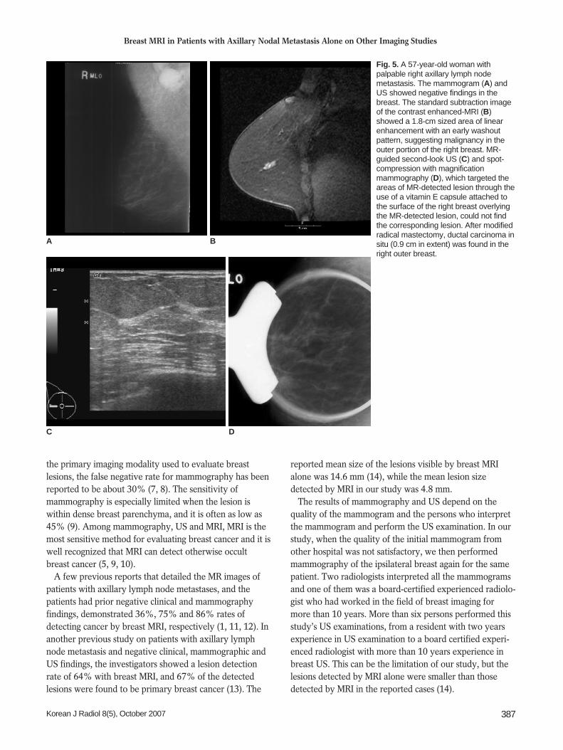
ment with focal wash out on MRI. This lesion was found tobe ductal carcinoma in situ (DCIS) after performingmodified radical mastectomy (Fig. 5).
After histologic confirmation of the primary breastmalignancies for 10 MR-detected lesions, two patientsunderwent lumpectomy following the US-guided localiza-tion, four patients were treated with breast conserving
surgery (BCS) after US or mammography-guided localiza-tion, and one patient underwent modified radical mastec-tomy (MRM) for multifocal lesions. As noted above, thepatient who had a suspicious lesion observed on MRI thatwas not otherwise localized, this patient received MRM aswell. Two patients were treated with chemotherapy andradiation therapy without surgical intervention due to their
Breast MRI in Patients with Axillary Nodal Metastasis Alone on Other Imaging Studies
Korean J Radiol 8(5), October 2007 385
A B C
Fig. 1. A 78-year-old woman withpalpable left axillary lymph nodemetastasis. The initial mammogram (A)and US showed negative findings in thebreast. Two small enhancing nodules(arrows) with a washout pattern werevisible in the left lower breast on thecontrast enhanced-MR images (B, C).Subsequent MR-guided second-look USexaminations revealed two small lowechoic nodules (3 4 mm) with indistinctmargins (arrows) that were not seen onthe prior US study (D, E). US-guidedcore needle biopsy revealed invasiveductal carcinomas.
D E
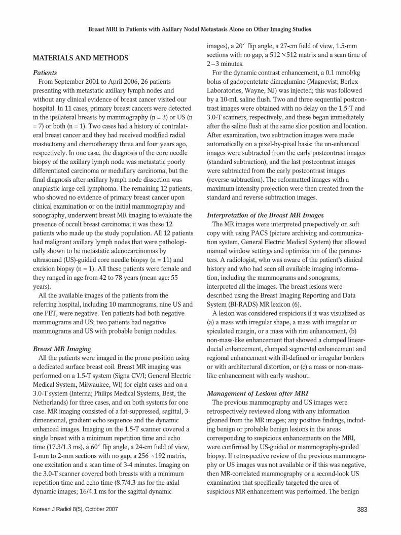
Fig. 2. A 43-year-old woman withpalpable left axillary lymph nodemetastasis. The mammogram and USimages from another hospital showednegative findings. On the maximunintensity projection image of the contrastenhanced-MRI (A), segmental clumpedor stippled enhancements (box) werenoted in the left lower breast. MR-correlated mammography (B) showedthe segmental distribution of faintamorphous microcalcifications in thatarea (box). Breast conserving surgeryafter mammography-guided wirelocalization revealed multifocal invasiveductal carcinomas up to 3 mm in size.A B
multifocal breast cancers with distant metastases. Of the 10 cases that underwent surgical treatment or
biopsy, five cases were invasive ductal carcinomas, onewas tubular carcinoma and one was ductal carcinoma insitu. Four of the patients with invasive ductal carcinomahad multiple lesions, and the patient with tubularcarcinoma had multiple areas of atypical ductal hyperplasiaaround the carcinoma. The mean size of the surgicallyremoved invasive carcinomas was 4.8 mm (range: 3 7mm).
In two patients with negative findings on both MRI and
the conventional images, there was still no evidence of theprimary malignancy in the ipsilateral breast on the conven-tional images or on the follow-up MRI during the follow-upperiods of 39 and 44 months, respectively.
DISCUSSION
With its recent technical advances, contrast-enhancedMRI plays an important role in identifying primary breastcancer, for defining the extent of tumor and for helpingdecide the therapeutic plans (1). While mammography is
Ko et al.
386 Korean J Radiol 8(5), October 2007
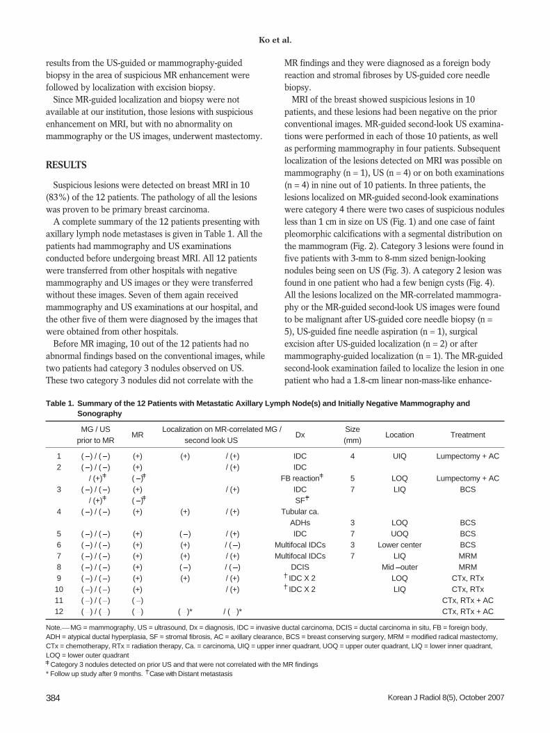
Fig. 3. A 53-year-old woman with palpable left axillary lymph node metastasis. The mammogram (A) and US showed no abnormalfindings in the breast. The contrast-enhanced MR image (B) showed 5 mm nodular enhancement without a washout pattern (arrow) inthe left lower outer breast. On the MR-guided second-look US examination (C), a well-defined flat nodular lesion (arrow) was identified,and invasive ductal carcinoma was diagnosed by US-guided localization and excision.
A B C
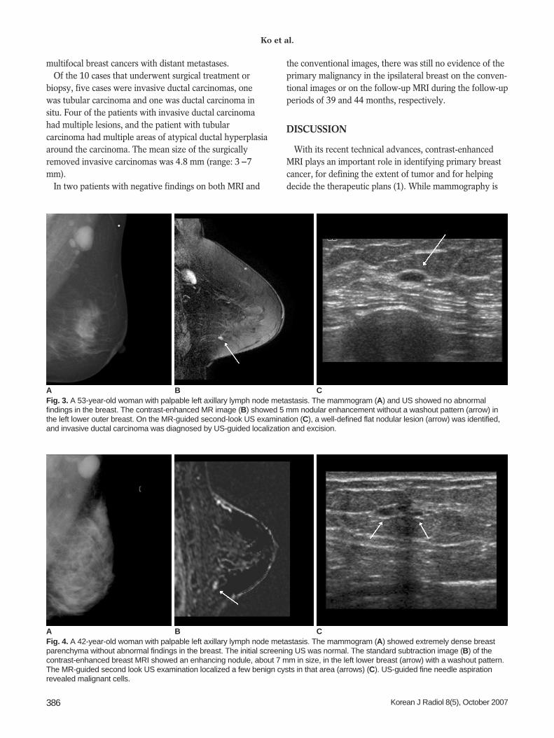
Fig. 4. A 42-year-old woman with palpable left axillary lymph node metastasis. The mammogram (A) showed extremely dense breastparenchyma without abnormal findings in the breast. The initial screening US was normal. The standard subtraction image (B) of thecontrast-enhanced breast MRI showed an enhancing nodule, about 7 mm in size, in the left lower breast (arrow) with a washout pattern.The MR-guided second look US examination localized a few benign cysts in that area (arrows) (C). US-guided fine needle aspirationrevealed malignant cells.
A B C
the primary imaging modality used to evaluate breastlesions, the false negative rate for mammography has beenreported to be about 30% (7, 8). The sensitivity ofmammography is especially limited when the lesion iswithin dense breast parenchyma, and it is often as low as45% (9). Among mammography, US and MRI, MRI is themost sensitive method for evaluating breast cancer and it iswell recognized that MRI can detect otherwise occultbreast cancer (5, 9, 10).
A few previous reports that detailed the MR images ofpatients with axillary lymph node metastases, and thepatients had prior negative clinical and mammographyfindings, demonstrated 36%, 75% and 86% rates ofdetecting cancer by breast MRI, respectively (1, 11, 12). Inanother previous study on patients with axillary lymphnode metastasis and negative clinical, mammographic andUS findings, the investigators showed a lesion detectionrate of 64% with breast MRI, and 67% of the detectedlesions were found to be primary breast cancer (13). The
reported mean size of the lesions visible by breast MRIalone was 14.6 mm (14), while the mean lesion sizedetected by MRI in our study was 4.8 mm.
The results of mammography and US depend on thequality of the mammogram and the persons who interpretthe mammogram and perform the US examination. In ourstudy, when the quality of the initial mammogram fromother hospital was not satisfactory, we then performedmammography of the ipsilateral breast again for the samepatient. Two radiologists interpreted all the mammogramsand one of them was a board-certified experienced radiolo-gist who had worked in the field of breast imaging formore than 10 years. More than six persons performed thisstudy’s US examinations, from a resident with two yearsexperience in US examination to a board certified experi-enced radiologist with more than 10 years experience inbreast US. This can be the limitation of our study, but thelesions detected by MRI alone were smaller than thosedetected by MRI in the reported cases (14).
Breast MRI in Patients with Axillary Nodal Metastasis Alone on Other Imaging Studies
Korean J Radiol 8(5), October 2007 387
A B
C D
Fig. 5. A 57-year-old woman withpalpable right axillary lymph nodemetastasis. The mammogram (A) andUS showed negative findings in thebreast. The standard subtraction imageof the contrast enhanced-MRI (B)showed a 1.8-cm sized area of linearenhancement with an early washoutpattern, suggesting malignancy in theouter portion of the right breast. MR-guided second-look US (C) and spot-compression with magnificationmammography (D), which targeted theareas of MR-detected lesion through theuse of a vitamin E capsule attached tothe surface of the right breast overlyingthe MR-detected lesion, could not findthe corresponding lesion. After modifiedradical mastectomy, ductal carcinoma insitu (0.9 cm in extent) was found in theright outer breast.

In our study, breast MRI showed suspicious lesions in83% (10/12) of the patients who had prior negativeclinical, mammography and US findings. Instead ofperforming the traditional blind mastectomy or upperouter quadrantectomy, eight patients received surgery: twolumpectomies, four BCSs and two MRMs. In addition, twopatients were treated with chemotherapy and radiationtherapy based on the information regarding the site andextent of their primary malignancy. As a result of theconfirmed information about the lesions, all the patientswere treated appropriately and conservatively, with theexception of one patient who received MRM for asuspicious lesion on MRI that was not otherwise localized.It is worthwhile to note that six patients were able to avoidunnecessary mastectomy or upper outer quadrantectomy,as the primary breast carcinoma was located in the upperouter quadrant in only one patient.
The one case that was localized only on MRI and thepatient underwent MRM was pure DCIS. Historically, thereported incidence of axillary metastasis in patients withDCIS is 1-2%, but the prevalence of positive lymph nodesin patients with pure DCIS is approximately 2 13% withperforming immunohistochemical staining (15, 16). Nodemetastasis is usually observed in the sentinel lymph nodeonly in patients with DCIS (17), but in our case, fourlymph nodes were revealed to have malignant cells afterMRM.
Since MRI has a very low false-negative rate (9, 15), wedid not perform blind mastectomy or quadrantectomy inthe two patients who had negative findings on MRI. Theyhave shown no evidence of malignancy in their breastsduring the follow-up period (39 and 44 months, respec-tively) to date.
Therefore, we suggest that before developing a therapeu-tic plan, breast MRI must be performed for those patientswith axillary lymph node metastasis and who are withoutevidence of primary breast cancer.
Although contrast-enhanced MRI has the highestsensitivity among the many imaging modalities for thebreast, the specificity of breast MRI is low (9, 10, 16).Therefore, preoperative biopsy or localization of the MR-detected lesion is necessary (17). Yet MR imaging-guidedbiopsy or localization requires commercially available MRIguiding equipment. Also, the cost-effectiveness of MRI-guided biopsy or localization has not yet been fullyassessed (18). Even when MRI guiding equipment isavailable, lesions in the medial breast are difficult to accessand small enhancing lesions (around 5 mm) may bedifficult to localize due to the transient nature of contrastenhancement and the obscuring that occurs when placingthe biopsy needle. MR imaging-guided biopsy or localiza-
tion is also a time-consuming procedure that lasts as long as45 to 60 minutes (14, 17, 18).
A few studies have reported excellent performance ofMR-guided second-look US localization (11, 19). In ourstudy, MR-guided second-look examinations localized allthe lesions that were detected on breast MRI alone, withthe exception of one case of DCIS. Therefore, localizationwith using MR-correlated mammography or MR-guidedsecond-look US can be a cheaper, practical imagingalternative.
In our experience, the lesions that were negative on priorUS or mammography, but that were localized on MR-correlated mammography or second-look sonography,included not only suspicious malignant lesions, but alsobenign-looking lesions (five category 3 lesions and onecategory 2 lesion) that were small in size; all of these werefound to be malignant. However, two category 3 nodulesthat were detected on prior US, but that did not correlatewith the MR findings, were revealed as benign lesions afterbiopsy. When BI-RADS category 2 or 3 lesions aredetected on conventional images, but they do not correlatewith the MR findings of the patients with suspicious occultbreast cancer, then we should not neglect these lesions.However, biopsy for all benign-looking lesions is notdesirable or even necessarily meaningful (6). Based on ourexperience, we suggest that when there is a benign orbenign-looking lesion on the conventional images of thepatients with metastatic axillary lymph node(s) and anunknown primary malignancy, then the conventionalimages should first be correlated with MR findings beforeperforming a biopsy of the lesions.
In conclusion, the high sensitivity of contrast-enhancedbreast MRI played an essential role in evaluating occultbreast cancer in patients who presented with a metastaticaxillary lymph node with an unknown primarymalignancy, and contrast-enhanced breast MRI also playedan essential role for determining the therapeutic plan. In90% of our cases, histologic work-up for the lesionsdetected on the breast MRI was practically feasible withsuccessful localization being achieved under MR-guidedsecond-look US or MR-correlated mammography.
References1. Orel SG, Weinstein SP, Schnall MD, Reynolds CA, Schuchter
LM, Fraker DL, et al. Breast MR imaging in patients withaxillary node metastases and unknown primary malignancy.Radiology 1999;212:543-549
2. Ellerbroek N, Holmes F, Singletary E, Evans H, Oswald M,McNeese M. Treatment of patients with isolated axillary nodalmetastases from an occult primary carcinoma consistent withbreast origin. Cancer 1990;66:1461-1467
3. Jackson B, Scott-Conner C, Moulder J. Axillary metastasis fromoccult breast carcinoma: diagnosis and management. Am J Surg
Ko et al.
388 Korean J Radiol 8(5), October 2007

Breast MRI in Patients with Axillary Nodal Metastasis Alone on Other Imaging Studies
Korean J Radiol 8(5), October 2007 389
1995;61:431-4344. Kemeny MM, Rivera DE, Terz JJ, Benfield JR. Occult primary
adenocarcinoma with axillary metastases. Am J Surg1986;152:43-47
5. Orel SG, Schnall MD. MR imaging of the breast for thedetection, diagnosis, and staging of breast cancer. Radiology2001;220:13-30
6. American College of Radiology. BI-RADS: Breast ImagingReporting and Data System Atlas(BI-RADS ) atlas, 4th ed.Reston: American College of Radiology, 2003
7. Harvey JA, Fajardo LL, Innis CA. Previous mammograms inpatients with impalpable breast carcinoma: retrospective vsblinded interpretation. 1993 ARRS President’s Award. AJR AmJ Roentgenol 1993;161:1167-1172
8. Majid AS, de Paredes ES, Doherty RD, Sharma NR, Salvador X.Missed breast carcinoma: pitfalls and pearls. Radiographics2003;23:881-895
9. Berg WA, Gutierrez L, NessAiver MS, Carter WB, BhargavanM, Lewis RS, et al. Diagnostic accuracy of mammography,clinical examination, US, and MR imaging in preoperativeassessment of breast cancer. Radiology 2004;233:830-849
10. Dershaw DD. Magnetic resonance imaging as a clinical tool. In:Morris EA, Liberman L, eds. Breast MRI; Diagnosis andintervention. New York, NY: Springer, 2004:256-264
11. Obdeijn IM, Brouwers-Kuyper EM, Tilanus-Linthorst MM,Wiggers T, Oudkerk M. MR imaging-guided sonographyfollowed by fine-needle aspiration cytology in occult carcinomaof the breast. AJR Am J Roentgenol 2000;174:1079-1084
12. Morris EA, Schwartz LH, Dershaw DD, van Zee KJ, AbramsonAF, Liberman L. MR imaging of the breast in patients withoccult primary breast carcinoma. Radiology 1997;205:437-440
13. Schorn C, Fischer U, Luftner-Nagel S, Westerhof JP, Grabbe E.MRI of the breast in patients with metastatic disease ofunknown primary. Eur Radiol 1999;9:470-473
14. Kuhl CK, Morakkabati N, Leutner CC, Schmiedel A,Wardelmann E, Schild HH. MR imaging—guided large-core (14-gauge) needle biopsy of small lesions visible at breast MRimaging alone. Radiology 2001;220:31-39
15. Zujewski J, Eng-Wong J. Sentinel lymph node biopsy in themanagement of ductal carcinoma in situ. Clin Breast Cancer2005;6:216-222
16. Sakr R, Barranger E, Antoine M, Prugnolle H, Darai E, Uzan S.Ductal carcinoma in situ: value of sentinel lymph node biopsy. JSurg Oncol 2006;94:426-430
17. Lara JF, Young SM, Velilla RE, Santoro EJ, Templeton SF. Therelevance of occult axillary micrometastasis in ductal carcinomain situ: a clinicopathologic study with long-term follow-up.Cancer 2003;98:2105-2113
18. Harms SE, Flamig DP, Hesley KL, Meiches MD, Jensen RA,Evans WP, et al. MR imaging of the breast with rotatingdelivery of excitation off resonance: clinical experience withpathologic correlation. Radiology 1993;187:493-501
19. Friedman P, Sanders L, Russo J, Sharo R, Swaminathan S,Smith R. Detection and localization of occult lesions using breastmagnetic resonance imaging: initial experience in a communityhospital. Acad Radiol 2005;12:728-738