Breast Milk Oligosaccharides: Structure-Function Relationships in … · 2015-12-14 ·...
27
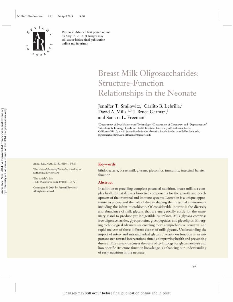
NU34CH14-Freeman ARI 24 April 2014 14:20 R E V I E W S I N A D V A N C E Breast Milk Oligosaccharides: Structure-Function Relationships in the Neonate Jennifer T. Smilowitz, 1 Carlito B. Lebrilla, 2 David A. Mills, 1, 3 J. Bruce German, 1 and Samara L. Freeman 1 1 Department of Food Science and Technology, 2 Department of Chemistry, and 3 Department of Viticulture & Enology, Foods for Health Institute, University of California, Davis, California 95616; email: [email protected], [email protected], [email protected], [email protected], [email protected] Annu. Rev. Nutr. 2014. 34:14.1–14.27 The Annual Review of Nutrition is online at nutr.annualreviews.org This article’s doi: 10.1146/annurev-nutr-071813-105721 Copyright c 2014 by Annual Reviews. All rights reserved Keywords bifidobacteria, breast milk glycans, glycomics, immunity, intestinal barrier function Abstract In addition to providing complete postnatal nutrition, breast milk is a com- plex biofluid that delivers bioactive components for the growth and devel- opment of the intestinal and immune systems. Lactation is a unique oppor- tunity to understand the role of diet in shaping the intestinal environment including the infant microbiome. Of considerable interest is the diversity and abundance of milk glycans that are energetically costly for the mam- mary gland to produce yet indigestible by infants. Milk glycans comprise free oligosaccharides, glycoproteins, glycopeptides, and glycolipids. Emerg- ing technological advances are enabling more comprehensive, sensitive, and rapid analyses of these different classes of milk glycans. Understanding the impact of inter- and intraindividual glycan diversity on function is an im- portant step toward interventions aimed at improving health and preventing disease. This review discusses the state of technology for glycan analysis and how specific structure-function knowledge is enhancing our understanding of early nutrition in the neonate. 14.1 Review in Advance first posted online on May 15, 2014. (Changes may still occur before final publication online and in print.) Changes may still occur before final publication online and in print Annu. Rev. Nutr. 2014.34. Downloaded from www.annualreviews.org by University of California - Davis on 05/30/14. For personal use only.
Transcript of Breast Milk Oligosaccharides: Structure-Function Relationships in … · 2015-12-14 ·...
NU34CH14-Freeman ARI 24 April 2014 14:20
RE V I E W
S
IN
AD V A
NC
E
Breast Milk Oligosaccharides:Structure-FunctionRelationships in the NeonateJennifer T. Smilowitz,1 Carlito B. Lebrilla,2
David A. Mills,1,3 J. Bruce German,1
and Samara L. Freeman1
1Department of Food Science and Technology, 2Department of Chemistry, and 3Department ofViticulture & Enology, Foods for Health Institute, University of California, Davis,California 95616; email: [email protected], [email protected], [email protected],[email protected], [email protected]
Annu. Rev. Nutr. 2014. 34:14.1–14.27
The Annual Review of Nutrition is online atnutr.annualreviews.org
This article’s doi:10.1146/annurev-nutr-071813-105721
Copyright c© 2014 by Annual Reviews.All rights reserved
Keywords
bifidobacteria, breast milk glycans, glycomics, immunity, intestinal barrierfunction
Abstract
In addition to providing complete postnatal nutrition, breast milk is a com-plex biofluid that delivers bioactive components for the growth and devel-opment of the intestinal and immune systems. Lactation is a unique oppor-tunity to understand the role of diet in shaping the intestinal environmentincluding the infant microbiome. Of considerable interest is the diversityand abundance of milk glycans that are energetically costly for the mam-mary gland to produce yet indigestible by infants. Milk glycans comprisefree oligosaccharides, glycoproteins, glycopeptides, and glycolipids. Emerg-ing technological advances are enabling more comprehensive, sensitive, andrapid analyses of these different classes of milk glycans. Understanding theimpact of inter- and intraindividual glycan diversity on function is an im-portant step toward interventions aimed at improving health and preventingdisease. This review discusses the state of technology for glycan analysis andhow specific structure-function knowledge is enhancing our understandingof early nutrition in the neonate.
14.1
Review in Advance first posted online on May 15, 2014. (Changes may still occur before final publication online and in print.)
Changes may still occur before final publication online and in print
Ann
u. R
ev. N
utr.
201
4.34
. Dow
nloa
ded
from
ww
w.a
nnua
lrev
iew
s.or
gby
Uni
vers
ity o
f C
alif
orni
a -
Dav
is o
n 05
/30/
14. F
or p
erso
nal u
se o
nly.

NU34CH14-Freeman ARI 24 April 2014 14:20
Contents
INTRODUCTION . . . . . . . . . . . . . . . . . . . . . . . . . . . . . . . . . . . . . . . . . . . . . . . . . . . . . . . . . . . . . . . 14.2MILK GLYCOMICS: MEASURING HUMAN MILK GLYCANS . . . . . . . . . . . . . . . . 14.3
State of Technology: Human Milk Oligosaccharides. . . . . . . . . . . . . . . . . . . . . . . . . . . . . . 14.3Human Milk Oligosaccharide Structures and Compositions . . . . . . . . . . . . . . . . . . . . . . . 14.5State of Technology: Milk Glycoproteins and Glycopeptides . . . . . . . . . . . . . . . . . . . . . . 14.6State of Technology: Glycolipids . . . . . . . . . . . . . . . . . . . . . . . . . . . . . . . . . . . . . . . . . . . . . . . . 14.8Human Milk Glycolipids Structures and Compositions . . . . . . . . . . . . . . . . . . . . . . . . . . . 14.8
ESTABLISHING STRUCTURE-FUNCTION RELATIONSHIPSOF HUMAN MILK GLYCANS . . . . . . . . . . . . . . . . . . . . . . . . . . . . . . . . . . . . . . . . . . . . . . . 14.9Prebiotics for Infant Gut Bifidobacteria . . . . . . . . . . . . . . . . . . . . . . . . . . . . . . . . . . . . . . . . . . 14.9Pathogen Deflection . . . . . . . . . . . . . . . . . . . . . . . . . . . . . . . . . . . . . . . . . . . . . . . . . . . . . . . . . . . .14.11Intestinal Barrier Function and Immune Modulation . . . . . . . . . . . . . . . . . . . . . . . . . . . . .14.13
UNDERSTANDING THE DIVERSITY OF MILK GLYCANS. . . . . . . . . . . . . . . . . . .14.13The Value of Glycomics for Nutrition Research. . . . . . . . . . . . . . . . . . . . . . . . . . . . . . . . . .14.13Human Milk Oligosaccharides Genetically Determined By Secretor
Status and Lewis Blood Group . . . . . . . . . . . . . . . . . . . . . . . . . . . . . . . . . . . . . . . . . . . . . . .14.14Lactation Stage . . . . . . . . . . . . . . . . . . . . . . . . . . . . . . . . . . . . . . . . . . . . . . . . . . . . . . . . . . . . . . . . .14.16Gestational Age of the Infant . . . . . . . . . . . . . . . . . . . . . . . . . . . . . . . . . . . . . . . . . . . . . . . . . . . .14.16Maternal Health and Phenotype . . . . . . . . . . . . . . . . . . . . . . . . . . . . . . . . . . . . . . . . . . . . . . . . .14.18
CONCLUSIONS AND FUTURE DIRECTIONS . . . . . . . . . . . . . . . . . . . . . . . . . . . . . . . .14.19
INTRODUCTION
The first several months of life are a unique window in time to understand how diet affects thegrowth, development, and protection of the neonate. The gastrointestinal tract is a focal pointfor the remarkable transitions that occur from birth through weaning. As a result of 120 millionyears of evolution, mammals have acquired a means of providing complete postnatal nutrition anddelivery of bioactive components for growth and maturation—lactation (13). Notably, lactation isproviding all these components precisely during the period in which infants develop their innateimmunity and gut microbiota.
Breastfeeding is associated with overt benefits to the neonate. In a thorough review of over 400individual studies, breastfeeding was associated with a reduction in the risk of acute ear infections,asthma (in young children), atopic dermatitis, gastrointestinal infections, respiratory tract diseases,obesity, type 1 and 2 diabetes, childhood leukemia, sudden infant death syndrome in term infants,and necrotizing enterocolitis (NEC) in preterm infants (66). However, one of the long-standingresearch challenges has been to understand how specific structures delivered within breast milkcan explain its diverse functions in vivo. The era of integrating multi-omic technologies and datasets is enabling more comprehensive analyses. Understanding the triad of diet, immunity, and theintestinal microbial ecosystems represents a new frontier for early infant nutrition.
One of the most striking features of breast milk is the diversity and abundance of complex gly-cans that include free human milk oligosaccharides (HMOs): glycoproteins, glycopeptides, andglycolipids. More striking is that these diverse glycan structures are indigestible to the infant andcan reach the large intestine; they can often be found in the stool (32). These diverse milk glycansserve many functions, including protection and development ranging from selectively enriching
14.2 Smilowitz et al.
Changes may still occur before final publication online and in print
Ann
u. R
ev. N
utr.
201
4.34
. Dow
nloa
ded
from
ww
w.a
nnua
lrev
iew
s.or
gby
Uni
vers
ity o
f C
alif
orni
a -
Dav
is o
n 05
/30/
14. F
or p
erso
nal u
se o
nly.
NU34CH14-Freeman ARI 24 April 2014 14:20
gut bifidobacteria; prophylactically binding bacteria, viruses, and toxins; promoting the immunesystem; and enhancing intestinal epithelial barrier function. Taken together, milk glycan functionsshape the intestinal microbiome, from the sterile uterine environment through the chaotic intro-duction of environmental bacteria at birth, through a stable milk-oriented microbiome (MOM)prior to the transition to a more adult-like phenotype after weaning (152). This presents an oppor-tunity to study the structure-function relationships between human milk glycans and their impacton specific aspects of intestinal development. However, elucidating these relationships cannot beachieved without the comprehensive and accurate measurement of the structures and composi-tions of milk, examination of how these elements are degraded in the gastrointestinal tract, anddetermination of how they interact in the intestinal environment.
Advances in analytical chemistry—which have made it possible to accurately and compre-hensively measure the free oligosaccharides and those bound to proteins, peptides, and lipids inmilk—coupled to the powerful and enabling tool sets of microbial meta-genomic, metatranscrip-tomic, and metabolomics in vitro and in vivo provide insight into how the mother-infant dyadfunctions to protect the vulnerable neonate. Milk glycans are variable across lactation and amongwomen. This milk glycan diversity has been found to influence immunity and microbiota in theneonate (44, 78, 90, 91, 113, 123, 128, 135).
This review highlights the milestones in research on glycan structures in breast milk, thefunctions of glycans in the infant, and the impact of maternal phenotype on the structure-functionrelationships; these findings are instructive for nutrition professionals as they begin to unravel theeffect of diet on the intestinal microbiome in health and disease.
MILK GLYCOMICS: MEASURING HUMAN MILK GLYCANS
Glycans have long been recognized as an important determinant of health or disease states. How-ever, the hallmark of glycans is their complexity compared with other polymeric biomoleculessuch as DNA and proteins, whose primary structures are linear with predictable linkages. Thecomplexity of a glycan refers to several different factors including its nonlinear nature, the num-ber of different sugars, the molecular structure of each sugar residue, the linkages between thosesugars leading to multiple isomers for a single mass, and whether they are free or bound to pro-teins, peptides, or lipids in a heterogeneous manner. This complexity contributes to the ongoingresearch in glycomic technologies and methodologies to routinely and comprehensively measurethe glycans in biological and clinical samples. Although breastfeeding has been linked to a vari-ety of functional benefits associated with glycan structures, initial studies provided limited or nostructural information on milk-borne oligosaccharides and/or glycoconjugates. Early studies onthe glycobiology of milk focused on measuring individual sugars, and the technology platformsprovided a piece of the story but were not comprehensive or rapid enough for routine application.Thus, glycomics is defined as the systematic study of the total complement of sugars present inan organism in their free or bound state (6). Glycomics represents a critical gap in knowledge innutrition and clinical research, especially in an era of applied omic technologies. Elucidating theglycan structures that are covalently bound to peptides, proteins, and lipids requires merging thefields of glycomics with proteomics and lipidomics to truly elucidate molecular structure (108).Documenting structural diversity through comprehensive glycan analysis provides an importantopportunity for understanding diet and health in the neonate.
State of Technology: Human Milk Oligosaccharides
HMOs are a particularly interesting class of molecules that have gained considerable attentionbecause they are an abundant (1–2% w/v) and structurally diverse component of breast milk,
www.annualreviews.org • Milk Glycans: Structure and Function 14.3
Changes may still occur before final publication online and in print
Ann
u. R
ev. N
utr.
201
4.34
. Dow
nloa
ded
from
ww
w.a
nnua
lrev
iew
s.or
gby
Uni
vers
ity o
f C
alif
orni
a -
Dav
is o
n 05
/30/
14. F
or p
erso
nal u
se o
nly.

NU34CH14-Freeman ARI 24 April 2014 14:20
yet they are indigestible by the neonate (30, 31). The widespread application of comprehensivemilk glycan analysis is dependent on sensitivity, reproducibility, high-throughput and speed of thetechnologies used to identify specific structural isomers in complex biological samples. MonitoringHMO abundances have traditionally been performed with several separation methods includingvarious types of high-performance liquid chromatography (HPLC) and capillary electrophoresisusing standard compounds (31, 121). Liquid chromatographic (LC) methods such as hydrophilicinteraction LC, reverse phase (C18), and porous graphitized carbon have been used to separateoligosaccharide isomers (147, 148). Porous graphitized carbon on native compounds has emergedas the best method for separating isomers (147, 148). However, LC methods are severely limitedby the small number of HMO standards that are commercially available. The lack of standardsis complicated by the difficulty in elucidating HMO structures. Nuclear magnetic resonance haspreviously been the major method for structural elucidation (122). However, it requires relativelylarge amounts of pure compounds making it impractical for all except the most abundant species.
Mass spectrometry (MS) is both a sensitive detector for LC methods and a tool for struc-tural elucidation (58). Structural elucidation provides information on the linkages between sugarresidues within each HMO molecule to determine the structural isomers present, whereas HMOcomposition provides information on the different sugar residues present but lacks linkage infor-mation. Tandem MS methods provide structural information and have been used for the structuralelucidation of many types of oligosaccharides. MS has been used to obtain complete structures(7, 132); however, this method also relies on known standard structural features. Linkages can beobtained with tandem MS, but distinguishing stereoisomers, such as glucose (Glc) versus galactose(Gal) versus mannose, is not possible. In general, de novo structural elucidation with tandem MSis not feasible with current methods. An effective method for the structural elucidation of HMOsemploys the combination of MS, tandem MS, and exoglycosidase digestion. In a systematic studyof neutral milk oligosaccharides (147) and anionic oligosaccharides (148), 75 HMO structuralisomers were determined and annotated. This library has been extended to include more than200 complete structures, including milks from other mammals. It also has been shown that only50 structures represent 99% of the abundances in human milks (4, 97, 147, 148). This approachleverages on oligosaccharide analysis by using accurate mass, reproducible retention times througheffective isomer separation (HPLC) and tandem MS (for less than 5% of the structures).
The quantitation of HMO structures in a complex mixture, i.e., biofluids, is necessary forphenotyping health and disease in the field of nutrition. Relative abundances of structures (orcompositions) provide quantitative information that can be used to obtain fold changes betweengroups of samples because of the high precision of the method. Absolute quantitation can beobtained by comparing peak areas to known standards. LC/MS quantitation is performed using anumber of methods. The most direct is to use ion count, which can be calibrated to some knownamount of compound. This approach assumes that all ionization efficiencies are equal, which is notstrictly true for oligosaccharides. Nonetheless, the method is sufficient for determining specificchanges in abundances. HMOs are often labeled with chromophoric tags such as anthranilic acidor 2-aminobenzamide for quantitation. Deuterium labeling has also been used for HMOs. Nano-LC MS is useful for performing quantitation using both deuterium labeling and total ion countsmethods. Whereas deuterium labeling provides very precise and accurate values (99), total ioncount by chromatography produces a suitable level of quantitation, as validated by deuteriumlabeling (124). More recently, multiple reaction monitoring using triple quadrupole MS has beenused for bovine milk oligosaccharides (43). The method was used to monitor a small number ofsialylated structures, which make up the major portion of bovine milk oligosaccharides. Multiplereaction monitoring of oligosaccharides is still relatively new but will be used for many moreapplications in the future.
14.4 Smilowitz et al.
Changes may still occur before final publication online and in print
Ann
u. R
ev. N
utr.
201
4.34
. Dow
nloa
ded
from
ww
w.a
nnua
lrev
iew
s.or
gby
Uni
vers
ity o
f C
alif
orni
a -
Dav
is o
n 05
/30/
14. F
or p
erso
nal u
se o
nly.
NU34CH14-Freeman ARI 24 April 2014 14:20
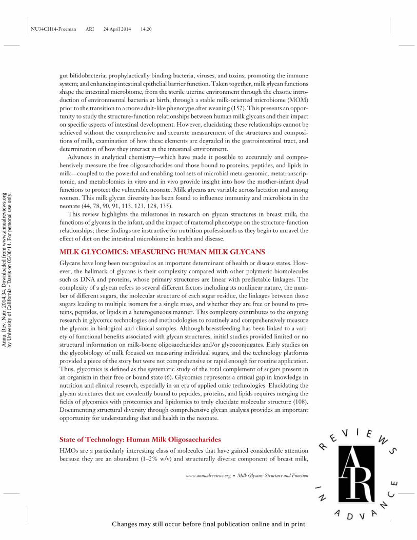
Human Milk Oligosaccharide Structures and Compositions
Human milk contains higher amounts and more complex structures of soluble oligosaccharidesthan any other mammalian milk (127); average amounts range from 7 g/L in mature breast milkto 23 g/L in colostrum (31, 46). The functional implications of the structural diversity are animportant area of research. HMOs are soluble complex and diverse sugars derived of Glc, Gal,N-acetylglucosamine (GlcNAc), fucose (Fuc), or sialic acid (Neu5Ac) monosaccharides. Thebiosynthesis of HMOs in the mammary gland begins with the formation of a lactose core from Galand Glc catalyzed by β-galactotransferase in the presence of α-lactalbumin. With few exceptions,all HMO structures consist of a lactose core (74). Lactose can be elongated enzymatically by β1-3linkage to lacto-N-biose or by β1-6 linkage to N-acetyllactosamine. The core HMO structurecan be further elongated by the addition of lacto-N-biose and N-acetyllactosamine units byβ1-3 and β1-6 linkages; Fuc connected with α1-2, α1-3, or α1-4 linkages, and/or sialic acidresidues attached by α2-3 or α2-6 linkages at the terminal positions (Figure 1). The proportion
Glucose
Galactose
N-acetylglucosamine
Fucose
Sialic acid
Monosaccharide key:
β1-4
α1-2
2'-FL 3FLβ1-3 β1-4
β1-4
β1-3
β1-6
Lacto-N-biose Lactose
N-acetyllactosamine
n = 0–15
α1-2α1-3α1-4
a b
e
β1-4
α1-3
α2-3α2-6
β1-4
α2-3
3'-SL 6'-SL
β1-4
α2-6
dβ1-4
β1-3
α1-2
β1-3
LNFP I
cβ1-4
β1-3
β1-3 β1-3
β1-4
β1-4
LNH
β1-4
β1-6 β1-4
β1-3
β1-3
LNT(type I)
LNnT(type II)
β1-4
β1-3
α1-4
β1-3
LNFP II
β1-4
β1-4
α1-3
β1-3
LNFP III
β1-4
β1-3
α1-3
β1-3
LNFP V
β1-4
β1-3
α2-3
β1-3
LST a
β1-4
β1-3
α2-6
β1-3
LST b
β1-4
β1-4β1-3
LST c
α2-6
Figure 1An example of the structural diversity of human milk oligosaccharides (HMOs). (a) With few exceptions, all HMO structures consist ofa lactose core linked to lacto-N-biose or to N-acetyllactosamine with n = 0–15 units. (b) Lactose can be fucosylated or sialylated bydifferent linkages. (c) Lactose can be elongated enzymatically in repeats of lacto-N-biose (type I) or N-acetyllactosamine (type II).(d ) Elongated type I or II chains can be fucosylated in different linkages to form structures that phenotypically describe secretor statusand the Lewis blood group. (e) The elongated core HMO structures can be sialylated by α2-3 or α2-6 linkages at the terminal positionsforming structural isomers. Abbreviations: 2′-FL, 2′-fucosyllactose; 3FL, 3-fucosyllactose; 3′-SL, 3′ sialyllactose; 6′-SL, 6′-sialyllactose;LNFP I, II, III, V, lacto-N-fucopentaose I, II, III, V; LNH, lacto-N-hexaose; LNnT, lacto-N-neotetraose; LNT, lacto-N-tetraose;LST a, b, c, sialyl-lacto-N-tetraoses a–c. Adapted from Reference 15 with permission.
www.annualreviews.org • Milk Glycans: Structure and Function 14.5
Changes may still occur before final publication online and in print
Ann
u. R
ev. N
utr.
201
4.34
. Dow
nloa
ded
from
ww
w.a
nnua
lrev
iew
s.or
gby
Uni
vers
ity o
f C
alif
orni
a -
Dav
is o
n 05
/30/
14. F
or p
erso
nal u
se o
nly.
NU34CH14-Freeman ARI 24 April 2014 14:20
of fucosylated, sialylated, and nonfucosylated neutral HMOs in term breast milk was recentlyreported as 35–50%, 12–14% and 42–55%, respectively (131).
State of Technology: Milk Glycoproteins and Glycopeptides
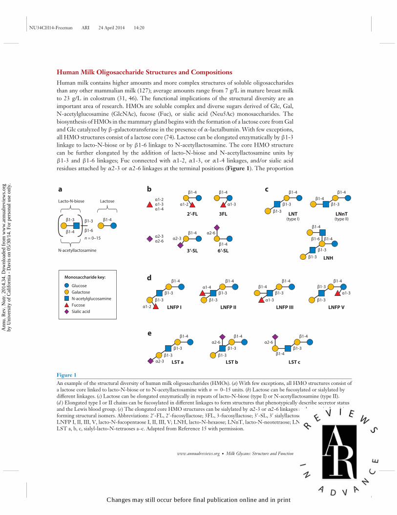
Protein glycosylation is a common and complicated type of posttranslational modification that di-rectly affects glycoprotein structure and protective functions through cell signaling and cell-to-cellrecognition events, enrichment of protective gut microbiota, modulation of pathogen adhesion andinvasion of the infant intestinal mucosa, and neutralization of antigens (1). The protein portion iscleaved by digestive proteases in the neonatal intestine, yet its glycosylation restricts digestion suchthat the digestive products and their absorption rates are dependent on the diversity, abundance,and location of oligosaccharide structures present on the protein. The level of proteolytic cleav-age therefore has both nutritive and biological consequences. The release of bioactive peptidesfrom proteins may be affected by glycosylation in at least two ways. Glycosylation limits access toprotease and peptidase cleavage sites and determines the ability of the remaining glycopeptide topersist and transit to lower parts of the intestine. The glycosylation of immune protective proteinsin milk render them partially resistant to digestion and supports their biological functions in thedistal gut. In addition, these milk glycoproteins (72) and glycopeptides (79, 103) possess glycans,which structurally resemble HMOs and can serve as growth substrates for infant-borne bifidobac-teria. Indeed, some bifidobacteria possess specific endoglycosidases that enable these strains tocleave the glycan portion away from the cognate glycoproteins, thus freeing the glycan substratefor consumption (Figure 2) (50, 72).
Unlike free milk oligosaccharides, glycoconjugates are complicated due to the respective pro-tein or lipid moieties. Two types of protein glycosylation exist: N-linked and O-linked. N-glycansare found on proteins with a consensus sequence NXT/S where N is asparagine, X is any aminoacid except proline, and the third amino acid can be either threonine (T) or serine (S) and, inrare cases, cysteine. O-glycosylation may occur at any serine or threonine residue with no singlecommon core structure or consensus protein sequence. Because glycosylation is dictated by a set ofcompeting glycosyl transferases, the glycosylation patterns of glycoproteins are very complicated.The population of glycans occurring at a given glycosylation site is often heterogeneous such thata specific N-glycosylation and O-glycosylation site may be occupied by a number of structurallydistinct glycans and described as microheterogeneous (4). For example, a protein containing threeglycosylation sites with 10 different glycans in each site can result in approximately 1,000 differentglycoforms of the protein.
Quantitative methods that monitor both protein abundances and site-specific glycosylationremain a significant analytical challenge; however, solving this problem will provide greater un-derstanding of protein functions in vivo. Several approaches are used to study the complexitiesinvolved in protein glycosylation. The first is to enzymatically release glycan moieties from theprotein molecule to observe the total glycan structures present. This method provides the glycanheterogeneity. This method can also yield site occupancy when the peptide is captured before gly-can release. However, relating glycan structure to specific sites is not possible. Another approachis to determine site-specific glycosylation where the intact glycopeptide is measured and is usedto retain information on location and microheterogeneity. Although this glycoproteomic methodis complicated considerably by the analysis of glycopeptides, it may ultimately provide the level ofdetail required to measure biological differences in milk across lactation and among women andto determine glycan metabolism within the infant (34).
The large number of glycoforms and the presence of multiple glycan-protein linkage siteshave made comprehensive glycoproteomics—the simultaneous determination of glycosites and
14.6 Smilowitz et al.
Changes may still occur before final publication online and in print
Ann
u. R
ev. N
utr.
201
4.34
. Dow
nloa
ded
from
ww
w.a
nnua
lrev
iew
s.or
gby
Uni
vers
ity o
f C
alif
orni
a -
Dav
is o
n 05
/30/
14. F
or p
erso
nal u
se o
nly.

NU34CH14-Freeman ARI 24 April 2014 14:20
β1-3
β1-4
β1-3
β1-4
β1-6
β1-4
β1-2
α2-3
α1-3/4α1-2α2-6
β1-4
β1-2
α2-6
α2-8 α2-3
α1-6
α1-6
α1-3
α2-3
α2-6
β1-4 β1-3
β1-3
β1-4
β1-3/4
β1-3
β1-6
α1-3
β1-4
HMO
1
3
2
5 6
4
7/10
98
Ceramide14 6 13 9
Glycolipid
Complex N-glycan O-linked glycans
Asn
16
17
18 18
19 19 2
4 4
5
15
12 8/10
5
3/4
Core 3
11
3
Core 16
Glucose
Galactose
N-acetylglucosamine
Fucose
Sialic acid
Monosaccharide key:
1
2
3
4
5
6
7
8
9
10
α1-2 fucosidase
α1-3/4 fucosidase
β1-3 galactosidase
β1-4 galactosidase
α2-6 sialidase
α2-3 sialidase
β1-3 N-acetylglucosaminidase
β1-6 N-acetylglucosaminidase
β1-4 galactosidase (lactase)
Lacto-N-biosidase
11
12
13
14
15
16
17
18
19
Endo-α-N-acetylgalactosaminidase (EngBF)
Endo-α-N-acetylgalactosaminidase (NagBb)
α2-8 sialidase
Endoglucosylceraminidase
α1-6 fucosidase
Endo-β-N-acetylglucosaminidase
β1-4 mannosidase
α-mannosidase
β1-2 N-acetylglucosaminidase
Enzyme key:
Ser/Thr
Ser/Thr
Ser/Thr11/12
4
Core 2 3
8
Figure 2Infant gut-associated bifidobacteria cleave a diverse range of specific linkages within human milk glycansusing a variety of glycosyl hydrolases. Legend at bottom indicates the monosaccharide composition andcorresponding potential glycolytic enzymes in bifidobacteria that cleave the specific linkages. The figuredepicts the structure of HMO, a complex N-glycan, three different cores found in human O-linked glycans,and the glycolipid structure of ganglioside GD3. Adapted from Reference 51 with permission.
site-specific microheterogeneity—currently impossible. For this reason, the focus has been oneither determining the site of glycosylation on many proteins or determining the site-specificheterogeneity of a few proteins. It is important to determine the glycan microheterogeneity ofthe protein to understand the role of glycosylation in protein function. Common glycoproteomicmethods used for determining site occupancy rely on methods that enrich tryptic glycopeptides(2). To identify the peptide, the glycans are released by the enzyme peptide-N-glycosidase F,thereby losing all information regarding glycan structures. Peptide-N-glycosidase F is an ami-dase that cleaves between the innermost GlcNAc and asparagine residues of high mannose, hy-brid, and complex oligosaccharides from N-linked glycoproteins. The alternative is to determinesite-specific glycosylation with microheterogeneity of a targeted group of proteins. Site-specificmapping can be performed using a procedure with nonspecific proteases to digest the proteinsinto short glycopeptide chains (101). Structural elucidation is possible by coupling accurate mass,tandem MS with bioinformatic tools (125). This highly targeted analysis can be performed on asmall number of proteins in a complex sample.
www.annualreviews.org • Milk Glycans: Structure and Function 14.7
Changes may still occur before final publication online and in print
Ann
u. R
ev. N
utr.
201
4.34
. Dow
nloa
ded
from
ww
w.a
nnua
lrev
iew
s.or
gby
Uni
vers
ity o
f C
alif
orni
a -
Dav
is o
n 05
/30/
14. F
or p
erso
nal u
se o
nly.
NU34CH14-Freeman ARI 24 April 2014 14:20
Several proteomic studies have attempted to examine the breadth of proteins in human milk.However, there are very few glycoproteomic analyses of milk. Mechref et al. (89) examined the N-glycans on bile salt–stimulated lipase (BSSL). The glycosylation varied both in absolute quantity ofmonosaccharide residues and their composition between the first and the sixth month of lactation.BSSL was found to eventually lose all glycosylation during late lactation. Charlwood et al. (23)reported the N-glycosylation of four abundant proteins in milk fat globule membrane. Froehlichet al. (44) have shown that several human milk glycoproteins including lactoferrin, BSSL, tenascin,immunoglobulin A (IgA), and xanthine dehydrogenase exhibit dynamic glycosylation behaviorduring the lactation period.
State of Technology: Glycolipids
In milk, glycolipids are found almost exclusively in the outer part of the milk fat globule mem-brane. These glycolipids occur mainly in the form of glycosphingolipids with a dominance ofNeu5Ac-containing gangliosides. The combination of the polar head (the oligosaccharide) andthe nonpolar lipid tail (ceramide) provides technical challenges from either the free oligosac-charides or the glycoproteins. The catalog of glycolipid structures has been achieved throughthe release and separation of the polar head group from the nonpolar tail using multiple chro-matographic methods. The lipid moieties are best separated by reverse-phase LC, whereas theglycan head group is best separated by hydrophilic interaction LC. The different methods separateoligosaccharides or the lipid moiety, but not both simultaneously. Numerous reviews have pub-lished information on human milk glycolipids (see, e.g., 94, 105, 140). These structures have beenelucidated through complex and laborious analytical workflows that have been the status quo inthe field. However, to achieve high-throughput, routine analysis in determining structural diver-sity in complex biological fluids including milk, new methods must take fundamentally differentapproaches to separate, detect, and identify the intact glycolipids. There is a risk of underesti-mating sialic acid residues because they can be labile and lost as a result of sample preparation.Furthermore, measurement of low-abundant gangliosides that might be functionally active couldbe missed owing to the presence of more abundant lipids that are more easily ionized. Rapidprofiling of breast milk glycolipids is possible using matrix-assisted desorption/ionization Fouriertransform ion cyclotron resonance tandem MS (76). Comprehensive methods that overcome theinherent challenges of glycolipid structure will improve our understanding of biological variationand increase the ability to monitor changes in these specific molecules as a function of time orhealth status or postconsumption.
Human Milk Glycolipids Structures and Compositions
The heterogeneity of the oligosaccharide structures conjugated to milk lipids is lower comparedto free oligosaccharides and glycoproteins and is limited to a few known structures. The variationsin glycolipids are primarily due to the heterogeneity of the lipid tail, which varies in carbon lengthand in the number and location of double bonds (16). The concentration of gangliosides (sialylatedglycolipids) in human colostrum and mature human breast milk is roughly the same ∼9 mg/L (104),with GD3 [Neu5Ac(α2-8)Neu5Ac(α2-3)Gal(β1-4)Glc(β1-1) ceramide] and GM3 [Neu5Ac(α2-3)Gal(β1-4)Glc(β1-1) ceramide] as the most abundant structures (76). Yet, the composition ofsialylated gangliosides varies across lactation. The ganglioside GD3 is highest in human colostrum,whereas GM3 is highest in mature breast milk (105). The gangliosides GM2 [GalNAc(β1-4)Neu5Ac(α2-3)Gal(β1-4)Glc(β1-1) ceramide] and GM1 [Gal(β1-3)GalNAc(β1-4)Neu5Ac(α2-3)Gal(β1-4)Glc(β1-1) ceramide] are found in human milk in microgram concentrations (94).
14.8 Smilowitz et al.
Changes may still occur before final publication online and in print
Ann
u. R
ev. N
utr.
201
4.34
. Dow
nloa
ded
from
ww
w.a
nnua
lrev
iew
s.or
gby
Uni
vers
ity o
f C
alif
orni
a -
Dav
is o
n 05
/30/
14. F
or p
erso
nal u
se o
nly.
NU34CH14-Freeman ARI 24 April 2014 14:20
ESTABLISHING STRUCTURE-FUNCTION RELATIONSHIPSOF HUMAN MILK GLYCANS
As discussed previously, HMOs are abundant in human milk and are indigestible by humans, yetevidence suggests at least three major functions for these sugars. First, HMOs promote growthof a milk-oriented microbiota (152), often dominated by bifidobacteria (149). Second, HMOs(especially fucosylated structures) resemble the host epithelial cell surface glycans and thus func-tion as soluble receptor analogs that compete for bacterial binding against the intestinal mucosa,preventing intestinal pathogen adhesion to epithelial surfaces and translocation (95). Third, milkglycans improve host defense by modulating immunity and promoting intestinal barrier function(27). Enrichment of intestinal bifidobacteria, pathogen deflection, and direct cell-surface medi-ated regulatory events involving the growth and maintenance of epithelia all contribute to hostprotection and are mediated by milk glycans.
Glycosylation and glycan diversity are directly related to the modulation of microbial adhesionand invasion during infection (87). Indeed, the first step in bacterial infection is the recognitionof host glycans by bacterial lectins or vice versa. Thus, glycans of human milk proteins can blockor modulate pathogen association to epithelial surfaces, which, given the large amount of glycansdelivered to the infant, partially explains the protection of breastfed infants against gastrointestinaltract infections. Human milk lactoferrin, the major milk glycoprotein, binds to pathogenic gram-positive (107) and gram-negative (102) bacteria, exerting antimicrobial activity via iron-depletionand/or bacterial membrane disruption. Human lactoferrin contains three N-linked glycosylationsites at asparagine 138, 479, and 624 (138), and its glycosylation influences the glycoprotein’ssusceptibility to proteolysis (137), thus affecting the production of potent active peptides(e.g., lactoferricin) and glycopeptides involved in its biological activities (9, 77). Gangliosidesare widely distributed in cellular membranes including the milk fat globule membrane. Withtheir oligosaccharide head group facing the external environment, gangliosides function inhost-pathogen interactions, cell-cell recognition, and modulation of membrane protein function.Additionally, milk gangliosides are proposed to modulate immunity and to prevent infection by act-ing as decoys that interfere with pathogenic binding to host cell receptors of the intestinal mucosa(110).
Prebiotics for Infant Gut Bifidobacteria
Breastfed infants are typically colonized by protective strains of bacteria that are thought to protect,feed, and communicate with the developing intestine (38). More than 100 years ago, Henry Tissierfirst demonstrated that the feces of breastfed infants contained a bacterial isolate he termed Bacillusbifidus communis (130). Since that time, numerous culture-based studies, and more recently, DNA-based culture-independent methods (61, 109, 114, 149), clearly demonstrated a predominance ofbifidobacterial species within the first months of breastfeeding prior to weaning and a transition toa more adult-like microbiota profile (62). Of the bifidobacterial species common to the breastfedintestinal tract, Bifidobacterium longum and B. breve are most frequently observed; B. bifidum,B. pseudocatenulatum, and B. catenulatum are found less often (134).
Gyorgy and colleagues (57) first showed that B. bifidum (then termed Lactobacillus bifidus) wasuniquely able to grow on human milk glycan fractions. Humans lack the various glycolytic enzymesthat break down HMOs, and various researchers have shown that these glycans reach the colonintact (25, 30, 39). Thus enrichment of bifidobacteria is believed to be driven in part throughthe prebiotic effect of free and bound glycans present in human milk (53, 75). Ward et al. (142,143) first demonstrated the selective growth of bifidobacterial species on intact HMO in vitro.Subsequent studies have confirmed that only certain bifidobacterial species vigorously consume
www.annualreviews.org • Milk Glycans: Structure and Function 14.9
Changes may still occur before final publication online and in print
Ann
u. R
ev. N
utr.
201
4.34
. Dow
nloa
ded
from
ww
w.a
nnua
lrev
iew
s.or
gby
Uni
vers
ity o
f C
alif
orni
a -
Dav
is o
n 05
/30/
14. F
or p
erso
nal u
se o
nly.

NU34CH14-Freeman ARI 24 April 2014 14:20
HMOs (53, 83, 115, 123, 133). B. longum subsp. infantis (B. infantis) (54, 77, 114, 137, 139) andselect B. breve (111, 149) preferentially consume smaller fucosylated and sialylated HMOs. It isclear the bifidobacterial strains that grow well on HMOs have acquired these specific geneticadaptations for select growth on human milk glycans (111, 117). Supporting a prebiotic conceptfor HMOs, Yu et al. (150) recently showed that certain HMO species promote bifidobacterialgrowth within in vitro fecal enrichment assays.
A number of studies have characterized the bifidobacterial moieties that specifically bind andcatabolize HMOs (51, 82, 86, 111). Different bifidobacterial species grow on HMO by differ-ent catalytic mechanisms. For example, B. infantis, a predominant bifidobacterial species in thebreastfed infant colon, possesses a 43-kb gene cluster that encodes transport systems (49) andintracellular glycosyl hydrolases (49, 117–119), which suggests that HMOs are internalized anddegraded by this subspecies (51, 52). However, B. bifidum employs a different mode of catalyticactivity toward HMO consumption by exporting sialidases, fucosidases, and a lacto-N-biosidase toliberate lacto-N-biose from HMO structures; lacto-N-biose is then transported and metabolized(71) (Figure 3).
Milk-derived secretory IgA (sIgA), lactoferrin, and haptocorrin are generally believed to bepartially resistant to proteolysis and remain partially intact through the gastrointestinal tract(84). Numerous researchers have shown that milk glycoproteins provide some enrichment forbifidobacteria in vitro (8, 63, 70, 106) and in vivo (26, 141). However, it is not always clear ifthe protein or glycan components (or both) are responsible for the enrichment. Studies haveidentified milk peptides with bifidobacterial growth-enhancing capacities (79, 103); however,enrichment via these peptides remains to be determined in vivo. The contribution of the glycanportion of these glycoconjugates to the ascribed activities has not been extensively studied becauseof methodological limitations for the comprehensive structural elucidation and quantitation.Degradation of the glycan portion of complex human milk glycoproteins requires a repertoireof endo- and exoglycosidases and cognate transport systems to make milk glycans available totheir central metabolic pathways. Recently, specific cell wall–associated endoglycosidases thatare employed by different bifidobacteria initially to degrade O- and N-linked glycoproteins havebeen identified. Garrido et al. (50) showed that select infant-borne bifidobacteria possess an endo-β-N-acetylglucosaminidase that releases glycans from N-linked glycoproteins at the chitobiosecore. An endo-β-acetylgalactosaminidase that cleaves O-linked glycans has also been identifiedin B. bifidum and select other bifidobacterial strains (72). This latter enzyme is likely involvedin both milk and mucin degradation. These endoglycosidases release the free glycans, which arethen catabolized via the endogenous HMO consumption pathways for a particular bifidobacterialstrain.
Growth on milk glycans confers a specific HMO phenotype to B. infantis that is mechanisti-cally linked to its success in establishing itself and persisting in the infant intestine. Chichlowskiet al. (27) showed that growth of B. infantis ATCC15697 on HMOs increases binding to intestinalepithelial cells in vitro, decreases release of inflammatory cytokines, and increases release of anti-inflammatory cytokines in response to an inflammatory stimulus. A similar increase in binding wasdetermined using sialyllactose (69). These studies suggest that the specific growth phenotype ofmilk glycan–enriched bifidobacterial populations promotes persistence in situ and positively mod-ulates the host epithelium. The promotion of the milk glycan–enriched bifidobacterial populationis also supported by in vivo administration of B. infantis to premature infants fed either formulaor breast milk (136). Importantly, when delivered in combination with breastfeeding, B. infantiswas shown to dominate the premature infant gastrointestinal tract, whereas B. lactis, a strain thatdoes not grow on HMO, did not persist at all. This is the first evidence of the importance ofHMO catabolism in bifidobacterial persistence in vivo. Moreover, the combination of B. infantis
14.10 Smilowitz et al.
Changes may still occur before final publication online and in print
Ann
u. R
ev. N
utr.
201
4.34
. Dow
nloa
ded
from
ww
w.a
nnua
lrev
iew
s.or
gby
Uni
vers
ity o
f C
alif
orni
a -
Dav
is o
n 05
/30/
14. F
or p
erso
nal u
se o
nly.

NU34CH14-Freeman ARI 24 April 2014 14:20
β1-3
β1-4
β1-3β1-4
β1-6
α2-3
α1-3/4α1-2
α2-6
B. bifidum
B. infantisB. breveB. longum
LNT-type 1 HMOABC importer
Extracellulardigestion
Membranetransport
Type 2 HMO-mucin glycansABC importers
LNB-GNB and blood group glycansABC importer
2' FL and H-disaccharideABC importers
α1-2 fucosidase
α1-3/4 fucosidase
β1-3/6 hexosaminidase
α2-3/6 sialidase
β1-3 galactosidase
β1-4 galactosidase
α1-2 fucosidase
α1-3/4 fucosidase
β1-3/6 hexosaminidase
α2-3/6 sialidase
β1-3/4 galactosidase
Lacto-N-biosidase
Centralmetabolism
LNBphosphorylase
Centralmetabolism
GNB/LNB ABC importer
Fucose permeaseGalactoside symporter
Phosphotransferase system(N-acetylglucosamine)
HMO
Glucose
Galactose
N-acetylglucosamine
Fucose
Sialic acid
Monosaccharide key:
Figure 3Possible strategies for human milk oligosaccharide (HMO) consumption in Bifidobacterium bifidum, B. infantis, B. breve, and B. longum.Dashed lines in the HMO panel represent potential linkages. Abbreviations: GNB, galacto-N-biose; LNB, lacto-N-biose. Adaptedfrom Reference 48 with permission.
supplementation and breastfeeding led to decreases in Gammaproteobacteria compared with amatched formula-fed group. Follow-up clinical trials are warranted in premature infants who areat risk for developing NEC, an inflammatory bowel disease, to determine if proliferation of milkglycan–enriched bifidobacterial populations leads to the anti-inflammatory and intestinal barrierfunctions established in vitro.
Pathogen Deflection
In addition to their prebiotic functions, HMOs also compete for specific pathogen binding withsites in the infant gut (93). Many viral, bacterial, or protozoan pathogens need to adhere to intestinal
www.annualreviews.org • Milk Glycans: Structure and Function 14.11
Changes may still occur before final publication online and in print
Ann
u. R
ev. N
utr.
201
4.34
. Dow
nloa
ded
from
ww
w.a
nnua
lrev
iew
s.or
gby
Uni
vers
ity o
f C
alif
orni
a -
Dav
is o
n 05
/30/
14. F
or p
erso
nal u
se o
nly.
NU34CH14-Freeman ARI 24 April 2014 14:20
epithelial surfaces to colonize or invade the host and cause disease. Human milk glycans andintestinal epithelial glycans are synthesized by similar glycosyltransferases and thus have commonepitopes. Ingested milk glycans act as pathogen decoys by binding to pathogens and their toxins,thereby limiting their binding to intestinal epithelial mucosal surfaces (95). Milk glycans bindviruses such as HIV (64) and rotavirus (65), pathogens such as Vibrio cholerae, Salmonella fyris,and enteropathogenic Escherichia coli (33), and enterotoxigenic Escherichia coli and caliciviruses(93) and Streptococcus pneumoniae (5). The large diversity of HMO structures suggests a largediversity of decoy functions (Figure 1) (17). Separated HMO fractions have been shown to havediffering activities. For instance, fucosylated HMOs inhibit the binding of Campylobacter jejuni tointestinal cells (93), whereas sialylated HMOs block the adhesion of E. coli to human erythrocytes(88).
Milk glycoproteins are well recognized for their protective functions in the neonate (84). Re-cently, human milk lactoferrin was found to significantly inhibit pathogen adhesion to colonicepithelial cells, and purified human milk lactoferrin glycans significantly reduced Salmonella in-vasion of colonic epithelial cells to levels associated with noninvasive deletion mutants (9). Thesedata suggest that glycan variation of human milk lactoferrin is involved in modulating pathogenassociation. sIgA, the predominant immunoglobulin in human milk (28), provides protection toneonates by coating microorganisms and inhibiting colonization and neutralizing viral and bac-terial endotoxins (19). sIgA is heavily glycosylated with N- and O-linked oligosaccharides (151),which vary according to the isotype and allotype of the immunoglobulin. Its oligosaccharidestructural complexity includes its role in immune protection in cell signaling, cell-cell recogni-tion, and microbial adhesion and invasion (1). sIgA is essential in providing passive immunity toinfants against infections (60). For example, specific binding between sialylated glycans of sIgAand pathogens protects newborns from sepsis and meningitis caused by infection by S-fimbriatedE. coli (115). In particular, the large diversity of N-glycan structures at seven potential sites on thesecretory component of the immunoglobulin creates many glycan epitopes that are potent decoysfor lectins on bacterial surfaces, thereby inhibiting infection through attachment with epithelialsurfaces (18, 41, 146).
The human milk fat globule membrane contains many membrane-associated glycoconjugatesproposed to protect the nursing infant by offering an alternate pathogen binding site (96). Gan-glioside composition of milk is implicated in binding and neutralizing various pathogens andtheir toxins such as enteropathogenic E. coli, C. jejuni, L. monocytogenes, Salmonella enterica (Typhi),S. sonnei, Helicobacter pylori vacuolating toxin, and cholera toxin (36, 105). The interaction betweenthe membrane-associated gangliosides GM1 and cholera toxin protein subunit B is a well-studiedsystem and is frequently employed for assessing the capabilities of any new analytical technique(20, 36). For example, gangliosides from pooled human milk exhibited the ability to reduce thebinding of cholera toxin to GM1 by 93%, and structures from goat milk completely inhibitedthe binding of botulinum type A neurotoxin to trisialoganglioside-GT1b (67). Gangliosides alsoexert immune-modulating effects on immune cells such as dendritic cells. Treatment of bonemarrow–derived dendritic cells with GD3 before lipopolysaccharide-induced maturation signif-icantly reduced the production of various proinflammatory cytokines and reduced the activationof CD4+ cell proliferation compared with treatment with GM3 (21). These data suggest thatGD3, which is more abundant in colostrum than GM3, may function to reduce the excessiveimmune responses against the large amounts of foreign antigens, albeit harmless, the infant en-counters during the first weeks of life. Human studies designed to confirm the effects of glycanstructures for their antiadhesive and anti-inflammatory and immunomodulatory functions arewarranted.
14.12 Smilowitz et al.
Changes may still occur before final publication online and in print
Ann
u. R
ev. N
utr.
201
4.34
. Dow
nloa
ded
from
ww
w.a
nnua
lrev
iew
s.or
gby
Uni
vers
ity o
f C
alif
orni
a -
Dav
is o
n 05
/30/
14. F
or p
erso
nal u
se o
nly.

NU34CH14-Freeman ARI 24 April 2014 14:20
Intestinal Barrier Function and Immune Modulation
The gastrointestinal tract is a complex organ system with local and regional morpholog-ical and physiological differences that lead to digestion and absorption of nutrients whileproviding a barrier against foreign agents. However, at birth the neonate’s gastrointestinal tractis functionally immature and immune incompetent (112), although it is confronted with an in-creasing antigenic load in the form of dietary proteins, commensal organisms, and pathogens. Thesingle layer of enterocytes that line the intestinal epithelium forms a functional barrier betweenthe luminal contents of the gut and the infant’s circulatory system. Permeability across the in-testinal epithelium is determined in part by the rate-limiting barrier of the paracellular pathwayinvolving tight junctions (100). This critical barrier is responsible for allowing nutrients and ben-eficial macromolecules to cross through the intestinal epithelium while preventing paracellulartranslocation of bacteria and bacterial products.
Human milk glycans support intestinal barrier function and modulate immunity through avariety of mechanisms. First, the abundance of infant-dominated gut bifidobacteria that resultsfrom the prebiotic function of HMOs and glycans leads to lower gastrointestinal pH through theproduction of short-chain fatty acids (in particular, acetate), which enhances gut barrier function(45). Second, binding of B. infantis grown on HMOs to intestinal cells in vitro enhanced tightjunction protein expression and immunomodulatory interleukin (IL)-10 (27). Third, B. infantis–conditioned media prevented tumor necrosis factor alpha and interferon gamma reduction intransepithelial resistance (a measurement of intestinal barrier function) and rearrangement of tightjunction proteins in intestinal epithelial cells and attenuated inflammation, normalized colonic per-meability, and decreased colonic and splenic interferon gamma secretion in a colitis mouse model(40). Ganguli et al. (47) demonstrated that B. infantis and Lactobacillus acidophilus conditionedmedia-attenuated lipopolysaccharide- and IL-1β-induced IL-8 and IL-6 expression; decreasedtoll-like receptor 2 and 4 mRNA; and increased specific negative regulators of inflammation ofimmature human enterocytes, immature human intestinal xenografts, and primary enterocytecultures of NEC tissue, with superior effects by B. infantis. These data suggest strain specificityin reducing intestinal inflammation and could explain one mechanism for the reduced rates ofNEC in low-birth-weight infants fed breast milk and supplemented with probiotics containingB. infantis (81). Fourth, one of the major milk-fat globule membrane-associated proteins, milk-fat globule EGF factor 8 (MFG-E8) protein, a 66kDa glycoprotein, plays a central role incell-surface-mediated regulatory events in the growth and maintenance of epithelia, and bind-ing apoptotic cells and cellular debris (59). For instance, depleting MFG-E8 in mice by ad-ministration of anti-MFG-E8 antibody or targeted deletion of the MFG-E8 gene resulted ina slower rate of enterocyte migration along the crypt-villus axis during mucosal injury. Treat-ment with recombinant MFG-E8 restored enterocyte migration, whereas deletion of MFG-E8 impeded mucosal healing in mice with sepsis (22). The improved barrier function ex-erted by human milk glycans is likely a result of their complex structures and multifarioustargets.
UNDERSTANDING THE DIVERSITY OF MILK GLYCANS
The Value of Glycomics for Nutrition Research
The glycans of breast milk are recognized as the vital structures that deliver protection to andpromote development of the neonate. Major advances in the field of glycomics in milk research,
www.annualreviews.org • Milk Glycans: Structure and Function 14.13
Changes may still occur before final publication online and in print
Ann
u. R
ev. N
utr.
201
4.34
. Dow
nloa
ded
from
ww
w.a
nnua
lrev
iew
s.or
gby
Uni
vers
ity o
f C
alif
orni
a -
Dav
is o
n 05
/30/
14. F
or p
erso
nal u
se o
nly.
NU34CH14-Freeman ARI 24 April 2014 14:20
including high mass accuracy and identification and quantitation of the diverse glycan structures(10, 85, 126, 127, 147), and novel methods for elucidating site-specific glycosylation of glycopep-tides (4, 37, 116), including nano-chip technology (11, 98), have enabled our understanding of thefunctional relationships of breast milk glycans on gut microbiota, pathogen binding, and intesti-nal barrier function in vitro. These analytical advances offer nutrition research powerful tools toelucidate the in vivo functional outcomes of breast milk glycan structure heterogeneity and vari-ation due to lactation stage, maternal diet, genetics, and phenotype on infant health outcomes.Maternal phenotype largely influences milk composition and structure. Yet functional in vivo out-come studies in infants are scarce and are required to fully understand the mechanistic link betweenglycan structural variation and infant intestinal health. Such health outcomes in the neonate shouldencompass gut microbiota composition and function, protection against pathogens, and support ofbarrier and immune functions. Characterizing, annotating, and evaluating the structure-functionof the mother-infant dyad is dependent on documenting and comparing the variable structureswithin breast milk prospectively for nutritional and health outcomes. Cataloguing the structuraland compositional diversity of breast milk as a function of maternal markers, gestational age, andtime of lactation provides the experimental controls and depth of information to contribute to ro-bust legacy multi-omic data sets that can truly begin to unravel the impact of structure on function.
Human Milk Oligosaccharides Genetically Determined By SecretorStatus and Lewis Blood Group
Sensitive and targeted methods are used to capture biological variation and differences in milkglycans concentrations among individuals. For example, the biological variation calculated as thecoefficient of variation (% CV) for total HMOs in mature term breast milk is as high as 28%(31, 131). Yet the biological variation is greater for specific structures than for total HMOs. Forinstance, the% CV ranges from 42% to 84.3% for the specific HMO structures 2′-fucosyllactose(2′-FL), 3-fucosyllactose (3FL), lacto-N-fucopentaose I and II (LNFP I and LNFP II), and lactod-ifucotetraose (LDFT) in mature term breast milk (122, 131). The variation of HMO-borne Fuccomposition is dependent on maternal genetics that encode the activities of three or more distinctfucosyltransferases phenotypically described as secretor (Se) status and Lewis (Le) blood group(74). Fuc residues may be attached by an α1-2 linkage through the action of the secretor gene forα-1,2-fucosyltransferase (FUT2) to a terminal Gal of the type 1 chain of HMOs. Fuc may also beattached by an α1-4 linkage through the action of the Lewis gene for α-1,3/4-fucosyltransferase(FUT3) to a subterminal GlcNAc of the type I chain of HMOs. HMO variation is largely ex-plained by maternal Se and Le blood type. The attachment of Fuc to a subterminal GlcNAc ofthe type I chain by an α1-4 linkage through FUT3 results in the presence of the Le b sugars in Semilk (Le a−b+) and the Le a sugars in nonsecretor milk (Le a+b−) or an absence of these sugarsin Le-negative women (Le a−b−), who can be either secretors or nonsecretors, thus resulting infour groups: Se+Le+, Se−Le+, Se+Le−, and Se−Le− (17) (Figure 4).
Women who express α-1,2-fucosyltransferase in body fluids such as tears, milk, and saliva andon mucosal surfaces, i.e., people who secrete soluble blood group substances [A, B, and O(H)] thatmatch their specific blood group type are phenotypically described as secretors, whereas those withundetectable levels are described as nonsecretors (55). Many allelic variants of the Se phenotypehave been found across the FUT2 gene worldwide. In 732 individuals from 39 human populations,55 different single-nucleotide polymorphisms were identified for the FUT2 gene. The percentageof nonsecretors in Europe was 43%, in individuals from the Middle East and North Africa, 54%;in Asians, 42% to 45%; and in sub-Saharan Africans, 26% (42). The allelic variation in the FUT2gene results in low or undetectable levels of α1,2-linked fucosylated HMOs such as 2′-FL, LDFT,
14.14 Smilowitz et al.
Changes may still occur before final publication online and in print
Ann
u. R
ev. N
utr.
201
4.34
. Dow
nloa
ded
from
ww
w.a
nnua
lrev
iew
s.or
gby
Uni
vers
ity o
f C
alif
orni
a -
Dav
is o
n 05
/30/
14. F
or p
erso
nal u
se o
nly.

NU34CH14-Freeman ARI 24 April 2014 14:20
FUT3
FUT3
FUT2
FUT2
FUT2
FUT3
FUT2
Secretor, Lewis-positive (Se+Le+)
FUT3
Lewis a–b–
Lewis a+b–
Lewis a–b+
Lewis a–b–
α1-2
β1-3
R
Secretor, Lewis-negative (Se+Le–)
β1-3
R
Nonsecretor, Lewis-positive (Se–Le+)
β1-3
R
Nonsecretor, Lewis-negative (Se–Le–)
β1-3
R
β1-3
R
α1-2
β1-3
R
α1-2
α1-4
α1-4
β1-3
R
β1-3
R
Glucose
Galactose
N-acetylglucosamine
Fucose
Sialic acid
Monosaccharide key:
Figure 4The variation of human milk oligosaccharide (HMO) fucose composition is dependent on maternal geneticsthat dictate the activities of distinct fucosyltransferases phenotypically described as Secretor status and Lewisblood group. Abbreviations: FL, fucosyllactose; LNH, lacto-N-hexaose; LNnT, lacto-N-neotetraose;LNFP I, II, III, V, lacto-N-fucopentaose I, II, III, V; LNT, lacto-N-tetraose; LST a–c, sialyl-lacto-N-tetraoses a–c. Adapted from Reference 17 with permission.
LNFP I, or lacto-N-difucohexaose I (24, 34, 55, 120, 122, 129, 131). Infants who were breastfedby Se women were protected against diarrhea caused by Campylobacter, caliciviruses, and stabletoxin of enterotoxigenic E. coli as well as moderate-to-severe diarrhea of all causes. The α1,2-linked fucosylated glycans in human milk act as pathogen antiadhesion agents competing withepithelial cell–receptor binding (93). Milk from non-Se women contains lower amounts of totaloligosaccharides than does milk from Se women (131); LNFP III (131), 3FL, and nonfucosylatedHMOs such as lacto-N-tetraose (LNT) (122, 129, 131) are higher in milk from non-Se comparedwith Se women (122, 129, 131). Although breast milk from non-Se women contains less HMOdiversity and no or negligible concentrations of α1,2-linked fucosylated HMOs compared to milkfrom Se women, the increased concentrations of LNFP III, 3FL, and LNT offer protection toinfants as prebiotics for specific gut bifidobacterial strains (111) and prevent binding to the hostintestinal cell and cytotoxicity by protozoan parasites (68).
Women who are classified as Se+Le+ have higher concentrations of HMOs and more complexprofiles than do women who are Se−Le− (46). Additionally, milk from Le− women have unde-tectable levels of α1,4-linked fucosylated HMOs such as LNFP II and lacto-N-difucohexaose I
www.annualreviews.org • Milk Glycans: Structure and Function 14.15
Changes may still occur before final publication online and in print
Ann
u. R
ev. N
utr.
201
4.34
. Dow
nloa
ded
from
ww
w.a
nnua
lrev
iew
s.or
gby
Uni
vers
ity o
f C
alif
orni
a -
Dav
is o
n 05
/30/
14. F
or p
erso
nal u
se o
nly.
NU34CH14-Freeman ARI 24 April 2014 14:20
and II (LNDFH I and II). Yet, Le blood group epitope based on serological tests may not be areliable marker of HMO composition (14). For example, in 60 Gambian women, the Le sugarsLeb, LNDFH I, and LNDFH II were present in 70% of Le-negative Se women and equaledabundances similar to women identified as Le a+b− and Le a−b+. Additionally, LNFP II, whichcontains an Lea epitope, was found in substantial amounts in nearly 90% of Le-negative women(131). These data suggest that HMO variation is more reliable on maternal Se status than onLe blood type. The growing recognition of the abundant functions and health consequences ofoligosaccharides has resulted in studies that are beginning to address the diversity and function ofHMOs.
Lactation Stage
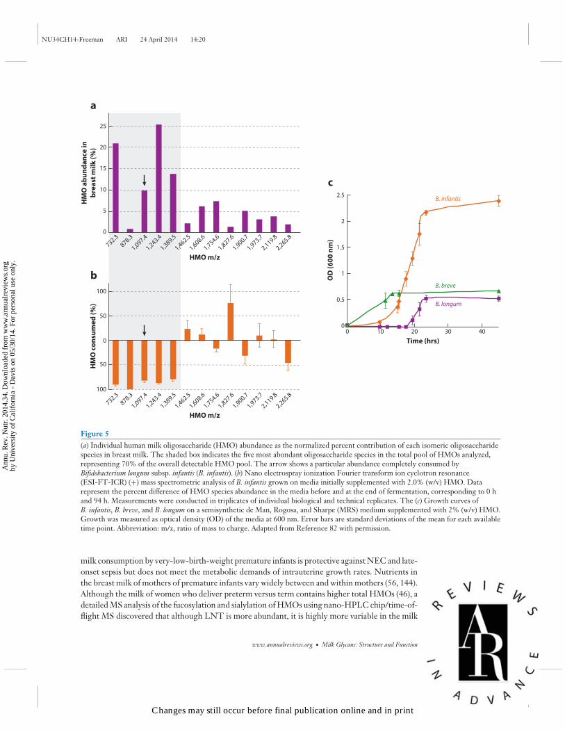
HMO structures and composition vary over the course of lactation (24, 129). HPLC was used in 46mothers who delivered term infants, and mean total concentration of HMOs was approximately20 g/L on days 4 and 10 postpartum decreasing to approximately 12 g/L by day 120 postpar-tum; the concentration of lactose increased from 56 g/L to 69 g/L over the same period (31).The higher concentration of total HMOs in breast milk in the early postpartum period is con-sistent with the protective functions of colostrum and early milk at a time when the neonate isimmunologically immature and the gut microbiota is not yet fully established. Although the totalHMO concentration is reduced across lactation, the direction of the change varies among thedifferent HMO compositions. High-performance anion-exchange chromatography showed thatconcentrations of α1,2-linked fucosyloligosaccharides, 2′-FL, and LNFP I were highest duringearly lactation (days 3–4) and were reduced by 37% and 53%, respectively, by day 90. In contrast,3FL—the structural isomer of 2′-FL—increased across lactation (days 3–4 to day 90 postpar-tum) by 1.8-fold in Se+Le+ women (129). These data suggest reduced activity of the Se enzyme(FUT2) and increased activity of Se- and Le-independent fucosyltransferases (FUT3, 4, 5, 6, 7,or 9) across lactation; however, human trials involving large numbers of women are required totest this hypothesis. Using MS, investigators determined that the major small neutral HMOs andtheir isomers (m/z 709.3, 855.3, 1074.4, 1220.4, and 1366.5) were the least varied across lactation(days 3 to 72) (98). These major HMO structures are likely selective growth substrates for specificbifidobacteria in the establishment of an infant’s healthy intestinal microbiota. Glycomic profilingof HMO consumption by bifidobacteria using Fourier transform ion cyclotron resonance MSrevealed that B. infantis (58) preferentially consumes small mass oligosaccharides with a degreeof polymerization ≤7 (m/z 1389 and below), representing 63.9% of the total HMOs, which aremostly fucosylated (82), whereas other bifidobacterial strains tested—B. longum subsp. longum,B. adolescentis, B. breve, and B. bifidum—showed only low or moderate growth ability (83)(Figure 5). These data suggest the ability of glycans to drive enrichment of a glycan-consumingbifidobacterial population in the infant gut. The development of glycomics analytics has enabledbreakthroughs in this research.
Protein glycosylation is also dynamic and varies during lactation. Many breast milk proteinsand the extent of their glycosylation as well as the specific glycans vary across lactation (9, 44). Theglycosylation composition of lactoferrin influences the function of the glycoprotein in preventingvarious pathogens from binding to and infecting intestinal epithelial cells (9).
Gestational Age of the Infant
Premature infants are at increased risk for infections compared with infants born at term owingto the immaturity of the gastrointestinal tract and innate and adaptive immune responses. Breast
14.16 Smilowitz et al.
Changes may still occur before final publication online and in print
Ann
u. R
ev. N
utr.
201
4.34
. Dow
nloa
ded
from
ww
w.a
nnua
lrev
iew
s.or
gby
Uni
vers
ity o
f C
alif
orni
a -
Dav
is o
n 05
/30/
14. F
or p
erso
nal u
se o
nly.
NU34CH14-Freeman ARI 24 April 2014 14:20
0
732.3
878.3
1,097.4
1,243.4
1,389.5
1,462.5
1,608.6
1,754.6
1,827.6
1,900.7
1,973.7
2,119.8
2,265.8
5
10
15
20
25
HM
O a
bu
nd
an
ce in
bre
ast
mil
k (
%)
HMO m/z
732.3
878.3
1,097.4
1,243.4
1,389.5
1,462.5
1,608.6
1,754.6
1,827.6
1,900.7
1,973.7
2,119.8
2,265.8
HMO m/z
100
50
0
50
100
HM
O c
on
sum
ed
(%
)
a
b OD
(6
00
nm
)
B. infantis
B. breve
B. longum
Time (hrs)
0.5
00 10 20 30 40
1
1.5
2
2.5
c
Figure 5(a) Individual human milk oligosaccharide (HMO) abundance as the normalized percent contribution of each isomeric oligosaccharidespecies in breast milk. The shaded box indicates the five most abundant oligosaccharide species in the total pool of HMOs analyzed,representing 70% of the overall detectable HMO pool. The arrow shows a particular abundance completely consumed byBifidobacterium longum subsp. infantis (B. infantis). (b) Nano electrospray ionization Fourier transform ion cyclotron resonance(ESI-FT-ICR) (+) mass spectrometric analysis of B. infantis grown on media initially supplemented with 2.0% (w/v) HMO. Datarepresent the percent difference of HMO species abundance in the media before and at the end of fermentation, corresponding to 0 hand 94 h. Measurements were conducted in triplicates of individual biological and technical replicates. The (c) Growth curves ofB. infantis, B. breve, and B. longum on a semisynthetic de Man, Rogosa, and Sharpe (MRS) medium supplemented with 2% (w/v) HMO.Growth was measured as optical density (OD) of the media at 600 nm. Error bars are standard deviations of the mean for each availabletime point. Abbreviation: m/z, ratio of mass to charge. Adapted from Reference 82 with permission.
milk consumption by very-low-birth-weight premature infants is protective against NEC and late-onset sepsis but does not meet the metabolic demands of intrauterine growth rates. Nutrients inthe breast milk of mothers of premature infants vary widely between and within mothers (56, 144).Although the milk of women who deliver preterm versus term contains higher total HMOs (46), adetailed MS analysis of the fucosylation and sialylation of HMOs using nano-HPLC chip/time-of-flight MS discovered that although LNT is more abundant, it is highly more variable in the milk
www.annualreviews.org • Milk Glycans: Structure and Function 14.17
Changes may still occur before final publication online and in print
Ann
u. R
ev. N
utr.
201
4.34
. Dow
nloa
ded
from
ww
w.a
nnua
lrev
iew
s.or
gby
Uni
vers
ity o
f C
alif
orni
a -
Dav
is o
n 05
/30/
14. F
or p
erso
nal u
se o
nly.
NU34CH14-Freeman ARI 24 April 2014 14:20
of women who deliver preterm. Furthermore, fucosylation was not as well regulated, resultingin higher within- and between-mother variations in milk from women delivering preterm versusterm. Of particular clinical interest, the concentration of 2′-FL was not consistent across lactationof several mothers who delivered preterm (35). Thus, Se status of preterm milk changed in a givenmother. Fluctuations in fucosylated HMOs in mothers’ milk are important because of the role thatfucosylated HMOs play in pathogen binding. A high degree of variability suggests immaturity inthe regulation of HMO fucosylation in the “premature breast.” Yet, milk from Se women in com-parison with non-Se women imparts protection against NEC and gram-negative sepsis (92), whichmay in part be explained by potential enrichment of gut bifidobacteria. Indeed, milk from Se moth-ers has specific linkages (α1,2-fucosylated HMOs) and higher amounts of total fucosylated HMOs(131), which are likely to enrich only those bifidobacterial species/strains that can catabolize theseglycans (118). Premature infants generally have very low numbers of fecal bifidobacteria (145),and probiotic supplementation with bifidobacteria is protective against NEC (1, 80, 81). Fucosy-lation of HMOs of preterm milk is unpredictable and raises the interesting question of whetherfortification with preterm milk HMOs would further reduce the risk of NEC and late-onsetsepsis.
Maternal Health and Phenotype
Maternal health ranging from malnutrition to overnutrition and metabolic syndrome leads tolong-term health consequences in the neonate. For example, HMO total concentrations weresignificantly lower in women with a body mass index between 14 and 18 than in women witha body mass index between 24 and 28 (17). In the United States, gestational diabetes mellitus(GDM), a complex disease characterized by elevated blood Glc, affects on average 7% and upto 14% of pregnancies (3). GDM has immediate and lasting consequences in women and toinfants exposed to maternal diabetes in utero (12, 29, 73). Hyperglycemia results in aberrantcarbohydrate metabolism that contributes to the pathogenesis of the disease in part throughincreased flux through the hexosamine biosynthetic pathway (38, 62) and altered activities ofcellular glycosyltransferases and glycosidases (53, 75). Very little is understood regarding therole of GDM in lactation and breast milk components, and much of what is known about theeffects of glycemic dysregulation on milk components comes from research on lactating womendiagnosed with insulin-dependent diabetes mellitus and from animal models. Recently, glycomicprofiling using nano-HPLC chip/time-of-flight MS and multivariate modeling was used toinvestigate the effects of GDM on the glycosylation of HMOs, sIgA, and lactoferrin of breastmilk (123). Although total HMOs and their composition were not different in milk from womenwith and without GDM, the N-linked glycosylation of specific milk proteins differed betweenthe two groups. The total N-linked glycosylation, mannose, Fuc, and sialylated residues of sIgAin transitional milk were up to 43% lower in milk from women with GDM in comparison withwomen without GDM. Reduced sialylation of immunoglobulins secreted by mammary epithelialcells has implications on the nonspecific innate immune defense against pathogen adhesion andinfection (115). The total N-linked glycosylation, Fuc, and sialylated residues of lactoferrin weresignificantly higher, by up to 72% (Figure 6). The higher N-linked glycosylation of lactoferrinobserved in milk from women with GDM could result from altered glycan-metabolizing enzymeslinked to the pathophysiology of diabetes mellitus during pregnancy (53). The glycosylationlevel of a glycoprotein influences its susceptibility to proteolysis (137, 139), thus affecting theproduction of potent active peptides and glycopeptides involved in its biological activities (77).These data suggest that diabetes mellitus during pregnancy alters the glycosylation of protective
14.18 Smilowitz et al.
Changes may still occur before final publication online and in print
Ann
u. R
ev. N
utr.
201
4.34
. Dow
nloa
ded
from
ww
w.a
nnua
lrev
iew
s.or
gby
Uni
vers
ity o
f C
alif
orni
a -
Dav
is o
n 05
/30/
14. F
or p
erso
nal u
se o
nly.

NU34CH14-Freeman ARI 24 April 2014 14:20
–0.4 –0.2 0
slgA total glycans
slgA F
slgA HM
slgA S
slgA FS
slgA protein
LF CH
LF S
LF FS
LF 5411
LF total glycans
LF F
LV[1]
0.2 0.4
Higher inGDM
Lower inGDM
Figure 6N-Glycosylation of milk proteins is altered in gestational diabetes mellitus (GDM). Milk proteins frommothers with and without GDM were compared using orthogonal signal corrected partial least-squaresdiscriminant analysis. The maximum difference between milk glycans from women with and without GDMwas captured in the first dimension or latent variable (LV1) of the model. Positive values indicate higherglycosylation in GDM compared to controls; conversely, negative values indicate lower glycosylation inGDM compared to controls. LF 5411 is an N-glycan that contains 5 hexoses, 4 N-acetyl-hexosamines,1 fucose, and 1 sialic acid. Abbreviations: CH, complex hybrid; F, fucose; FS, fucose and sialic acid; HM,high mannose; LF, lactoferrin; S, sialic acid; sIgA, secretory immunoglobulin A. Adapted from Reference122 with permission.
proteins in milk; future research is needed to discover the effects of these alterations on neonataloutcomes.
CONCLUSIONS AND FUTURE DIRECTIONS
The first proof of principle from integrating the science of glycomics into microbiota researchis that stereospecific glycans are key modulators of the intestinal environment in the narrowlydefined successful feeding of breastfed infants. The accuracy and specificity of analytical glycomicsnow enable researchers to determine interindividual and intraindividual milk variations; theability to track and monitor the fate of glycomic structures assists investigators in understandinghow they shape the intestine. The tool sets of systems biology, including genomics, metabolomics,proteomics, and glycomics, which are being used to investigate the complexity of milk andare revealing how colonization and development of the infant microbiota occurs, can now beapplied to the more complex challenges of the adult intestinal microbiota. Determining howto standardize, integrate, and use glycomic information in large multi-omic, annotated datasets will be the next milestone in elucidating structure-function relationships in breast milkoligosaccharides.
www.annualreviews.org • Milk Glycans: Structure and Function 14.19
Changes may still occur before final publication online and in print
Ann
u. R
ev. N
utr.
201
4.34
. Dow
nloa
ded
from
ww
w.a
nnua
lrev
iew
s.or
gby
Uni
vers
ity o
f C
alif
orni
a -
Dav
is o
n 05
/30/
14. F
or p
erso
nal u
se o
nly.
NU34CH14-Freeman ARI 24 April 2014 14:20
SUMMARY POINTS
Milk glycan research is providing a wealth of information regarding the impact of diet onhost and intestinal microbiota.
1. Major advances in the field of glycomics in milk research, including high mass accu-racy for the identification and quantitation of diverse glycan structures, contribute tothe understanding of the functional relationships between breast milk glycans and gutmicrobiota, pathogen binding, and intestinal barrier function.
2. Glycan heterogeneity on milk proteins influences their structures, functions, anddigestibility.
3. Effective tools for the structural elucidation of human milk oligosaccharides, glycol-ipids, and glycoproteins combine exoglycosidase digestion, separation technologies, MS,structural libraries, and bioinformatics.
4. Analysis of breast milk glycolipids is now achievable using matrix-assisted desorption/ionization Fourier transform ion cyclotron resonance tandem MS.
5. The glycans of breast milk are substrates for the selective enrichment of bifidobacterialspecies in the gastrointestinal tract of breastfed infants.
6. Growth of B. infantis on human milk glycans promotes intestinal barrier and immuno-logical development and function.
DISCLOSURE STATEMENT
J.B.C. is a past or present consultant for Nestle, Abbott, Dairy Research Inc., Arla Foods, andFrieslandCampina. The other authors are not aware of any affiliations, memberships, funding, orfinancial holdings that might be perceived as affecting the objectivity of this review.
ACKNOWLEDGMENTS
We acknowledge all of the researchers in the UC Davis Foods for Health Institute and theMilk Bioactives Program for their enthusiasm, imagination, and collective contribution to thissubject matter. Work by the Milk Bioactives Program has been supported by the UC Davis Re-search Investments in the Sciences and Engineering Program; the UC Discovery Grant Program;the California Dairy Research Foundation; the Dairy Research Institute; the Bill & MelindaGates Foundation; and the National Institutes of Health awards R01HD059127, R01HD065122,R01HD061923, R21AT006180, and R01AT007079. D.A.M. acknowledges support as the PeterJ. Shields Endowed Chair in Dairy Food Science.
LITERATURE CITED
1. Alfaleh K, Anabrees J, Bassler D, Al-Kharfi T. 2011. Probiotics for prevention of necrotizing enterocolitisin preterm infants. Cochrane Database Syst. Rev. 3:CD005496
2. Alvarez-Manilla G, Atwood J III, Guo Y, Warren NL, Orlando R, Pierce M. 2006. Tools for glyco-proteomic analysis: size exclusion chromatography facilitates identification of tryptic glycopeptides withN-linked glycosylation sites. J. Proteome Res. 5:701–8
3. Am. Diabetes Assoc. 2011. Diagnosis and classification of diabetes mellitus. Diabetes Care 34:S62–69
14.20 Smilowitz et al.
Changes may still occur before final publication online and in print
Ann
u. R
ev. N
utr.
201
4.34
. Dow
nloa
ded
from
ww
w.a
nnua
lrev
iew
s.or
gby
Uni
vers
ity o
f C
alif
orni
a -
Dav
is o
n 05
/30/
14. F
or p
erso
nal u
se o
nly.
NU34CH14-Freeman ARI 24 April 2014 14:20
4. An HJ, Froehlich JW, Lebrilla CB. 2009. Determination of glycosylation sites and site-specific hetero-geneity in glycoproteins. Curr. Opin. Chem. Biol. 13:421–26
5. Andersson B, Porras O, Hanson LA, Lagergard T, Svanborg-Eden C. 1986. Inhibition of attachmentof Streptococcus pneumoniae and Haemophilus influenzae by human milk and receptor oligosaccharides.J. Infect. Dis. 153:232–37
6. Aoki-Kinoshita KF. 2008. An introduction to bioinformatics for glycomics research. PLoS Comput. Biol.4:e1000075
7. Ashline D, Singh S, Hanneman A, Reinhold V. 2005. Congruent strategies for carbohydrate sequencing.1. Mining structural details by MSn. Anal. Chem. 77:6250–62
8. Azuma N, Yamauchi K, Mitsuoka T. 1984. Bifidus growth-promoting activity of a glycomacropeptidederived from human K-casein. Agric. Biol. Chem. 48:2159–62
9. Barboza M, Pinzon J, Wickramasinghe S, Froehlich JW, Moeller I, et al. 2012. Glycosylation of humanmilk lactoferrin exhibits dynamic changes during early lactation enhancing its role in pathogenic bacteria-host interactions. Mol. Cell Proteomics 11:M111.015248
10. Barboza M, Sela DA, Pirim C, Locascio RG, Freeman SL, et al. 2009. Glycoprofiling bifidobacterialconsumption of galacto-oligosaccharides by mass spectrometry reveals strain-specific, preferential con-sumption of glycans. Appl. Environ. Microbiol. 75:7319–25
11. Barile D, Marotta M, Chu C, Mehra R, Grimm R, et al. 2010. Neutral and acidic oligosaccharides inHolstein-Friesian colostrum during the first 3 days of lactation measured by high performance liquidchromatography on a microfluidic chip and time-of-flight mass spectrometry. J. Dairy Sci. 93:3940–49
12. Bellamy L, Casas JP, Hingorani AD, Williams D. 2009. Type 2 diabetes mellitus after gestationaldiabetes: a systematic review and meta-analysis. Lancet 373:1773–79
13. Blackburn D. 1993. Lactation: historical patterns and potential for manipulation. J. Dairy Sci. 76:3195–212
14. Blank D, Dotz V, Geyer R, Kunz C. 2012. Human milk oligosaccharides and Lewis blood group:individual high-throughput sample profiling to enhance conclusions from functional studies. Adv. Nutr.3:440–49S
15. Bode L. 2012. Human milk oligosaccharides: Every baby needs a sugar mama. Glycobiology 22:1147–6216. Bode L, Beermann C, Mank M, Kohn G, Boehm G. 2004. Human and bovine milk gangliosides differ
in their fatty acid composition. J. Nutr. 134:3016–2017. Bode L , Jantscher-Krenn E. 2012. Structure-function relationships of human milk oligosaccharides.
Adv. Nutr. 3:383–91S18. Boren T, Falk P, Roth KA, Larson G, Normark S. 1993. Attachment of Helicobacter pylori to human
gastric epithelium mediated by blood group antigens. Science 262:1892–9519. Brandtzaeg P. 2010. The mucosal immune system and its integration with the mammary glands.
J. Pediatr. 156:S8–1520. Bricarello DA, Smilowitz JT, Zivkovic AM, German JB, Parikh AN. 2011. Reconstituted lipoprotein: a
versatile class of biologically-inspired nanostructures. ACS Nano 5:42–5721. Brønnum H, Seested T, Hellgren L, Brix S, Frøkiær H. 2005. Milk-derived GM3 and GD3 differentially
inhibit dendritic cell maturation and effector functionalities. Scand. J. Immunol. 61:551–5722. Bu H, Zuo X, Wang X, Ensslin MA, Koti V, et al. 2007. Milk fat globule-EGF factor 8/lactadherin plays
a crucial role in maintenance and repair of murine intestinal epithelium. J. Clin. Invest. 117:3673–8323. Charlwood J, Hanrahan S, Tyldesley R, Langridge J, Dwek M, Camilleri P. 2002. Use of proteomic
methodology for the characterization of human milk fat globular membrane proteins. Anal. Biochem.301:314–24
24. Chaturvedi P, Warren CD, Altaye M, Morrow AL, Ruiz-Palacios G, et al. 2001. Fucosylated humanmilk oligosaccharides vary between individuals and over the course of lactation. Glycobiology 11:365–72
25. Chaturvedi P, Warren CD, Buescher CR, Pickering LK, Newburg DS. 2001. Survival of human milkoligosaccharides in the intestine of infants. In Bioactive Components of Human Milk, pp. 315–23, ed. DSNewburg, pp. 315–24. New York: Springer
26. Chen Q, Cao J, Jia Y, Liu X, Yan Y, Pang G. 2012. Modulation of mice fecal microbiota by administrationof casein glycomacropeptide. Microbiol. Res. 3:e3
www.annualreviews.org • Milk Glycans: Structure and Function 14.21
Changes may still occur before final publication online and in print
Ann
u. R
ev. N
utr.
201
4.34
. Dow
nloa
ded
from
ww
w.a
nnua
lrev
iew
s.or
gby
Uni
vers
ity o
f C
alif
orni
a -
Dav
is o
n 05
/30/
14. F
or p
erso
nal u
se o
nly.

NU34CH14-Freeman ARI 24 April 2014 14:20
27. Chichlowski M, De Lartigue G, German JB, Raybould HE, Mills DA. 2012. Bifidobacteria isolatedfrom infants and cultured on human milk oligosaccharides affect intestinal epithelial function. J. Pediatr.Gastroenterol. Nutr. 55:321–27
28. Chirico G, Marzollo R, Cortinovis S, Fonte C, Gasparoni A. 2008. Antiinfective properties of humanmilk. J. Nutr. 138:1801–6S
29. Clausen TD, Mathiesen ER, Hansen T, Pedersen O, Jensen DM, et al. 2008. High prevalence of type2 diabetes and pre-diabetes in adult offspring of women with gestational diabetes mellitus or type 1diabetes. Diabetes Care 31:340–46
30. Coppa G, Pierani P, Zampini L, Bruni S, Carloni I, Gabrielli O. 2001. Characterization of oligosaccha-rides in milk and feces of breast-fed infants by high-performance anion-exchange chromatography. InBioactive Components of Human Milk, ed. DS Newburg, pp. 307–14. New York: Springer
31. Coppa GV, Gabrielli O, Pierani P, Catassi C, Carlucci A, Giorgi PL. 1993. Changes in carbohydratecomposition in human milk over 4 months of lactation. Pediatrics 91:637–41
32. Coppa GV, Pierani P, Zampini L, Bruni S, Carloni I, Gabrielli O. 2001. Characterization of oligosac-charides in milk and feces of breast-fed infants by high-performance anion-exchange chromatography.Adv. Exp. Med. Biol. 501:307–14
33. Coppa GV, Zampini L, Galeazzi T, Facinelli B, Ferrante L, et al. 2006. Human milk oligosaccharidesinhibit the adhesion to Caco-2 cells of diarrheal pathogens: Escherichia coli, Vibrio cholerae, and Salmonellafyris. Pediatr. Res. 59:377–82
34. De Leoz ML, Wu S, Strum JS, Ninonuevo MR, Gaerlan SC, et al. 2013. A quantitative and compre-hensive method to analyze human milk oligosaccharide structures in the urine and feces of infants. Anal.Bioanal. Chem. 405:4089–105
35. De Leoz MLA, Gaerlan SC, Strum JS, Dimapasoc LM, Mirmiran M, et al. 2012. Lacto-N-tetraose, fu-cosylation, and secretor status are highly variable in human milk oligosaccharides from women deliveringpreterm. J. Proteome Res. 11:4662–72
36. Desai PT, Walsh MK, Weimer BC. 2008. Solid phase capture of pathogenic bacteria using gangliosidesand detection with real time PCR. Appl. Environ. Microbiol. 74:2254–58
37. Dodds ED, Seipert RR, Clowers BH, German JB, Lebrilla CB. 2009. Analytical performance of immobi-lized pronase for glycopeptide footprinting and implications for surpassing reductionist glycoproteomics.J. Proteome Res. 8:502–12
38. Du X-L, Edelstein D, Rossetti L, Fantus IG, Goldberg H, et al. 2000. Hyperglycemia-induced mitochon-drial superoxide overproduction activates the hexosamine pathway and induces plasminogen activatorinhibitor-1 expression by increasing Sp1 glycosylation. Proc. Natl. Acad. Sci. 97:12222–26
39. Engfer MB, Stahl B, Finke B, Sawatzki G, Daniel H. 2000. Human milk oligosaccharides are resistantto enzymatic hydrolysis in the upper gastrointestinal tract. Am. J. Clin. Nutr. 71:1589–96
40. Ewaschuk JB, Diaz H, Meddings L, Diederichs B, Dmytrash A, et al. 2008. Secreted bioactive factorsfrom Bifidobacterium infantis enhance epithelial cell barrier function. Am. J. Physiol. Gastrointest. LiverPhysiol. 295:G1025–34
41. Falk P, Roth KA, Boren T, Westblom TU, Gordon JI, Normark S. 1993. An in vitro adherence assayreveals that Helicobacter pylori exhibits cell lineage specific tropism in the human gastric epithelium. Proc.Natl. Acad. Sci. USA 90:2035–39
42. Ferrer-Admetlla A, Sikora M, Laayouni H, Esteve A, Roubinet F, et al. 2009. A natural history of FUT2polymorphism in humans. Mol. Biol. Evol. 26:1993–2003
43. Fong B, Ma K, McJarrow P. 2011. Quantification of bovine milk oligosaccharides using liquidchromatography-selected reaction monitoring-mass spectrometry. J. Agric. Food Chem. 59:9788–95
44. Froehlich J, Dodds E, Barboza M, McJimpsey E, Seipert R, et al. 2010. Glycoprotein expression inhuman milk during lactation. J. Agric. Food Chem. 58:6440–48
45. Fukuda S, Toh H, Hase K, Oshima K, Nakanishi Y, et al. 2011. Bifidobacteria can protect from en-teropathogenic infection through production of acetate. Nature 469:543–47
46. Gabrielli O, Zampini L, Galeazzi T, Padella L, Santoro L, et al. 2011. Preterm milk oligosaccharidesduring the first month of lactation. Pediatrics 128:e1520–31
14.22 Smilowitz et al.
Changes may still occur before final publication online and in print
Ann
u. R
ev. N
utr.
201
4.34
. Dow
nloa
ded
from
ww
w.a
nnua
lrev
iew
s.or
gby
Uni
vers
ity o
f C
alif
orni
a -
Dav
is o
n 05
/30/
14. F
or p
erso
nal u
se o
nly.

NU34CH14-Freeman ARI 24 April 2014 14:20
47. Ganguli K, Meng D, Rautava S, Lu L, Walker WA, Nanthakumar N. 2013. Probiotics prevent necrotiz-ing enterocolitis by modulating enterocyte genes that regulate innate immune-mediated inflammation.Am. J. Physiol. Gastrointest. Liver Physiol. 304:G132–41
48. Garrido D, Barile D, Mills DA. 2012. A molecular basis for bifidobacterial enrichment in the infantgastrointestinal tract. Adv. Nutr. 3:415–21S
49. Garrido D, Kim JH, German JB, Raybould HE, Mills DA. 2011. Oligosaccharide binding proteins fromBifidobacterium longum subsp. infantis reveal a preference for host glycans. PLoS ONE 6:e17315
50. Garrido D, Nwosu C, Ruiz-Moyano S, Aldredge D, German JB, et al. 2012. Endo-β-N-acetylglucosaminidases from infant gut-associated bifidobacteria release complex N-glycans from humanmilk glycoproteins. Mol. Cell Proteomics 11:775–85
51. Garrido D, Ruiz-Moyano S, Jimenez-Espinoza R, Eom HJ, Block DE, Mills DA. 2013. Utilization ofgalactooligosaccharides by Bifidobacterium longum subsp. infantis isolates. Food Microbiol. 33:262–70
52. Garrido D, Ruiz-Moyano S, Mills DA. 2012. Release and utilization of N-acetyl-D-glucosamine fromhuman milk oligosaccharides by Bifidobacterium longum subsp. infantis. Anaerobe 18:430–35
53. Georgieff M, Petry C, Mills M, McKay H, Wobken J. 1997. Increased N-glycosylation and reducedtransferrin-binding capacity of transferrin receptor isolated from placentae of diabetic women. Placenta18:563–68
54. Gordon JI, Dewey KG, Mills DA, Medzhitov RM. 2012. The human gut microbiota and undernutrition.Sci. Transl. Med. 4:137ps12
55. Grollman EF, Ginsburg V. 1967. Correlation between secretor status and the occurrence of 2′-fucosyllactose in human milk. Biochem. Biophys. Res. Commun. 28:50–53
56. Gross SJ, Geller J, Tomarelli R. 1981. Composition of breast milk from mothers of preterm infants.Pediatrics 68:490–93
57. Gyorgy P, Norris RF, Rose CS. 1954. Bifidus factor. I. A variant of Lactobacillus bifidus requiring a specialgrowth factor. Arch. Biochem. Biophys. 48:193–201
58. Haarman M, Knol J. 2005. Quantitative real-time PCR assays to identify and quantify fecal Bifidobacteriumspecies in infants receiving a prebiotic infant formula. Appl. Environ. Microbiol. 71:2318–24
59. Hanayama R, Tanaka M, Miwa K, Shinohara A, Iwamatsu A, Nagata S. 2002. Identification of a factorthat links apoptotic cells to phagocytes. Nature 417:182–87
60. Hanson LA. 1998. Breastfeeding provides passive and likely long-lasting active immunity. Ann. AllergyAsthma Immunol. 81:523–37
61. Harmsen HJ, Wildeboer-Veloo AC, Raangs GC, Wagendorp AA, Klijn N, et al. 2000. Analysis ofintestinal flora development in breast-fed and formula-fed infants by using molecular identification anddetection methods. J. Pediatr. Gastroenterol. Nutr. 30:61–67
62. Hebert LF Jr, Daniels MC, Zhou J, Crook ED, Turner RL, et al. 1996. Overexpression ofglutamine:fructose-6-phosphate amidotransferase in transgenic mice leads to insulin resistance. J. Clin.Invest. 98:930–36
63. Hernandez-Hernandez O, Sanz ML, Kolida S, Rastall RA, Moreno FJ. 2011. In vitro fermentation byhuman gut bacteria of proteolytically digested caseinomacropeptide nonenzymatically glycosylated withprebiotic carbohydrates. J. Agric. Food Chem. 59:11949–55
64. Hong P, Ninonuevo MR, Lee B, Lebrilla C, Bode L. 2009. Human milk oligosaccharides reduce HIV-1-gp120 binding to dendritic cell-specific ICAM3-grabbing non-integrin (DC-SIGN). Br. J. Nutr.101:482–86
65. Huang P, Xia M, Tan M, Zhong W, Wei C, et al. 2012. Spike protein VP8∗ of human rotavirus recognizeshisto-blood group antigens in a type-specific manner. J. Virol. 86:4833–43
66. Ip S, Chung M, Raman G, Chew P, Magula N, et al. 2007. Breastfeeding and maternal and infant healthoutcomes in developed countries. Evid. Rep. Technol. Assess. 153:1–186
67. Iwamori M, Takamizawa K, Momoeda M, Iwamori Y, Taketani Y. 2008. Gangliosides in human, cowand goat milk, and their abilities as to neutralization of cholera toxin and botulinum type A neurotoxin.Glycoconj. J. 25:675–83
68. Jantscher-Krenn E, Lauwaet T, Bliss LA, Reed SL, Gillin FD, Bode L. 2012. Human milk oligosaccha-rides reduce Entamoeba histolytica attachment and cytotoxicity in vitro. Br. J. Nutr. 108:1839–46
www.annualreviews.org • Milk Glycans: Structure and Function 14.23
Changes may still occur before final publication online and in print
Ann
u. R
ev. N
utr.
201
4.34
. Dow
nloa
ded
from
ww
w.a
nnua
lrev
iew
s.or
gby
Uni
vers
ity o
f C
alif
orni
a -
Dav
is o
n 05
/30/
14. F
or p
erso
nal u
se o
nly.

NU34CH14-Freeman ARI 24 April 2014 14:20
69. Kavanaugh DW, O’Callaghan J, Butto LF, Slattery H, Lane J, et al. 2013. Exposure of Bifidobacteriumlongum subsp. infantis to milk oligosaccharides increases adhesion to epithelial cells and induces a sub-stantial transcriptional response. PLoS ONE 8:e67224
70. Kim W-S, Ohashi M, Tanaka T, Kumura H, Kim G-Y, et al. 2004. Growth-promoting effects oflactoferrin on L. acidophilus and Bifidobacterium spp. Biometals 17:279–83
71. Kitaoka M. 2012. Bifidobacterial enzymes involved in the metabolism of human milk oligosaccharides.Adv. Nutr. 3:422–29S
72. Kiyohara M, Nakatomi T, Kurihara S, Fushinobu S, Suzuki H, et al. 2012. α-N-acetylgalactosaminidasefrom infant-associated bifidobacteria belonging to novel glycoside hydrolase family 129 is implicated inalternative mucin degradation pathway. J. Biol. Chem. 287:693–700
73. Kumar R, Ouyang F, Story RE, Pongracic JA, Hong X, et al. 2009. Gestational diabetes, atopic dermatitis,and allergen sensitization in early childhood. J. Allergy Clin. Immunol. 124:1031–38.e4
74. Kunz C, Rudloff S, Baier W, Klein N, Strobel S. 2000. Oligosaccharides in human milk: structural,functional, and metabolic aspects. Annu. Rev. Nutr. 20:699–722
75. Lee CL, Chiu PCN, Pang PC, Chu IK, Lee KF, et al. 2011. Glycosylation failure extends to glycoproteinsin gestational diabetes mellitus. Diabetes 60:909–17
76. Lee H, An HJ, Lerno LA, German JB, Lebrilla CB. 2011. Rapid profiling of bovine and human milkgangliosides by matrix-assisted laser desorption/ionization Fourier transform ion cyclotron resonancemass spectrometry. Int. J. Mass Spectrom. 305:138–50
77. Legrand D, Pierce A, Elass E, Carpentier M, Mariller C, Mazurier J. 2008. Lactoferrin structure andfunctions. Adv. Exp. Med. Biol. 606:163–94
78. Liao Y, Alvarado R, Phinney B, Lonnerdal B. 2011. Proteomic characterization of human milk wheyproteins during a twelve-month lactation period. J. Proteome Res. 10:1746–54
79. Liepke C, Adermann K, Raida M, Magert HJ, Forssmann WG, Zucht HD. 2002. Human milk providespeptides highly stimulating the growth of bifidobacteria. Eur. J. Biochem. 269:712–18
80. Lin H-C, Hsu C-H, Chen H-L, Chung M-Y, Hsu J-F, et al. 2008. Oral probiotics prevent necrotiz-ing enterocolitis in very low birth weight preterm infants: a multicenter, randomized, controlled trial.Pediatrics 122:693–700
81. Lin H-C, Su B-H, Chen A-C, Lin T-W, Tsai C-H, et al. 2005. Oral probiotics reduce the incidenceand severity of necrotizing enterocolitis in very low birth weight infants. Pediatrics 115:1–4
82. LoCascio R, Ninonuevo M, Freeman S, Sela D, Grimm R, et al. 2007. Glycoprofiling of bifidobacterialconsumption of human milk oligosaccharides demonstrates strain specific, preferential consumption ofsmall chain glycans secreted in early human lactation. J. Agric. Food Chem. 55:8914–19
83. Locascio RG, Ninonuevo MR, Kronewitter SR, Freeman SL, German JB, et al. 2009. A versatile andscalable strategy for glycoprofiling bifidobacterial consumption of human milk oligosaccharides. Microb.Biotechnol. 2:333–42
84. Lonnerdal B. 2010. Bioactive proteins in human milk: mechanisms of action. J. Pediatr. 156:S26–3085. Marcobal A, Barboza M, Froehlich J, Block D, German JB, et al. 2010. Consumption of human milk
oligosaccharides by gut-related microbes. J. Agric. Food Chem. 58:5334–4086. Marcobal A, Barboza M, Sonnenburg ED, Pudlo N, Martens EC, et al. 2011. Bacteroides in the infant
gut consume milk oligosaccharides via mucus-utilization pathways. Cell Host Microbe 10:507–1487. Marth JD, Grewal PK. 2008. Mammalian glycosylation in immunity. Nat. Rev. Immunol. 8:874–8788. Martin-Sosa S, Martin MJ, Hueso P. 2002. The sialylated fraction of milk oligosaccharides is partially
responsible for binding to enterotoxigenic and uropathogenic Escherichia coli human strains. J. Nutr.132:3067–72
89. Mechref Y, Chen P, Novotny MV. 1999. Structural characterization of the N-linked oligosaccharides inbile salt-stimulated lipase originated from human breast milk. Glycobiology 9:227–34
90. Molinari CE, Casadio YS, Hartmann BT, Arthur PG, Hartmann PE. 2013. Longitudinal analysis ofprotein glycosylation and β-casein phosphorylation in term and preterm human milk during the first2 months of lactation. Br. J. Nutr. 110:105–15
91. Molinari CE, Casadio YS, Hartmann BT, Livk A, Bringans S, et al. 2012. Proteome mapping of humanskim milk proteins in term and preterm milk. J. Proteome Res. 11:1696–714
14.24 Smilowitz et al.
Changes may still occur before final publication online and in print
Ann
u. R
ev. N
utr.
201
4.34
. Dow
nloa
ded
from
ww
w.a
nnua
lrev
iew
s.or
gby
Uni
vers
ity o
f C
alif
orni
a -
Dav
is o
n 05
/30/
14. F
or p
erso
nal u
se o
nly.

NU34CH14-Freeman ARI 24 April 2014 14:20
92. Morrow AL, Meinzen-Derr J, Huang P, Schibler KR, Cahill T, et al. 2011. Fucosyltransferase 2 non-secretor and low secretor status predicts severe outcomes in premature infants. J. Pediatr. 158:745–51
93. Morrow AL, Ruiz-Palacios GM, Jiang X, Newburg DS. 2005. Human-milk glycans that inhibit pathogenbinding protect breast-feeding infants against infectious diarrhea. J. Nutr. 135:1304–7
94. Newburg DS. 2013. Glycobiology of human milk. Biochemistry (Moscow) 78:771–8595. Newburg DS, Ruiz-Palacios GM, Morrow AL. 2005. Human milk glycans protect infants against enteric
pathogens. Annu. Rev. Nutr. 25:37–5896. Newburg DS, Walker WA. 2007. Protection of the neonate by the innate immune system of developing
gut and of human milk. Pediatr. Res. 61:2–897. Ninonuevo MR, Park Y, Yin H, Zhang J, Ward RE, et al. 2006. A strategy for annotating the human
milk glycome. J. Agric. Food Chem. 54:7471–8098. Ninonuevo MR, Perkins PD, Francis J, Lamotte LM, LoCascio RG, et al. 2008. Daily variations in
oligosaccharides of human milk determined by microfluidic chips and mass spectrometry. J. Agric. FoodChem. 56:618–26
99. Ninonuevo MR, Ward RE, LoCascio RG, German JB, Freeman SL, et al. 2007. Methods for the quan-titation of human milk oligosaccharides in bacterial fermentation by mass spectrometry. Anal. Biochem.361:15–23
100. Nusrat A, Turner J, Madara J. 2000. IV. Regulation of tight junctions by extracellular stimuli: nutrients,cytokines, and immune cells. Am. J. Physiol. Gastrointest. Liver Physiol. 279:G851–57
101. Nwosu CC, Huang J, Aldredge DL, Strum JS, Hua S, et al. 2012. In-gel nonspecific proteolysis for elu-cidating glycoproteins: a method for targeted protein-specific glycosylation analysis in complex proteinmixtures. Anal. Chem. 85:956–63
102. Ochoa TJ, Noguera-Obenza M, Ebel F, Guzman CA, Gomez HF, Cleary TG. 2003. Lactoferrin impairstype III secretory system function in enteropathogenic Escherichia coli. Infect. Immun. 71:5149–55
103. Oda H, Wakabayashi H, Yamauchi K, Sato T, Xiao J-Z, et al. 2013. Isolation of a bifidogenic peptidefrom the pepsin hydrolysate of bovine lactoferrin. Appl. Environ. Microbiol. 79:1843–49
104. Pan XL, Izumi T. 2000. Variation of the ganglioside compositions of human milk, cow’s milk and infantformulas. Early Hum. Dev. 57:25–31
105. Peterson R, Cheah WY, Grinyer J, Packer N. 2013. Glycoconjugates in human milk: protecting infantsfrom disease. Glycobiology 23:1425–38
106. Petshow BW, Talbottt RD, Batema RP. 1999. Ability of lactoferrin to promote the growth of Bifi-dobacterium spp. in vitro is independent of receptor binding capacity and iron saturation level. J. Med.Microbiol. 48:541–49
107. Qiu J, Hendrixson DR, Baker EN, Murphy TF, Geme JWS, Plaut AG. 1998. Human milk lactoferrininactivates two putative colonization factors expressed by Haemophilus influenzae. Proc. Natl. Acad. Sci.95:12641–46
108. Reinhold V, Zhang H, Hanneman A, Ashline D. 2013. Toward a platform for comprehensive glycansequencing. Mol. Cell. Proteomics 12:866–73
109. Roger LC, McCartney AL. 2010. Longitudinal investigation of the faecal microbiota of healthy full-terminfants using fluorescence in situ hybridization and denaturing gradient gel electrophoresis. Microbiology156:3317–28
110. Rueda R. 2007. The role of dietary gangliosides on immunity and the prevention of infection. Br. J.Nutr. 98:S68–73
111. Ruiz-Moyano S, Totten SM, Garrido DA, Smilowitz JT, German JB, et al. 2013. Variation in con-sumption of human milk oligosaccharides by infant gut-associated strains of Bifidobacterium breve. Appl.Environ. Microbiol. 79:6040–49
112. Rumbo M, Schiffrin EJ. 2005. Ontogeny of intestinal epithelium immune functions: developmental andenvironmental regulation. Cell Mol Life Sci 62:1288–96
113. Ruvoen-Clouet N, Le Pendu J, Mas E, Marionneau S, Guillon P, Lombardo D. 2006. Bile-salt-stimulatedlipase and mucins from milk of “secretor” mothers inhibit the binding of Norwalk virus capsids to theircarbohydrate ligands. Biochem. J 393:627–34
www.annualreviews.org • Milk Glycans: Structure and Function 14.25
Changes may still occur before final publication online and in print
Ann
u. R
ev. N
utr.
201
4.34
. Dow
nloa
ded
from
ww
w.a
nnua
lrev
iew
s.or
gby
Uni
vers
ity o
f C
alif
orni
a -
Dav
is o
n 05
/30/
14. F
or p
erso
nal u
se o
nly.
NU34CH14-Freeman ARI 24 April 2014 14:20
114. Sakata S, Tonooka T, Ishizeki S, Takada M, Sakamoto M, et al. 2005. Culture-independent analysisof fecal microbiota in infants, with special reference to Bifidobacterium species. FEMS Microbiol. Lett.243:417–23
115. Schroten H, Stapper C, Plogmann R, Kohler H, Hacker J, Hanisch FG. 1998. Fab-independent anti-adhesion effects of secretory immunoglobulin A on S-fimbriated Escherichia coli are mediated by sialy-loligosaccharides. Infect. Immun. 66:3971–73
116. Seipert RR, Dodds ED, Clowers BH, Beecroft SM, German JB, Lebrilla CB. 2008. Factors that influencefragmentation behavior of N-linked glycopeptide ions. Anal. Chem. 80:3684–92
117. Sela DA, Chapman J, Adeuya A, Kim J, Chen F, et al. 2008. The genome sequence of Bifidobacteriumlongum subsp. infantis reveals adaptations for milk utilization within the infant microbiome. Proc. Natl.Acad. Sci. USA 105:18964–69
118. Sela DA, Garrido D, Lerno L, Wu S, Tan K, et al. 2012. Bifidobacterium longum subsp. infantis ATCC15697 α-fucosidases are active on fucosylated human milk oligosaccharides. Appl. Environ. Microbiol.78:795–803
119. Sela DA, Li Y, Lerno L, Wu S, Marcobal AM, et al. 2011. An infant-associated bacterial commensalutilizes breast milk sialyloligosaccharides. J. Biol. Chem. 286:11909–18
120. Shen L, Grollman EF, Ginsburg V. 1968. An enzymatic basis for secretor status and blood groupsubstance specificity in humans. Proc. Natl. Acad. Sci. USA 59:224–30
121. Shen Z, Warren CD, Newburg DS. 2000. High-performance capillary electrophoresis of sialylatedoligosaccharides of human milk. Anal. Biochem. 279:37–45
122. Smilowitz JT, O’Sullivan A, Barile D, German JB, Lonnerdal B, Slupsky CM. 2013. The human milkmetabolome reveals diverse oligosaccharide profiles. J. Nutr. 143:1709–18
123. Smilowitz JT, Totten SM, Huang J, Grapov D, Durham HA, et al. 2013. Human milk secretory im-munoglobulin A and lactoferrin N-glycans are altered in women with gestational diabetes mellitus.J. Nutr. 143:1906–12
124. Strum JS, Kim J, Wu S, De Leoz ML, Peacock K, et al. 2012. Identification and accurate quantitationof biological oligosaccharide mixtures. Anal. Chem. 84:7793–801
125. Strum JS, Nwosu CC, Hua S, Kronewitter SR, Seipert RR, et al. 2013. Automated assignments of N- andO-site specific glycosylation with extensive glycan heterogeneity of glycoprotein mixtures. Anal. Chem.85:5666–75
126. Tao N, Ochonicky KL, German JB, Donovan SM, Lebrilla CB. 2010. Structural determination anddaily variations of porcine milk oligosaccharides. J. Agric. Food Chem. 58:4653–59
127. Tao N, Wu S, Kim J, An HJ, Hinde K, et al. 2011. Evolutionary glycomics: characterization of milkoligosaccharides in primates. J. Proteome Res. 10:1548–57
128. Thakkar SK, Giuffrida F, Cristina CH, Castro CA, Mukherjee R, et al. 2013. Dynamics of humanmilk nutrient composition of women from Singapore with a special focus on lipids. Am. J. Hum. Biol.25:770–79
129. Thurl S, Munzert M, Henker J, Boehm G, Muller-Werner B, et al. 2010. Variation of human milkoligosaccharides in relation to milk groups and lactational periods. Br. J. Nutr. 104:1261–71
130. Tissier H. 1900. Recherches sur la flore intestinale des nourrissons: etat normal et pathologique. Paris: G. Carreet C. Naud
131. Totten SM, Zivkovic AM, Wu S, Ngyuen U, Freeman SL, et al. 2012. Comprehensive profiles ofhuman milk oligosaccharides yield highly sensitive and specific markers for determining secretor statusin lactating mothers. J. Proteome Res. 11:6124–33
132. Tseng K, Hedrick JL, Lebrilla CB. 1999. Catalog-library approach for the rapid and sensitive structuralelucidation of oligosaccharides. Anal. Chem. 71:3747–54
133. Turroni F, Bottacini F, Foroni E, Mulder I, Kim JH, et al. 2010. Genome analysis of Bifidobacteriumbifidum PRL2010 reveals metabolic pathways for host-derived glycan foraging. Proc. Natl. Acad. Sci. USA107:19514–19
134. Turroni F, Van Sinderen D, Ventura M. 2011. Genomics and ecological overview of the genus Bifidobac-terium. Int. J. Food Microbiol. 149:37–44
135. Uchiyama S-i, Sekiguchi K, Akaishi M, Anan A, Maeda T, Izumi T. 2011. Characterization and chrono-logical changes of preterm human milk gangliosides. Nutrition 27:998–1001
14.26 Smilowitz et al.
Changes may still occur before final publication online and in print
Ann
u. R
ev. N
utr.
201
4.34
. Dow
nloa
ded
from
ww
w.a
nnua
lrev
iew
s.or
gby
Uni
vers
ity o
f C
alif
orni
a -
Dav
is o
n 05
/30/
14. F
or p
erso
nal u
se o
nly.

NU34CH14-Freeman ARI 24 April 2014 14:20
136. Underwood MA, Kalanetra KM, Bokulich NA, Lewis ZT, Mirmiran M, et al. 2013. A comparison oftwo probiotic strains of bifidobacteria in premature infants. J. Pediatr. 163:1585–91
137. Van Berkel P, Geerts M, Van Veen H, Kooiman P, Pieper F, et al. 1995. Glycosylated and unglycosylatedhuman lactoferrins both bind iron and show identical affinities towards human lysozyme and bacteriallipopolysaccharide, but differ in their susceptibilities towards tryptic proteolysis. Biochem. J. 312:107–14
138. Van Berkel P, Van Veen H, Geerts M, De Boer H, Nuijens J. 1996. Heterogeneity in utilization ofN-glycosylation sites Asn624 and Asn138 in human lactoferrin: a study with glycosylation-site mutants.Biochem. J. 319:117–22
139. van Veen H, Geerts M, van Berkel P, Nuijens J. 2004. The role of N-linked glycosylation in the protectionof human and bovine lactoferrin against tryptic proteolysis. Eur. J. Biochem. 271:678–84
140. Varki A. 2011. Evolutionary forces shaping the Golgi glycosylation machinery: why cell surface glycansare universal to living cells. Cold Spring Harb. Perspect. Biol. 3:6
141. Wang Y-Z, Shan T-Z, Xu Z-R, Feng J, Wang Z-Q. 2007. Effects of the lactoferrin (LF) on the growthperformance, intestinal microflora and morphology of weanling pigs. Anim. Feed Sci. Technol. 135:263–72
142. Ward RE, Ninonuevo M, Mills DA, Lebrilla CB, German JB. 2006. In vitro fermentation of breast milkoligosaccharides by Bifidobacterium infantis and Lactobacillus gasseri. Appl. Environ. Microbiol. 72:4497–99
143. Ward RE, Ninonuevo M, Mills DA, Lebrilla CB, German JB. 2007. In vitro fermentability of humanmilk oligosaccharides by several strains of bifidobacteria. Mol. Nutr. Food Res. 51:1398–405
144. Weber A, Loui A, Jochum F, Buhrer C, Obladen M. 2001. Breast milk from mothers of very lowbirthweight infants: variability in fat and protein content. Acta Paediatr. 90:772–75
145. Westerbeek EA, van den Berg A, Lafeber HN, Knol J, Fetter WP, van Elburg RM. 2006. The intestinalbacterial colonisation in preterm infants: a review of the literature. Clin. Nutr. 25:361–68
146. Wold AE, Mestecky J, Tomana M, Kobata A, Ohbayashi H. 1990. Secretory immunoglobulin A carriesoligosaccharide receptors for Escherichia coli type 1 fimbrial lectin. Infect. Immun. 58:3073–77
147. Wu S, Tao N, German JB, Grimm R, Lebrilla CB. 2010. Development of an annotated library of neutralhuman milk oligosaccharides. J. Proteome Res. 9:4138–51
148. Wu SA, Grimm R, German JB, Lebrilla CB. 2011. Annotation and structural analysis of sialylated humanmilk oligosaccharides. J. Proteome Res. 10:856–68
149. Yatsunenko T, Rey FE, Manary MJ, Trehan I, Dominguez-Bello MG, et al. 2012. Human gut micro-biome viewed across age and geography. Nature 486:222–27
150. Yu Z-T, Chen C, Kling DE, Liu B, McCoy JM, et al. 2013. The principal fucosylated oligosaccharidesof human milk exhibit prebiotic properties on cultured infant microbiota. Glycobiology 23:169–77
151. Zauner G, Selman MHJ, Bondt A, Rombouts Y, Blank D, et al. 2013. Glycoproteomic analysis ofantibodies. Mol. Cell. Proteomics 12:856–65
152. Zivkovic AM, Lewis ZT, German JB, Mills DA. 2013. Establishment of a milk-oriented microbiota(MOM) in early life: how babies meet their MOMs. Funct. Food Rev. 5:3–12
www.annualreviews.org • Milk Glycans: Structure and Function 14.27
Changes may still occur before final publication online and in print
Ann
u. R
ev. N
utr.
201
4.34
. Dow
nloa
ded
from
ww
w.a
nnua
lrev
iew
s.or
gby
Uni
vers
ity o
f C
alif
orni
a -
Dav
is o
n 05
/30/
14. F
or p
erso
nal u
se o
nly.