Brain evolution by brain pathway duplication
12
rstb.royalsocietypublishing.org Opinion piece Cite this article: Chakraborty M, Jarvis ED. 2015 Brain evolution by brain pathway duplication. Phil. Trans. R. Soc. B 370: 20150056. http://dx.doi.org/10.1098/rstb.2015.0056 Accepted: 4 September 2015 One contribution of 16 to a discussion meeting issue ‘Origin and evolution of the nervous system’. Subject Areas: behaviour, cellular biology, cognition, developmental biology, evolution, neuroscience Keywords: brain pathway, duplication, parrots, song systems, brain evolution, speech Authors for correspondence: Mukta Chakraborty e-mail: [email protected] Erich D. Jarvis e-mail: [email protected] Brain evolution by brain pathway duplication Mukta Chakraborty 1,2 and Erich D. Jarvis 1,2 1 Department of Neurobiology, Duke University Medical Center, Durham, NC 27713, USA 2 Howard Hughes Medical Institute, Chevy Chase, MD 20815, USA Understanding the mechanisms of evolution of brain pathways for complex behaviours is still in its infancy. Making further advances requires a deeper understanding of brain homologies, novelties and analogies. It also requires an understanding of how adaptive genetic modifications lead to restructur- ing of the brain. Recent advances in genomic and molecular biology techniques applied to brain research have provided exciting insights into how complex behaviours are shaped by selection of novel brain pathways and functions of the nervous system. Here, we review and further develop some insights to a new hypothesis on one mechanism that may contribute to nervous system evolution, in particular by brain pathway duplication. Like gene duplication, we propose that whole brain pathways can dupli- cate and the duplicated pathway diverge to take on new functions. We suggest that one mechanism of brain pathway duplication could be through gene duplication, although other mechanisms are possible. We focus on brain pathways for vocal learning and spoken language in song- learning birds and humans as example systems. This view presents a new framework for future research in our understanding of brain evolution and novel behavioural traits. 1. Introduction The evolution of brain pathways for generation of complex behavioural traits remains an enigmatic and fundamental question in biology. Therefore, examin- ing the proximate and ultimate mechanisms driving changes in brain structure and function provides an exciting opportunity to understand the evolution of complex behavioural traits. In this regard, various hypotheses have been pro- posed to explain evolution of complex behavioural traits, including increases in brain or brain region size relative to body size, increases in total number of neurons or neuron density, and presence versus absence of particular neural networks that control a specific type of behaviour [1–5]. Some such changes may have occurred with the emergence of the telencephalon during the invertebrate to vertebrate transition, indicating that the central nervous system has been an important target of selection [4,6–8]. However, current empirical evidence for such models and theories are few and wanting. Another fundamental problem in explaining the evolution of complex beha- viours and brain pathways is understanding the contributing cellular and molecular mechanisms. One overall hypothesis is that significant changes in the brain can be generated by novel gene functions owing to gene duplications or expansion of gene regulatory networks [7,9–12]. One of the duplicated genes may then acquire a mutation in coding or regulatory sequences, which enables it to acquire a new function that then undergoes selection, a process known as neofunctionalization [12 –15]. Other hypotheses posit that existing genes are modified, including changes in coding sequence, cis-regulatory motifs [16,17] or new alternative mRNA splice variants [18–21], a process known as subfunc- tionalization [12–14]. However, the origin and evolution of such molecular changes in the evolution of the nervous system and behavioural complexity are not well understood. & 2015 The Authors. Published by the Royal Society under the terms of the Creative Commons Attribution License http://creativecommons.org/licenses/by/4.0/, which permits unrestricted use, provided the original author and source are credited.
Transcript of Brain evolution by brain pathway duplication

rstb.royalsocietypublishing.org
Opinion pieceCite this article: Chakraborty M, Jarvis ED.
2015 Brain evolution by brain pathway
duplication. Phil. Trans. R. Soc. B 370:
20150056.
http://dx.doi.org/10.1098/rstb.2015.0056
Accepted: 4 September 2015
One contribution of 16 to a discussion meeting
issue ‘Origin and evolution of the nervous
system’.
Subject Areas:behaviour, cellular biology, cognition,
developmental biology, evolution, neuroscience
Keywords:brain pathway, duplication, parrots,
song systems, brain evolution, speech
Authors for correspondence:Mukta Chakraborty
e-mail: [email protected]
Erich D. Jarvis
e-mail: [email protected]
& 2015 The Authors. Published by the Royal Society under the terms of the Creative Commons AttributionLicense http://creativecommons.org/licenses/by/4.0/, which permits unrestricted use, provided the originalauthor and source are credited.
Brain evolution by brain pathwayduplication
Mukta Chakraborty1,2 and Erich D. Jarvis1,2
1Department of Neurobiology, Duke University Medical Center, Durham, NC 27713, USA2Howard Hughes Medical Institute, Chevy Chase, MD 20815, USA
Understanding the mechanisms of evolution of brain pathways for complex
behaviours is still in its infancy. Making further advances requires a deeper
understanding of brain homologies, novelties and analogies. It also requires
an understanding of how adaptive genetic modifications lead to restructur-
ing of the brain. Recent advances in genomic and molecular biology
techniques applied to brain research have provided exciting insights into
how complex behaviours are shaped by selection of novel brain pathways
and functions of the nervous system. Here, we review and further develop
some insights to a new hypothesis on one mechanism that may contribute
to nervous system evolution, in particular by brain pathway duplication.
Like gene duplication, we propose that whole brain pathways can dupli-
cate and the duplicated pathway diverge to take on new functions.
We suggest that one mechanism of brain pathway duplication could be
through gene duplication, although other mechanisms are possible. We
focus on brain pathways for vocal learning and spoken language in song-
learning birds and humans as example systems. This view presents a new
framework for future research in our understanding of brain evolution
and novel behavioural traits.
1. IntroductionThe evolution of brain pathways for generation of complex behavioural traits
remains an enigmatic and fundamental question in biology. Therefore, examin-
ing the proximate and ultimate mechanisms driving changes in brain structure
and function provides an exciting opportunity to understand the evolution of
complex behavioural traits. In this regard, various hypotheses have been pro-
posed to explain evolution of complex behavioural traits, including increases
in brain or brain region size relative to body size, increases in total number
of neurons or neuron density, and presence versus absence of particular
neural networks that control a specific type of behaviour [1–5]. Some such
changes may have occurred with the emergence of the telencephalon during
the invertebrate to vertebrate transition, indicating that the central nervous
system has been an important target of selection [4,6–8]. However, current
empirical evidence for such models and theories are few and wanting.
Another fundamental problem in explaining the evolution of complex beha-
viours and brain pathways is understanding the contributing cellular and
molecular mechanisms. One overall hypothesis is that significant changes in
the brain can be generated by novel gene functions owing to gene duplications
or expansion of gene regulatory networks [7,9–12]. One of the duplicated genes
may then acquire a mutation in coding or regulatory sequences, which enables
it to acquire a new function that then undergoes selection, a process known as
neofunctionalization [12–15]. Other hypotheses posit that existing genes are
modified, including changes in coding sequence, cis-regulatory motifs [16,17]
or new alternative mRNA splice variants [18–21], a process known as subfunc-
tionalization [12–14]. However, the origin and evolution of such molecular
changes in the evolution of the nervous system and behavioural complexity
are not well understood.

rstb.royalsocietypublishing
2
Here, we review and expand upon an underappreciatedtheory of evolution of brain complexity, namely by brain
region or whole brain pathway duplication from pre-existing
brain circuits. We propose hypotheses on cellular and molecu-
lar mechanisms for brain region and pathway duplication,
including by gene duplication. We believe that such mechan-
isms may form a cornerstone of evolution of brain and
behaviour complexity, which enable adaptations to new
environments and social situations.
.orgPhil.Trans.R.Soc.B
370:20150056
2. Theories on brain region and brain pathwayevolution for brain complexity
Comparative neurobiology studies indicate that many primitive
features of brain organization have been preserved to varying
degrees in extant species [22]. The brain also has evolved in a
mosaic pattern, with some regions changing dramatically,
while others have remained little changed through the course
of evolution [23]. While it is still unclear how brains evolve,
past theories posit that brain evolution could be understood
by examining how brains develop embryonically and how
such development can be modified [22]. It is thought that the
early embryonic state of the brain across species represents a
more similar and thus ancestral state, and that during develop-
ment, brain cells, regions and pathways diverge towards
lineage- or species-specific states. This is one way in which hom-
ologous brain regions can become diverse in adults across
species. Based on this view, the vertebrate brain is proposed
to consist of three basic divisions, with the spinal cord and
brainstem (hindbrain, midbrain and thalamus) having more
conserved organization, and the telencephalon more divergent
organization [24]. In turn, the telencephalon consists of three
major subdivisions, with the pallidum and striatum having
more conserved organization and the pallium or cortex a
more divergent organization. The pallium is largely layered in
mammals, and mostly nuclear in birds, reptiles and other
vertebrates, but with divergences among them [24,25].
With these fundamental principles, one can argue that
divergences may occur in many forms leading to more behav-
ioural complexity, including: (i) larger brain-to-body size ratios
endowing those animals with more advanced abilities [3];
(ii) novel connectivity within a pre-existing brain circuit that
enhances that particular circuit’s function for complex beha-
viours [26,27]; and (iii) the de novo presence of a specific
brain region or circuit that controls a newly evolved behaviour,
as has been proposed for the evolution of brain pathways for
vocal learning and spoken language [2,28,29]. It is this latter
theory that requires greater explanation.
A long proposed explanation for generating increased
cortical complexity is that a single region gradually differen-
tiates into two or more areas [30–35]. This could occur by
expansion of an existing region and then selectively partition-
ing part of the older region to the new function, while the
other part maintains the old function [36]. Allman and
Kaas also proposed that development of the brain could be
altered owing to a gene mutation so that a given area is dupli-
cated [33,37]. The duplication event would modify the
function of either one or both of the ancestral and duplicated
areas to take on a new function. Duplication itself may
modify the selection pressure on both structures, thereby
allowing the individual structures to use the once single func-
tional space in a mechanism reminiscent of adaptive
radiation [38]. More recently, Feenders et al. [39] suggested
that whole brain pathways could duplicate, followed by
divergence of one of the duplicated copies. This idea was pro-
posed as a mechanism to explain what they called the MotorTheory of Vocal Learning Origin, which we review next.
(a) The motor theory of vocal learning origin and brainpathway duplication
Vocal learning, a critical component of spoken language
acquisition, is the ability to modify acoustic and/or syntactic
features of sounds produced, including vocal imitation and
improvization. Vocal learning is a rare trait, so far discovered
in five distantly related groups of mammals (humans, bats,
elephants, cetaceans (dolphins and whales) and pinnipeds
(seals and sea lions)) and three distantly related groups of
birds (parrots, songbirds and hummingbirds) [1,40–42]. In
the past few decades, significant advances have been made
in guiding our understanding of the evolution and mechan-
isms of brain pathways for vocal learning in birds and
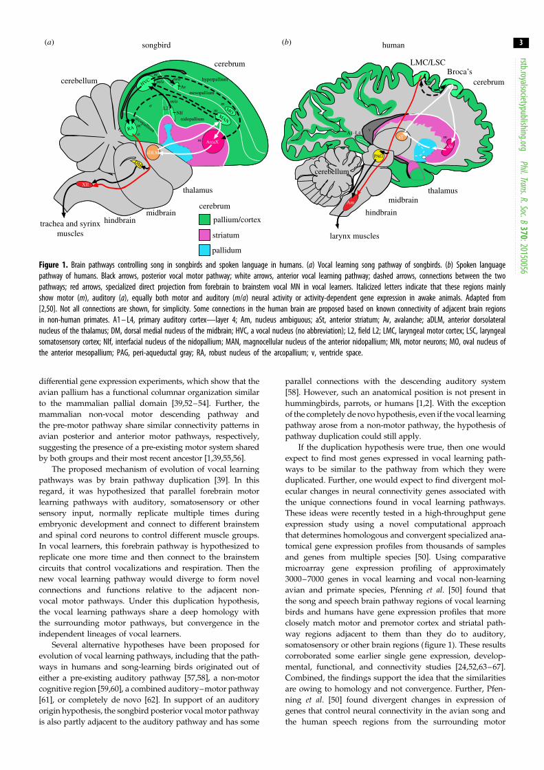
humans [2,40,42–49] (figure 1).
The independently evolved lineages of vocal learning
birds and humans share distinct forebrain pathways that con-
trol the acquisition and production of learned vocalizations
[2]. Within these pathways, all three avian lineages contain
seven cerebral (telencephalic) vocal nuclei and several thal-
amic nuclei. These nuclei, best characterized in songbirds
and parrots, are distributed between two subpathways
(figure 1a): (i) the vocal production, or posterior, pathway
that influences the production of learned song—which
includes an arcopallium nucleus (songbird RA (robust
nucleus of the arcopallium), parrot AAC (central nucleus of
the anterior arcopallium), hummingbird VA (vocal nucleus
of the arcopallium)), analogous to the laryngeal motor
cortex (LMC) in humans (figure 1b) that makes a specialized
direct projection to brainstem vocal motor neurons (MN),
which in turn controls the vocal organs, the syrinx (birds)
and larynx (humans); and (ii) the vocal learning, or anterior,
pathway that is primarily responsible for vocal imitation and
plasticity, which forms a pallial–basal ganglia–thalamic
loop, analogous to such loops in the mammalian brain that
presumably include Broca’s speech area in humans. The
song and speech regions in both these pathways are
embedded in or adjacent to non-vocal motor brain areas
[39,50]. These non-vocal motor regions are present in other
vertebrate species examined thus far, and are thought to be
involved in the production and learning of non-vocal motor
behaviours. Based on these findings, Feenders et al. [39] pro-
posed a motor theory of vocal learning origin, which stated that
‘cerebral brain pathways for vocal learning in distantly related
animals evolved independently as specializations of a pre-
existing motor system inherited from their common ancestor’
([39], p.1). This was a more general theory of the motor theoryof language origin [51], but with specific brain regions identified
and a proposed mechanism. The motor theory of vocal learn-
ing origin suggested that the last common ancestor of birds
and mammals had a motor forebrain pathway, including a
motor cortex or pallium region. This is because although the
motor pallial domain in mammals consists of six layers of
cells (layered) and nuclear subdivisions in birds and reptiles
(clustered), they function similarly and developed from the
same embryonic primordium. This is supported by results
obtained from activity-dependent gene expression and
human
cerebellum
hindbrain
thalamusmidbrain
v
Am
larynx muscles
PAG
aT
Broca’sLMC/LSC
aSt
A1- L4m
m cerebrum
songbird
DM
Av
NIf
XII
HVC
RA
MOMAN
aDLM
AreaX
thalamus
L2
hyperpallium
mesopallium
nidopalliumarcopallium
m
m
m
m/a
m/a
m/a
a
m
m
a
(b)
cerebellum
v
Am
PAG
aT
aSt
A1–L4m
m
(a)
cerebrum
DM
Av
NIf
XII
cerebellum HVC
RA
MOMAN
aDLM
AreaX
hindbrain
cerebrum
midbrainpallium/cortex
striatum
pallidum
L2
hyperpallium
mesopallium
nidopalliumarcopallium
m
m
m
m/a
m/a
m/a
a
m
m
a
trachea and syrinxmuscles
Figure 1. Brain pathways controlling song in songbirds and spoken language in humans. (a) Vocal learning song pathway of songbirds. (b) Spoken languagepathway of humans. Black arrows, posterior vocal motor pathway; white arrows, anterior vocal learning pathway; dashed arrows, connections between the twopathways; red arrows, specialized direct projection from forebrain to brainstem vocal MN in vocal learners. Italicized letters indicate that these regions mainlyshow motor (m), auditory (a), equally both motor and auditory (m/a) neural activity or activity-dependent gene expression in awake animals. Adapted from[2,50]. Not all connections are shown, for simplicity. Some connections in the human brain are proposed based on known connectivity of adjacent brain regionsin non-human primates. A1 – L4, primary auditory cortex—layer 4; Am, nucleus ambiguous; aSt, anterior striatum; Av, avalanche; aDLM, anterior dorsolateralnucleus of the thalamus; DM, dorsal medial nucleus of the midbrain; HVC, a vocal nucleus (no abbreviation); L2, field L2; LMC, laryngeal motor cortex; LSC, laryngealsomatosensory cortex; NIf, interfacial nucleus of the nidopallium; MAN, magnocellular nucleus of the anterior nidopallium; MN, motor neurons; MO, oval nucleus ofthe anterior mesopallium; PAG, peri-aqueductal gray; RA, robust nucleus of the arcopallium; v, ventricle space.
rstb.royalsocietypublishing.orgPhil.Trans.R.Soc.B
370:20150056
3
differential gene expression experiments, which show that the
avian pallium has a functional columnar organization similar
to the mammalian pallial domain [39,52–54]. Further, the
mammalian non-vocal motor descending pathway and
the pre-motor pathway share similar connectivity patterns in
avian posterior and anterior motor pathways, respectively,
suggesting the presence of a pre-existing motor system shared
by both groups and their most recent ancestor [1,39,55,56].
The proposed mechanism of evolution of vocal learning
pathways was by brain pathway duplication [39]. In this
regard, it was hypothesized that parallel forebrain motor
learning pathways with auditory, somatosensory or other
sensory input, normally replicate multiple times during
embryonic development and connect to different brainstem
and spinal cord neurons to control different muscle groups.
In vocal learners, this forebrain pathway is hypothesized to
replicate one more time and then connect to the brainstem
circuits that control vocalizations and respiration. Then the
new vocal learning pathway would diverge to form novel
connections and functions relative to the adjacent non-
vocal motor pathways. Under this duplication hypothesis,
the vocal learning pathways share a deep homology with
the surrounding motor pathways, but convergence in the
independent lineages of vocal learners.
Several alternative hypotheses have been proposed for
evolution of vocal learning pathways, including that the path-
ways in humans and song-learning birds originated out of
either a pre-existing auditory pathway [57,58], a non-motor
cognitive region [59,60], a combined auditory–motor pathway
[61], or completely de novo [62]. In support of an auditory
origin hypothesis, the songbird posterior vocal motor pathway
is also partly adjacent to the auditory pathway and has some
parallel connections with the descending auditory system
[58]. However, such an anatomical position is not present in
hummingbirds, parrots, or humans [1,2]. With the exception
of the completely de novo hypothesis, even if the vocal learning
pathway arose from a non-motor pathway, the hypothesis of
pathway duplication could still apply.
If the duplication hypothesis were true, then one would
expect to find most genes expressed in vocal learning path-
ways to be similar to the pathway from which they were
duplicated. Further, one would expect to find divergent mol-
ecular changes in neural connectivity genes associated with
the unique connections found in vocal learning pathways.
These ideas were recently tested in a high-throughput gene
expression study using a novel computational approach
that determines homologous and convergent specialized ana-
tomical gene expression profiles from thousands of samples
and genes from multiple species [50]. Using comparative
microarray gene expression profiling of approximately
3000–7000 genes in vocal learning and vocal non-learning
avian and primate species, Pfenning et al. [50] found that
the song and speech brain pathway regions of vocal learning
birds and humans have gene expression profiles that more
closely match motor and premotor cortex and striatal path-
way regions adjacent to them than they do to auditory,
somatosensory or other brain regions (figure 1). These results
corroborated some earlier single gene expression, develop-
mental, functional, and connectivity studies [24,52,63–67].
Combined, the findings support the idea that the similarities
are owing to homology and not convergence. Further, Pfen-
ning et al. [50] found divergent changes in expression of
genes that control neural connectivity in the avian song and
the human speech regions from the surrounding motor

rstb.royalsociet
4
areas, but that were convergent among the vocal learning birdsand humans. There were also convergent changes in some genes
involved in neural development, neuroprotection and synaptic
communication functions. The brain expression characteriz-
ation of these genes [50,68] led to the discovery of a further
apparent duplication in the parrot brain [56], as described next.
ypublishing.orgPhil.Trans.R.Soc.B370:20150056
(b) Parrots contain a song system within a song systemParrots surpass other vocal learning avian species in their abil-
ity to imitate human speech and also rival non-human
primates in their display of advanced cognitive skills and abil-
ity for tool use [2,69–74]. From 1981 until recently in 2015
(approx. 34 years), the neural pathways for vocal learning
had been studied in only one parrot species, the budgerigar
(Melopsittacus undulatus) [55,75–79]. From these studies, it
was apparent that the budgerigar song system shows some
differences from the songbird and hummingbird song systems
[55,75,76,79,80]. The posterior song system of songbirds and
parrots (and presumably hummingbirds) receives auditory
input from the posterior auditory pathway (e.g. auditory
Field L), but in parrots it receives additional auditory input
from a small part of the nucleus basorostralis (B), the remain-
der of which is somatosensory [81]. Neural tracing and
singing-regulated immediate early gene studies revealed some
differences in connectivity and position or shape of song
nuclei in parrots, but no clear differences were noted in the
presence or absence of song system structures [55,75,76,78–80].
Recently, partly based on the high-throughput gene
expression [50,68] and other findings, we led a study [56]
characterizing gene expression profiles that are specialized
in avian and/or human song/speech vocal learning circuits
(e.g. PVALB, SLIT1, FOXP1, NR2A, GLUR1, NADPH-d, AR,
mENK, TH, CGRP-LI) to understand the organization of the
song system in diverse groups of parrots representing all
the three superfamilies, Strigopoidea, Cacatuoidea and
Psittacoidea [82]. We found that the parrot pallial (cortical)
song nuclei had core regions that differed in gene expression
from surrounding shell regions, and both in turn differed
from the surrounding cortical motor areas (figure 2) [56].
Surprisingly, a subset of the genes (including PVALB) had
moderate specialized expression in the immediate surround-
ing non-vocal motor areas. Both the core and shell song
regions were functionally active in the production of learned
vocalizations, as revealed by vocalizing-driven immediate
early gene expression (EGR-1, C-FOS and DUSP-1), whereas
the surrounding brain regions were active in production of
non-singing motor behaviour, as revealed by rhythmic
controlled hopping-driven gene expression [39,56,79,85,86].
The connectivity of the core and shell systems were similar
to each other, but with some significant differences. One funda-
mental difference was that the core system made the rare direct
projection to brainstem vocal MN (via the AAC core nucleus),
whereas the parallel shell song system made mostly intra-
cortical projections (via AAC shell; figure 2b,c). The direct
projection to the brainstem vocal MN is considered critical
for the evolution of vocal learning and spoken language, as
it is either absent or very sparse in vocal non-learners
[2,42,87–92]. There were sparse connections between the
parrot cores and shells within and among each song nucleus.
The presence of these song nuclei in the kea, the most dis-
tantly related to the other extant parrot species [82,93],
indicates that parrots evolved the core and shell song systems
over 29 Ma before the kea split from the other parrot lineages.
The kea shell system, however, was less well differentiated in
terms of its gene expression specializations. There were also
large species differences in relative sizes of the core and shell
regions, where the shell had a significant log-linear relationship
with their brain section size, but the core did not. This meant
that shell regions were relatively larger in species with bigger
brains such as the gold and blue macaw, and the African
Grey and Amazon parrots that are considered to have more
advanced communication and cognitive capacities.
The fact that the shell system AAC nucleus does not have
the direct projection to the vocal MN (which is restricted to
the core region of AAC), but may be correlated with more
complex vocal learning behaviour, indicates that such direct
projections may not be the only means to increase learned
motor behavioural complexity over innate motor behaviours.
We speculate that it is possible that the direct projection may
not be required for the ability to imitate complex vocaliza-
tions, but strictly for the production of those learned
vocalizations. Clearly, further studies will be required to
explicitly test this hypothesis in parrots.
The gene expression specializations and neural connectivity
of the parrot core song nuclei were most similar to the song
nuclei of songbirds and hummingbirds, whereas the shells
were unique to parrots. The shell specializations appear to be
restricted to the cortical parrot song nuclei, as no shells have
yet been found for the striatal or thalamic song nuclei. The song-
bird and hummingbird species examined to date do not have
parallel vocal motor shell regions for any of their nuclei.
These findings indicate that the core and shell are two
parallel, partially independent systems, performing similar
and some unique functions for vocalizations. They support
a partial brain pathway duplication hypothesis of brain evol-
ution. In particular, we suggest that the core song system
evolved convergently in parrots, songbirds and humming-
birds as a duplication event in each lineage from the
surrounding motor areas (figure 2a,b). Thereafter, the parrot
core cortical song nuclei underwent a further partial dupli-
cation event to evolve the shell song system (figure 2c). The
shell song system went on to evolve specializations that
allow more complex vocal communication abilities in parrots
compared to other avian vocal learners. This dual system
evolved early in the parrot lineage, and has lasted and
expanded for millions of years in different species. In
addition, changes in the regulation of some genes that may
allow greater vocal–motor–auditory integration in vocal
learning systems could have influenced changes in the sur-
rounding motor areas to allow greater auditory–motor
entrainment and synchronizing of body movements to the
rhythm of music for dance in parrots [94–96].
It would be exciting to determine if similar duplications
of brain pathways have occurred in humans, not only in
the speech pathways but also for other advanced motor
movements such as dancing [97–99]. In the human brain,
areas 44 and 45 constitute Broca’s area, the ventrolatreal fron-
tal region critical for spoken language acquisition and
production [100]. Petrides et al. [101,102] showed that there
is a comparable area 44-like region involved in orofacial
musculature functions in macaque monkeys. What is still
unknown is whether area 44 in the human and the macaque
brain share common ancestry since there is lack of an out-
group comparison so far. It is tempting to speculate that
Broca’s area could constitute one or more duplications of a

(b) evolution of the core song systemfrom duplication of the motor pathway
thalamic motor system
core song system
shell song system
pallial motor system
striatal motor system
trachea and syrinxmuscles
L2
BNLC
DM
LAN
LAM
MMSt
NAO
DMM
MO
hindbrainXII
midbrain
thalamus
nidopallium
mesopallium
hyperpallium
AAC
arcopallium
(a) general motor pathwayof ancestral parrot species
cerebrumpallium/cortex
striatum
pallidum
L2
B
DM
hindbrain midbrain
thalamus
nidopallium
mesopallium
hyperpallium
trachea and syrinxmuscles
XII
limb and bodymuscles
MN
PMN
ASt
AN
DT
AM
arcopallium
NCLPLN
PLM
Ai
(c) evolution of the shell song systemfrom partial duplication of the core system
trachea and syrinxmuscles
L2
BNLC
DM
AAC
hindbrainXII
midbrain
nidopallium
mesopallium
hyperpallium
LAM
MMSt
DMM
thalamus
arcopallium
NAO
MOLAN
Figure 2. Hypothesis of evolution of song system in parrots owing to sequential pathway duplications. (a) The parrot ancestral motor pathway (light green) with theposterior motor connections (in black arrows) and the anterior motor connections (in white arrows). (b) The parrot core song system (red), proposed to have evolved outof the pre-existing motor pathway through duplication. (c) The parrot shell song system (yellow), proposed to have evolved out of a partial duplication of the core songsystem. Black arrows, posterior vocal motor pathway; white arrows, anterior vocal motor pathway; dashed arrows, connections between the two pathways; red arrow,specialized direct projection from forebrain to brainstem vocal MN in vocal learners. Connectivity based on summaries in [39,55,56,76,83,84]. See fig. 20 from Chakra-borty et al. [56] for additional connections of the core and shell song pathways. Not all connections are shown for simplicity, including reciprocal connections andadditional thalamic projections. AAC, central nucleus of the anterior arcopallium; Ai, intermediate arcopallium; AM, anterior mesopallium; AN, anterior nidopallium;ASt, anterior striatum; B, basorostralis; DM, dorsal medial nucleus of the midbrain; DMM, magnocellular nucleus of the dorsomedial thalamus; DT, dorsal thalamus; L2,L4, auditory areas; NAO, oval nucleus of the anterior nidopallium; NCL, nidopallium caudal lateral; NLC, central nucleus of the lateral nidopallium; PMN, premotorneurons; LAN, lateral nucleus of the anterior nidopallium; LAM, lateral nucleus of the anterior mesopallium; MMSt, magnocellular nucleus of the medial striatum;MO, oval nucleus of the anterior mesopallium; PLM, posterior lateral mesopallium; PLN, posterior lateral nidopallium; XII, 12th motor nucleus.
rstb.royalsocietypublishing.orgPhil.Trans.R.Soc.B
370:20150056
5
more ancestral area 44, with divergent specializations for
learned vocalizations and thus spoken language.
3. Some alternative hypotheses to the motortheory and brain pathway duplications
Expanding upon the alternative hypotheses, it is possible that
the core and shell systems in parrots arose simultaneously, in
which case the shell would not be a duplication of the core. How-
ever, for this alternative, it would be difficult to explain why the
parrot core pathway is more similar to the songbird and
hummingbird song systems, than an apparently evolutionary
older state (i.e. appearing first). The shell song system of parrots
does not appear to be a functionally differentiated region of the
core system, since the core system still exists in them and other
vocal learners.
A second alternative is that the shell is an independent dupli-
cation of the adjacent motor pathway, as the shell has a more
similar gene expression profile to the adjacent motor pathway
than the core. However, if this were the case, it would be hard
to explain how evolution of the shell is more independent of
the core, considering that the shell song nuclei surround the
cores and are not located elsewhere in the motor pathway and

rstb.royalsocietypublishing.orgPhil.Trans.R.Soc.B
370:20150056
6
that both core and shell are interconnected without notablemajor connections to the surrounding motor areas.
A third alternative is that the parrot shell and core song
nuclei, as well as the songbird, hummingbird and human
song/speech region analogues, are all specialized transform-
ations of an existing motor pathway, i.e. subfunctionalizations,
rather than duplications from it, i.e. neofunctionalizations.
This would mean that the vocal learning species lost non-
vocal motor learning pathway neurons to gain vocal motor
pathway neurons. However, there is as yet no evidence for
loss of non-vocal motor or other functions in vocal learn-
ing species, but rather gain of functions even beyond vocal
learning (such as learning how to dance) [94,96,97,99] and
increases in relative brain-to-body sizes [103].
A fourth alternative is that vocal learning pathway neurons
migrate into their adult locations from developing non-adjacent
and even non-motor brain areas, and then byadjacent association
they adopt some of the motor learning pathway phenotypes.
Although this alternative is theoretically possible, one would
expect that the vocal learning pathway cells would have some
vestigial properties of their non-motor origin. Thus far, the evi-
dence has not revealed such an alternative origin, although
only in songbirds does the HVC (a letter-based name) motor
pathway nucleus share some secondary profiles in gene
expression with the human secondary auditory cortex [50].
A bigger challenge to the motor theory and duplication
hypotheses might at first glance be derived from a recently pro-
posed ‘continuum hypothesis’ of vocal learning. Based on
findings that mice have a rudimentary forebrain circuit involved
in modulating vocalizations but with a very sparse direct projec-
tion to brainstem vocal MN, buried within a motor region that
also controls other behaviours, and that mice and non-human
primates have at least a very limited ability to modify their voca-
lizations based on auditory experience, Arriaga & Jarvis [104]
and Petkov & Jarvis [2] proposed a continuum hypothesis of
vocal learning. In this hypothesis, vocal learning is considered
to range from complex (e.g. humans and parrots), moderate
(some songbirds), to limited or none (mice and non-human
primates) [105–108]. In this model, there would be a rudimen-
tary pre-existing forebrain vocal pathway within the vertebrate
motor learning pathway, but in the more complex vocal lear-
ners these vocal pathway neurons independently expanded
and segregated out of the motor learning pathway. However,
it is also possible that initially the motor learning pathway
duplicates within the non-vocal motor learning circuits to
form a limited vocal learning circuit, which subsequently
evolves enhanced functions and moves outside of to become
adjacent to the motor learning circuit.
Resolving these hypotheses will require more compara-
tive research. The nuances for limited vocal plasticity and
an associated neural circuit in mice and non-human primates
are also still an ongoing debate that requires further investi-
gation [104–111]. Thus far, of all the hypotheses proposed,
we believe the existing data most support the motor origin
and duplication hypothesis for vocal learning pathways.
4. Other examples of duplicated morphologicalstructures and structural subdivisions in theevolution of functional complexity
If brain pathway evolution by duplication were possible for
vocal learning circuits, then it could be one broad mechanism
of brain evolution. The presence of the well-known parallel
cortical–basal ganglia–thalamic–cortical loops through the
anterior forebrain of mammals and birds is consistent with
such an idea. These parallel loops could be replicates of a
basic motor learning pathway design. Since all of the cortex is
connected with all of the basal ganglia and thalamus [112],
when a cortex region is duplicated, one would expect to see a
concomitant duplication in the connected basal ganglia and
thalamic regions [22,33]. However, our finding of only cortical
shell song nuclei duplications thus far in parrots indicates that
it could be possible that the duplicated cortical regions co-
innervate the non-duplicated striatal and thalamic regions
(figure 2c). Such flexibility would allow for greater diversity in
neural circuit evolution balanced with the constraint of limited
cranial space to accommodate increases in brain size owing
to duplications.
Studies of non-human primate motor cortex are consistent
with the idea of duplicated brain components. For example,
studies using retrograde transneuronal transport of the rabies
virus from single muscles of rhesus macaque monkeys to ident-
ify layer 5 cortico-motoneuronal (CM) cells in the primary
motor cortex (M1) have shown that the M1 region has two sub-
divisions [113]. The rostral subdivision of macaque M1 has
been proposed to be an ‘older’ region as it contains fewer
CM cells that make indirect projections to spinal cord MN
and is present in most mammals requiring the indirect use of
the spinal cord to influence motor output. The caudal subdivi-
sion of macaque M1 is proposed to be ‘newer’ as it contains the
more rarely found CM cells that make direct projections to MN
in the spinal cord and brainstem, including controlling
shoulder, elbow and finger muscles to produce highly skilled
motor actions. Based on these and other findings, it is generally
proposed that the direct CM system of M1 is a recently evolved
brain structure that conferred evolutionarily novel functions in
the motor system in primates, including independent voluntary
control of finger movements, which are more advanced in pri-
mates compared with non-primates [114–120]. Assuming that
the evidence continues to support differences between species,
one hypothesis, like the one we propose for vocal learning path-
ways, is that the newer caudal M1 region is a duplication of the
older rostral M1 region, but with a divergent connection of the
CM cells from cortical layer 5.
Examples of morphological duplications or subdivisions to
enhance complexity also exist outside of the nervous system.
Many animals, such as annelids, have repeated parallel body
segments or specialized limb types among species, where the
replicated parts are thought to be owing to a repeating devel-
opmental genomic programme [121,122]. A striking example
of independently evolved morphological duplications is the
diversification of the adductor mandibular muscles in teleost
fish jaws, which have independently subdivided several
times during tetraodontiform evolution [123–125]. Most of
these divisions have been incomplete, which suggests that
some parts were subfunctionalized instead of duplicated. The
duplicated adductor mandibulae muscles continue to maintain
similar morphological characteristics, but with increased mor-
phological complexity associated with their functional
complexity resulting in finer motor control for feeding [125].
Overall, structural duplications have been proposed to be
one mechanism that allows for morphological decoupling
[126]. Making structures, such as brain circuits, functionally
independent of one another may provide increased comple-
xity and opportunity for modification and diversification

rstb.royalsocietypu
7
[126–128]. In this regard, brain evolution by brain regionduplication, brain pathway duplication or structure subdivi-
sion may follow a general mechanism of morphological
evolution to enhance functional complexity. Testing these
hypotheses will be best informed by deciphering the cellular
and molecular mechanisms for the development of additional,
parallel circuits in the brain.
blishing.orgPhil.Trans.R.Soc.B370:20150056
5. Proposed cellular and molecular mechanismsfor evolution of brain pathway duplications
During development, neural stem cell/progenitor cells that give
rise to forebrain circuits derive mainly from stem cells in the
ventricular zone [129–131]. The daughter cells travel to their
positions either by radial migration perpendicularly away
from the ventricle (such as excitatory neurons within layers of
the mammalian cortex), and/or by tangential migration parallel
to the ventricle (such as inhibitory neurons that migrate from
the basal ganglia into the cortex) [132–134]. Their local brain
region identity is thought to be controlled by patterning tran-
scription factors, such as the homeobox (Hox) genes [135,136].
Once daughter cells reach their target location in the brain,
they find their connecting partners in a process that requires
cell adhesion and axon guidance genes [137,138]. Given these
principles, we propose that one possible mechanism for the
evolution and development of duplicated segmented brain cir-
cuits is that there is a set of transcription factors that not only
control the position of such circuits, but also the number of
replicates of that circuit. A genetic change in such transcription
factors could result in a new parallel circuit, such as that for
vocal learning. Thereafter, changes in axon guidance genes in
the new circuit would control divergence in connectivity rela-
tive to the older circuit. This begs the question of what kind
of genetic change would this be?
We propose that gene duplication could be one such mech-
anism. Gene duplications have been found to influence the
development and function of many organs and tissues, includ-
ing brains, eyes and wings [11,139–147]. As proposed originally
by Ohno [13,14], many consider gene duplication to be one of
the most important factors in evolution, including neofunctio-
nalization, subfunctionalization and evolutionary innovations.
Gene duplication allows the old gene copy to maintain its func-
tion and the new copy to evolve new functions. Even theories
on gene evolution through gene duplication have influenced
the theories on brain evolution by morphological duplication
[139,141]. The concept of neofunctionalization of genes [14]
and subfunctionalization of genes [148] match those proposed
for structural duplications [128,149,150].
One of the most well-studied and significant examples of
duplicated genes controlling duplicated, repeated or segmen-
ted morphological structures are the Hox genes. These are
transcription factors that control the anterior–posterior body
plan axis and are situated in the genome in the same order as
the body plan they control [136,151,152]. They are duplicated
to different degrees in different animal lineages, with greater
complexity and more anatomical segments correlated with
more duplications [153]. Many invertebrates and Amphioxuspossess one Hox gene cluster, whereas the remaining ver-
tebrates have four Hox gene clusters, in part owing to two
whole-genome duplication events that occurred early in ver-
tebrate evolution [151,154–158]. Within the brain, the Hoxgenes and the greater Hox gene transcription factor
superfamily (including OTX, EMX, DMBX, GBX and EN) are
involved in brain division and subdivision segmentation
[135]. They do so by controlling regional neuronal identity,
stem cell progenitors, cell migration and cell death [159,160].
We propose that one possible mechanism for brain
pathway duplication could be a local duplication of Hox super-
family genes in the genome segments that control forebrain
development. One prediction of this hypothesis is that one
should find such genes uniquely duplicated in vocal learning
species that control brain development. Recently, based on
comparative genomic analyses across the bird family tree,
unique gene duplications were found in the songbird lineage
and some of these genes had enriched or nearly exclusive
expression in the song learning nuclei [161]. It remains to be
determined, however, if any of these transcription factors
belong to the Hox gene family.
We caution that we are not suggesting a one-to-
one relationship of gene duplication with morphological
duplication. There are many examples of gene duplications
resulting in modifications of existing structures and functions.
An example relevant to the topic of vocal learning and
cognition is the Slit-Robo GTPase 2 gene (SRGAP2), which has
undergone two partial duplications (SRGAP2B and
SRGAP2C) uniquely in humans relative to other mammals
[26,27,162]. The duplicated copies act as competitive inhibitors
to slow cortical dendritic development of already existing brain
pathways, which in turn allow greater neural plasticity into
adulthood. SRGAP2 modulates activity of the ROBO axon
guidance receptors, which are in turn activated by the SLIT
family of protein ligands to modulate axonal/dendritic
migration and branching in various brain regions [163–167].
Intriguingly, the SLIT1 ligand is uniquely downregulated in
the song production nucleus RA analogue of vocal learning
birds (songbird RA, parrot AAC and hummingbird VA)
[56,68] and the analogous human LMC [50], which would
mean that there could be a synergistic effect of the duplicated
SRGAP2 GTPase and lower SLIT1 levels in the duplicated
vocal motor pathways in humans. Another recent example of
partial duplication includes another GTPase, the ARHGAP11Bgene, which arose from ARHGAP11A in humans after separ-
ation from the chimpanzee lineage [168]. The duplicated copy
of the ARHGAP11A gene causes cortical area expansion, and
this expansion causes folding, which we surmise could be
owing to the duplication.
Advances in genetic technologies have also allowed scien-
tists to test some hypotheses on duplicating or eliminating
neural structures genetically. For example, ectopic visual
responsive eyes were induced in Drosophila with the addition
of an extra copy of one transcription factor, Pax6, expressed
during development in another part of the body [169]. Another
study showed that electroporating an extra copy of the fibro-
blast growth factor 8 (FGF8) gene locally in the posterior
cortical primordium of mouse embryos causes a partial dupli-
cation of the primary somatosensory cortex, with concomitant
input from the thalamus to its layer 4 cortical cells, as shown
by the presence of ectopic somatosensory barrel fields [170].
In vertebrates, the expression of the Hox1a gene marks the ear-
liest stages of regionalization of the developing hindbrain. Mice
mutant for the Hoxa1 gene lack the developing rhombomere 2
(r2) brain region, but the r2 neurons escape apoptosis and
develop within r3 and r4, to still incorporate into appropriate
circuits to drive the rhythm of breathing [171]. This suggests
to us that Hox1a is needed for development of a separate,

rstb.royalsocietypublishing.orgPhil.Trans.R.Soc.B
370:20150056
8
repeated rhombomere region, but that other factors are suffi-cient to develop the associated circuit within another circuit.
An example of loss of gene function and functional redundancy
leading to duplication of structure is the Mauthner (M) cells, a
pair of reticulospinal neurons that control escape behaviour in
zebra fishes [139,172–174]. During the escape response, if the
threatening stimulus arrives from the left side, the left M
cell fires, and its action potential travels to the right side so
that the fish swerves to the right side owing to the contraction of
the muscles on the right side to avoid the threat. Mutation
of the notch1a/deadly seven (des) in zebra fish results in the devel-
opment of extra M cells in r4 [175]. All extra copies of the M cells
are responsive to the escape stimuli, suggesting that when
duplication of the cells takes place, they receive the appropriate
sensory information and respond in a normal way indicating
adaptive plasticity of the escape-response circuit.
Other plausible hypotheses of molecular mechanisms that
could lead to brain pathway duplication include: (i) changes
in splice variants of a gene [176], which we propose could
switch on and off at different developmental times to control
the generation of parallel circuits; (ii) changes in the cis-
regulatory elements of genes, which we propose again
would change the reiterative use of a gene network in parallel
developing circuits; and (iii) loss of function in a gene that
may normally inhibit development of some circuits.
Overall, the various hypotheses may be tested with recent
advances in genomics, transcriptomics and gene manipula-
tions, using complete genome sequences from multiple
species with and without the brain pathways of interest
[93]. Until then, the existing evidence supports the possibility
that brain pathway evolution through brain pathway duplicationcould be one mechanism to generate higher-order complexity
in highly evolved animals.
6. ConclusionIn this review, we discussed new evidence from studies
in birds, primates and other species that suggests that brain
pathways for a novel convergent trait, vocal learning, possi-
bly evolved by duplication from adjacent motor learning
pathways. The continuum hypothesis of a pre-existing vocal
learning pathway that was independently enhanced in
vocal learners is an alternative, but could be compatible with
the duplication hypothesis if the duplication occurs within an
existing pathway, as seen with Hox1a r2 manipulations.
Whether by duplication or enhancement, the pathways have
diverged from their adjacent brain regions by specializations
of genes involved in neural connectivity. These divergences
may have been heavily selected upon for immediate and
substantial phenotypic benefits. Despite these divergences,
the vocal learning circuits share many properties with the adja-
cent motor pathways. The findings of the parrot core and shell
song system lead us to wonder if humans evolved consecutive
or simultaneous multiple duplications of a vocal learning path-
way leading to more advanced spoken language abilities.
Moreover, findings from studies outside of the vocal learning
systems indicate that brain region or pathway duplication
could be a general mechanism of brain evolution.
Answers to these questions can now be determined through
comparative neurobiology and comparative genomics research.
With the recent availability of genomes across the avian
[93,177,178] and eventually primate [179,180] family trees, it
becomes possible to discover candidate genes. They can then
be studied with advanced technologies, such as transcriptomics
and genome editing tools, including CRISPR-Cas9, RNAi,
TALENs, Cre-Lox systems and more. The theoretical framework
presented here will help guide use of these technologies.
Authors’ contributions. E.D.J. and M.C. conceived and wrote the paper.
Competing interests. We have no competing interests.
Funding. The authors were supported by funds from the HowardHughes Medical Institute.
Acknowledgements. The authors thank members of the Jarvis Lab (MattBiegler, Greg Gedman, Ha Na Choe, Lindsey Catlin, Joshua Robinsonand Jonathan Chabout) for critical discussions on the paper.
References
1. Jarvis ED. 2004 Learned birdsong and theneurobiology of human language. Ann. NY Acad. Sci.1016, 749 – 777. (doi:10.1196/annals.1298.038)
2. Petkov CI, Jarvis ED. 2012 Birds, primates, andspoken language origins: behavioral phenotypesand neurobiological substrates. Front. Evol. Neurosci.4, 12. (doi:10.3389/fnevo.2012.00012)
3. Merker B. 2012 The vocal learning constellation:imitation, ritual culture, encephalization. In Music,language, and human evolution (ed. N Bannan),pp. 215 – 259. Oxford, UK: Oxford University Press.
4. Williams RW, Herrup K. 1988 The control of neuronnumber. Annu. Rev. Neurosci. 11, 423 – 453.(doi:10.1146/annurev.ne.11.030188.002231)
5. Joseph-Harrigan W, Commons LM. 2014 The stageof development of a species predicts the number ofneurons. Behav. Dev. Bull. 19, 12 – 19. (doi:10.1037/h0101077)
6. Holland LZ, Holland ND. 1999 Chordate origins ofthe vertebrate central nervous system. Curr. Opin.
Neurobiol. 9, 596 – 602. (doi:10.1016/S0959-4388(99)00003-3)
7. Holland LZ, Short S. 2008 Gene duplication, co-option and recruitment during the origin of thevertebrate brain from the invertebrate chordatebrain. Brain Behav. Evol. 72, 91 – 105. (doi:10.1159/000151470)
8. Chen JY. 2008 Early crest animals and the insight theyprovide into the evolutionary origin of craniates.Genesis 46, 623 – 639. (doi:10.1002/dvg.20445)
9. Emes RD, Grant SG. 2012 Evolution of synapsecomplexity and diversity. Annu. Rev. Neurosci.35, 111 – 131. (doi:10.1146/annurev-neuro-062111-150433)
10. Davidson EH, Erwin DH. 2006 Gene regulatorynetworks and the evolution of animal bodyplans. Science 311, 796 – 800. (doi:10.1126/science.1113832)
11. Taylor JS, Raes J. 2004 Duplication and divergence:the evolution of new genes and old ideas. Annu.
Rev. Genet. 38, 615 – 643. (doi:10.1146/annurev.genet.38.072902.092831)
12. Prince VE, Pickett FB. 2002 Splitting pairs:the diverging fates of duplicated genes.Nat. Rev. Genet. 3, 827 – 837. (doi:10.1038/nrg928)
13. Ohno S. 1967 Sex chromosomes and sex-linkedgenes. Berlin, Germany: Springer.
14. Ohno S. 1970 Evolution by gene duplication.New York, NY: Springer.
15. Zhang J. 2003 Evolution by gene duplication: anupdate. Trends Ecol. Evol. 18, 292 – 298. (doi:10.1016/S0169-5347(03)00033-8)
16. Johnson R, Samuel J, Ng CK, Jauch R, Stanton LW,Wood IC. 2009 Evolution of the vertebrate generegulatory network controlled by the transcriptionalrepressor REST. Mol. Biol. Evol. 26, 1491 – 1507.(doi:10.1093/molbev/msp058)
17. Ono H, Kozmik Z, Yu JK, Wada H. 2014 A novelN-terminal motif is responsible for the evolution of

rstb.royalsocietypublishing.orgPhil.Trans.R.Soc.B
370:20150056
9
neural crest-specific gene-regulatory activity invertebrate FoxD3. Dev. Biol. 385, 396 – 404. (doi:10.1016/j.ydbio.2013.11.010)18. Goymer P. 2007 Alternative splicing switches on thebrain. Nat. Rev. Neurosci. 8, 576. (doi:10.1038/nrn2200)
19. Chen M, Manley JL. 2009 Mechanisms of alternativesplicing regulation: insights from molecular andgenomics approaches. Nat. Rev. Mol. Cell Biol. 10,741 – 754. (doi:10.1038/nrm2777)
20. Irimia M et al. 2011 Stepwise assembly of theNova-regulated alternative splicing network in thevertebrate brain. Proc. Natl Acad. Sci. USA 108,5319 – 5324. (doi:10.1073/pnas.1012333108)
21. Gueroussov S, Gonatopoulos-Pournatzis T, Irimia M,Raj B, Lin ZY, Gingras AC, Blencowe BJ. 2015 RNAsplicing. An alternative splicing event amplifiesevolutionary differences between vertebrates. Science349, 868 – 873. (doi:10.1126/science.aaa8381)
22. Kaas JH. 1989 The evolution of complex sensorysystems in mammals. J. Exp. Biol. 146, 165 – 176.
23. Striedter GF. 2005 Principles of brain evolution.Sunderland, MA: Sinauer Associates.
24. Jarvis ED et al. 2005 Avian brains and a newunderstanding of vertebrate brain evolution. Nat.Rev. Neurosci. 6, 151 – 159. (doi:10.1038/nrn1606)
25. Northcutt RG. 1984 The evolution of the vertebratecentral nervous system: patterns and processes. Am.Zool. 24, 701 – 716. (doi:10.1093/icb/24.3.701)
26. Charrier C et al. 2012 Inhibition of SRGAP2 functionby its human-specific paralogs induces neotenyduring spine maturation. Cell 149, 923 – 935.(doi:10.1016/j.cell.2012.03.034)
27. Dennis MY et al. 2012 Evolution of human-specificneural SRGAP2 genes by incomplete segmentalduplication. Cell 149, 912 – 922. (doi:10.1016/j.cell.2012.03.033)
28. Fodor JA. 1983 The modularity of mind. Cambridge,MA: MIT Press.
29. Pinker S. 1994 The language instinct: how the mindcreates language. New York, NY: W. Morrow.
30. Brodmann K. 1909 Vergleichende Lokalisationslehreder Grosshirnrinde. Leipzig, Germany: Barth.
31. Diamond IT, Hall WC. 1969 Evolution of neocortex.Science 164, 251 – 262. (doi:10.1126/science.164.3877.251)
32. von Economo C. 1929 The cytoarchitectonics of thehuman cortex. Oxford, UK: Oxford University Press.
33. Kaas JH. 1982 The segregation of function in thenervous system: why do sensory systems have somany subdivisions? In Contributions to SensoryPhysiology, vol. 7 (ed. WP Neff ), pp. 201 – 240.New York, NY: Academic Press.
34. Ebbesson SOE. 1984 Evolution and ontogeny ofneural circuits. Behav. Brain Sci. 7, 321 – 331.(doi:10.1017/S0140525X00018379)
35. Lende RA. 1969 A comparative approach to neocortex:localization in Monotremes, marsupials andinsectivores. Ann. NY Acad. Sci. 167, 262 – 276.(doi:10.1111/J.1749-6632.1969.Tb20449.X)
36. Finlay BL, Cheung D, Darlington RB. 2005Developmental constraints on or developmentalstructure in brain evolution? In Attention andperformance XXI ‘process of change in brain and
cognitive development’ (eds Y Munakata, MJohnson), pp. 131 – 162. Oxford, UK: OxfordUniversity Press.
37. Allman JK, Kaas JH. 1971 A representation of thevisual field in the caudal third of the middletemporal gyrus of the owl monkey (Aotustrivirgatus). Brain Res. 31, 85 – 105. (doi:10.1016/0006-8993(71)90635-4)
38. Hughes AL. 1994 The evolution of functionallynovel proteins after gene duplication. Proc. R. Soc.Lond. B 256, 119 – 124. (doi:10.1098/rspb.1994.0058)
39. Feenders G, Liedvogel M, Rivas M, Zapka M, HoritaH, Hara E, Wada K, Mouritsen H, Jarvis ED. 2008Molecular mapping of movement-associated areasin the avian brain: a motor theory for vocal learningorigin. PLoS ONE 3, e1768. (doi:10.1371/journal.pone.0001768)
40. Nottebohm F. 1972 The origins of vocal learning.Am. Nat. 106, 116 – 140. (doi:10.1086/282756)
41. Janik VM, Slater PJB. 1997 Vocal learning inmammals. Adv. Study Behav. 26, 59 – 99. (doi:10.1016/S0065-3454(08)60377-0)
42. Fitch WT, Jarvis ED. 2013 Birdsong and otheranimals models for human speech, song, and vocallearning. In Language, music, and the brain (ed. MAArbib), pp. 499 – 539. Cambridge, MA: MIT Press.
43. Brainard MS, Doupe AJ. 2013 Translating birdsong:songbirds as a model for basic and applied medicalresearch. Annu. Rev. Neurosci. 36, 489 – 517.(doi:10.1146/annurev-neuro-060909-152826)
44. Margoliash D. 1997 Functional organization offorebrain pathways for song production andperception. J. Neurobiol. 33, 671 – 693. (doi:10.1002/(SICI)1097-4695(19971105)33:5,671::AID-NEU12.3.0.CO;2-C)
45. Fee MS, Goldberg JH. 2011 A hypothesis for basalganglia-dependent reinforcement learning in thesongbird. Neuroscience 198, 152 – 170. (doi:10.1016/j.neuroscience.2011.09.069)
46. Mooney R. 1999 Sensitive periods and circuits forlearned birdsong. Curr. Opin. Neurobiol. 9,121 – 127. (doi:10.1016/S0959-4388(99)80015-4)
47. Mooney R. 2009 Neural mechanisms for learnedbirdsong. Learn. Mem. 16, 655 – 669. (doi:10.1101/lm.1065209)
48. Simonyan K, Horwitz B. 2011 Laryngeal motorcortex and control of speech in humans.Neuroscientist 17, 197 – 208. (doi:10.1177/1073858410386727)
49. Simonyan K, Horwitz B, Jarvis ED. 2012 Dopamineregulation of human speech and bird song: a criticalreview. Brain Lang. 122, 142 – 150. (doi:10.1016/j.bandl.2011.12.009)
50. Pfenning AR et al. 2014 Convergent transcriptionalspecializations in the brains of humans and song-learning birds. Science 346, 1256846. (doi 10.1126/science.1256846)
51. Allott R. 1992 The motor theory of language: originand function. In Language origin: a multidisciplinaryapproach (eds J Wind, BH Bichakjian, A Nocentini, BChiarelli), pp. 105 – 119. Dordrecht, TheNetherlands: Kluwer.
52. Jarvis ED et al. 2013 Global view of the functionalmolecular organization of the avian cerebrum:mirror images and functional columns. J. Comp.Neurol. 521, 3614 – 3665. (doi:10.1002/cne.23404)
53. Wang Y, Brzozowska-Prechtl A, Karten HJ. 2010Laminar and columnar auditory cortex in avianbrain. Proc. Natl Acad. Sci. USA 107, 12 676 –12 681. (doi:10.1073/pnas.1006645107)
54. Shimizu T, Patton TB, Husband SA. 2010 Avianvisual behavior and the organization of thetelencephalon. Brain Behav. Evol. 75, 204 – 217.(doi:10.1159/000314283)
55. Durand SE, Heaton JT, Amateau SK, Brauth SE. 1997Vocal control pathways through the anteriorforebrain of a parrot (Melopsittacus undulatus).J. Comp. Neurol. 377, 179 – 206. (doi:10.1002/(SICI)1096-9861(19970113)377:2,179::AID-CNE3.3.0.CO;2-0)
56. Chakraborty M et al. 2015 Core and shellsong systems unique to the parrot brain. PLoSONE 10, e0118496. (doi:10.1371/journal.pone.0118496)
57. Margoliash D, Fortune ES, Sutter ML, Yu AC, Wren-Hardin BD, Dave A. 1994 Distributed representation inthe song system of oscines: evolutionary implicationsand functional consequences. Brain Behav. Evol. 44,247 – 264. (doi:10.1159/000113580)
58. Mello CV, Vates GE, Okuhata S, Nottebohm F. 1998Descending auditory pathways in the adult malezebra finch (Taeniopygia guttata). J. Comp. Neurol.395, 137 – 160. (doi:10.1002/(SICI)1096-9861(19980601)395:2,137::AID-CNE1.3.0.CO;2-3)
59. Edelman DB, Baars BJ, Seth AK. 2005 Identifyinghallmarks of consciousness in non-mammalianspecies. Conscious. Cogn. 14, 169 – 187. (doi:10.1016/j.concog.2004.09.001)
60. Rendell L, Whitehead H. 2001 Culture in whalesand dolphins. Behav. Brain Sci. 24, 309 – 324;discussion 324 – 382. (doi:10.1017/S0140525X0100396X)
61. Okanoya K. 2007 Language evolution and anemergent property. Curr. Opin. Neurobiol. 17,271 – 276. (doi:10.1016/j.conb.2007.03.011)
62. Fisher SE, Marcus GF. 2006 The eloquent ape:genes, brains and the evolution of language. Nat.Rev. Genet. 7, 9 – 20. (doi:10.1038/nrg1747)
63. Dugas-Ford J, Rowell JJ, Ragsdale CW. 2012 Cell-type homologies and the origins of the neocortex.Proc. Natl Acad. Sci. USA 109, 16 974 – 16 979.(doi:10.1073/pnas.1204773109)
64. Wang C et al. 2010 High throughput sequencingreveals a complex pattern of dynamicinterrelationships among human T cell subsets.Proc. Natl Acad. Sci. USA 107, 1518 – 1523. (doi:10.1073/pnas.0913939107)
65. Reiner A et al. 2004 Revised nomenclature for aviantelencephalon and some related brainstem nuclei.J. Comp. Neurol. 473, 377 – 414. (doi:10.1002/cne.20118)
66. Reiner A, Perkel DJ, Mello CV, Jarvis ED. 2004Songbirds and the revised avian brainnomenclature. Ann. NY Acad. Sci. 1016, 77 – 108.(doi:10.1196/annals.1298.013)

rstb.royalsocietypublishing.orgPhil.Trans.R.Soc.B
370:20150056
10
67. Chen CC, Winkler CM, Pfenning AR, Jarvis ED. 2013Molecular profiling of the developing aviantelencephalon: regional timing and brainsubdivision continuities. J. Comp. Neurol. 521,3666 – 3701. (doi:10.1002/cne.23406)68. Wang R, Chen CC, Hara E, Rivas MV, Roulhac PL,Howard JT, Chakraborty M, Audet J, Jarvis ED. 2014Convergent differential regulation of SLIT-ROBOaxon guidance genes in the brains of vocallearners. J. Comp. Neurol. 523, 892 – 906.(doi:10.1002/cne.23719)
69. Pepperberg I. 1999 The Alex studies: cognitive andcommunicative abilities of grey parrots. Cambridge,MA: Harvard University Press.
70. Pepperberg IM. 2010 Vocal learning in grey parrots:a brief review of perception, production, and cross-species comparisons. Brain Lang. 115, 81 – 91.(doi:10.1016/j.bandl.2009.11.002)
71. Auersperg AM, Oswald N, Domanegg M, Gajdon GK,Bugnyar T. 2014 Unrewarded object combinations incaptive parrots. Anim. Behav. Cogn. 1, 470 – 488.(doi:10.12966/abc.11.05.2014)
72. Striedter GF. 2013 Bird brains and tool use: beyondinstrumental conditioning. Brain Behav. Evol. 82,55 – 67. (doi:10.1159/000352003)
73. Emery NJ. 2006 Cognitive ornithology: the evolutionof avian intelligence. Phil. Trans. R. Soc. B 361,23 – 43. (doi:10.1098/rstb.2005.1736)
74. Auersperg AM, von Bayern AM, Gajdon GK, Huber L,Kacelnik A. 2011 Flexibility in problem solving andtool use of kea and New Caledonian crows in amulti access box paradigm. PLoS ONE 6, e20231.(doi:10.1371/journal.pone.0020231)
75. Paton JA, Manogue KR, Nottebohm F. 1981 Bilateralorganization of the vocal control pathway in thebudgerigar, Melopsittacus undulatus. J. Neurosci. 1,1279 – 1288.
76. Striedter GF. 1994 The vocal control pathwaysin budgerigars differ from those in songbirds.J. Comp. Neurol. 343, 35 – 56. (doi:10.1002/cne.903430104)
77. Striedter GF, Lei K. 2006 Vocal performance andplasticity functions are segregated into dorsal andventral subdivisions of a single nucleus inbudgerigars (Melopsittacus undulatus). Posternumber 818.10/X22. Society for Neuroscience,Atlanta, GA, USA.
78. Brauth SE, Liang W, Roberts TF. 2001 Projections ofthe oval nucleus of the hyperstriatum ventrale inthe budgerigar: relationships with the auditorysystem. J. Comp. Neurol. 432, 481 – 511. (doi:10.1002/cne.1115)
79. Jarvis ED, Mello CV. 2000 Molecular mapping ofbrain areas involved in parrot vocal communication.J. Comp. Neurol. 419, 1 – 31. (doi:10.1002/(SICI)1096-9861(20000327)419:1,1::AID-CNE1.3.0.CO;2-M)
80. Jarvis ED, Ribeiro S, da Silva ML, Ventura D, Vielliard J,Mello CV. 2000 Behaviourally driven gene expressionreveals song nuclei in hummingbird brain. Nature 406,628 – 632. (doi:10.1038/35020570)
81. Farabaugh SM, Wild JM. 1997 Reciprocalconnections between primary and secondary
auditory pathways in the telencephalon of thebudgerigar (Melopsittacus undulatus). Brain Res.747, 18 – 25. (doi:10.1016/S0006-8993(96)01143-2)
82. Joseph L, Toon A, Schirtzinger EE, Wright TF,Schodde R. 2012 A revised nomenclature andclassification for family-group taxa of parrots(Psittaciformes). Zootaxa 3205, 26 – 40.
83. Veenman CL, Wild JM, Reiner A. 1995 Organizationof the avian ‘corticostriatal’ projection system: aretrograde and anterograde pathway tracing studyin pigeons. J. Comp. Neurol. 354, 87 – 126. (doi:10.1002/cne.903540108)
84. Atoji Y, Wild JM. 2012 Afferent and efferentprojections of the mesopallium in the pigeon(Columba livia). J. Comp. Neurol. 520, 717 – 741.(doi:10.1002/cne.22763)
85. Horita H, Kobayashi M, Liu WC, Oka K, Jarvis ED,Wada K. 2012 Specialized motor-driven dusp1expression in the song systems of multiple lineagesof vocal learning birds. PLoS ONE 7, e42173.(doi:10.1371/journal.pone.0042173)
86. Jarvis ED. 2007 Neural systems for vocal learning inbirds and humans: a synopsis. J. Ornithol. 143,S35 – S44. (doi:10.1007/s10336-007-0243-0)
87. Kuypers HGJM. 1958 Some projections from theperi-central cortex to the pons and lower brain stemin monkey and chimpanzee. J. Comp. Neurol. 100,221 – 255. (doi:10.1002/cne.901100205)
88. Jurgens U. 2002 Neural pathways underlying vocalcontrol. Neurosci. Biobehav. Rev. 26, 235 – 258.(doi:10.1016/S0149-7634(01)00068-9)
89. Simonyan K, Jurgens U. 2003 Efferent subcorticalprojections of the laryngeal motorcortex in therhesus monkey. Brain Res. 974, 43 – 59. (doi:10.1016/S0006-8993(03)02548-4)
90. Fitch WT, Huber L, Bugnyar T. 2010 Social cognitionand the evolution of language: constructingcognitive phylogenies. Neuron 65, 795 – 814.(doi:10.1016/j.neuron.2010.03.011)
91. Fitch WT. 2000 The evolution of speech: acomparative review. Trends Cogn. Sci. 4, 258 – 267.(doi:10.1016/S1364-6613(00)01494-7)
92. Arriaga G, Zhou EP, Jarvis ED. 2012 Of mice, birds,and men: the mouse ultrasonic song system hassome features similar to humans and song learningbirds. PLoS ONE 7, e46610. (doi:10.1371/journal.pone.0046610)
93. Jarvis ED et al. 2014 Whole-genome analysesresolve early branches in the tree of life of modernbirds. Science 346, 1320 – 1331. (doi:10.1126/science.1253451)
94. Schachner A, Brady TF, Pepperberg IM, Hauser MD.2009 Spontaneous motor entrainment to music inmultiple vocal mimicking species. Curr. Biol. 19,831 – 836. (doi:10.1016/j.cub.2009.03.061)
95. Schachner A. 2010 Auditory-motor entrainment invocal mimicking species: additional ontogeneticand phylogenetic factors. Commun. Integr. Biol. 3,290 – 293. (doi:10.4161/cib.3.3.11708)
96. Patel AD, Iversen JR, Bregman MR, Schulz I. 2009Experimental evidence for synchronization to amusical beat in a nonhuman animal. Curr. Biol. 19,827 – 830. (doi:10.1016/j.cub.2009.03.038)
97. Nettl B. 2000 An ethnomusicologist contemplatesuniversals in musical sound and musical culture. InThe origins of music (eds NL Wallin, B Merker, SBrown), pp. 463 – 472. Cambridge, MA: MIT Press.
98. Fitch WT. 2015 Four principles of bio-musicology.Phil. Trans. R. Soc. B 370, 20140091. (doi:10.1098/rstb.2014.0091)
99. Karpati FJ, Giacosa C, Foster NE, Penhune VB,Hyde KL. 2015 Dance and the brain: a review.Ann. NY Acad. Sci. 1337, 140 – 146. (doi:10.1111/nyas.12632)
100. Amunts K, Schleicher A, Burgel U, Mohlberg H,Uylings HB, Zilles K. 1999 Broca’s region revisited:cytoarchitecture and intersubject variability.J. Comp. Neurol. 412, 319 – 341. (doi:10.1002/(SICI)1096-9861(19990920)412:2,319::AID-CNE10.3.0.CO;2-7)
101. Petrides M, Cadoret G, Mackey S. 2005 Orofacialsomatomotor responses in the macaque monkeyhomologue of Broca’s area. Nature 435,1235 – 1238. (doi:10.1038/nature03628)
102. Petrides M, Pandya DN. 2009 Distinct parietal andtemporal pathways to the homologues of Broca’sarea in the monkey. PLoS Biol. 7, e1000170. (doi:10.1371/journal.pbio.1000170)
103. Iwaniuk AN, Dean KM, Nelson JE. 2005 Interspecificallometry of the brain and brain regions in parrots( psittaciformes): comparisons with other birds andprimates. Brain Behav. Evol. 65, 40 – 59. (doi:10.1159/000081110)
104. Arriaga G, Jarvis ED. 2013 Mouse vocalcommunication system: are ultrasounds learned orinnate? Brain Lang. 124, 96 – 116. (doi:10.1016/j.bandl.2012.10.002)
105. Hammerschmidt K, Reisinger E, Westekemper K,Ehrenreich L, Strenzke N, Fischer J. 2012 Mice do notrequire auditory input for the normal development oftheir ultrasonic vocalizations. BMC Neurosci. 13,1471 – 2202. (doi:10.1186/1471-2202-13-40)
106. Hammerschmidt K, Whelan G, Eichele G, Fischer J.2015 Mice lacking the cerebral cortex developnormal song: insights into the foundations of vocallearning. Sci. Rep. 5, 8808. (doi:10.1038/srep08808)
107. Portfors CV, Perkel DJ. 2014 The role of ultrasonicvocalizations in mouse communication. Curr. Opin.Neurobiol. 28, 115 – 120. (doi:10.1016/j.conb.2014.07.002)
108. Kikusui T, Nakanishi K, Nakagawa R, Nagasawa M,Mogi K, Okanoya K. 2011 Cross fosteringexperiments suggest that mice songs are innate.PLoS ONE 6, e17721. (doi:10.1371/journal.pone.0017721)
109. Hauser MD, Chomsky N, Fitch WT. 2002 The facultyof language: what is it, who has it, and how did itevolve? Science 298, 1569 – 1579. (doi:10.1126/science.298.5598.1569)
110. Hauser MD, Evans CS, Marler P. 1993 The role ofarticulation in the production of rhesus monkey,Macaca mulatta, vocalizations. Anim. Behav. 45,423 – 433. (doi:10.1006/anbe.1993.1054)
111. Takahashi DY, Fenley AR, Teramoto Y, Narayanan DZ,Borjon JI, Holmes P, Ghazanfar AA. 2015 Languagedevelopment. The developmental dynamics of

rstb.royalsocietypublishing.orgPhil.Trans.R.Soc.B
370:20150056
11
marmoset monkey vocal production. Science 349,734 – 738. (doi:10.1126/science.aab1058)112. Swanson L. 2000 What is the brain? TrendsNeurosci. 23, 519 – 527. (doi:10.1016/S0166-2236(00)01639-8)
113. Rathelot JA, Strick PL. 2009 Subdivisions of primarymotor cortex based on cortico-motoneuronal cells.Proc. Natl Acad. Sci. USA 106, 918 – 923. (doi:10.1073/pnas.0808362106)
114. Maier MA, Armand J, Kirkwood PA, Yang HW, DavisJN, Lemon RN. 2002 Differences in the corticospinalprojection from primary motor cortex andsupplementary motor area to macaque upper limbmotoneurons: an anatomical andelectrophysiological study. Cereb. Cortex 12,281 – 296. (doi:10.1093/cercor/12.3.281)
115. Schmidlin E, Brochier T, Maier MA, Kirkwood PA,Lemon RN. 2008 Pronounced reduction of digit motorresponses evoked from macaque ventral premotorcortex after reversible inactivation of the primarymotor cortex hand area. J. Neurosci. 28, 5772 – 5783.(doi:10.1523/JNEUROSCI.0944-08.2008)
116. Dum RP, Strick PL. 2005 Frontal lobe inputs to thedigit representations of the motor areas on thelateral surface of the hemisphere. J. Neurosci. 25,1375 – 1386. (doi:10.1523/JNEUROSCI.3902-04.2005)
117. Lemon RN, Kirkwood PA, Maier MA, Nakajima K,Nathan P. 2004 Direct and indirect pathways forcorticospinal control of upper limb motoneurons inthe primate. Prog. Brain Res. 143, 263 – 279.(doi:10.1016/S0079-6123(03)43026-4)
118. Nakajima K, Maier MA, Kirkwood PA, Lemon RN.2000 Striking differences in transmission ofcorticospinal excitation to upper limb motoneuronsin two primate species. J. Neurophysiol. 84,698 – 709.
119. Bortoff GA, Strick PL. 1993 Corticospinalterminations in two new-world primates: furtherevidence that corticomotoneuronal connectionsprovide part of the neural substrate for manualdexterity. J. Neurosci. 13, 5105 – 5118.
120. Lemon RN. 2008 Descending pathways in motorcontrol. Annu. Rev. Neurosci. 31, 195 – 218. (doi:10.1146/annurev.neuro.31.060407.125547)
121. Carroll SB, Grenier K, Weatherbee SD. 2005 FromDNA to diversity: molecular genetics and theevolution of animal design. Oxford, UK: Blackwell.
122. Tusscher Ten KHWJ. 2013 Mechanisms andconstraints shaping the evolution of body plansegmentation. Eur. Phys. J. E. Soft Matter 36, 54.(doi:10.1140/epje/i2013-13054-7)
123. Friel JP, Wainwright PC. 1997 A model system ofstructural duplication: homologies of adductormandibulae muscles in tetraodontiform fishes. Syst.Biol. 46, 441 – 463. (doi:10.1093/sysbio/46.3.441)
124. Friel JP, Wainwright PC. 1998 Evolution of motorpatterns in tetraodontiform fishes: does muscleduplication lead to functional diversification? BrainBehav. Evol. 52, 159 – 170. (doi:10.1159/000006560)
125. Friel JP, Wainwright PC. 1999 Evolution of complexityin motor patterns and jaw musculature oftetraodontiform fishes. J. Exp. Biol. 202, 867 – 880.
126. Schaefer SA, Lauder GV. 1996 Testing historicalhypotheses of morphological change: biomechanicaldecoupling in loricarioid catfishes. Evolution 50,1661 – 1675. (doi:10.2307/2410902)
127. Lauder GV. 1993 Design of the aquatic vertebrateskull: major patterns and their evolutionaryinterpretations. In The skull (eds J Hanken, BK Hall),pp. 113 – 149. Chicago, IL: University of ChicagoPress.
128. Lauder GV. 1990 Functional morphology andsystematics: studying functional patterns in anhistorial context. Ann. Rev. Ecol. Syst. 21, 317 – 340.(doi:10.1146/annurev.es.21.110190.001533)
129. Gage FH. 2002 Neurogenesis in the adult brain.J. Neurosci. 22, 612 – 613.
130. Alvarez-Buylla A, Lois C. 1995 Neuronal stem cellsin the brain of adult vertebrates. Stem Cells 13,263 – 272. (doi:10.1002/stem.5530130307)
131. Hatten ME. 1999 Central nervous system neuronalmigration. Annu. Rev. Neurosci. 22, 511 – 539.(doi:10.1146/annurev.neuro.22.1.511)
132. Nomura T, Hattori M, Osumi N. 2009 Reelin, radialfibers and cortical evolution: insights fromcomparative analysis of the mammalian and aviantelencephalon. Dev. Growth Differ. 51, 287 – 297.(doi:10.1111/j.1440-169X.2008.01073.x)
133. Tanaka DH, Oiwa R, Sasaki E, Nakajima K. 2011Changes in cortical interneuron migration contributeto the evolution of the neocortex. Proc. Natl Acad.Sci. USA 108, 8015 – 8020. (doi:10.1073/pnas.1102153108)
134. Suzuki IK, Kawasaki T, Gojobori T, Hirata T. 2012 Thetemporal sequence of the mammalian neocorticalneurogenetic program drives mediolateral pattern inthe chick pallium. Dev. Cell 22, 863 – 870. (doi:10.1016/j.devcel.2012.01.004)
135. Holland PW, Takahashi T. 2005 The evolution ofhomeobox genes: implications for the study of braindevelopment. Brain Res. Bull. 66, 484 – 490.(doi:10.1016/j.brainresbull.2005.06.003)
136. Mallo M, Alonso CR. 2013 The regulation of Hoxgene expression during animal development.Development 140, 3951 – 3963. (doi:10.1242/dev.068346)
137. Brose K, Tessier-Lavigne M. 2000 Slit proteins: keyregulators of axon guidance, axonal branching, andcell migration. Curr. Opin. Neurobiol. 10, 95 – 102.(doi:10.1016/S0959-4388(99)00066-5)
138. Marın O, Valiente M, Ge X, Tsai L. 2010 Guidingneuronal cell migrations. Cold Spring Harb.Perspect. Biol. 2, a001834. (doi:10.1101/cshperspect.a001834)
139. Hurley I, Hale ME, Prince VE. 2005 Duplicationevents and the evolution of segmental identity.Evol. Dev. 7, 556 – 567. (doi:10.1111/j.1525-142X.2005.05059.x)
140. Innan H, Kondrashov F. 2010 The evolution of geneduplications: classifying and distinguishing betweenmodels. Nat. Rev. Genet. 11, 97 – 108. (doi:10.1038/nrg2689)
141. Oakley TH, Rivera AS. 2008 Genomics and theevolutionary origins of nervous system complexity.
Curr. Opin. Genet. Dev. 18, 479 – 492. (doi:10.1016/j.gde.2008.12.002)
142. Teichmann S, Patel NH. 2008 Genomes andevolution: multidimensional approaches tounderstanding diversity. Curr. Opin. Genet. Dev. 18,469 – 471. (doi:10.1016/j.gde.2008.11.003)
143. Rivera AS, Pankey MS, Plachetzki DC, Villacorta C,Syme AE, Serb JM, Omilian AR, Oakley TH. 2010Gene duplication and the origins of morphologicalcomplexity in pancrustacean eyes, a genomicapproach. BMC Evol. Biol. 10, 123. (doi:10.1186/1471-2148-10-123)
144. Baker CR, Hanson-Smith V, Johnson AD. 2013Following gene duplication, paralog interferenceconstrains transcriptional circuit evolution. Science342, 104 – 108. (doi:10.1126/science.1240810)
145. Magadum S, Banerjee U, Murugan P, Gangapur D,Ravikesavan R. 2013 Gene duplication as a majorforce in evolution. J. Genet. 92, 155 – 161. (doi:10.1007/s12041-013-0212-8)
146. Shukla V, Habib F, Kulkarni A, Ratnaparkhi GS. 2014Gene duplication, lineage-specific expansion, andsubfunctionalization in the MADF-BESS familypatterns the Drosophila wing hinge. Genetics 196,481 – 496. (doi:10.1534/genetics.113.160531)
147. Larhammar D, Nordstrom K, Larsson TA. 2009Evolution of vertebrate rod and conephototransduction genes. Phil. Trans. R. Soc. B 364,2867 – 2880. (doi:10.1098/rstb.2009.0077)
148. Liem KF. 1973 Evolutionary strategies andmorphological innovations: cichlid pharyngeal jaws.Syst. Zool. 22, 425 – 441. (doi:10.2307/2412950)
149. Lauder GV. 1981 Intraspecific functional repertoiresin the feeding mechanism of the characoid fishesLebiasina, Hoplias and Chalceus. Copeia, 1981,154 – 168. (doi:10.2307/1444050)
150. Lauder GV. 1982 Historical biology and the problemof design. J. Theor. Biol. 97, 57 – 67. (doi:10.1016/0022-5193(82)90276-4)
151. Ruddle FH, Bartels JL, Bentley KL, Kappen C, MurthaMT, Pendleton JW. 1994 Evolution of Hox genes.Annu. Rev. Genet. 28, 423 – 442. (doi:10.1146/annurev.ge.28.120194.002231)
152. Pearson JC, Lemons D, McGinnis W. 2005 ModulatingHox gene functions during animal body patterning.Nat. Rev. Genet. 6, 893 – 904. (doi:10.1038/nrg1726)
153. Wagner GP, Amemiya C, Ruddle F. 2003 Hox clusterduplications and the opportunity for evolutionarynovelties. Proc. Natl Acad. Sci. USA 100, 14 603 –14 606. (doi:10.1073/pnas.2536656100)
154. Garcia-Fernandez J, Holland PW. 1994 Archetypicalorganization of the amphioxus Hox gene cluster.Nature 370, 563 – 566. (doi:10.1038/370563a0)
155. Powers TP, Amemiya CT. 2004 Evidence for a Hox14paralog group in vertebrates. Curr. Biol. 14,R183 – R184. (doi:10.1016/j.cub.2004.02.015)
156. Holland PW. 1997 Vertebrate evolution: somethingfishy about Hox genes. Curr. Biol. 7, R570 – R572.(doi:10.1016/S0960-9822(06)00284-3)
157. Holland PW, Garcia-Fernandez J. 1996 Hox genesand chordate evolution. Dev. Biol. 173, 382 – 395.(doi:10.1006/dbio.1996.0034)

rstb.royalsocietypublishing.orgPhil.Trans.R.Soc.B
370:20150056
12
158. Schughart K, Kappen C, Ruddle FH. 1989 Duplicationof large genomic regions during the evolution ofvertebrate homeobox genes. Proc. Natl Acad. Sci. USA86, 7067 – 7071. (doi:10.1073/pnas.86.18.7067)159. Reichert H, Bello B. 2010 Hox genes and braindevelopment in Drosophila. Adv. Exp. Med. Biol. 689,145 – 153. (doi:10.1007/978-1-4419-6673-5_11)
160. Tumpel S, Wiedemann LM, Krumlauf R. 2009 Hoxgenes and segmentation of the vertebratehindbrain. Curr. Top. Dev. Biol. 88, 103 – 137.(doi:10.1016/S0070-2153(09)88004-6)
161. Wirthlin M, Lovell PV, Jarvis ED, Mello CV. 2014Comparative genomics reveals molecular featuresunique to the songbird lineage. BMC Genomics 15,1082. (doi:10.1186/1471-2164-15-1082)
162. Tyler-Smith C, Xue YL. 2012 Sibling rivalryamong paralogs promotes evolution of thehuman brain. Cell 149, 737 – 739. (doi:10.1016/J.Cell.2012.04.020)
163. Bagri A, Marin O, Plump AS, Mak J, Pleasure SJ,Rubenstein JL, Tessier-Lavigne M. 2002 Slit proteinsprevent midline crossing and determine thedorsoventral position of major axonal pathways inthe mammalian forebrain. Neuron 33, 233 – 248.(doi:10.1016/S0896-6273(02)00561-5)
164. Brose K, Bland KS, Wang KH, Arnott D, Henzel W,Goodman CS, Tessier-Lavigne M, Kidd T. 1999 Slitproteins bind Robo receptors and have anevolutionarily conserved role in repulsive axonguidance. Cell 96, 795 – 806. (doi:10.1016/S0092-8674(00)80590-5)
165. Ma L, Tessier-Lavigne M. 2007 Dual branch-promoting and branch-repelling actions of Slit/Robosignaling on peripheral and central branches ofdeveloping sensory axons. J. Neurosci. 27, 6843 –6851. (doi:10.1523/JNEUROSCI.1479-07.2007)
166. Ypsilanti AR, Zagar Y, Chedotal A. 2010 Movingaway from the midline: new developments for Slitand Robo. Development 137, 1939 – 1952. (doi:10.1242/dev.044511)
167. Long H et al. 2004 Conserved roles for Slit and Roboproteins in midline commissural axon guidance.Neuron 42, 213 – 223. (doi:10.1016/S0896-6273(04)00179-5)
168. Florio M et al. 2015 Human-specific geneARHGAP11B promotes basal progenitoramplification and neocortex expansion. Science 347,1465 – 1470. (doi:10.1126/science.aaa1975)
169. Gehring WJ. 2001 The genetic control of eyedevelopment and its implications for the evolutionof the various eye-types. Zoology 104, 171 – 183.(doi:10.1078/0944-2006-00022)
170. Fukuchi-Shimogori T, Grove EA. 2001 Neocortexpatterning by the secreted signaling molecule FGF8.Science 294, 1071 – 1074. (doi:10.1126/science.1064252)
171. del Toro ED, Borday V, Davenne M, Neun R, Rijli FM,Champagnat J. 2001 Generation of a novelfunctional neuronal circuit in Hoxa1 mutant mice.J. Neurosci. 21, 5637 – 5642.
172. Liu KS, Fetcho JR. 1999 Laser ablations revealfunctional relationships of segmental hindbrain
neurons in zebrafish. Neuron 23, 325 – 335. (doi:10.1016/S0896-6273(00)80783-7)
173. Zottoli SJ, Faber DS. 2000 The Mauthner cell: whathas it taught us? Neuroscientist 6, 26 – 38. (doi:10.1177/107385840000600111)
174. Eaton RC, Lee RK, Foreman MB. 2001 TheMauthner cell and other identified neurons ofthe brainstem escape network of fish. Prog.Neurobiol. 63, 467 – 485. (doi:10.1016/S0301-0082(00)00047-2)
175. Liu KS, Gray M, Otto SJ, Fetcho JR, Beattie CE. 2003Mutations in deadly seven/notch1a revealdevelopmental plasticity in the escape responsecircuit. J. Neurosci. 23, 8159 – 8166.
176. Johnson MB et al. 2009 Functional and evolutionaryinsights into human brain development throughglobal transcriptome analysis. Neuron 62, 494 – 509.(doi:10.1016/j.neuron.2009.03.027)
177. Zhang G. 2015 Genomics: bird sequencing projecttakes off. Nature 522, 34. (doi:10.1038/522034d)
178. Koepfli KP, Paten B, Genome KCOS, O’Brien SJ. 2015The genome 10K project: a way forward. Annu. Rev.Anim. Biosci. 3, 57 – 111. (doi:10.1146/annurev-animal-090414-014900)
179. Scally A et al. 2012 Insights into hominid evolutionfrom the gorilla genome sequence. Nature 483,169 – 175. (doi:10.1038/nature10842)
180. Xue Y et al. 2015 Mountain gorilla genomes revealthe impact of long-term population decline andinbreeding. Science 348, 242 – 245. (doi:10.1126/science.aaa3952)