
Brain Dopamine Transporter in Spontaneously Hypertensive Rats
6
"C-flumazenil (FMZ) and noted that the distribution volume (receptor binding) of FMZ was not affected in the parietal cortex where metabolic defect was detected with FDG in Alzheimer's subjects. The reason for the difference between the FMZ binding data and our data is unclear. The difference in binding characteristics between the two ligands, IMZ and FMZ must be considered. For example, IMZ has a tenfold higher affinity for its binding than FMZ at 37°Cand the nonspecific uptake of FMZ is much higher than that for IMZ (24). We could not include an adequate number of normal subjects, since all the patients were studied as a part of a Phase II or III clinical trial of IMZ in Japan (25,26). Therefore, to assess the diagnostic value of this new radiopharmaceutical, a greater number of normal individuals should be studied. CONCLUSION IMZ-SPECT may be useful for the evaluation of the disease. The decline in Bz receptor density might provide a more accurate estimate of disease progression than reduction in rCBF. ACKNOWLEDGMENTS The authors thank Nihon Medi-Physic Co., Ltd. for providing the [123I] lomazenil. REFERENCES 1. Hulmán BL. Johnson KA. Gerada B, Carvalho PA, Satlin A. The scintigraphic appearance of Alzheimer's disease: A prospective study using technetium-99m- HMPAO-SPECT. J NucÃ-Med 1992:33:181-185. 2. Cross AJ. Crow TJ. Ferner IN. Johnson JA. The selectivity of the reduction of serotonin S2 receptors in Alzheimer-type dementia. Neurobiol Aging 1985;7:3-7. 3. Griffiths PD. Crossman AR. Receptor changes in the neocortex of postmortem tissue in Parkinson's disease and Alzheimer's disease. Dementia 1992:3:239-246. 4. Beer H. Bläuenstein PA. Hasler PH. et al. In vitro and in vivo evaluation of iodine-I23-Ro 16-0154: A new imaging agent for SPECT investigations of benzo- diazepine receptors. J NucÃ-Med 1990:31:1007-1014. 5. Cordes M. Hcnkes H. Persil F. et al. Evaluation of focal epilepsy: A SPECT scanning comparison of '-'-1-iomazenil versus HMPAO. AJNR 1992:13:249-253. 6. Huffclcn AC. Veelen JW. Identification of the side of epileptic focus with I23I- iomazenil SPECT. A comparison with I8F-FDG-PET and ictal EEC findings in patients with medically intractable complex partial seizures. Acta Neurochir 1990; 50(suppl):95-99. 7. Shimohama S. Taniguchi T. Fujiwara M. Kameyama M. Changes in benzodiazepine receptors in Alzheimer-type dementia. Ann Neurol 1988:23:404-406. 8. Mckhann G. Drachman D. Folsteinal M. Katzman R. Price P. Stadlar EM. Clinical diagnosis of Alzheimer's disease: Report of the NINCDS-ADRDA work group under the auspices of the department of health and human services task force on Alzheimer's disease. Neurology 1984:34:939-944. 9. Kimura K. liashikawa K. Etani H. et al. A new apparatus for brain imaging: four-head rotating gamma camera single-photon emission computed tomography. J NucÃ-Med 1990:31:603-609. 10. Schubiger PA. Hasler PH. Beer-Wohlfahrt H. et al. Evaluation of a multicenter study with iomazenil—a benzodiazepine receptor ligand. Ear J NucÃ- Med 1991 ; 12:569-582. 11. Yonekura Y, Nishizawa S. Tanaka F. et al. Phase I clinical study of I23l-iomazenil: a new probe to evaluate central-type benzodiazepine receptor with SPECT. Jpn J NucÃ- Med 1995:32:87-97. 12. Aquiloniws S. Eckerns A. A color atlas of the human brain. New York: Raven Press, 1980. 13. Onishi Y. Yonekura Y, Nishizawa, et al. Noninvasive quantification of iodine-123- iomazenil SPECT. J NucÃ-Med 1996:37:374-378. 14. Woods SW, Seibyl JP. Goddard AW. et al. Dynamic SPECT imaging after injection of the benzodiazepine receptor ligand [I2JI] iomazenil in healthy human subjects. Psych Res Neuroimag 1992:45:67-77. 15. Lassen NA. Andersen AR. Friberg L, Paulson OB. The retention of [99mTc]-d,l-HM- PAO in the human brain after intracarotid bolus injection: a kinetic analysis. J Cereb Blood Flow Metab 1988;8:S13-S22. 16. Lear JL. Ackermann RF. Kameyama M. Kühl DE. Evaluation of [12JI] isopropyliodo- amphetamine as a tracer for local cerebral blood (low using direct autoradiographic comparison. J Cereh Blood Flow Me/ah 1982:2:179-185. 17. Friedland RP. Brun A. Budinger TF. Pathological and positron emission tomographic correlation in Alzheimer's disease. Lancet 1985:1:228. 18. Hendry SHC, Schwark HD. Jones EG, Yan J. Numbers and proportions of GABA- immunoreactive neurons in different areas of monkey cerebral cortex. J Neurosa 1987:7:1503-1519. 19. Lamelle M, Baldwin RM. Ranner Z, et al. SPECT quantification of [1231] iomazenil binding to benzodiazepine receptors in nonhuman primates: 1. kinetic modeling of single bolus experiments. J Cereh Blood Flow Melab 1994:14:439-452. 20. Abi-Dargham A. Laruelle M. Seibyl J. et al. SPECT measurement of benzodiazepine receptors in human brain with iodine-123-iomazenil: kinetic and equilibrium para digms. J NucÃ-Med 1994:35:228-238. 21. Onishi Y. Yonekura Y. Mukai T, et al. Simple quantification of benzodiazepine receptor binding and ligand transport using iodine-123-iomazenil and two SPECT scans. J NucÃ-Med 1995:36:1201-1210. 22. Weinberger DR. Gibson R. Coppola R. et al. The distribution of cerebral muscarinic acetylcholtne receptors in vivo in patients with dementia: a controlled study with I2'IQNB and single-photon emission computed tomography. Arch Neural 1991 ;48: 169-176. 23. Meyer MA. Koeppc RA, Frey KA, Foster NL, Kühl DE. Positron emission tomogra phy measures of benzodiazepine binding in Alzheimer's disease. Arch Neural 1995:52:314-317. 24. Johnson EW. Woods SW, Zoghbi S, McBride BJ. Baldwin RM. Innis RB. Receptor binding characterization of the benzodiazepine radioligand '-5l-Rol6-0154: potential probe for SPECT brain imaging. Life Sci 1990:47:1535-1546. 25. Torizuka K, Uemura K, Tohru M, et al. Phase 2 clinical study of I23l-iomazenil in various cerebral disease: part 2. Clinical evaluation of central-type benzodiazepine receptor imaging with I2'l-iomazenil SPECT. Jpn J NucÃ-Med 1996:33:191-205. 26. Torizuka K, Uemura K. Tohru M, et al. Phase 3 clinical study of I2'l-Iomazenil, a new central-type benzodiazepine receptor imaging agent (part 2): report on clinical usefulness in diagnosis of degenerative neurological diseases and mental disorders. Jpn J NucÃ-Med 1996:33:303-318. Brain Dopamine Transporter in Spontaneously Hypertensive Rats Yoshiyuki Watanabe, Masahiro Fujita, Yasushi Ito, Tomoya Okada, Hideo Kusuoka and Tsunehiko Nishimura Division of Tracer Kinetics. Biomedicai Research Center Osaka University Medical School. Osaka, Japan The brain dopamine system plays an important role in the develop ment of hypertension. Methods: The amounts of the dopamine transporter (DAT) and dopamine D1 and D2 receptors in the brain were assessed by in vitro autoradiography with the ligands [125l]/3- CIT, [125I]SCH23982 and [1Z5l]iodospiperone, respectively. Changes in this transporter and the two receptors were evaluated in sponta neously hypertensive (SH) rats and control (Wistar-Kyoto) rats at the prehypertensive (2-wk-old, n = 5) and posthypertensive (15-wk-old, n = 5)stages. Results: The ß-CITbinding for the DAT was increased significantly in the caudate-putamen (CPu) of SH rats compared with Received Jan. 16, 1996; revision accepted Jun. 13, 1996. For correspondence or reprints contact: Yoshiyuki Watanabe, MD, Division of Tracer Kinetics, Biomedicai Research Center, Osaka University Medical School, 2-2 Yamad- aoka Suita, Osaka 565, Japan. that of Wistar-Kyoto (WKY) rats at both pre- and posthypertensive stages. In the evaluation of the lateral-to-medial CPu, the ß-CIT binding on the lateral side was significantly higher than that on the medial side in SH rats at 2 wk. The SCH23982 binding for D1 receptor was increased significantly in CPu at posthypertensive SH rats. Conclusion: Increased DAT was found before the development of hypertension, and the increased DAT and D1 receptor were found at posthypertensive SH rats. The abnormal dopamine system contrib utes the development of hypertension, suggesting the possibility of diagnostic imaging for the essential hypertension. Key Words: hypertension;¡odine-125-ß-CIT; dopamine transporter; dopamine receptor J NucÃ- Med 1997; 38:470-474 470 THE JOURNALOF NUCLEARMEDICINE• Vol. 38 • No. 3 • March 1997 by on February 15, 2018. For personal use only. jnm.snmjournals.org Downloaded from
Transcript of Brain Dopamine Transporter in Spontaneously Hypertensive Rats
"C-flumazenil (FMZ) and noted that the distribution volume
(receptor binding) of FMZ was not affected in the parietalcortex where metabolic defect was detected with FDG inAlzheimer's subjects. The reason for the difference between the
FMZ binding data and our data is unclear. The difference inbinding characteristics between the two ligands, IMZ and FMZmust be considered. For example, IMZ has a tenfold higheraffinity for its binding than FMZ at 37°Cand the nonspecific
uptake of FMZ is much higher than that for IMZ (24).We could not include an adequate number of normal subjects,
since all the patients were studied as a part of a Phase II or IIIclinical trial of IMZ in Japan (25,26). Therefore, to assess thediagnostic value of this new radiopharmaceutical, a greaternumber of normal individuals should be studied.
CONCLUSIONIMZ-SPECT may be useful for the evaluation of the disease.
The decline in Bz receptor density might provide a moreaccurate estimate of disease progression than reduction inrCBF.
ACKNOWLEDGMENTSThe authors thank Nihon Medi-Physic Co., Ltd. for providing
the [123I] lomazenil.
REFERENCES1. Hulmán BL. Johnson KA. Gerada B, Carvalho PA, Satlin A. The scintigraphic
appearance of Alzheimer's disease: A prospective study using technetium-99m-
HMPAO-SPECT. J NucÃMed 1992:33:181-185.
2. Cross AJ. Crow TJ. Ferner IN. Johnson JA. The selectivity of the reduction ofserotonin S2 receptors in Alzheimer-type dementia. Neurobiol Aging 1985;7:3-7.
3. Griffiths PD. Crossman AR. Receptor changes in the neocortex of postmortem tissuein Parkinson's disease and Alzheimer's disease. Dementia 1992:3:239-246.
4. Beer H. Bläuenstein PA. Hasler PH. et al. In vitro and in vivo evaluation ofiodine-I23-Ro 16-0154: A new imaging agent for SPECT investigations of benzo-diazepine receptors. J NucÃMed 1990:31:1007-1014.
5. Cordes M. Hcnkes H. Persil F. et al. Evaluation of focal epilepsy: A SPECT scanningcomparison of '-'-1-iomazenil versus HMPAO. AJNR 1992:13:249-253.
6. Huffclcn AC. Veelen JW. Identification of the side of epileptic focus with I23I-iomazenil SPECT. A comparison with I8F-FDG-PET and ictal EEC findings in
patients with medically intractable complex partial seizures. Acta Neurochir 1990;50(suppl):95-99.
7. Shimohama S. Taniguchi T. Fujiwara M. Kameyama M. Changes in benzodiazepinereceptors in Alzheimer-type dementia. Ann Neurol 1988:23:404-406.
8. Mckhann G. Drachman D. Folsteinal M. Katzman R. Price P. Stadlar EM. Clinicaldiagnosis of Alzheimer's disease: Report of the NINCDS-ADRDA work group under
the auspices of the department of health and human services task force on Alzheimer's
disease. Neurology 1984:34:939-944.9. Kimura K. liashikawa K. Etani H. et al. A new apparatus for brain imaging: four-head
rotating gamma camera single-photon emission computed tomography. J NucÃMed1990:31:603-609.
10. Schubiger PA. Hasler PH. Beer-Wohlfahrt H. et al. Evaluation of a multicenter studywith iomazenil—a benzodiazepine receptor ligand. Ear J NucÃMed 1991 ; 12:569-582.
11. Yonekura Y, Nishizawa S. Tanaka F. et al. Phase I clinical study of I23l-iomazenil: a
new probe to evaluate central-type benzodiazepine receptor with SPECT. Jpn J NucÃMed 1995:32:87-97.
12. Aquiloniws S. Eckerns A. A color atlas of the human brain. New York: Raven Press,1980.
13. Onishi Y. Yonekura Y, Nishizawa, et al. Noninvasive quantification of iodine-123-iomazenil SPECT. J NucÃMed 1996:37:374-378.
14. Woods SW, Seibyl JP. Goddard AW. et al. Dynamic SPECT imaging after injectionof the benzodiazepine receptor ligand [I2JI] iomazenil in healthy human subjects.
Psych Res Neuroimag 1992:45:67-77.15. Lassen NA. Andersen AR. Friberg L, Paulson OB. The retention of [99mTc]-d,l-HM-
PAO in the human brain after intracarotid bolus injection: a kinetic analysis. J CerebBlood Flow Metab 1988;8:S13-S22.
16. Lear JL. Ackermann RF. Kameyama M. KühlDE. Evaluation of [12JI] isopropyliodo-
amphetamine as a tracer for local cerebral blood (low using direct autoradiographiccomparison. J Cereh Blood Flow Me/ah 1982:2:179-185.
17. Friedland RP. Brun A. Budinger TF. Pathological and positron emission tomographiccorrelation in Alzheimer's disease. Lancet 1985:1:228.
18. Hendry SHC, Schwark HD. Jones EG, Yan J. Numbers and proportions of GABA-
immunoreactive neurons in different areas of monkey cerebral cortex. J Neurosa1987:7:1503-1519.
19. Lamelle M, Baldwin RM. Ranner Z, et al. SPECT quantification of [1231] iomazenilbinding to benzodiazepine receptors in nonhuman primates: 1. kinetic modeling ofsingle bolus experiments. J Cereh Blood Flow Melab 1994:14:439-452.
20. Abi-Dargham A. Laruelle M. Seibyl J. et al. SPECT measurement of benzodiazepinereceptors in human brain with iodine-123-iomazenil: kinetic and equilibrium paradigms. J NucÃMed 1994:35:228-238.
21. Onishi Y. Yonekura Y. Mukai T, et al. Simple quantification of benzodiazepinereceptor binding and ligand transport using iodine-123-iomazenil and two SPECTscans. J NucÃMed 1995:36:1201-1210.
22. Weinberger DR. Gibson R. Coppola R. et al. The distribution of cerebral muscarinicacetylcholtne receptors in vivo in patients with dementia: a controlled study withI2'IQNB and single-photon emission computed tomography. Arch Neural 1991 ;48:
169-176.
23. Meyer MA. Koeppc RA, Frey KA, Foster NL, KühlDE. Positron emission tomography measures of benzodiazepine binding in Alzheimer's disease. Arch Neural
1995:52:314-317.
24. Johnson EW. Woods SW, Zoghbi S, McBride BJ. Baldwin RM. Innis RB. Receptorbinding characterization of the benzodiazepine radioligand '-5l-Rol6-0154: potential
probe for SPECT brain imaging. Life Sci 1990:47:1535-1546.25. Torizuka K, Uemura K, Tohru M, et al. Phase 2 clinical study of I23l-iomazenil in
various cerebral disease: part 2. Clinical evaluation of central-type benzodiazepinereceptor imaging with I2'l-iomazenil SPECT. Jpn J NucÃMed 1996:33:191-205.
26. Torizuka K, Uemura K. Tohru M, et al. Phase 3 clinical study of I2'l-Iomazenil, a new
central-type benzodiazepine receptor imaging agent (part 2): report on clinicalusefulness in diagnosis of degenerative neurological diseases and mental disorders. JpnJ NucÃMed 1996:33:303-318.
Brain Dopamine Transporter in SpontaneouslyHypertensive RatsYoshiyuki Watanabe, Masahiro Fujita, Yasushi Ito, Tomoya Okada, Hideo Kusuoka and Tsunehiko NishimuraDivision of Tracer Kinetics. Biomedicai Research Center Osaka University Medical School. Osaka, Japan
The brain dopamine system plays an important role in the development of hypertension. Methods: The amounts of the dopaminetransporter (DAT) and dopamine D1 and D2 receptors in the brainwere assessed by in vitro autoradiography with the ligands [125l]/3-CIT, [125I]SCH23982 and [1Z5l]iodospiperone, respectively. Changes
in this transporter and the two receptors were evaluated in spontaneously hypertensive (SH) rats and control (Wistar-Kyoto) rats at theprehypertensive (2-wk-old, n = 5) and posthypertensive (15-wk-old,n = 5) stages. Results: The ß-CITbinding for the DAT was increasedsignificantly in the caudate-putamen (CPu) of SH rats compared with
Received Jan. 16, 1996; revision accepted Jun. 13, 1996.For correspondence or reprints contact: Yoshiyuki Watanabe, MD, Division of Tracer
Kinetics, Biomedicai Research Center, Osaka University Medical School, 2-2 Yamad-aoka Suita, Osaka 565, Japan.
that of Wistar-Kyoto (WKY) rats at both pre- and posthypertensivestages. In the evaluation of the lateral-to-medial CPu, the ß-CITbinding on the lateral side was significantly higher than that on themedial side in SH rats at 2 wk. The SCH23982 binding for D1 receptorwas increased significantly in CPu at posthypertensive SH rats.Conclusion: Increased DAT was found before the development ofhypertension, and the increased DAT and D1 receptor were found atposthypertensive SH rats. The abnormal dopamine system contributes the development of hypertension, suggesting the possibility ofdiagnostic imaging for the essential hypertension.
Key Words: hypertension;¡odine-125-ß-CIT;dopamine transporter;dopamine receptor
J NucÃMed 1997; 38:470-474
470 THE JOURNALOF NUCLEARMEDICINE•Vol. 38 •No. 3 •March 1997
by on February 15, 2018. For personal use only. jnm.snmjournals.org Downloaded from
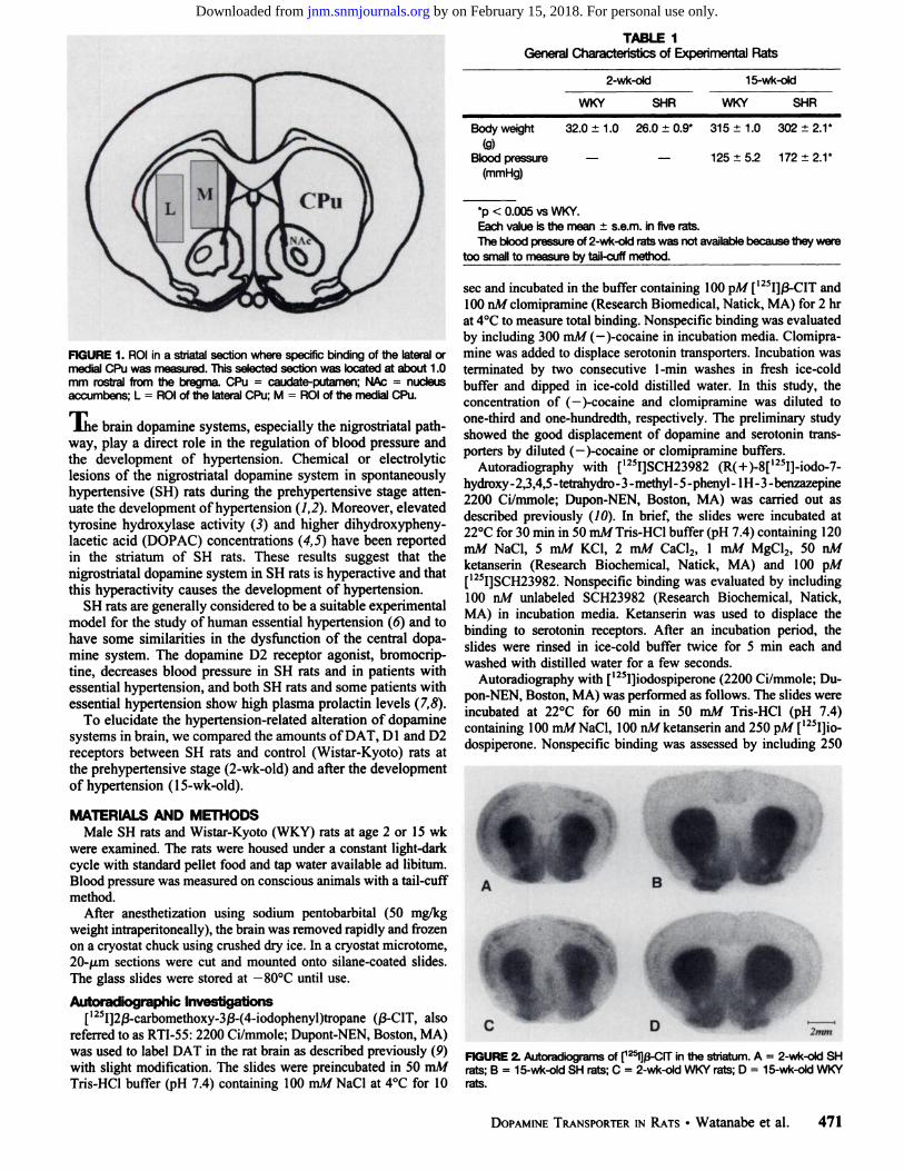
TABLE 1General Characteristics of Experimental Rats
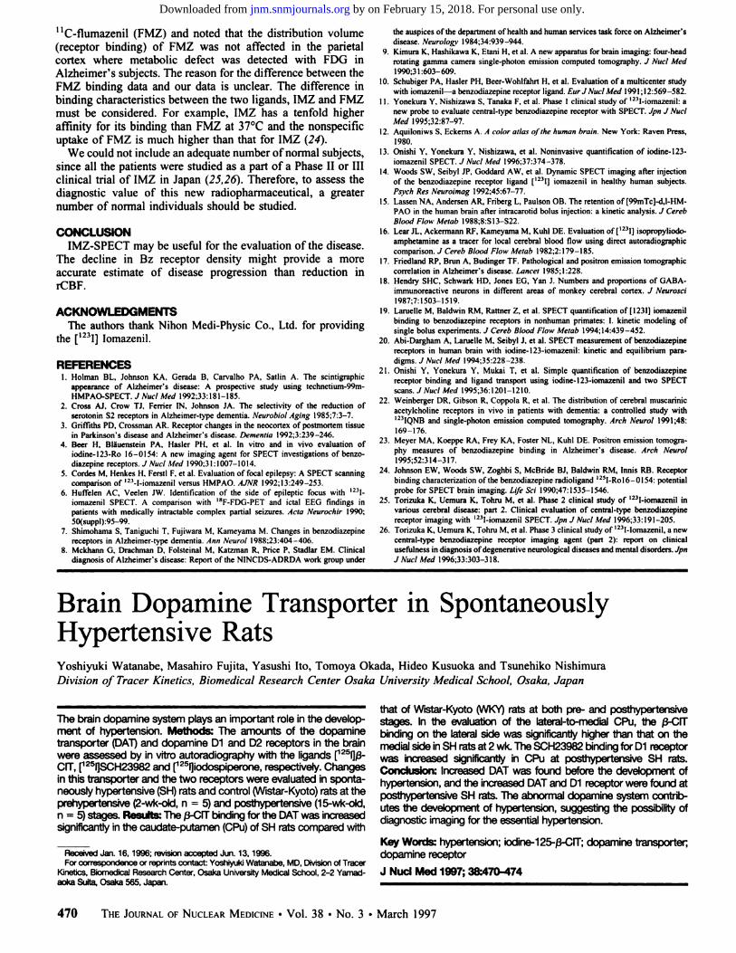
FIGURE 1. ROI in a striatal section where specific binding of the lateral ormedial CPu was measured. This selected section was located at about 1.0mm rostral from the bregma. CPu = caudate-putamen; NAc = nucleusaccumbens; L = ROI of the lateral CPu; M = ROI of the medial CPu.
J.he brain dopamine systems, especially the nigrostriatal pathway, play a direct role in the regulation of blood pressure andthe development of hypertension. Chemical or electrolyticlesions of the nigrostriatal dopamine system in spontaneouslyhypertensive (SH) rats during the prehypertensive stage attenuate the development of hypertension (1,2). Moreover, elevatedtyrosine hydroxylase activity (3) and higher dihydroxypheny-lacetic acid (DOPAC) concentrations (4,5) have been reportedin the striatum of SH rats. These results suggest that thenigrostriatal dopamine system in SH rats is hyperactive and thatthis hyperactivity causes the development of hypertension.
SH rats are generally considered to be a suitable experimentalmodel for the study of human essential hypertension (6) and tohave some similarities in the dysfunction of the central dopamine system. The dopamine D2 receptor agonist, bromocrip-tine, decreases blood pressure in SH rats and in patients withessential hypertension, and both SH rats and some patients withessential hypertension show high plasma prolactin levels (7,8).
To elucidate the hypertension-related alteration of dopaminesystems in brain, we compared the amounts of DAT, D1 and D2receptors between SH rats and control (Wistar-Kyoto) rats atthe prehypertensive stage (2-wk-old) and after the developmentof hypertension ( 15-wk-old).
MATERIALS AND METHODSMale SH rats and Wistar-Kyoto (WKY) rats at age 2 or 15 wk
were examined. The rats were housed under a constant light-darkcycle with standard pellet food and tap water available ad libitum.Blood pressure was measured on conscious animals with a tail-cuffmethod.
After anesthetization using sodium pentobarbital (50 mg/kgweight intraperitoneally), the brain was removed rapidly and frozenon a cryostat chuck using crushed dry ice. In a cryostat microtome,20-/xm sections were cut and mounted onto silane-coated slides.The glass slides were stored at —80°Cuntil use.
Autoradiographic Investigations['25I]2ß-carbomethoxy-3ß-(4-iodophenyl)tropane (ß-CIT,also
referred to as RTI-55: 2200 Ci/mmole; Dupont-NEN, Boston, MA)was used to label DAT in the rat brain as described previously (9)with slight modification. The slides were preincubated in 50 mMTris-HCl buffer (pH 7.4) containing 100 mM NaCl at 4°Cfor 10
2-wk-old 15-wk-old
WKY SHR WKY SHR
Body weight 32.0 ±1.0 26.0 ±0.9* 315 ±1.0 302 ±2.1*
(9)Blood pressure — — 125 ±5.2 172 ±2.1*
(mmHg)
*p < 0.005 vs WKY.
Each value is the mean ±s.e.m. in five rats.The blood pressure of 2-wk-old rats was not available because they were
too small to measure by tail-cuff method.
sec and incubated in the buffer containing 100 pA/ [125I]ß-CITand
100 nM clomipramine (Research Biomédical,Natick, MA) for 2 hrat 4°Cto measure total binding. Nonspecific binding was evaluatedby including 300 mM (—)-cocaine in incubation media. Clomipra-
mine was added to displace serotonin transporters. Incubation wasterminated by two consecutive 1-min washes in fresh ice-coldbuffer and dipped in ice-cold distilled water. In this study, theconcentration of ( —)-cocaine and clomipramine was diluted toone-third and one-hundredth, respectively. The preliminary studyshowed the good displacement of dopamine and serotonin transporters by diluted (—(-cocaineor clomipramine buffers.
Autoradiography with [125I]SCH23982 (R( + )-8[125I]-iodo-7-
hydroxy -2,3,4,5 - tetrahydro -3-methy1- 5-pheny1-1H -3-benzazepine2200 Ci/mmole; Dupon-NEN, Boston, MA) was carried out asdescribed previously (70). In brief, the slides were incubated at22°Cfor 30 min in 50 mM Tris-HCl buffer (pH 7.4) containing 120
mM NaCl, 5 mM KC1, 2 mM CaCl2, 1 mM MgCU, 50 nMketanserin (Research Biochemical, Natick, MA) and 100 pM[125I]SCH23982. Nonspecific binding was evaluated by including
100 nM unlabeled SCH23982 (Research Biochemical, Natick,MA) in incubation media. Ketanserin was used to displace thebinding to serotonin receptors. After an incubation period, theslides were rinsed in ice-cold buffer twice for 5 min each andwashed with distilled water for a few seconds.
Autoradiography with [125I]iodospiperone (2200 Ci/mmole; Du
pon-NEN, Boston, MA) was performed as follows. The slides wereincubated at 22°Cfor 60 min in 50 mM Tris-HCl (pH 7.4)containing 100 mM NaCl, 100 nM ketanserin and 250 pM [I25l]io-
dospiperone. Nonspecific binding was assessed by including 250
%f*f ff
C D 2mm
RGURE 2. Autoradiograms of [125I]0-CITin the striatum. A = 2-wk-old SHrats; B = 15-wk-old SH rats; C = 2-wk-old WKY rats; D = 15-wk-old WKY
rats.
DOPAMINETRANSPORTERIN RATS•Watanabe et al. 471
by on February 15, 2018. For personal use only. jnm.snmjournals.org Downloaded from
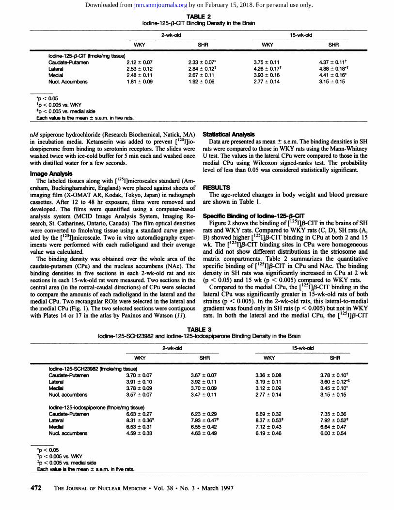
TABLE 2lodine-125-ß-CIT Binding Density in the Brain
2-wk-old 15-wk-old
WKY SHR WKY SHR
Iodine-125-ß-CIT (fmole/mg tissue)Caudate-Putamen 2.12 ±0.07
Lateral 2.53 ±0.12Medial 2.48 ±0.11NucÃ.Accumbens 1.81 ±0.09
2.33 ±0.07*
2.84 ±0.12*
2.67 ±0.111.92 ±0.06
3.75 ±0.114.26 ±0.17*
3.93 ±0.162.77 ±0.14
4.37 ±0.11t4.88 ±0.18**4.41 ±0.16*
3.15 ±0.15
*p < 0.05
*p < 0.005 vs. WKY*p < 0.005 vs. medial side
Each value is the mean ±s.e.m. in five rats.
nM spiperone hydrochloride (Research Biochemical, Natick, MA)in incubation media. Ketanserin was added to prevent [125I]io-
dospiperone from binding to serotonin receptors. The slides werewashed twice with ice-cold buffer for 5 min each and washed oncewith distilled water for a few seconds.
Image AnalysisThe labeled tissues along with [125I]microscales standard (Am-
ersham, Buckinghamshire, England) were placed against sheets ofimaging film (X-OMAT AR, Kodak, Tokyo, Japan) in radiographcassettes. After 12 to 48 hr exposure, films were removed anddeveloped. The films were quantified using a computer-basedanalysis system (MC ID Image Analysis System, Imaging Research, St. Catharines, Ontario, Canada). The film optical densitieswere converted to fmole/mg tissue using a standard curve generated by the [I25l]microscale. Two in vitro autoradiography exper
iments were performed with each radioligand and their averagevalue was calculated.
The binding density was obtained over the whole area of thecaudate-putamen (CPu) and the nucleus accumbens (NAc). Thebinding densities in five sections in each 2-wk-old rat and sixsections in each 15-wk-old rat were measured. Two sections in thecentral area (in the rostral-caudal directions) of CPu were selectedto compare the amounts of each radioligand in the lateral and themedial CPu. Two rectangular ROIs were selected in the lateral andthe medial CPu (Fig. 1). The two selected sections were contiguouswith Plates 14 or 17 in the atlas by Paxinos and Watson (11).
Statistical AnalysisData are presented as mean ±s.e.m. The binding densities in SH
rats were compared to those in WKY rats using the Mann-WhitneyU test. The values in the lateral CPu were compared to those in themedial CPu using Wilcoxon signed-ranks test. The probabilitylevel of less than 0.05 was considered statistically significant.
RESULTSThe age-related changes in body weight and blood pressure
are shown in Table 1.
Specific Binding of lodine-125-ß-CITFigure 2 shows the binding of [125I]ß-CITin the brains of SH
rats and WKY rats. Compared to WKY rats (C, D), SH rats (A,B) showed higher [125I]ß-CITbinding in CPu at both 2 and 15wk. The [125I]/3-CIT binding sites in CPu were homogeneous
and did not show different distributions in the striosome andmatrix compartments. Table 2 summarizes the quantitativespecific binding of [125I]ß-CITin CPu and NAc. The binding
density in SH rats was significantly increased in CPu at 2 wk(p < 0.05) and 15 wk (p < 0.005) compared to WKY rats.
Compared to the medial CPu, the [125I]ß-CITbinding in the
lateral CPu was significantly greater in 15-wk-old rats of bothstrains (p < 0.005). In the 2-wk-old rats, this lateral-to-medialgradient was found only in SH rats (p < 0.005) but not in WKYrats. In both the lateral and the medial CPu, the [l25I]ß-CIT
TABLE 3lodine-125-SCH23982 and lodine-125-lodospiperone Binding Density in the Brain
2-wk-old 15-wk-old
WKY SHR WKY SHR
lodine-125-SCH23982 (fmole/mg tissue)Caudate-Putamen 3.70 ±0.07LateralMedialNucÃ,
accumbens3.91
±0.103.78±0.093.57±0.073.67
±0.073.92±0.113.70
±0.093.47±0.113.36
±0.083.19±0.113.12
±0.092.77±0.143.78
±0.1Of3.60±0.12**3.45±0.10*3.15
±0.15
lodine-125-iodospiperone (fmole/mg tissue)Caudate-Putamen 6.63 ±0.27Lateral 8.31 ±0.36*
Medial 6.53 ±0.31NucÃ,accumbens 4.59 ±0.33
6.23 ±0.297.93 ±0.47*
6.55 ±0.424.63 ±0.49
6.69 ±0.328.37 ±0.53*
7.12 ±0.436.19 ±0.46
7.35 ±0.367.92 ±0.52*
6.64 ±0.476.00 ±0.54
*p < 0.05
fp < 0.005 vs. WKY*p < 0.005 vs. medial side
Each value is the mean ±s.e.m. in five rats.
472 THE JOURNALOFNUCLEARMEDICINE•Vol. 38 •No. 3 •March 1997
by on February 15, 2018. For personal use only. jnm.snmjournals.org Downloaded from

binding in SH rats was significantly greater than that in WKYrats at 15 wk (p < 0.05).
Specific Binding of lodine-125 SCH23982 andIodine-125 lodospiperone
Table 3 summarizes the quantitative specific binding of[125I]SCH23982 and [I25l]iodospiperone in CPu and NAc. The[125I]SCH23982 binding in CPu was significantly increased in
SH rats only at age 15 wk (p < 0.005) compared to WKY rats.There was no significant difference between SH rats and WKYrats at either age in [125I]iodospiperone binding. In NAc, the
binding densities showed no difference in the two strains foreither tracer.
In both the lateral and the medial CPu, the [125I]SCH23982
binding in 15-wk-old SH rats was significantly greater compared to WKY rats (p < 0.05). In addition, the lateral-to-medialgradient of [I25I]SCH23982 binding was detected only in15-wk-old SH rats (p < 0.005). The [125I]iodospiperone bind
ing in the lateral CPu was significantly greater than that in themedial CPu at both ages and in both strains (p < 0.005).
DISCUSSIONThis study demonstrates the increased DAT in the CPu of
both pre- and posthypertensive SH rats. In the prehypertensiveSH rats, the difference between SH rats and WKY rats wasfound only by the ligand of DAT. That is, the increase of totalamount in CPu and the expression of lateral-to-medial gradient.These results suggest that these changes may be inherent andpathogenetic to hypertension and indicate that it may bepossible to detect an abnormality in DAT with in vivo imagingeven before the development of hypertension.
SH rats were bred from the WKY rats by selective brother-to-sister inbreeding and uniformly result in offspring thatdevelop hypertension (72). SH rats are similar to humans withrespect to essential hypertension in several ways. Both haveapparent onsets very early in life. Their elevated arterialpressure is mediated through a slow and progressively increasedtotal peripheral resistance that demands cardiac and vascularadaptation (13).
In contrast to our findings, it was reported that there was nosignificant difference in DAT labeled with [3H]mazindol be
tween adult age-matched SH rats and Sprague-Dawley rats (14).The discrepancy between two studies may be due to thedifferences in radioligands and in the strain selected as thenormotensive rat. Iodine-125-ß-CIT may be different from[3H]mazindol in the binding site and the affinity to DAT. The[3H]mazindol binding in the striatum is differentially distrib
uted in the striosome and matrix compartments (75). Thisinhomogeneity in the striatum is not observed in [125I]ß-CIT(9,16,17). Furthermore, [125I]ß-CITbinds to DAT at both high-
and low-affinity sites in the striatum, similar to cocaine (76),but [3H]mazindol binds to DAT only at one high-affinity
binding site (18).The lateral-to-medial gradient of DAT in CPu was found only
in SH rats at age 2 wk but in both strains at 15 wk (Table 2). Thelateral or medial portions of CPu differ in neurogenesis and inthe development of dopaminergic innervation (19,20). In thestriatum, the ingrowth of the mesencephalo-prosencephalicdopaminergic fibers is predominantly located laterally. Fromlateral portion, the outgrowth of the dopaminergic fibers proceeds in the medial direction (79). Thus, early expression of thelateral-to-medial gradient in DAT indicates an abnormal onto-genic development of the dopamine system in SH rats.
There are many previous reports describing dopamine Dl orD2 receptor binding studies in SH rats, but these results are
confusing. Some researchers reported an increase of D1 or D2receptor densities in the striatum of SH rats (14,21-23),
whereas others reported that there was no difference betweenSH and WKY rats (24-26). The discrepancies in Dl and D2receptor data have been attributed to the difference of radioligands and experimental procedures and to genetic drift resultingin biological variability among the substrains of SH rats (27).
Iodine-125-ß-CIT has been used as a tracer for SPECTstudies in baboons and humans (28-33). In recent years, the invivo tracer kinetics (34) or age-related decline (35) in humanstriatum of [125I]ß-CIThave been investigated. Iodine-125-ß-
CIT is a promising SPECT agent for imaging the DAT inhumans. Our study suggests the possibility of diagnostic imaging for essential hypertension.
CONCLUSIONThe increased DAT was found before the development of
hypertension in SH rats, and increased DAT and Dl receptorwere found in posthypertensive SH rats. These results suggestthat the dopamine system in the striatum plays an important rolein the pathogenesis and development of hypertension.
REFERENCES1. Buuse MV, Versteeg DHG. Jong WD. Brain dopamine depletion by lesion in the
substantia nigra attenuates the development of hypertension in the spontaneouslyhypertensive rat. Brain Res !986;368:69-78.
2. Linthorst ACE. Giersbergen PLM. Gras M. Versteeg DHG. Jong WD. The nigrostri-atal dopamine system: role in the development of hypertension in spontaneouslyhypertensive rat. Brain Res 1994;639:261-268.
3. Nagaoka A. Lovenberg W. Regional changes in the activities of aminergic biosyntheticenzymes in the brains of hypertensive rats. Eur J Pharmacol 1977:43:297-306.
4. Howes LG. Rowe PR. Summers RL. Louis WJ. Age-related changes of cat-echolamines and their metabolites in central nervous system regions of spontaneouslyhypertensive and normotensive Wistar-Kyoto rats. Clin Exp Hypert 1984;A6:2263-
2277.5. McKeon TW. Hendly ED. Brain monoamines and metabolites in hypertensive and
hyperactive rat strains. Clin Exp Hypert 1988;A 10:971-994.
6. Yamori Y. Development of the spontaneously hypertensive rat and of variousspontaneous rat models and their implications. In: Jong WD. ed. Handbook ofhypertension: experimental and genetic model of hypertension, vol.4. Amsterdam, TheNetherlands: Elsevier Science: 1984:224-239.
7. Stumpe KO. Kolloch R. Higuchi M. Kruck F. Vetter H. Hyperproloctinemia. antihyper-tensive effect of bromocriptine in essential hypertension. Lancet 1977:211-214.
8. Sowers JR. Effect of bromocriptine on responses to stress in spontaneously hypertensive rats. Hypertension 198I;3:544-550.
9. Fujita M. Shimada S. Fukuchi K. Tohyama M. Nishimura T. Distribution of cocainerecognition sites in rat brain: in vitro and ex vivo autoradiography with [L25I]RTI-55.J Chem Neuroanat 1994;7:13-23.
10. Dawson TM. Barone P. Sidhu A, Wamsley JK. Chase TN. The Dl dopamine receptorsin the rat brain: quantitative autoradiographic localization using an iodinated ligand.Neuroscience 1988;26:83-100.
11. Paxinos G, Watson C. The ral brain in stereotaxic coordinales. San Diego, CA:Academic Press; 1986.
12. Okamoto K. Aoki K. Development of a strain of spontaneous hypertensive rats. JpnCircJ 1963:27:282-293.
13. Trippodo NC. FröhlichED. Similarities of genetic hypertension: man and rat. Ore Res1981:48:309-319.
14. Kujirai K. Przendborski S, Kostic V, et al. Autoradiography of dopamine receptors anddopamine uptake sites in the spontaneously hypertensive rat. Brain Res Bull 1990:25:703-709.
15. Graybiel AM, Moratalla R. Dopamine uptake sites in the striatum are distributeddifferentially in striosome and matrix compartment. Proc Nail Acad Sci USA1989:86:9020-9024.
16. Boja JW. Mitchell WM. Palei A, et al. High-affinity binding of [l25I]RTI-55 todopamine and serotonin transporters in rat brain. Synapse 1992:12:27-36.
17. Cline EJ, Scheffel U, Boja JW, et al. In vivo binding of ['-'l]RTI-55 to dopamine
transporters: pharmacology and regional distribution with autoradiography. Svnapse1992:12:37-46.
18. Javitch JA, Blaustein RO, Snyder SH. ['H]mazindol binding associated with neuronal
dopamine and norepinephrine uptake sites. Mol Pharmacol 1984:26:35-44.19. Voorn P, Kalsbeek A. Jorritsma-Byham B, Groenewegen HJ. The pre-. postnatal
development of the dopaminergic cell groups in the ventral mesencephalon of thestriatum of the rat. Neuroscience 1988:25:857-887.
20. Bayer SA. Neurogenesis in the rat striatum. Ini J Devi Neuroscience 1984:2:163-175.21. Kirouac GJ. Ganguly PK. Up-regulation of dopamine receptors in the brain of the
spontaneous hypertensive rat: an autoradiography analysis. Neuroscience 1993:52:135-141.
22. Lim DK, Ito Y, Hoskins B, Rockhold RW. Ho IK. Comparative studies of muscarinicand dopamine receptors in three strains of rat. Eur J Pharmacol 1989:165:279-287.
23. Lim DK. Yu ZJ, Hoskins B. Rockhoid RW. Ho IK. Effect of acute and subacutecocaine administration on the CNS dopaminergic system in Wistar-Kyoto and
DOPAMINETRANSPORTERIN RATS•Watanabe et al. 473
by on February 15, 2018. For personal use only. jnm.snmjournals.org Downloaded from
spontaneously hypertensive rats: 2 dopamine receptors. Neurochem Res 1990;15:621-
627.24. Watanabe M, Tsuruta S, Inoue Y, et al. Dopamine Dl and D2 receptors in
spontaneously hypertensive rat brain striatum. Can J Physiol Phamacol 1989;67:1596-1597.
25. Linthorst ACE, Jong WD, Boer TD, Versteeg DHG. Dopamine Dl and D2 receptorsin the caudate nucleus of spontaneously hypertensive rats and normotensive Wistar-Kyoto rats. Brain Res 1993;602:119-125.
26. Buuse MV, Jones R, Wagner J. Brain dopamine D2 receptor mechanisms inspontaneously hypertensive rats. Brain Res Bull 1992;28:289-297.
27. Versteeg DHG, Petty MA, Bohus B. The central nervous system and hypertension: therole of catechoamines and neuropeptides. In: Jong WD, ed. Handbook of hypertension:experimental and genetic model of hipertensión, vol. 4. Amsterdam. The Netherlands:Elsevier Science;1984:398-430.
28. Kaufman MJ. Madras BK. Distribution of cocaine recognition sites in monkey brain:2. Ex vivoautoradiography with ['HJCFT and [l25I]RTI-55. Synapse 1992;12:99-111.
29. Staley JK, Basile M, Flynn DD, Mash DC. Visualizing dopamine and serotonin
transporters in the human brain with the potent cocaine analog ['2!I]RTI-55: in vitro
binding and autoradiographic characterization. J Neurochem 1994;62:549-556.30. Seibyl JP, Wallace E, Smith EO, et al. Whole-body biodistribution, radiation absorbed
dose and brain SPECT imaging with iodine-123-ß-CIT in healthy human subjects.J NucÃMed 1994;35:764-770.
31. Kuikka JT, Bergstrom KA, Ahonen A, Lansimies E. The dosimetry of iodine-123-labeled 2ß-carbomethoxy-3ß-(4-iodophenyl)tropane. Ear J NucÃMed 1994;21:53-56.
32. Innis RB. Single-photon emission tomography imaging of dopamine terminal innervation: a potential clinical tool in Parkinson's disease. Ear J NucÃMed 1994;21:l-5.
33. Seibyl JP, Lamelle M, Dyck CH, et al. Reproducibility of iodine-123-ß-ClT SPECTbrain measurement of dopamine transporters. J NucÃMed 1996;37:222-228.
34. Lamelle M, Wallace E, Seibyl JP, et al. Graphical, kinetic and equilibrium analyses ofin vivo [123I]beta-CIT binding to dopamine transporters in healthy human subjects. JCereb Blood Flow Metab 1994;14:982-994.
35. Dyck CH, Seibyl JP, Malison RT, et al. Age-related decline in striatal dopamine transporter binding with iodine-123-ß-CIT SPECT. J NucÃMed 1995;36:1175-1181.
(continued from page 7A)
FIRST IMPRESSIONSTechnetium-99m-Macroaggregated Albumin in Superior Vena Cavai Obstruction
Figure 1.
SVC
I IV
Figure 2.
PURPOSEA 19-yr-old girl with acute lymphocytic leukemia and a known clot aroundher porta-cath central line was studied for suspected pulmonary embolismbecause of chest pain. After right arm intravenous injection of Tc-MAA,
the perfusion lung scan (Fig. 1, anterior view) showed abnormal traceractivity below the diaphragm, in the left lobe of the liver, suggestive ofsuperior vena cavai (SVC) obstruction with collateral drainage into thesystemic-portal venous blood flow to the left lobe of the liver. Right arm
venogram done the same day confirmed SVC obstruction with collateralsand flow via the internal thoracic vein.
The demonstration of the left lobe of the liver suggests that the mainroute for collateral drainage, in this patient, is through the internal thoracicvein, the superior epigastric veins, the periumbilical venous channels andthe umbilical and/or paraumbilical veins that drain into the left branch of theportal vein (Fig. 2, where AV = axillary vein, BV = brachial vein, EIV =external iliac vein, IEV = inferior epigastric vein, ITV = internal thoracicvein, IVC = inferior vena cava, L = left, LBPV = left branch of portal vein,PUV = paraumbilical vein, R = right, RBPV = right branch of portal vein,SEV = superior epigastric vein, SV = subclavian vein, SVC = superior venacava and site of obstruction, U = umbilicus and periumbilical venouschannels, UV = umbilical vein) and give rise to the tracer activity seen in
the left lobe of the liver. In addition, the appearance of the liver alsosuggests that the major deep collateral flow through the azygos ascendinglumbar pathway is less well developed.
TRACERTechnetium-99m-macroaggregatedalbumin,3mCi(111 MBq)
ROUTE OF ADMINISTRATIONIntravenous, right arm
TIME AFTER INJECTIONTen minutes
INSTRUMENTATIONGeneral Electric Starcam 3000 LFOV gamma camera with LEHRcollimator
CONTRIBUTORSHaim Golan, Judith M. Ash, Peter G. Chait and David L. Gilday, Divisionof Nuclear Medicine, Department of Diagnostic Imaging, The Hospital forSick Children, Medicine, Toronto, Ontario, Canada
474 THE JOURNALOFNUCLEARMEDICINE•Vol. 38 •No. 3 •March 1997
by on February 15, 2018. For personal use only. jnm.snmjournals.org Downloaded from
1997;38:470-474.J Nucl Med. Yoshiyuki Watanabe, Masahiro Fujita, Yasushi Ito, Tomoya Okada, Hideo Kusuoka and Tsunehiko Nishimura Brain Dopamine Transporter in Spontaneously Hypertensive Rats
http://jnm.snmjournals.org/content/38/3/470This article and updated information are available at:
http://jnm.snmjournals.org/site/subscriptions/online.xhtml
Information about subscriptions to JNM can be found at:
http://jnm.snmjournals.org/site/misc/permission.xhtmlInformation about reproducing figures, tables, or other portions of this article can be found online at:
(Print ISSN: 0161-5505, Online ISSN: 2159-662X)1850 Samuel Morse Drive, Reston, VA 20190.SNMMI | Society of Nuclear Medicine and Molecular Imaging
is published monthly.The Journal of Nuclear Medicine
© Copyright 1997 SNMMI; all rights reserved.
by on February 15, 2018. For personal use only. jnm.snmjournals.org Downloaded from


![Journal of Membrane Science in pdf/JMembraneS2017.pdf · solution, dopamine can spontaneously oxidize and self-polymerize to form polydopamine [12]. This self-polymerization reaction](https://static.fdocuments.net/doc/165x107/5ea10be7338e0c403956a7dc/journal-of-membrane-science-in-pdfjmembranes2017pdf-solution-dopamine-can-spontaneously.jpg)