Brain Circuitries in Basic and Clinical Neuroscience
104
Brain Circuitries in Basic and Clinical Neuroscience Seventh International Conference on Creative Content Technologies, ComputerWorld 2015, March 24, 2015 - Nice, France Nikos Makris, M.D., Ph.D. Center for Morphometric Analysis Departments of Psychiatry and Neurology Massachusetts General Hospital Harvard Medical School C e n t e r f o r M o r p h o m e t r i c A n a l y s i s M a s s a c h u s e t t s G e n e r a l H o s p i t a l ComputerWorld 2015, March 24, 2015 - Nice, France
Transcript of Brain Circuitries in Basic and Clinical Neuroscience

Brain Circuitries in Basic and ClinicalNeuroscience
Seventh International Conference on Creative Content Technologies,ComputerWorld 2015, March 24, 2015 - Nice, France
Nikos Makris, M.D., Ph.D.
Center for Morphometric Analysis
Departments of Psychiatry and Neurology
Massachusetts General Hospital
Harvard Medical School
Cente
rfo
r Morphometric
Ana
lysis
Massachusetts General Hosp
ital
ComputerWorld 2015, March 24, 2015 - Nice, France

Disclosures of Potential ConflictsSource Consultant Advisory
BoardStock orEquity
>$10,000
Speakers’Bureau
ResearchSupport
Honorariumfor this talk or
meeting
Expensesrelated to this
talk ormeeting
NIA-NIMH (RO1)
(Aging)
X
MGH-MAC Core
(Multiple Projects)
X
NIH (R21)
(Neurodegeneration-
FTD)
X
NIH (R21)
(Neurodegeneration-
FTD and ALS)
X
NIH (R21)
(Modeling Deep
Brain Stimulation
and MRI safety)
X
Departmental X

Center for Morphometric Analysis
(CMA)-MGH collaborating PIs
Marek Kubicki, M.D., Ph.D. (BWH/HMS)
Bradford Dickerson, M.D. (MGH/HMS)
Giorgio Bonmassar, Ph.D. (MGH/HMS)
Jill Goldstein, Ph.D. (BWH/HMS)
Larry Seidman, Ph.D.(BIDMC/MMHC/HMS)
Gordon Harris, Ph.D. (MGH/HMS)
Harvard Medical School affiliated Hospitals
Massachusetts General Hospital (MGH)Brigham and Women’s Hospital (BWH)McLean HospitalBeth Israel Deaconess Medical Center (BIDMC)Mass Mental Health Center (MMHC)Children’s Hospital
Gordon Harris, Ph.D. (MGH/HMS)
Scott Rauch, M.D. (McLean/HMS)
Scott Lukas, Ph.D. (McLean/HMS)
Joseph Biederman, M.D. (MGH/HMS)
Marlene Oscar-Berman, Ph.D.(BUSM)
Deepak Pandya, M.D. (BUSM)
Douglas Rosene, Ph.D. (BUSM)
Martha Shenton, Ph.D. (BWH/HMS)
David Kennedy, Ph.D.(UMassMS)
David Caplan, M.D., Ph.D. (MGH/HMS)
Bruce Rosen, M.D., Ph.D. (MGH/HMS)
Helen Grant, M.D., Ph.D.(Children’s//HMS)
Hans Breiter, M.D.(NWU)
Anne Blood, Ph.D. (MGH/HMS)
Children s Hospital
Center for Morphometric Analysis (CMA)-MGH Core
Nikos Makris, M.D., Ph.D. (Director)Verne Caviness, M.D., Ph.D. (Founding Director)George Papadimitriou, B.Sc. (Computer Science)Lichen Liang, Ph.D. (MRI Engineering, Modeling)Takeshi Takahashi, M.D., Ph.D. (Functional Conn.)Isaac Ng, B.A. (RA)Anni Zhu, B.A. (RA)Yeetzou Kao (RA)
Cente
rfo
r Morphometric
Ana
lysis
Massachusetts General Hosp
ital

Nikos Makris, M.D., Ph.D. (MGH/BWH/McLean/HMS) Brain Connectivity/Aging/Neurodegeneration/Addiction/ADHD/Stroke/ITD
Verne Caviness, M.D., Ph.D. (MGH/HMS) Brain Development/Laptop Imaging Technology Development/Stroke
Marek Kubicki, M.D., Ph.D. (BWH/HMS) Schizophrenia/Imaging Technology Development/Aging/Brain Connectivity
Bradford Dickerson, M.D. (MGH/HMS) Neurodegeneration/Aging/ITD/Brain Connectivity
Giorgio Bonmassar, Ph.D. (MGH/HMS) Technology Development/MRI Safety/Deep Brain Stimulation
Jill Goldstein, Ph.D. (BWH/HMS) Stress Response/Sexual Dimorphisms/Aging/MDD/Schizophrenia
Larry Seidman, Ph.D.(BIDMC/MMHC/HMS) Schizophrenia/MDD/BPD/ADHD/Resting State Brain Connectivity
Gordon Harris, Ph.D. (MGH/HMS) 3D Imaging Visualizations Technology Development/Alcohol DependenceGordon Harris, Ph.D. (MGH/HMS) 3D Imaging Visualizations Technology Development/Alcohol Dependence
Scott Rauch, M.D. (McLean/HMS) OCD/PTSD/Anxiety Disorder/Psychosurgery
Scott Lukas, Ph.D. (McLean/HMS) Nicotine Dependence/Alcohol Dependence
Joseph Biederman, M.D. (MGH/HMS) Attention Deficit/Hyperactivity Disorder (ADHD)/Bipolar Disorder (BPD)
Marlene Oscar-Berman, Ph.D.(BUSM) Alcohol Dependence
Deepak Pandya, M.D. (BUSM) Brain Connectivity
Douglas Rosene, Ph.D. (BUSM) Aging in the Rhesus Monkey/Monkey Brain Connectivity
Martha Shenton, Ph.D. (BWH/HMS) Traumatic Brain Injury/Imaging Technology Development
David Kennedy, Ph.D.(UMassMS) Neuroinformatics/Imaging Databasing
David Caplan, M.D., Ph.D. (MGH/HMS) Stroke/Aphasia Research
Bruce Rosen, M.D., Ph.D. (MGH/HMS) Imaging Technology Development (ITD)/Program in Acupuncture Research
Helen Grant, M.D., Ph.D.(Children’s//HMS) Brain Development
Hans Breiter, M.D.(NWU) Cocaine Dependence
Anne Blood, Ph.D. (MGH/HMS) Dystonia Research

Some discoveries are breakthroughs inknowledge and others in technology

Importance ofTechnology in
ScientificDiscovery
(Hypothesis:
Great Tools to
see lead to GreatDiscoveries)
Courtesy of Dr. Bruce Rosen
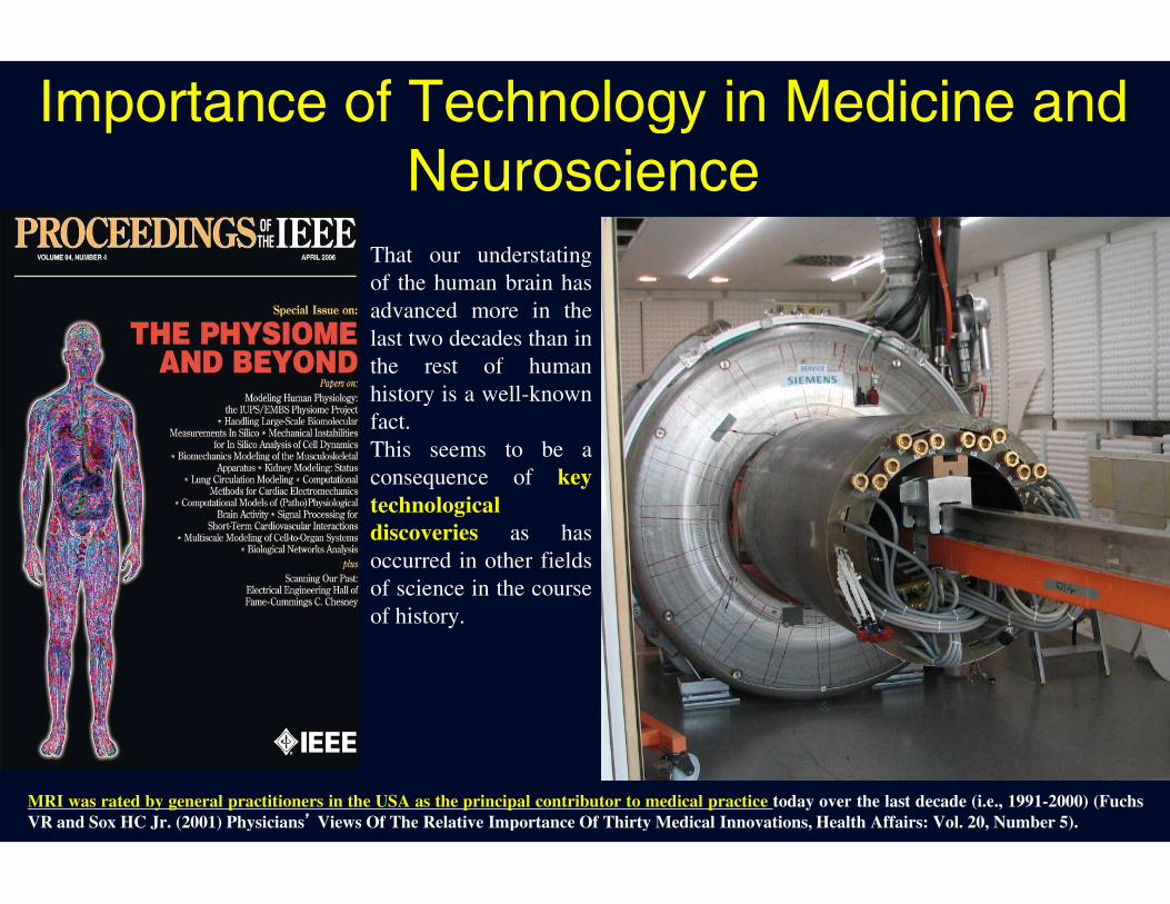
Importance of Technology in Medicine andNeuroscience
That our understatingof the human brain hasadvanced more in thelast two decades than inthe rest of humanhistory is a well-knownfact.This seems to be aThis seems to be aconsequence of keytechnologicaldiscoveries as hasoccurred in other fieldsof science in the courseof history.
MRI was rated by general practitioners in the USA as the principal contributor to medical practice today over the last decade (i.e., 1991-2000) (FuchsVR and Sox HC Jr. (2001) Physicians’ Views Of The Relative Importance Of Thirty Medical Innovations, Health Affairs: Vol. 20, Number 5).

Modeling Human Anatomy and Physiology(Integration across multiple levels of description is a major goal in medicine)


MRI-PET installations at MGHUnique Instruments in New England
Nuclear Imaging two ways
Whole Body Head-onlyCourtesy of Dr. Bruce RosenImportance of Technology in Neuroscience

Magnetic resonance imaging (MRI)technology had a critical role in understating
the human brain.
Besides the great cultural and intellectual value of the accrued information andnovel conceptualizations in brain science, these advances had a tremendousimpact on the way we are currently facing neurological and psychiatric
diseases and the neurosurgical approaches we adopt for diagnosis, surgicalplanning and treatment.

”Despite 4000 papers, not a single finding has changed routine
clinical care in psychiatry”
But this will change, and the change will revolve around a new
definition of psychiatric diseases as disorders of brain circuitry - that
this will change our definition of the illnesses, and “our
understanding of their causes, treatments, and preventions”
Paraphrasing Tom Insel, Director, National Institute of Mental Health

In Imaging Research, Brain Connectivityis done with Diffusion MRI

In astronomy, the way to sensitivity and resolution is mirror diameter
Corning 200" mirror for the Hale Telescope at Mt Palomar
Courtesy of Dr. Bruce Rosen
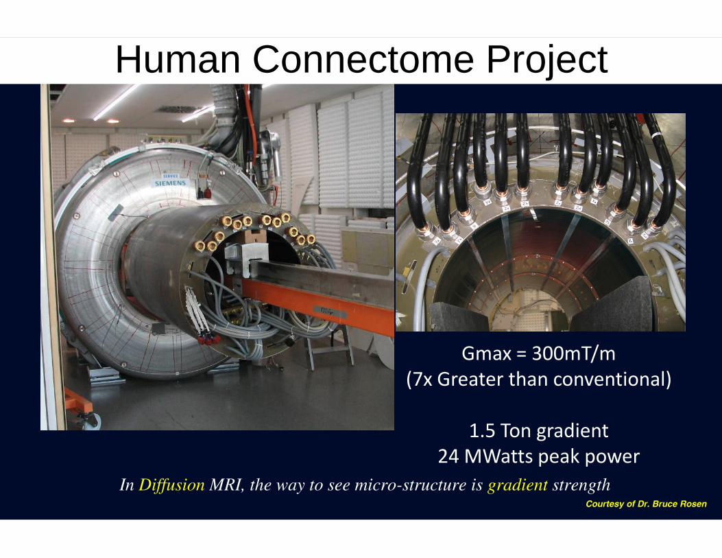
Human Connectome Project
Gmax = 300mT/m(7x Greater than conventional)
1.5 Ton gradient24 MWatts peak power
In Diffusion MRI, the way to see micro-structure is gradient strengthCourtesy of Dr. Bruce Rosen

Courtesy of Dr.Bruce Rosen

Diffusion Imaging of Microstructure
Courtesy of Dr. Bruce Rosen

Water Diffusion in the Brain has Directionality
Isotropic diffusionAnisotropic diffusion
Psychiatry Neuroimaging Laboratory
Possible sources of anisotropy:
• Axonal membranes of densely packed axons hinderdiffusion perpendicularly to the fiber long axis.
• Myelin may also modulate anisotropy.

Diffusion Tensor Imaging
Psychiatry Neuroimaging Laboratory
•At each location, the diffusion behavior of water ismodeled as an ellipsoid.
•In medical imaging this ellipsoid is called a diffusiontensor.

From Tensors to Tracts
Psychiatry Neuroimaging Laboratory
• Associate the majordiffusion direction withthe tangent to a curve.
• Estimate the curve fromits tangents.

Multimodal Brain Connectivity
Courtesy of:Steven Stufflebeam
Patric HagmannChristopher HoneyMatti Hämäläinen

How does assessment of large neuronal circuits usingfunctional connectivity MRI work?

While people are simply resting, the brain shows large, spontaneous, activity fluctuations
Spontaneous BOLD fluctuations within seed region
Per
cen
tsi
gn
alm
od
ula
tio
n
-40
-30
-20
-10
0
10
20
30
40
0 5 10 15 20 25 30 35 40 45 50 55 60 65 70 75 80 85 90
Biswal et al., MRM, 1995
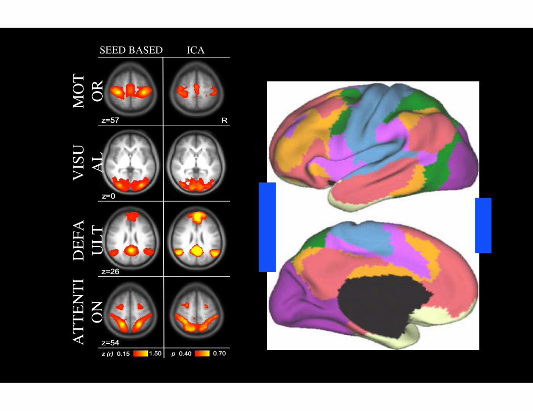
SEED BASED ICA
MO
TO
RV
ISU
AL
DE
FA
UL
TA
TT
EN
TI
ON
Van Dijk et al., 2010 Yeo et al., 2011

Yeo BT, Krienen FM, Sepulcre J, Sabuncu MR, Lashkari D, Hollinshead M, Roffman JL, Smoller JW, Zollei L, Polimeni JR, Fischl B, Liu H, Buckner RL. The organization of thehuman cerebral cortex estimated by intrinsic functional connectivity. Journal of neurophysiology. 106(3):1125-65. Pubmed PMID: 21653723; PMCID: PMC3174820.

Buckner RL, Krienen FM. (2013) The evolution of distributed association networks in the human brain. Trends in cognitive sciences. 17(12):648-65.Pubmed PMID: 24210963.

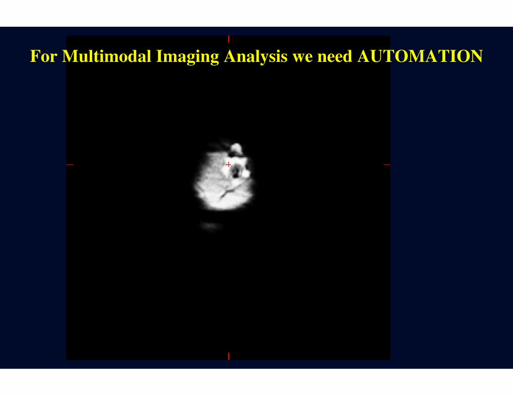
For Multimodal Imaging Analysis we need AUTOMATION

Methodologies
Volume - Morphometry Shape - Morphometry
PET
Diffusion Imaging
Functional Imaging
MR Spectroscopy
GeneticsPsychiatry Neuroimaging Lab
Data Infrastructure

PNL Pipeline
Download Scans and Organize within Network
MaskMask
Quality Control
MaskMask
T1W DTI
OtherModalities:
fMRI/MEG/M
EMSeg
CSF)
EMSeg(GM, WM,
CSF)
MaskMask
FreeSurfer
(Whole Brain)
FreeSurferSegmentation(Whole Brain)
2-TensorWhole BrainTractography
MaskMask
TBSS
WQML (querylanguage) to extract
connections of interest
Register FreeSurferlabels to DTI space
(Ants)
fMRI/MEG/MRS
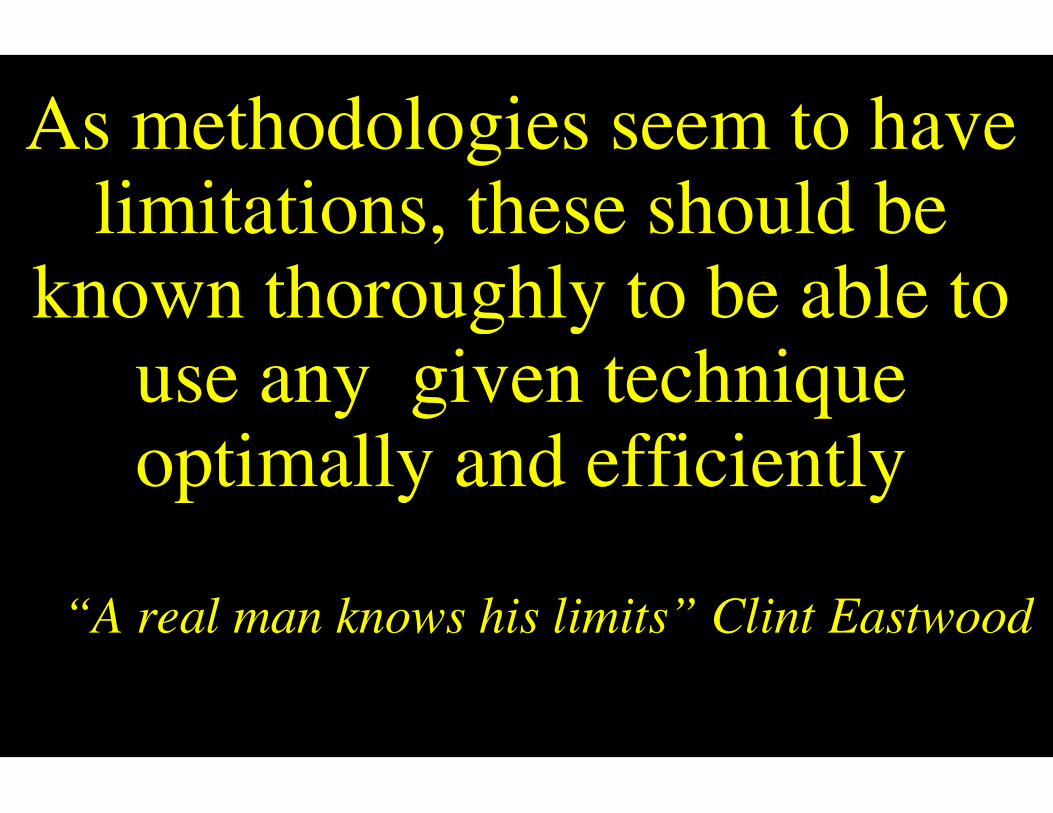
As methodologies seem to havelimitations, these should be
known thoroughly to be able touse any given techniqueuse any given techniqueoptimally and efficiently
“A real man knows his limits” Clint Eastwood

Imaging BrainCircuitries in BasicCircuitries in Basic
and ClinicalNeuroscience

Large-scale cognitive systemsunderstrood through resting state fMRI
Dorsal visuospatial attention system
Frontoparietal executive control system
Vincent et al., J Neurophys, 2008
Episodic memory/Default-mode system

The Language System


In 1997, our CMA/NMR team at MGH hasenvisioned and established what has become a
prevailing convention in the field of DTI
In 2008, Pandya and Makris proposed analternative model of language circuitry that
currently is under debate
Between 1999 (Makris PhD thesis) and 2012 (Makris et al., 2008, 2012, 2013), Makrisand Pandya have provided evidence for a novel fiber tract in humans, named, bythem, the “Middle Longitudinal Fascicle” (MdLF) (Makris, Pandya, et al., Cer Cor, 2008)

Diagrammaticrepresentation ofthe cerebrocerebellarcircuitry, includingcorticopontine connectionswhich carry higher-order
From Schmahmann 2000
which carry higher-order(Cognitive) information aswell as sensorimotor inputsto the cerebellar cortex.

Prefrontocerebellar circuitry: A) Prefrontoponine ipsilateral projection, B) c rossingpontocerebellar projection, C) crossing cerebellothalamic projection, and D)thalamoprefrontal ipsilateral projection

Imaging Circuitries in the Developing Brain
The above composite MRI brain images show top views of the sequence of gray matter maturation over the surface ofthe brain.
Courtesy of Dr. Scott Lukas

• Planning behavior• Use of strategies
• Cognitive flexibility (can you change your
Prefrontal Cortex
The Developing Brain
• Cognitive flexibility (can you change yourmind)
• Fluid methods of solving problems
--The Executive Office of the brain is still beingbuilt during the teenage years--
Courtesy of Dr. Scott Lukas

• The brain matures from the back to the front,so the frontal cortex is the last area to be
completed.
• Insight, judgment, decision-making, risk
The Developing Brain
• Insight, judgment, decision-making, risktaking, impulse control, are all impaired
in adolescents.
• During this most vulnerable period is whenteens are more likely to experiment with
drugs and other damaging activities.Courtesy of Dr. Scott Lukas

Executive and Attention cognitive systemsare critical during development
Dorsal visuospatial attention system Frontoparietal executive control system
Vincent et al., J Neurophys, 2008
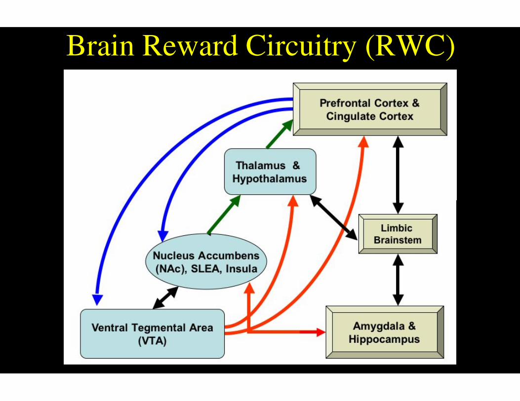
Brain Reward Circuitry (RWC)

Brain Reward Circuitry:Brain Reward Circuitry:
The Extended Reward andOversight System consists of corticaland subcortical structures involvedin controlling emotion andregulating sensitivity toreinforcements.
Makris et al., Biological Psychiatry, 2008

In cocaine abusethere are also
observed alterationsin the brain networkin the brain network
for reward
Breiter, Hyman, et al., Neuron, 1997

Amygdala in Cocaine Addiction
Superior
AnteriorPosterior
Right Lateral Ventricle = redLeft Lateral Ventricle = green
PatientsNormal ControlsCommon
Right Amygdala
Makris et al., Neuron, 200423% Volume Reduction
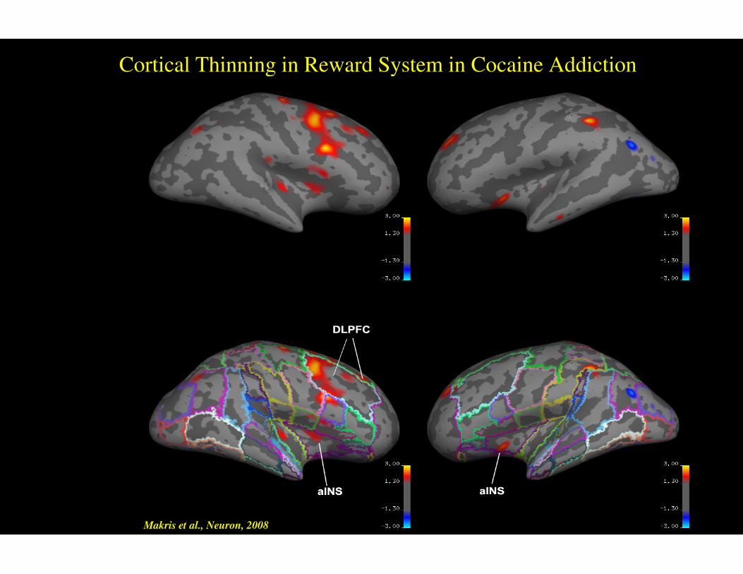
Cortical Thinning in Reward System in Cocaine Addiction
Makris et al., Neuron, 2008

Reduced brain metabolism in smokers
Tobacco: Role in Drug of Abuse
• Lower test scores• Poor athletic ability
• Lower cognitive function• Poor decision making
Courtesy of Dr. Scott Lukas

Smoked Marihuana and fMRI
GLM analyses revealsextensive orbitofrontal and
ventromedial prefrontalcortical regions with
significant group differences(marihuana > placebo
smoking (p<0.05 corrected,smoking (p<0.05 corrected,shown in red-yellow). The
single-group analysis showsthat marihuana activated
these regions and bilateralcaudate and nucleus
accumbens, while placebosmoking did not.
Courtesy of Dr. Scott Lukas
Imaging Neural Systems in AttentionDeficit-Hyperactivity DisorderDeficit-Hyperactivity Disorder
(ADHD)

Neural Systems approach addresses at leastthree questions
1) Are the structures shown to be altered in ADHD indeed componentparts of well-understood neural systems?parts of well-understood neural systems?
2) Do these structural neural systems correlate with specific behaviors?
3) Are these neural systems associated with specific genotypes?
Makris N., et al.,Dev Neurosci 2009
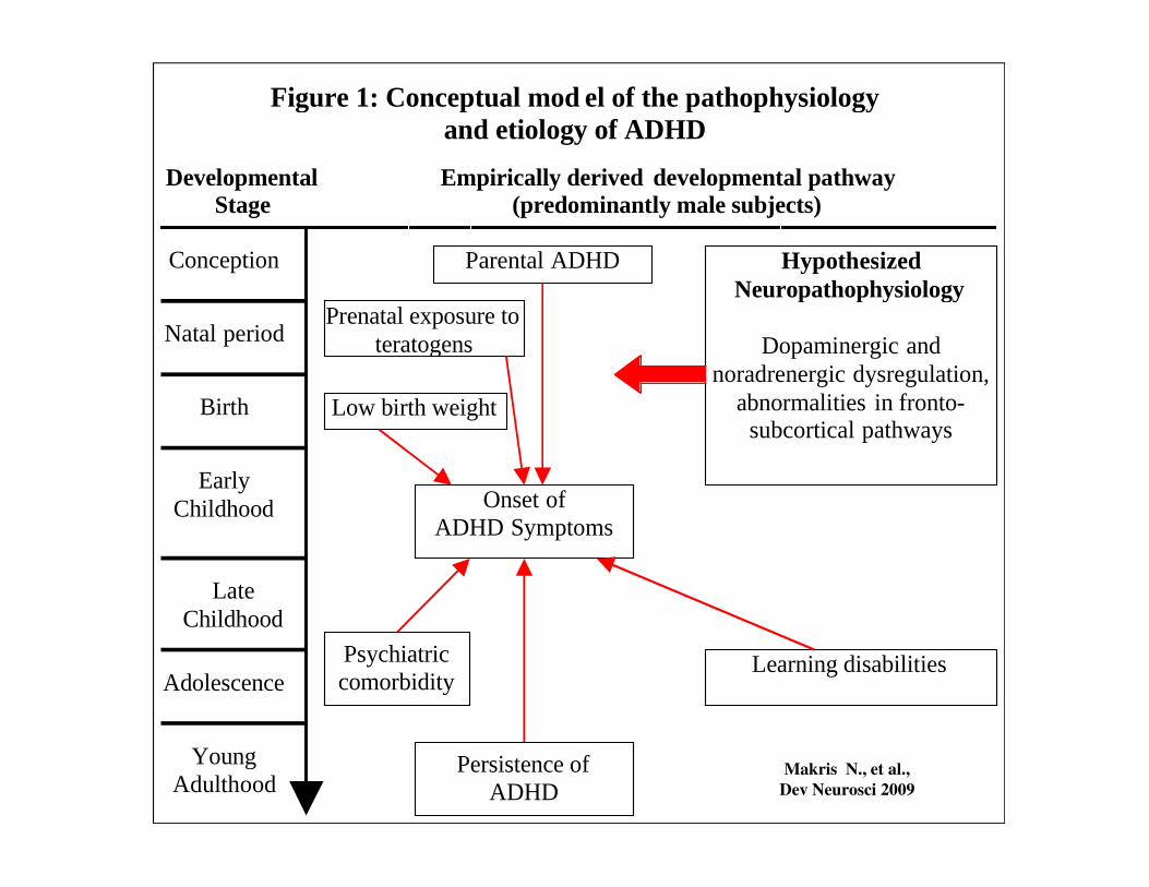
Empirically derived developmental pathway(predominantly male subjects)
DevelopmentalStage
Parental ADHD HypothesizedNeuropathophysiology
Dopaminergic andnoradrenergic dysregulation,
abnormalities in fronto-subcortical pathways
Figure 1: Conceptual mod el of the pathophysiologyand etiology of ADHD
Conception
Natal period
Birth
Prenatal exposure toteratogens
Low birth weight
Makris N., et al.,Dev Neurosci 2009
EarlyChildhood
Early
LateChildhood
Adolescence
YoungAdulthood
Learning disabilities
Persistence ofADHD
subcortical pathways
Psychiatriccomorbidity
Onset ofADHD Symptoms

Functional neuroanatomy of neural systems involved in psychiatric disorders
Makris N., et al.,Dev Neurosci 2009

Once we quantify imaging-based markers (or endo-phenotypes), for example, size of brain structures (such ascingulate cortex or prefrontal cortex) or neural systems (such as the reward, the attention or executive function systems),we may be able to diagnose psychiatric illnesses, assess treatment and identify genes that will lead to newmedications.
A major goal of MRI brain research is the study of endophenotypes
Makris N., et al.,Dev Neurosci 2009
Systems biology acts as an interface between the behavior/environment and genome/epigenome.

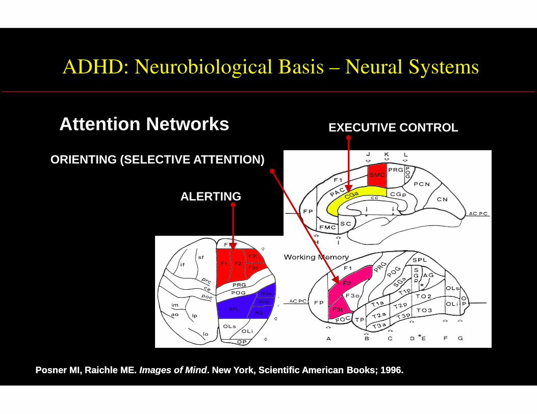
ADHD: Neurobiological Basis – Neural Systems
ALERTING
EXECUTIVE CONTROL
ORIENTING (SELECTIVE ATTENTION)
Attention Networks
Posner MI, Raichle ME.Posner MI, Raichle ME. Images of MindImages of Mind. New York, Scientific American Books; 1996.. New York, Scientific American Books; 1996.
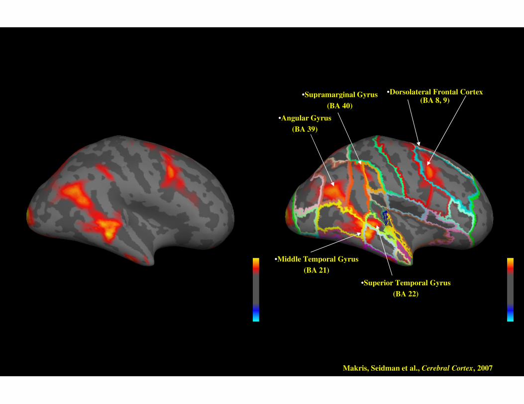
•Dorsolateral Frontal Cortex(BA 8, 9)
•Supramarginal Gyrus
(BA 40)
•Angular Gyrus
(BA 39)
•Superior Temporal Gyrus
(BA 22)
•Middle Temporal Gyrus
(BA 21)
Makris, Seidman et al., Cerebral Cortex, 2007

•Anterior Cingulate Gyrus(BA 24)
•Orbital Frontal Cortex
((BA 11, 12, 13, 14)•Orbital Frontal Cortex
((BA 11, 12, 13, 14)
Makris, Seidman et al., Cerebral Cortex, 2007

Fractional anisotropy decrease in ROIs for the cingulum bundle andthe superior longitudinal fascicle II (SLF II) in adults with ADHD
Makris, Seidman et al., Cerebral Cortex, 2008

Some challenges in psychiatric imagingSome challenges in psychiatric imaging
Diagnosis at a brain circuitry levelDiagnosis at a brain circuitry level
Clear localization of abnormalitiesClear localization of abnormalitiesClear localization of abnormalitiesClear localization of abnormalities
Instantaneous results with treatmentInstantaneous results with treatment
Use of advanced mathematicalUse of advanced mathematicalmodelingmodeling

Subgenual Cingulate Cortex (BA 25)
Increased activity with sadness anddepression (a, b) (using PET)
Decreased activity with chronicfluoxetine treatment for depression (c) or
natural placebo recovery (d)
Decreased activity in responders vs non-responders to CBT and citalopram for
social phobia (e, f)
Ressler and Mayberg, Nature Neuroscience, 2007
social phobia (e, f)

Deep Brain Stimulation (DBS)
One possible mechanism: DBS-inducedinhibition [e.g., DBS in PD at GPi: discharges
from high-frequency discharge neuronsshowing inhibitory periods after each
stimulus pulse (a, b)]
DBS at Cg25 for depression (c-f)Preoperative (d)
At 3 months in treatment responders (f)
Ressler and Mayberg, Nature Neuroscience, 2007

Invasive
Deep Brain Stimulation (DBS)
Vagal Nerve Stimulation (VNS)
NoninvasiveTranscranial Magnetic Stimulation (TMS)
Transcranial Direct Current Stimulation (tDCS)
Brain Stimulation-Neuromodulation

• Neural Pacemaker: Forces a population ofneurons to fire at a specific frequency,changing excitability and functionalconnectivity both locally and within a givennetwork
Neuromodulation
connectivity both locally and within a givennetwork
• Chronic vs. Discrete Stimulation• Invasive vs Noninvasive• All aim to induce adaptive neuroplasticity

Transcranial Magnetic Stimulation
Anthony Barker 1984Anthony Barker 1984
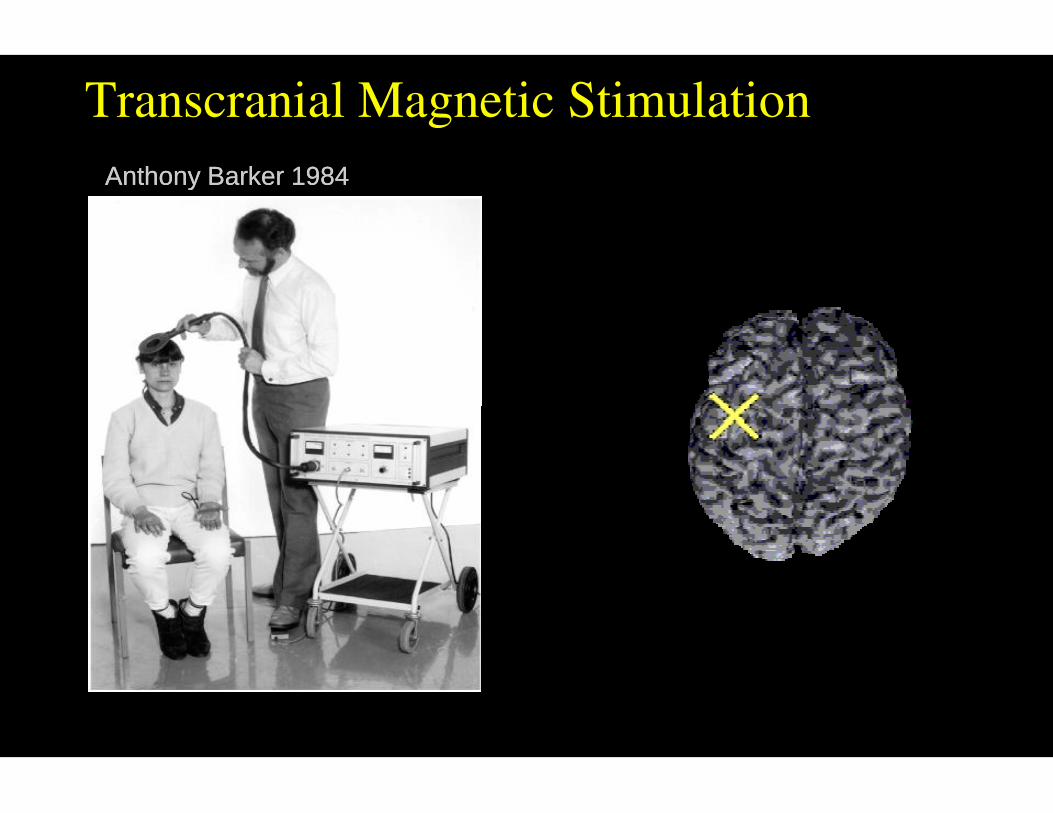
Transcranial Magnetic Stimulation
Anthony Barker 1984Anthony Barker 1984

Imaging inImaging inDeep Brain Stimulation
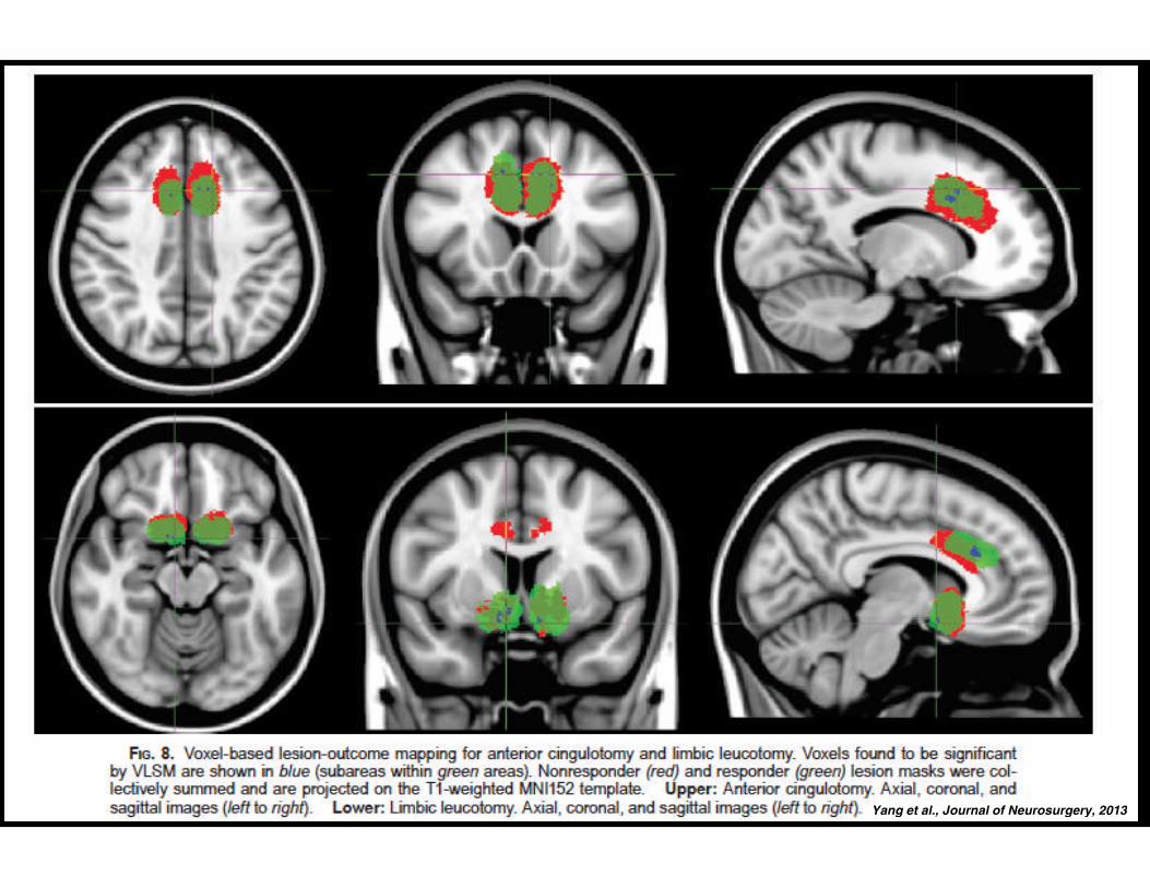
Yang et al., Journal of Neurosurgery, 2013
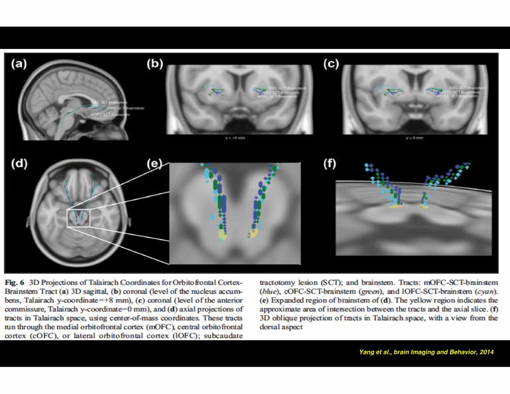
Yang et al., brain Imaging and Behavior, 2014
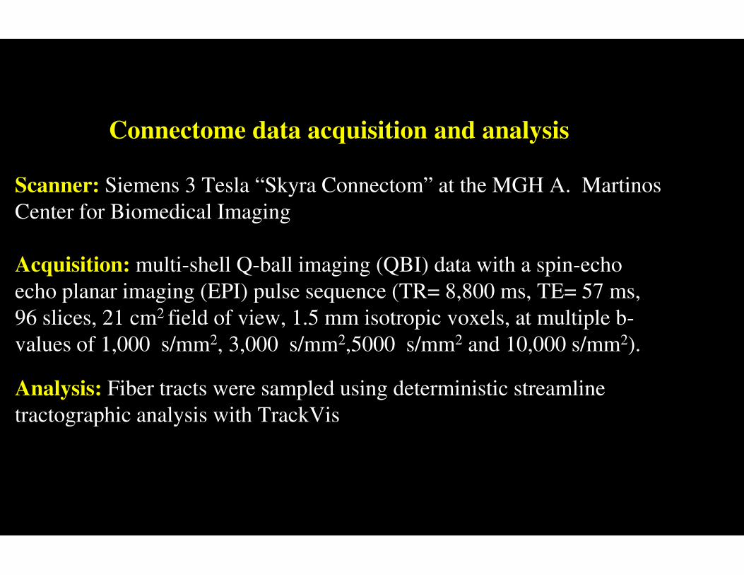
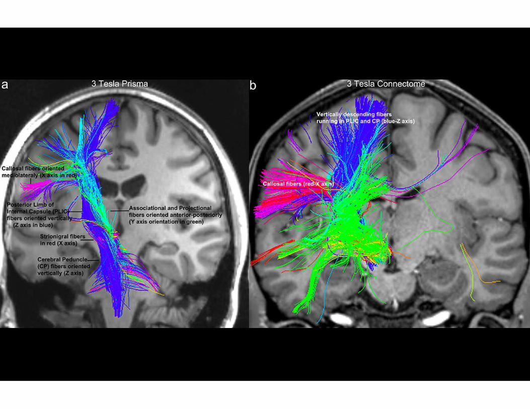
Connectome data acquisition and analysis
Scanner: Siemens 3 Tesla “Skyra Connectom” at the MGH A. MartinosCenter for Biomedical Imaging
Acquisition: multi-shell Q-ball imaging (QBI) data with a spin-echoecho planar imaging (EPI) pulse sequence (TR= 8,800 ms, TE= 57 ms,echo planar imaging (EPI) pulse sequence (TR= 8,800 ms, TE= 57 ms,96 slices, 21 cm2 field of view, 1.5 mm isotropic voxels, at multiple b-values of 1,000 s/mm2, 3,000 s/mm2,5000 s/mm2 and 10,000 s/mm2).
Analysis: Fiber tracts were sampled using deterministic streamlinetractographic analysis with TrackVis


DBSDBS
in OCDin OCD
usingusingConnectomeConnectome
TechnologyTechnology

The half-head sample sealed in a vacuumed plastic (top-right) and assembly of aright brain hemisphere opposite to the half-head sample to fill-in the head coil andprohibit image distortion (left images). The assembled half-head-half-brain samplewas used for imaging

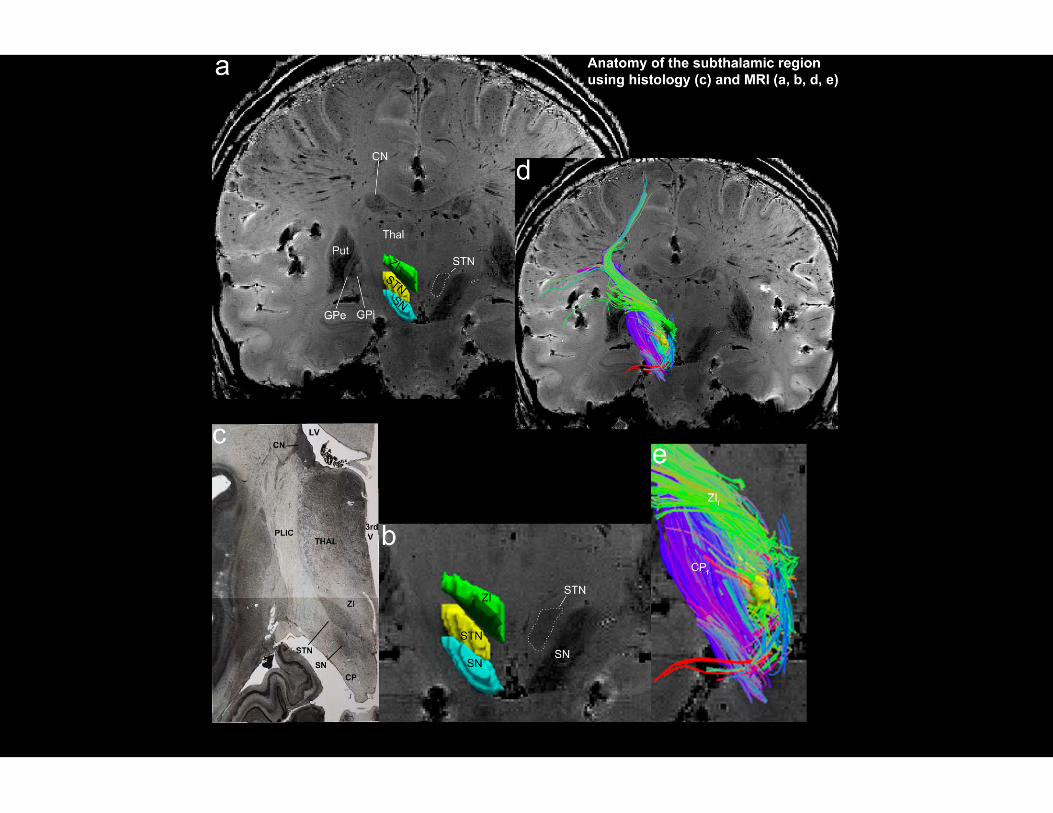
Upper-right and bottom images: Coronal sections of high-resolution structural MRI of right (upper-right image) andleft (bottom image) subthalamic nucleus (STN); lower-right images: outlined zoomed versions of STN.
7 Tesla Scanner @ Martinos Center for BiomedicalImaging; Sequence: FLASH;Voxel size: 400um isotropic; Total time: 1:09:08TR: 23 ms; TE: 10.2 ms; Echo spacing: 10 msFlip Angle: 30.0 degrees; PAT: off, Fat supp: off
Upper-left image: Axial section offractional anisotropy map (FA map)fractional anisotropy map (FA map)of the head sample obtained by Q-ball diffusion imaging
Connectome Scanner @ MartinosCenter for Biomedical Imaging;Sequence: 64 dir Q-ball imaging
Voxel size: 1.5 mm isotropicTotal time: 11mins 44sTR: 8800 ms; TE: 57.0 msEcho spacing: 0.63 msNumber of directions: 64b-value: 1400PAT: 3; Fat suppression: strong


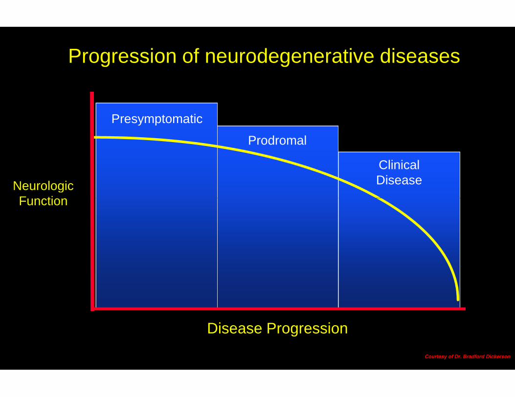
Neurodegeneration

CognitiveFunction
Presymptomatic
Prodromal
ClinicalDementia
Progression of Alzheimer’s Disease:similar model for many other diseases
Disease Progression
Function
MildCognitive
Impairment
ProbableAlzheimer’s
Disease
Accumulating pathology Courtesy of Dr. Bradford Dickerson

Large-scale cognitive systemsunderstrood through resting state fMRI
Dorsal visuospatial attention system
Frontoparietal executive control system
Vincent et al., J Neurophys, 2008
Episodic memory/Default-mode system

AD cortical signature:Cortical atrophy compared to normals
Dickerson et al, Cerebral Cortex 2009
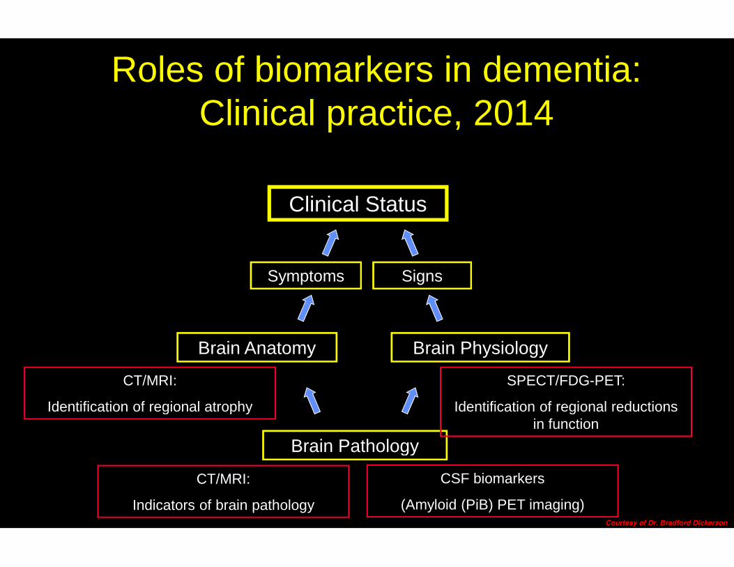
Clinical Status
Roles of biomarkers in dementia:Clinical practice, 2014
SignsSymptoms Signs
Brain Anatomy Brain Physiology
Symptoms
Brain Pathology
CT/MRI:
Indicators of brain pathology
CSF biomarkers
(Amyloid (PiB) PET imaging)
CT/MRI:
Identification of regional atrophy
SPECT/FDG-PET:
Identification of regional reductionsin function
Courtesy of Dr. Bradford Dickerson
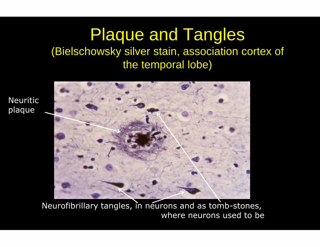
Plaque and Tangles(Bielschowsky silver stain, association cortex of
the temporal lobe)
Neuriticplaque
Neurofibrillary tangles, in neurons and as tomb-stones,where neurons used to be

Only Post-Mortem Tau Pathology
(McKee et al., 2009)Psychiatry Neuroimaging
Laboratory

Amyloid imaging with PETPittsburgh Compound-B (PiB)
Alzheimer’sdisease
Normalaging
Courtesy of Dr. Bradford Dickerson

Aging, MCI, AD: FDG-PET
Cognitively intactolder adult
Mild CognitiveImpairment
Alzheimer’sDisease
Courtesy of Dr. Bradford Dickerson

Case 1
FDG-PET

Case 2
FDG-PET Amyloid (PiB)-PET
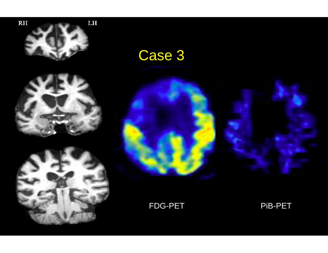
Case 3
FDG-PET PiB-PET

Pre-Clinical PET Tracer Used in Animals
Psychiatry NeuroimagingLaboratory
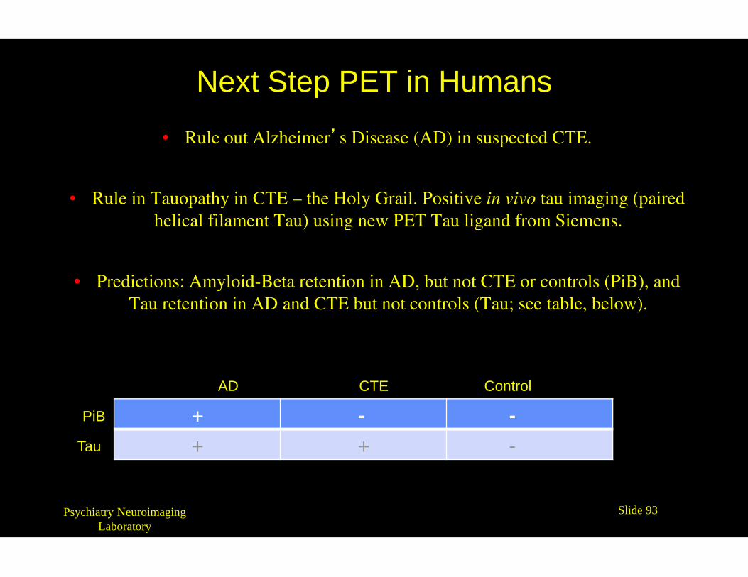
Next Step PET in Humans
• Rule out Alzheimer’s Disease (AD) in suspected CTE.
• Rule in Tauopathy in CTE – the Holy Grail. Positive in vivo tau imaging (pairedhelical filament Tau) using new PET Tau ligand from Siemens.
• Predictions: Amyloid-Beta retention in AD, but not CTE or controls (PiB), and
Psychiatry NeuroimagingLaboratory
• Predictions: Amyloid-Beta retention in AD, but not CTE or controls (PiB), andTau retention in AD and CTE but not controls (Tau; see table, below).
+ - -
+ + -
AD CTE Control
PiB
Tau
Slide 93

Imaging technology is not just making pretty picturesor even simply increasing our scientific understandingof Alzheimer’s and related diseases; it is in factenabling new revolutions in testing treatments thatmay lead to real benefits for patients, possibly evenforms of prevention.forms of prevention.
e.g., anti-amyloid treatment with monoclonalantibodies in mildly symptomatic patients (MCI) oreven asymptomatic individuals with brain amyloid.
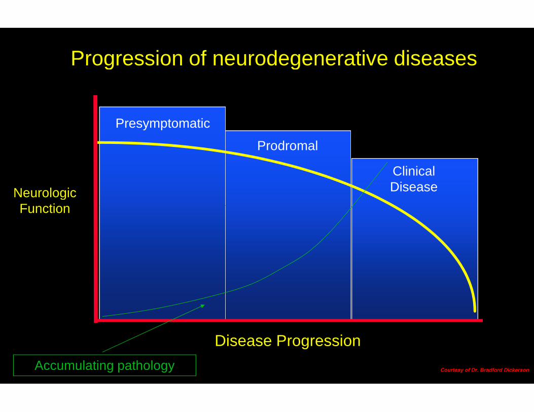
NeurologicFunction
Presymptomatic
Prodromal
ClinicalDisease
Progression of neurodegenerative diseases
Disease Progression
Function
Courtesy of Dr. Bradford Dickerson
NeurologicFunction
Presymptomatic
Prodromal
ClinicalDisease
Progression of neurodegenerative diseases
Disease Progression
Function
Accumulating pathology Courtesy of Dr. Bradford Dickerson

NeurologicFunction
Presymptomatic
Prodromal
ClinicalDisease
Progression of neurodegenerative diseases
Regional brain volume
Disease Progression
Function
Accumulating pathology Courtesy of Dr. Bradford Dickerson

NeurologicFunction
Presymptomatic
Prodromal
ClinicalDisease
Progression of neurodegenerative diseases
Regional brain volume
Hypometabolism
Disease Progression
Function
Accumulating pathology Courtesy of Dr. Bradford Dickerson

NeurologicFunction
Presymptomatic
Prodromal
ClinicalDisease
Progression of neurodegenerative diseases
Disease Progression
Function
Accumulating pathology
Disease-modifyingtherapy
Courtesy of Dr. Bradford Dickerson

NeurologicFunction
Presymptomatic
Prodromal
ClinicalDisease
Progression of neurodegenerative diseases
Disease Progression
FunctionDisease-modifyingtherapy
Courtesy of Dr. Bradford Dickerson

NeurologicFunction
Presymptomatic
Prodromal
ClinicalDisease
Progression of neurodegenerative diseases
Disease Progression
FunctionDisease-modifyingtherapy
Courtesy of Dr. Bradford Dickerson

NeurologicFunction
Presymptomatic
Prodromal
ClinicalDisease
Progression of neurodegenerative diseases
Disease Progression
FunctionDisease-modifyingtherapy
Courtesy of Dr. Bradford Dickerson
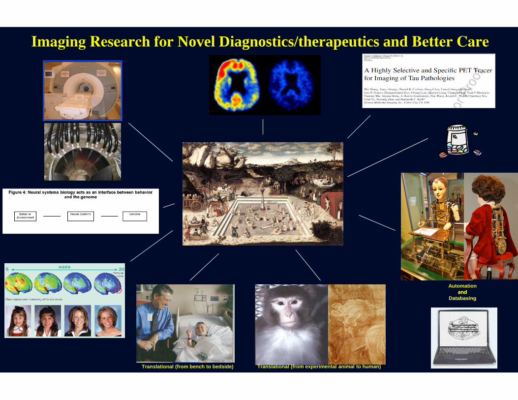
Imaging Research for Novel Diagnostics/therapeutics and Better Care
Translational (from experimental animal to human)Translational (from bench to bedside)
Automationaandnd
Databasing

THANK YOU!THANK YOU!