Bowel Screening Histology Data Standard · BOWEL SCREENING HISTOLOGY DATA STANDARD: DRAFT FOR...
33
Bowel Screening Histology Data Standard Draft for public comment HISO 10072.2: 2019
Transcript of Bowel Screening Histology Data Standard · BOWEL SCREENING HISTOLOGY DATA STANDARD: DRAFT FOR...
Bowel Screening
Histology Data
Standard
Draft for public comment
HISO 10072.2: 2019

Contributors The development of this standard was led by Dr Nicole Kramer, Lead Pathologist
National Bowel Screening Programme.
The Bowel Cancer Histopathology Subgroup provided significant input during the
development of this standard. The group was made up of the following members:
Dr Nicole Kramer, Lead Pathologist National Bowel Screening Programme,
Auckland District Health Board, LabPlus (Chair)
Professor Ian Bissett, National Bowel Cancer Working Group Chair, University of
Auckland
Dr Michael Lau, Pathologist, Southern Community Laboratories
Dr Harold Neale, Principal Scientific Advisor – Population Health and Prevention,
Clinician’s Screening
Dr Vladmir Osipov, Pathologist Auckland District Health Board, LabPlus
Associate Professor Susan Parry, Clinical Director National Bowel Screening
Programme, Ministry of Health / Auckland District Health Board
Dave Scarrow, Manager Information Systems, Pathlab
Dr Kerry Sexton, Clinical Lead, Monitoring and Evaluation, National Screening
Unit, Ministry of Health
Dr Nicholas Shaw, Anatomical Pathologist, Pathlab
Dr Martin Whitehead, Anatomical Pathologist, Canterbury Health Laboratories
Dr Masato Yozu, Histopathologist, Counties Manukau Laboratory Services.
The development of this standard was facilitated by Carrie Buckmaster, Senior
Business Analyst, National Bowel Screening Programme, Ministry of Health.
Citation: Ministry of Health. 2019. HISO 10072.2: 2019 Bowel Screening Histology
Data Standard: Draft for public comment. Wellington: Ministry of Health.
Published in February 2019 by the Ministry of Health
PO Box 5013, Wellington 6140, New Zealand
ISBN 978-1-98-856852-2 (online)
HP 7038
Health Information Standards Organisation (HISO) standards are published by the
Ministry of Health for the New Zealand health and disability sector.
This document is available at www.health.govt.nz/our-work/ehealth/digital-health-
standards-and-governance/health-information-standards
This work is licensed under the Creative Commons Attribution 4.0 International licence.
In essence, you are free to: share ie, copy and redistribute the material in any medium or
format; adapt ie, remix, transform and build upon the material. You must give
appropriate credit, provide a link to the licence and indicate if changes were made.

HISO 10072.2: 2019
BOWEL SCREENING HISTOLOGY DATA STANDARD: DRAFT FOR PUBLIC CONSULTATION iii
Keeping standards up to date HISO standards are regularly updated to reflect advances in health information science
and technology. See the Ministry of Health website at health.govt.nz for information
about the standards development process. We welcome your ideas for improving this
standard. Email [email protected] or write to HISO, Ministry of Health,
PO Box 5013, Wellington 6145.


HISO 10072.2: 2019
BOWEL SCREENING HISTOLOGY DATA STANDARD: DRAFT FOR PUBLIC CONSULTATION v
Contents 1 Introduction 1
1.1 Purpose 1
1.2 Scope 1
1.3 Implementation 2
1.4 SNOMED CT 2
1.5 New Zealand legislation 2
1.6 Data element definitions 3
2 Data elements 4
2.1 Report 5
2.2 Specimen 11
2.3 Other pathological findings 27


HISO 10072.2: 2019
BOWEL SCREENING HISTOLOGY DATA STANDARD: DRAFT FOR PUBLIC CONSULTATION 1
1 Introduction The National Bowel Screening Programme1 (NBSP) is a free programme for men and
women aged 60 – 74 years eligible for publically funded health care. The primary
objective of bowel screening is to reduce the mortality rate by diagnosing and treating
bowel cancer at an earlier more treatable stage. The introduction of the NBSP in New
Zealand followed a successful six-year pilot.
The new NBSP information technology system is called the National Screening Solution
(NSS). This system will enable easy management of the bowel screening pathway,
support planning and management of participants, monitor safety and quality, and
enable ongoing evaluation of the programme. The NSS is a long-term strategic
solution that is capable of being extended to support future population health
initiatives.
1.1 Purpose The standard identifies and describes the data elements that need to be captured in
information systems of the laboratories contracted to perform NBSP histology services.
This data will support the monitoring, operation and quality of the NBSP and may also
be used for research and education purposes.
The standard is designed to ensure that consistent information is sent from various
laboratories into the NSS.
Laboratory information systems must provide the data described in this standard to
the NSS in a way that does not significantly impact laboratory pathologists’ ease of
working (ie, pathologists should not be expected to manually enter SNOMED CT codes
into their information systems).
1.2 Scope The standard defines the data required to be sent to the NSS. This standard does not
define the data sent from the laboratory to the physician responsible for the patient’s
care.
1 National Bowel Screening Programme: https://www.timetoscreen.nz/bowel-screening/about-the-
national-bowel-screening-programme/

2 HISO 10072.2: 2019
BOWEL SCREENING HISTOLOGY DATA STANDARD: DRAFT FOR PUBLIC CONSULTATION
1.3 Implementation Laboratories performing NBSP histology services must update their information
systems to ensure that the data specified in this standard is able to be captured
accordingly.
1.4 SNOMED CT SNOMED CT is the endorsed terminology standard for clinical information systems and
electronic health records in New Zealand. SNOMED CT is developed by SNOMED
International, of which New Zealand is one of a number of member countries.
1.5 New Zealand legislation The following Acts of Parliament and Regulations have specific relevance to this
standard. Readers must consider other Acts and Regulations and their amendments
that are relevant to their own organisation, in the implementation or use of this
standard:
Health Act 1956
Health and Disability Commissioner (Code of Health and Disability Services
Consumers’ Rights) Regulations 1996
Health Information Privacy Code 1994
Health Practitioners Competence Assurance Act 2003
Privacy Act 1993 (revised 2008)
Public Records Act 2005
Retention of Health Information Regulations 1996.

HISO 10072.2: 2019
BOWEL SCREENING HISTOLOGY DATA STANDARD: DRAFT FOR PUBLIC CONSULTATION 3
1.6 Data element definitions Each data element in this standard is defined according to a set of metadata
components from ISO/IEC 11179 Information Technology – Specification and
standardization of data elements 2003.
Definition A statement that expresses the essential nature of the data element and its
differentiation from all other data elements in this standard
Source
standards
Established data definitions or guidelines pertaining to the data element
Data type Alphabetic (A)
Numeric (N)
Alphanumeric (X)
Boolean
Date
Representationa
l class
Code, free text, value or identifier
For date and time data types, use
full date or partial date
Does not apply to Boolean types
Field size Maximum
number of
characters
Representationa
l layout
The arrangement of characters in
the data element – eg,
‘A(50)’ means up to
50 alphabetic characters
‘NNAAAA’ means two numeric
followed by four alphabetic
characters.
Full date/time representation is
YYYYMMDD hh:mm:ss.
Obligation Indicates if the data element is mandatory or optional for the entity being
discussed. It can include conditional obligations of the data element.
Data domain The valid values or codes that are acceptable for the data element.
The data elements contained in this standard are dates, free text or coded.
Each coded data element has a specified code set.
Guide for use Additional guidance about using the data element.
Verification
rules
Quality control mechanisms that preclude invalid values.

4 HISO 10072.2: 2019
BOWEL SCREENING HISTOLOGY DATA STANDARD: DRAFT FOR PUBLIC CONSULTATION
2 Data elements This section describes the set of histology data that needs to be sent to the NSS for
use by the NBSP. The messages sent to the NSS are in addition and different to the
existing histology messages that laboratories already send to requesting physicians.
Each report must have one or more specimens. For each specimen, in addition to the
main diagnosis, there can be up to five other pathological findings.
Report BR Other Pathological Findings
Patient identifier
Patient name
Specimen identifier
Sample procedure
Patient date of birth
Programme identifier
Lab report identifier
Date of order
Date specimens received
Date of report
Requesting clinic identifier
Requesting clinician identifier
Laboratory facility identifier
Site
Main Diagnosis
Size of polyp
Dysplasia
Margin - polypectomy
Distance from anal verge
Histological Grade (tumour
differentiation)
Poor / undifferentiated tumour
Width of Tumour
Haggitt Level
Kikuchi Level
Pathologist identifier
Clinical Details
Other pathological finding
Lymphatic invasion
Deep Margin
Peripheral Margin
Depth of invasion
Venous invasion
Nuclear expression of MLH1
Nuclear expression of MSH2
Nuclear expression of MSH6
Nuclear expression of PMS2
protein
BRAFV600E mutation status
BRAF method of testing
MLH1 Promoter Methylation
Testing

HISO 10072.2: 2019
BOWEL SCREENING HISTOLOGY DATA STANDARD: DRAFT FOR PUBLIC CONSULTATION 5
2.1 Report This section lists the relevant data elements for a report.
2.1.1 Laboratory facility identifier
Definition The unique identifier for the facility (laboratory) that performed the
pathology work.
Source standards Information on the Health Provider Index is available at
https://www.health.govt.nz/our-work/health-identity/health-provider-
index
Data type Alphanumeric Representational class Identifier
Field size 8 Representational layout FXXNNN-C
Obligation Mandatory
Data domain A valid HPI Facility ID
Guide for use This must be the HPI Facility ID for the laboratory that performed the
pathology work.
For organisations using the Ministry of Health’s legacy Health Facility Codes,
refer to the Ministry’s current list of mappings to identify the relevant HPI
Facility ID. The current list is available at https://www.health.govt.nz/nz-
health-statistics/data-references/code-tables/common-code-
tables/facility-code-table.
Verification rules A valid HPI Facility ID
2.1.2 Laboratory report identifier
Definition A laboratory’s unique accession number or ‘day number’ for the report, ie, the
number under which the specimens or episode is documented in the
laboratory information system.
Source standards N/A
Data type Alphanumeric Representational class Identifier
Field size 30 Representational
layout
X(30)
Obligation Mandatory
Data domain As defined by the laboratory.
Guide for use N/A
Verification rules Each laboratory report identifier must be unique for all reports sent from that
laboratory.
The laboratory report identifier will be stored within the NSS to enable communication
with a laboratory about a particular report.

6 HISO 10072.2: 2019
BOWEL SCREENING HISTOLOGY DATA STANDARD: DRAFT FOR PUBLIC CONSULTATION
2.1.3 Pathologist identifier
Definition A unique identifier for the pathologist who extracted the samples which this
histology report relates to.
Source standards HPI documentation: www.health.govt.nz/our-work/health-
identity/health-practitioner-index
See also:
HISO 10005:2008 Health Practitioner Index Data Set:
www.health.govt.nz/publication/hiso-100052008-health-
practitioner-index-hpi-data-set
HISO 10006:2008 Health Practitioner Index Code Set:
www.health.govt.nz/publication/hiso-100062008-health-
practitioner-index-hpi-code-set
Data type Alphanumeric Representational class Identifier
Field size 6 Representational layout NNAAAA
Obligation Mandatory
Data domain HPI Common Person Number (CPN) generated by the HPI system.
Guide for use This field uses the Health Provider Index (HPI) CPN, a unique identifying
number for the health practitioner delivering the service. This field is only
for use where the practitioner is a member of a Responsible Authority
under the Health Practitioners Competence Assurance Act 2003.
Verification rules CPN can be obtained from the clinician but must be validated with the HPI
system.
2.1.4 Patient identifier
Definition National Health Index (NHI) number – a unique identifier assigned by the
NHI system to a patient.
Source standards HISO 10046 Consumer Health Identity Standard:
www.health.govt.nz/publication/hiso-10046-consumer-health-
identity-standard
See also NHI data dictionary: www.health.govt.nz/publication/national-
health-index-data-dictionary.
Data type Alphanumeric Representational class Identifier
Field size 7 Representational layout AAANNNN
Obligation Mandatory
Data domain NHI numbers
Guide for use Only the NHI system generates the NHI number assigned to a patient.
NHI numbers are not reused once assigned to a patient.
Where more than one number exists for a patient, one number is declared
‘live’ and all other numbers are made ‘dormant’ and attached to the live
record.
The NHI number is the primary key for patients’ records.
Verification rules See the source standards for the check digit algorithm and NHI number
validation rules.

HISO 10072.2: 2019
BOWEL SCREENING HISTOLOGY DATA STANDARD: DRAFT FOR PUBLIC CONSULTATION 7
2.1.5 Patient name
This is the name of the NSS participant whose specimens are being examined and
reported on. This is a complex field, and the report must contain the data elements
identified in the ‘Patient name’ section of the HISO 10046 Consumer Health Identity
standard.
2.1.6 Patient birth date
Definition The date when the patient was born.
Source standards HISO 10046 Consumer Health Identity Standard:
www.health.govt.nz/publication/hiso-10046-consumer-health-
identity-standard
Data type Date Representational class Full or partial date
Field size Max: 8 Representational layout CCYY[MM[DD]]
Obligation The year component of the date is mandatory.
Month is conditional and to be used if known.
Day is conditional and to be used if known and month has been recorded.
Data domain A valid date.
Guide for use Year of birth must be recorded as a minimum.
Verification rules The date of birth must be a valid day, month and year combination and
cannot be in the future.
For a partial date, the month of birth can be left blank if unknown. In this
case, the day of birth must be blank.
2.1.7 Programme identifier
Definition This will be ‘NBSP’ for histology sent to NSS as part of the National Bowel
Screening Programme.
Source standards N/A
Data type Alpha Representational class Code
Field size 4 Representational layout A(4)
Obligation Mandatory
Data domain
Code Description
NBSP National Bowel Screening Programme
Guide for use This is used by the NSS to determine what screening programme the
pathology results relate to.
Verification rules This must be NBSP.

8 HISO 10072.2: 2019
BOWEL SCREENING HISTOLOGY DATA STANDARD: DRAFT FOR PUBLIC CONSULTATION
2.1.8 Requesting clinic identifier
Definition This is the HPI Facility ID of the endoscopy clinic that performed the
colonoscopy, or other screening procedure, during which the specimens
were taken.
Source standards Information on the Health Provider Index is available at
https://www.health.govt.nz/our-work/health-identity/health-provider-
index.
Data type Alphanumeric Representational class Identifier
Field size 8 Representational layout FXXNNN-C
Obligation Mandatory
Data domain Valid HPI number only.
Guide for use Use the HPI Facility ID of the endoscopy clinic, hospital or surgery that sent
the specimens to the laboratory. Use the most specific HPI facility ID
available.
For organisations using the Ministry of Health’s legacy Health Facility
Codes, refer to the Ministry’s current list of mappings to identify the
relevant HPI Facility ID. The current list is available at
https://www.health.govt.nz/nz-health-statistics/data-references/code-
tables/common-code-tables/facility-code-table.
Verification rules: A valid HPI Facility ID.

HISO 10072.2: 2019
BOWEL SCREENING HISTOLOGY DATA STANDARD: DRAFT FOR PUBLIC CONSULTATION 9
2.1.9 Requesting clinician identifier
Definition Identifier for the endoscopist who performed the colonoscopy – this should
appear on the histology request form sent to the laboratory.
Source standards HPI documentation: www.health.govt.nz/our-work/health-
identity/health-practitioner-index
See also:
HISO 10005:2008 Health Practitioner Index Data Set:
www.health.govt.nz/publication/hiso-100052008-health-
practitioner-index-hpi-data-set.
HISO 10006:2008 Health Practitioner Index Code Set:
www.health.govt.nz/publication/hiso-100062008-health-
practitioner-index-hpi-code-set.
Data type Alphanumeric Representational class Identifier
Field size 6 Representational layout NNAAAA
Obligation Mandatory
Data domain HPI CPN numbers generated by the HPI system.
Guide for use This field uses the Health Provider Index (HPI) Common Person Number
(HPI_CPN): A unique identifying number pertaining to the health provider
that is delivering the service where that health practitioner is a member of a
Responsible Authority as set out in the Health Practitioners Competence
Assurance Act 2003.2
This field is only for use where the practitioner is a member of a
Responsible Authority under the Health Practitioners Competence
Assurance Act 2003.
Verification rules CPN can be obtained from the clinician but must be validated with the HPI
system.
2.1.10 Date of order
Definition The date when the histology order was made, as provided on the request
form, which should match the date of the endoscopy.
Source standards N/A
Data type Date Representational class Date
Field size 8 Representational layout CCYYMMDD
Obligation Mandatory
Data domain A valid date
Guide for use Use the date when the histology order was made
Verification rules A valid date that is less than or equal to the current date
2 www.health.govt.nz/our-work/regulation-health-and-disability-system/health-practitioners-
competence-assurance-act/responsible-authorities-under-act

10 HISO 10072.2: 2019
BOWEL SCREENING HISTOLOGY DATA STANDARD: DRAFT FOR PUBLIC CONSULTATION
2.1.11 Date specimens received
Definition The date when the specimen(s) were received in the laboratory
Source standards RCPA guideline and policy (8.2.l):
https://www.rcpa.edu.au/Library/College-
Policies/Guidelines/Turnaround-Time-in-Anatomical-Pathology
Data type Date Representational class Date
Field size 8 Representational layout CCYYMMDD
Obligation Mandatory
Data domain A valid date.
Guide for use Use the date when the tissue was received in the laboratory.
The interim quality standards require that turnaround times are accordant
with the RCPA guideline and policy (8.2.l).
Verification rules A valid date that is less than or equal to the current date.
2.1.12 Date of report
Definition The date when the laboratory report was finalised.
Source standards N/A
Data type Date Representational class Date
Field size 8 Representational layout CCYYMMDD
Obligation Mandatory
Data domain A valid date.
Guide for use Use the date when the laboratory report was finalised.
Verification rules A valid date that is less than or equal to the current date.
2.1.13 Clinical details
Definition Additional clinical information provided by the endoscopist.
Source standards N/A
Data type Alphanumeric Representational class Free text
Field size 2000 Representational layout X(2000)
Obligation Conditional. Required where the information is provided on the laboratory
request form.
Data domain Free text.
Guide for use A free-text description of the pathology or any details about it, not already
catered for by the elements in this report.
Verification rules N/A

HISO 10072.2: 2019
BOWEL SCREENING HISTOLOGY DATA STANDARD: DRAFT FOR PUBLIC CONSULTATION 11
2.2 Specimen There are one or more specimens for each report. The following identified the data
elements for a specimen.
2.2.1 Specimen identifier
Definition The identifier for the specimen for which the examination is being
described.
Source standards N/A
Data type Alphanumeric Representational class Identifier
Field size 30 Representational layout X(30)
Obligation Mandatory
Data domain N/A
Guide for use This is the same as the Pot ID provided on the pot which the specimen was
contained in, and on the laboratory request form.
Laboratories may use their own internal identifiers for the pot(s) in an order,
but the identifier used in the report must match that used to originally label
the pot.
Verification rules N/A

12 HISO 10072.2: 2019
BOWEL SCREENING HISTOLOGY DATA STANDARD: DRAFT FOR PUBLIC CONSULTATION
2.2.2 Site
Definition This is the location the tissue was taken from
Source standards N/A
Data type Numeric Representational class Code
Field size 18 Representational layout N(18)
Obligation Mandatory
Data domain
Clinical term SNOMED CT
Caecum 32713005
Appendiceal orifice 83856002
Ileocaecal valve 23153004
Ileum (excluding terminal ileum) 34516001
Terminal ileum 85774003
Right (ascending) colon 9040008
Hepatic flexure 48338005
Transverse colon 485005
Splenic flexure 72592005
Left (descending) colon 32622004
Sigmoid colon 60184004
Rectosigmoid junction 49832006
Rectum 34402009
Anal canal 34381000
Colon (NOS) 71854001
Unknown body region 87100004
Guide for use ‘Unknown body region’ should only be used when the histology request
form is not filled in correctly.
If the location where the specimen was removed from cannot categorically
be identified by the endoscopist, the distance from the anal verge should
be recorded instead on the histology request form. This should then be
provided in the ‘Distance from anal verge’ element and the site
documented as ’Colon (NOS)’.
Verification rules One of the options must be provided.

HISO 10072.2: 2019
BOWEL SCREENING HISTOLOGY DATA STANDARD: DRAFT FOR PUBLIC CONSULTATION 13
2.2.3 Distance from the anal verge
Definition The measurement, in millimetres, of the distance between the anal verge
and where the specimen was taken from.
Source standards N/A
Data type Numeric Representational class Number
Field size 2 Representational layout N(2)
Obligation Conditional. Required when provided on laboratory request form.
Data domain An integer.
Guide for use In some situations, it may not be possible to categorically specify the name
of the site where the specimen was taken from. In such cases the
endoscopist may provide the distance from the anal verge instead of the
location in the large bowel.
If the distance from the anal verge is provided on the laboratory request
form for the specimen then it should be provided here.
Verification rules If the site value is Colon (NOS) then the distance from the anal verge should
be provided.
2.2.4 Sample procedure
Definition This identifies how the specimen was removed.
Source standards N/A
Data type Numeric Representational class Code
Field size 18 Representational layout N(18)
Obligation Mandatory
Data domain
Clinical term SNOMED CT
Biopsy 274323008
Polypectomy 274025005
Unknown procedure 428119001
Other procedure on large intestine 118838009
Guide for use Refer to information in the histology request form.
Verification rules One of the provided options.

14 HISO 10072.2: 2019
BOWEL SCREENING HISTOLOGY DATA STANDARD: DRAFT FOR PUBLIC CONSULTATION
2.2.5 Size
Definition This is the size of the specimen in millimetres.
Source standards N/A
Data type Numeric Representational class Number
Field size 2 Representational layout N(2)
Obligation Conditional. Required if documented.
Data domain An integer.
Guide for use According to programme’s interim quality standard 8.2.c, the size of lesions
is generally accepted as that measured by the endoscopist and provided on
the request form. However, if there is a major discrepancy between the
provided size and the size of the lesion microscopically, the largest
dimension should be measured by the reporting pathologist to the nearest
millimetre on the haematoxylin and eosin slide.
Provided in mm.
Verification rules An integer.
HISO 10072.2: 2019
BOWEL SCREENING HISTOLOGY DATA STANDARD: DRAFT FOR PUBLIC CONSULTATION 15
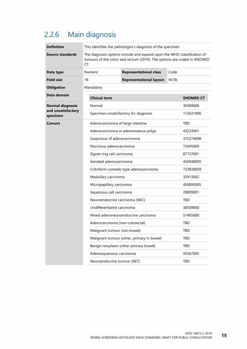
2.2.6 Main diagnosis
Definition This identifies the pathologist’s diagnosis of the specimen
Source standards The diagnosis options include and expand upon the WHO classification of
tumours of the colon and rectum (2010). The options are coded in SNOMED
CT.
Data type Numeric Representational class Code
Field size 18 Representational layout N(18)
Obligation Mandatory
Data domain
Clinical term SNOMED CT
Normal diagnosis
and unsatisfactory
specimen
Normal 30389008
Specimen unsatisfactory for diagnosis 112631006
Cancers Adenocarcinoma of large intestine TBD
Adenocarcinoma in adenomatous polyp 43233001
Suspicious of adenocarcinoma 315274008
Mucinous adenocarcinoma 72495009
Signet ring cell carcinoma 87737001
Serrated adenocarcinoma 450948005
Cribriform comedo type adenocarcinoma 733838009
Medullary carcinoma 32913002
Micropapillary carcinoma 450895005
Squamous cell carcinoma 28899001
Neuroendocrine carcinoma (NEC) TBD
Undifferentiated carcinoma 38549000
Mixed adenoneuroendocrine carcinoma 51465000
Adenocarcinoma (non-colorectal) TBD
Malignant tumour (non bowel) TBD
Malignant tumour (other, primary in bowel) TBD
Benign neoplasm (other primary bowel) TBD
Adenosquamous carcinoma 59367005
Neuroendocrine tumour (NET) TBD

16 HISO 10072.2: 2019
BOWEL SCREENING HISTOLOGY DATA STANDARD: DRAFT FOR PUBLIC CONSULTATION
Polyps Tubular adenoma 19665009
Tubulovillous adenoma 61722000
Villous adenoma 128859003
Hyperplastic polyp 62047007
Sessile serrated adenoma /polyp 443157008
Traditional serrated adenoma 443734007
Serrated polyp (not otherwise specified) 449854009
Inflammatory polyp 76235005
Mucosal prolapse 29696001
Mesenchymal tumours – Leiomyoma 44598004
Mesenchymal tumours – Lipoma 46720004
Mesenchymal tumours – Gastrointestinal stromal
tumour
128755003
Hamartomatous polyp 27391005
Lymphoid polyp 80297003
Other pathology Ulcerative colitis 64766004
Crohn’s disease 34000006
Chronic idiopathic inflammatory bowel disease,
unclassified
359664009
Inflammation, unspecified 23583003
Guide for use The members in this code set cover both polyps and cancers.
The main diagnosis for the specimen must be provided. Any additional
pathological findings can be provided using ‘other pathological findings’
data elements.
The pathologist should be able to enter the diagnosis in the same manner
as they always have, or in an intuitive manner when the laboratory
information systems are upgraded.
Colorectal adenocarcinoma is coded as adenocarcinoma of large intestine.
Guidance is currently being refined on how adenocarcinomas known to be
from other sites (such as ovarian or prostate adenocarcinoma) should be
coded.
Guidance is currently being refined on how malignant neoplasms such as a
high risk GIST should be coded.
Verification rules The value must be one of the agreed options.

HISO 10072.2: 2019
BOWEL SCREENING HISTOLOGY DATA STANDARD: DRAFT FOR PUBLIC CONSULTATION 17
2.2.7 Dysplasia
Definition Describes the presence or not of dysplasia and where present the degree.
Source standards The interim quality standards require that no more than 10% of adenomata
(including sessile serrated adenomata/polyps) are reported as high-grade
dysplasia by a pathologist.
Data type Numeric Representational class Code
Field size 18 Representational layout X(18)
Obligation Conditional. Required to be if the predisposing adenoma is present.
Data domain
Clinical term SNOMED CT
Low grade dysplasia 43185009
High grade dysplasia 55237006
Dysplasia (NOS) 25723000
Guide for use Low-grade dysplasia describes unequivocal neoplasia confined to the
epithelial glands, while high grade dysplasia incorporates architectural
changes with supporting cytologic changes.
Dysplasia must be graded for tubular adenomas, tubulovillous adenomas
and villous adenomas. It is not required to grade the dysplasia in sessile
serrated adenomas/polyps or traditional serrated adenomas. The ‘Dysplasia
(NOS)’ option should be used for sessile serrated adenomas/polyps.
Verification rules Required if the predisposing adenoma is present.
2.2.8 Margin –polypectomy
Definition This identifies whether there is dysplasia present at the margin of the polyp,
and what grade that dysplasia is.
Source standards N/A
Data type Numeric Representational class Code
Field size 1 Representational layout N
Obligation Conditional. Required for all specimens except biopsies.
Data domain Involvement by low grade 1
Involvement by high grade 2
No involvement 3
Not assessable 4
Guide for use If the margin cannot be determined because the specimen is in fragments
or the margin cannot be identified, use ‘Not assessable’.
Verification rules Not applicable for biopsies. For adenocarcinomas arising in polyps, the
peripheral and deep margin fields also apply.
18 HISO 10072.2: 2019
BOWEL SCREENING HISTOLOGY DATA STANDARD: DRAFT FOR PUBLIC CONSULTATION
2.2.9 Histological grade (tumour differentiation)
Definition The histologic grade or differentiation describes how much an
adenocarcinoma resembles the normal tissue from which it arose.
Source standards Based on the 2010 WHO classification which uses the degree of gland
formation to grade an adenocarcinoma.
Data type Numeric Representational class Code
Field size 18 Representational layout X(18)
Obligation Conditional
Data domain
Clinical term SNOMED CT
Well differentiated 263933003
Moderately differentiated 384812005
Poorly differentiated 263843001
Undifferentiated 263918006
Guide for use This is required for polypectomy specimens showing adenocarcinomas. It is
not required for special variants of adenocarcinoma such as mucinous,
medullary, micropapillary, serrated or signet-ring cell carcinoma.
Verification rules One of the options provided.
2.2.10 Poor/undifferentiated tumour
Definition The presence of any degree of poor differentiation/undifferentiated tumour
must be recorded.
Source standards RCPA structured reporting protocol for polypectomies:
https://www.rcpa.edu.au/getattachment/777b2f36-3b54-4d97-94c0-
040a31f97b2b/Protocol-Polypectomy-local-resections-CR.aspx
Data type Numeric Representational class Number
Field size 18 Representational layout N(18)
Obligation Conditional
Data domain
Clinical term SNOMED CT
Present 52101004
Absent 2667000
Not applicable 385432009
Guide for use This is required for polypectomy specimens with a diagnosis of
adenocarcinoma. It is not required for special variants of adenocarcinoma
such as mucinous, medullary, micropapillary, serrated or signet-ring cell
carcinoma.
Verification rules One of the options provided.

HISO 10072.2: 2019
BOWEL SCREENING HISTOLOGY DATA STANDARD: DRAFT FOR PUBLIC CONSULTATION 19
2.2.11 Lymphatic invasion
Definition This identifies whether there is lymphatic invasion.
Source standards N/A
Data type Numeric Representational class Code
Field size 18 Representational layout X(18)
Obligation Conditional. This is required for polypectomy specimens showing
adenocarcinoma.
Data domain Present
(SNOMED CT term: Lymphatic tumour invasion finding)
385414009
Not present
(SNOMED CT term: No tumour invasion)
370049004
Cannot be determined
(SNOMED CT term: Tumour invasion cannot be assessed)
370048007
Guide for use This is required for polypectomy specimens showing adenocarcinoma.
Verification rules One of the options provided.
2.2.12 Venous invasion
Definition This identifies whether there is venous invasion.
Source standards N/A
Data type Numeric Representational class Code
Field size 18 Representational layout X(18)
Obligation Conditional. This is required for polypectomy specimens showing
adenocarcinoma.
Data domain
Clinical term SNOMED CT
Present
(SNOMED CT term: Vascular invasion by tumour present)
372287009
Not present
(SNOMED CT term: No tumour invasion)
370049004
Cannot be determined
(SNOMED CT term: Tumour invasion cannot be assessed)
370048007
Guide for use This is required for polypectomy specimens showing adenocarcinoma.
Verification rules One of the options provided.

20 HISO 10072.2: 2019
BOWEL SCREENING HISTOLOGY DATA STANDARD: DRAFT FOR PUBLIC CONSULTATION
2.2.13 Deep margin status
Definition This field records the distance of the tumour from the deep margin (in mm).
Source standards N/A
Data type Numeric Representational class Numeric
Field size 2 Representational layout N(2)
Obligation Conditional. See guide for use.
Data domain An integer.
Guide for use This can be used to identify whether the deep margin of the polyp is
involved.
The distance from the deep margin (in mm) is required for adenocarcinoma
arising in polypectomy specimens.
If the tissue is received piecemeal, then it is not assessable and a
measurement is not required.
Verification rules N/A
2.2.14 Peripheral margin status
Definition This field records the distance of the tumour from the peripheral (mucosal)
margin (in mm).
Source standards N/A
Data type Numeric Data type Numeric
Field size 2 Field size N(2)
Obligation Conditional. See guide for use.
Data domain An integer.
Guide for use This can be used to identify whether the peripheral margin of the polyp is
involved.
This is required for adenocarcinoma arising in polypectomy specimens.
If the tissue is received piecemeal, then it is not assessable and a
measurement is not required.
Verification rules N/A

HISO 10072.2: 2019
BOWEL SCREENING HISTOLOGY DATA STANDARD: DRAFT FOR PUBLIC CONSULTATION 21
2.2.15 Depth of invasion
Definition This is the maximum depth of an invasive adenocarcinoma from the
muscularis mucosae in millimetres.
Source standards N/A
Data type Numeric Representational class Number
Field size 3 Representational layout N(3)
Obligation Optional
Data domain An integer.
Guide for use This is required for adenocarcinomas arising in polypectomy specimens. If
the muscularis mucosae is destroyed then the maximum tumour thickness
will suffice. In piecemeal resections, the maximum dimension of invasive
adenocarcinoma in any one piece should be recorded.
Verification rules N/A
2.2.16 Width of tumour
Definition This is the maximum width of the invasive adenocarcinoma in millimetres.
Source standards N/A
Data type Numeric Representational class Number
Field size 3 Representational layout N(3)
Obligation Conditional – required for adenocarcinomas.
Data domain An integer.
Guide for use This is required for adenocarcinomas in intact polypectomy specimens.

22 HISO 10072.2: 2019
BOWEL SCREENING HISTOLOGY DATA STANDARD: DRAFT FOR PUBLIC CONSULTATION
2.2.17 Haggitt level
Definition This identifies the Haggitt level for polypoid (pedunculated) tumours as
determined by the pathologist. Haggitt level can only be determined for a
resected polyp and not for a biopsy. It is a four level system.
Source standards N/A
Data type Numeric Representational class Code
Field size 1 Representational layout N
Obligation Conditional. This is required for adenocarcinomas arising in pedunculated
polyps removed by polypectomy (not biopsies).
Data domain Level 1 = carcinoma invades submucosa; limited to head of polyp 1
Level 2 = carcinoma invades neck of polyp 2
Level 3 = carcinoma invades any part of the stalk 3
Level 4 = carcinoma invades submucosa of bowel wall, below polyp
stalk but above muscularis propria
4
Cannot be determined 0
Guide for use This is required for adenocarcinomas removed by polypectomy (not
biopsies). The level cannot be determined if the tissue is received
piecemeal.
Verification rules One of the options provided.
2.2.18 Kikuchi level
Definition This identifies the Kikuchi level for sessile tumours as determined by the
pathologist. It is used for describing the degree of infiltration of a sessile
early invasive colorectal cancer. Kikuchi levels can only be determined for
resected intact polyps and not for biopsies.
Source standards N/A
Data type Alphanumeric Representational class Code
Field size 3 Representational layout XXX
Obligation Conditional. This is required for sessile adenocarcinomas removed by
polypectomy (not biopsies).
Data domain Slight submucosal invasion (200–300um (0.2–0.3mm) sm1
Invasion of the middle one-third of the submucosa or intermediate
between sm2 and sm3
sm2
Invasion of the deep one-third of the submucosa sm3
Cannot be determined X
Guide for use This is required for adenocarcinomas arising in sessile polyps removed by
polypectomy (not biopsies). The level cannot be determined if the tissue is
received piecemeal. The definitions are based on the RCPA Polypectomy
and Local Resections of the Colorectum Structured Reporting Protocol
(2013).
Verification rules One of the options provided.

HISO 10072.2: 2019
BOWEL SCREENING HISTOLOGY DATA STANDARD: DRAFT FOR PUBLIC CONSULTATION 23
2.2.19 Nuclear expression of MLH1
Definition Mismatch repair protein (MMR) immunohistochemistry helps identify one of
four potentially defective MMR genes responsible for a hereditary form of
colorectal cancer called Lynch syndrome. In addition, MMR status may predict
response to chemotherapy and provide information regarding prognosis. Loss
of nuclear expression of MLH1 indicates a need for further testing.
Source standards National Bowel Cancer Working Group proposal for standards in molecular
testing of colorectal cancer:
https://www.health.govt.nz/system/files/documents/publications/molec
ular-testing-colorectal-cancer-nz-jun18.pdf
Data type Numeric Representational class Code
Field size 1 Representational layout N
Obligation Conditional. Required for adenocarcinoma.
Data domain Intact nuclear expression 1
Loss of nuclear expression 2
Other abnormal pattern 3
Equivocal 4
Test failed 5
Not performed 6
Guide for use Other abnormal pattern includes but not limited to unequivocally weak or
subclonal (partial) loss of nuclear expression. Equivocal is used when the
staining is difficult to interpret whether it is normal or abnormal.
Verification rules One of the options provided.
2.2.20 Nuclear expression of MSH2
Definition Mismatch repair protein (MMR) immunohistochemistry helps identify one of
four potentially defective MMR genes responsible for a hereditary form of
colorectal cancer called Lynch syndrome. In addition, MMR status may predict
response to chemotherapy and provide information regarding prognosis. Loss
of MSH2 (usually accompanied by loss of MSH6) raises the possibility of Lynch
syndrome.
Source standards National Bowel Cancer Working Group proposal for standards in molecular
testing of CRC.
Data type Numeric Representational class Code
Field size 1 Representational layout N
Obligation Conditional. Required for adenocarcinoma.
Data domain Intact nuclear expression 1
Loss of nuclear expression 2
Other abnormal pattern 3
Equivocal 4
Test failed 5
Not performed 6
Guide for use Other abnormal pattern includes but not limited to unequivocally weak or
subclonal (partial) loss of nuclear expression. Equivocal is used when the
staining is difficult to interpret whether it is normal or abnormal.
Verification rules One of the options provided.

24 HISO 10072.2: 2019
BOWEL SCREENING HISTOLOGY DATA STANDARD: DRAFT FOR PUBLIC CONSULTATION
2.2.21 Nuclear expression of MSH6
Definition Mismatch repair protein (MMR) immunohistochemistry helps identify one of
four potentially defective MMR genes responsible for a hereditary form of
colorectal cancer called Lynch syndrome. In addition, MMR status may predict
response to chemotherapy and provide information regarding prognosis.
Isolated loss of expression raises the possibility of Lynch syndrome.
Source standards National Bowel Cancer Working Group proposal for standards in molecular
testing of CRC.
Data type Numeric Representational class Code
Field size 1 Representational layout N
Obligation Conditional. Required for adenocarcinoma
Data domain Intact nuclear expression 1
Loss of nuclear expression 2
Other abnormal pattern 3
Equivocal 4
Test failed 5
Not performed 6
Guide for use Other abnormal pattern includes but not limited to unequivocally weak or
subclonal (partial) loss of nuclear expression. Equivocal is used when the
staining is difficult to interpret whether it is normal or abnormal.
Verification rules One of the options provided.
2.2.22 Nuclear expression of PMS2 protein
Definition Mismatch repair protein (MMR) immunohistochemistry helps identify one of
four potentially defective MMR genes responsible for a hereditary form of
colorectal cancer called Lynch syndrome. In addition, MMR status may predict
response to chemotherapy and provide information regarding prognosis.
Isolated loss of expression suggests Lynch syndrome.
Source standards National Bowel Cancer Working Group proposal for standards in molecular
testing of CRC.
Data type Numeric Representational class Code
Field size 1 Representational layout N
Obligation Conditional. Required for adenocarcinoma.
Data domain Intact nuclear expression 1
Loss of nuclear expression 2
Other abnormal pattern 3
Equivocal 4
Test failed 5
Not performed 6
Guide for use Other abnormal pattern includes but not limited to unequivocally weak or
subclonal (partial) loss of nuclear expression. Equivocal is used when the
staining is difficult to interpret whether it is normal or abnormal.
Verification rules One of the options provided.

HISO 10072.2: 2019
BOWEL SCREENING HISTOLOGY DATA STANDARD: DRAFT FOR PUBLIC CONSULTATION 25
2.2.23 BRAFV600E mutation status
Definition BRAFV600E mutational analysis is performed when there is a loss of
expression of MLH1 and PMS2 to rule out the methylation pathway to
colorectal cancer.
The oncologists may also use this for prognosis and treatment selection.
Source standards NBCWG proposal for standards in molecular testing of CRC.
Data type Numeric Representational class Code
Field size 1 Representational layout N
Obligation Conditional. Required in those colorectal adenocarcinomas with MLH1 loss,
microsatellite instability or stage IV colorectal disease.
Data domain BRAFV600E mutation present 1
BRAFV600E mutation absent 2
Not tested 3
Test failed 4
Guide for use Lynch syndrome is unlikely if BRAFV600E mutation is present in
adenocarcinoma with loss of MLH1.
Verification rules Required in those colorectal adenocarcinomas with MLH1 loss, microsatellite
instability or stage IV colorectal disease.
2.2.24 BRAF method of testing
Definition This indicates the means by which BRAFV600E mutation status was
determined.
Source standards N/A
Data type Numeric Representational class Code
Field size 18 Representational layout N(18)
Obligation Conditional. Required if BRAFV600E mutation status documented as present,
absent or failed.
Data domain Immunohistochemistry 117617002
Non-immunohistochemical assay (eg, RT-PCR, Sanger
sequencing, NGS, FA test etc)
(SNOMED clinical term: Molecular genetics procedure)
116148004
Guide for use Only required if BRAFV600E mutation status documented as present, absent
or failed.
Verification rules Only required if BRAFV600E mutation status documented as present, absent
or failed.

26 HISO 10072.2: 2019
BOWEL SCREENING HISTOLOGY DATA STANDARD: DRAFT FOR PUBLIC CONSULTATION
2.2.25 MLH1 promoter methylation testing
Definition Analysis for MLH1 promoter methylation should be performed when
BRAFV600E mutation is absent in adenocarcinoma with loss of MLH1.
Source standards National Bowel Cancer Working Group proposal for standards in molecular
testing of colorectal cancer.
Data type Numeric Representational class code
Field size 1 Representational layout N
Obligation Conditional. Required if MLH1 and PMS2 show absent nuclear expression and
BRAFV600E mutation is absent.
Data domain MLH1 promoter hypermethylation present 1
MLH1 promoter hypermethylation absent 2
Not tested 3
Test failed 4
Guide for use Lynch syndrome is unlikely if MLH1 promoter hypermethylation is present in
adenocarcinoma with loss of MLH1.
Verification rules Only required if: MLH1 and PMS2 show absent nuclear expression and
BRAFV600E mutation is absent.

HISO 10072.2: 2019
BOWEL SCREENING HISTOLOGY DATA STANDARD: DRAFT FOR PUBLIC CONSULTATION 27
2.3 Other pathological findings For each specimen, in addition to a main pathological finding, there can be up to five
other pathological findings, or no other pathological findings.
2.3.1 Other pathological finding
Definition This identifies the pathologist’s other pathological finding(s) in addition to
the main diagnosis of the specimen. The members in this code set cover both
polyps and cancers.
Source standards The diagnosis options include and expand upon the WHO classification of
tumours of the colon and rectum (2010). The options are coded in SNOMED CT.
Data type Numeric Representational class Code
Field size 18 Representational layout N(18)
Obligation Optional
Data domain The clinical terms and corresponding SNOMED CT values that are used for
this field are the same as those used in the ‘Main diagnosis field’.
Guide for use This field can be used to provide a pathological finding in addition to the
main diagnosis for a specimen. There can be up to five instances of this field
for each specimen.
The pathologist should be able to enter the diagnosis in the same manner as
they always have, or in an intuitive manner when the laboratory information
systems are upgraded.
Colorectal adenocarcinoma is coded as adenocarcinoma of large intestine,
while adenocarcinomas known to be from other sites (such as ovarian or
prostate adenocarcinoma) should be coded as adenocarcinoma, no subtype
for the purposes of this data. Malignant neoplasms such as a high risk GIST
should be coded as ‘malignant, tumour, other’.
Verification rules The value must be one of the agreed options.


![Histology Slides - mediconotes.commediconotes.com/freenotes/basic/histology_laboratory_slides.pdf[Histology] Histology Slides MedicoNotes provides real laboratory Histological slides](https://static.fdocuments.net/doc/165x107/5ae110e87f8b9a5a668e6aa3/histology-slides-histology-histology-slides-mediconotes-provides-real-laboratory.jpg)