
Bowel! Puja Chopra PGY-1 Emergency Medicine. Case History: 50 yo male complains of periumbilical and...
109
Bowel! Puja Chopra PGY-1 Emergency Medicine
-
Upload
andrea-shaw -
Category
Documents
-
view
223 -
download
0
Transcript of Bowel! Puja Chopra PGY-1 Emergency Medicine. Case History: 50 yo male complains of periumbilical and...

Bowel!
Puja ChopraPGY-1
Emergency Medicine

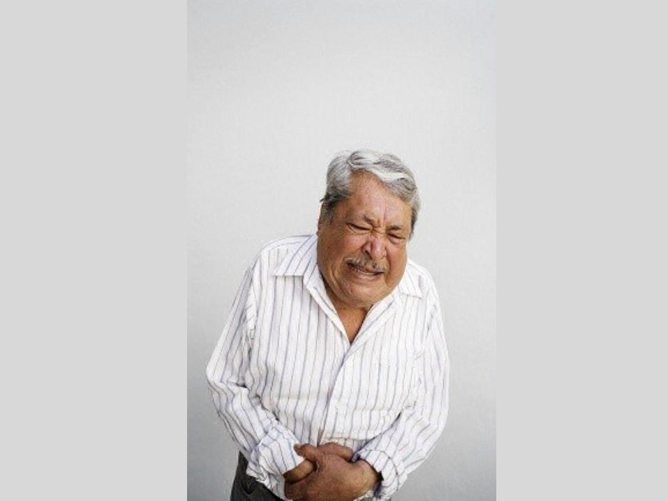
Case
History: • 50 yo male complains of periumbilical and left
lower quadrant abdominal pain that began earlier that day.
• Intermittent and crampy pain, accompanied by anorexia and vomiting
• Normal BM yesterday• No History of this pain has had prior abdominal
surgery

…continued
Physical Exam: • Afebrile• Moderate distress due to his abdominal pain• Bowel sounds present• Abdomen: mildly distended with periumbilical
tenderness but no rebound

DDx


Small Bowel Obstruction

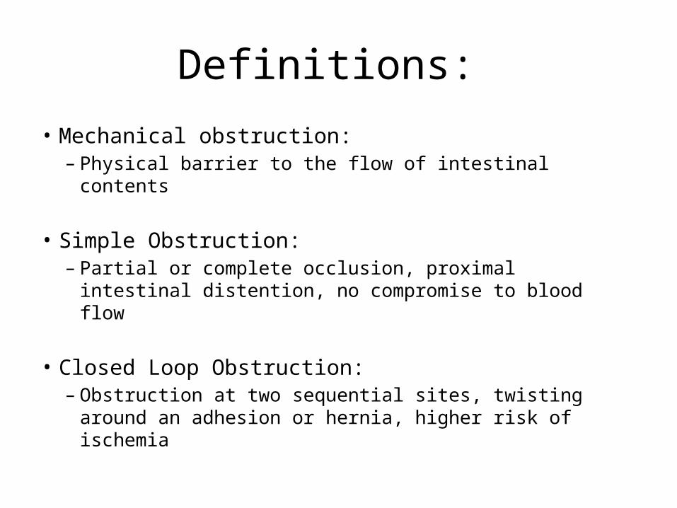
Definitions:
• Mechanical obstruction: – Physical barrier to the flow of intestinal contents
• Simple Obstruction: – Partial or complete occlusion, proximal intestinal
distention, no compromise to blood flow
• Closed Loop Obstruction: – Obstruction at two sequential sites, twisting around an
adhesion or hernia, higher risk of ischemia
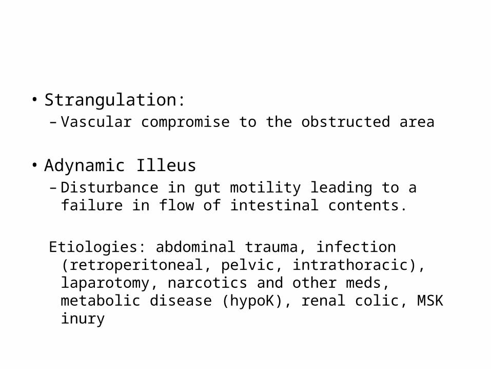
• Strangulation: – Vascular compromise to the obstructed area
• Adynamic Illeus– Disturbance in gut motility leading to a failure in flow
of intestinal contents.
Etiologies: abdominal trauma, infection (retroperitoneal, pelvic, intrathoracic), laparotomy, narcotics and other meds, metabolic disease (hypoK), renal colic, MSK inury

Etiology• Extraluminal Causes (Most common)
– Adhesions• Post pelvic surgery, appendectomy, colorectal surgery
– Hernia– Cancer
• Intrinsic Causes: – Congenital (stenosis, atresia) – Neoplasm– Infection from chrones/colitis– Intuscception
• Intraluminal Causes: – Gallstones– Foreign body– Barium – Cancer
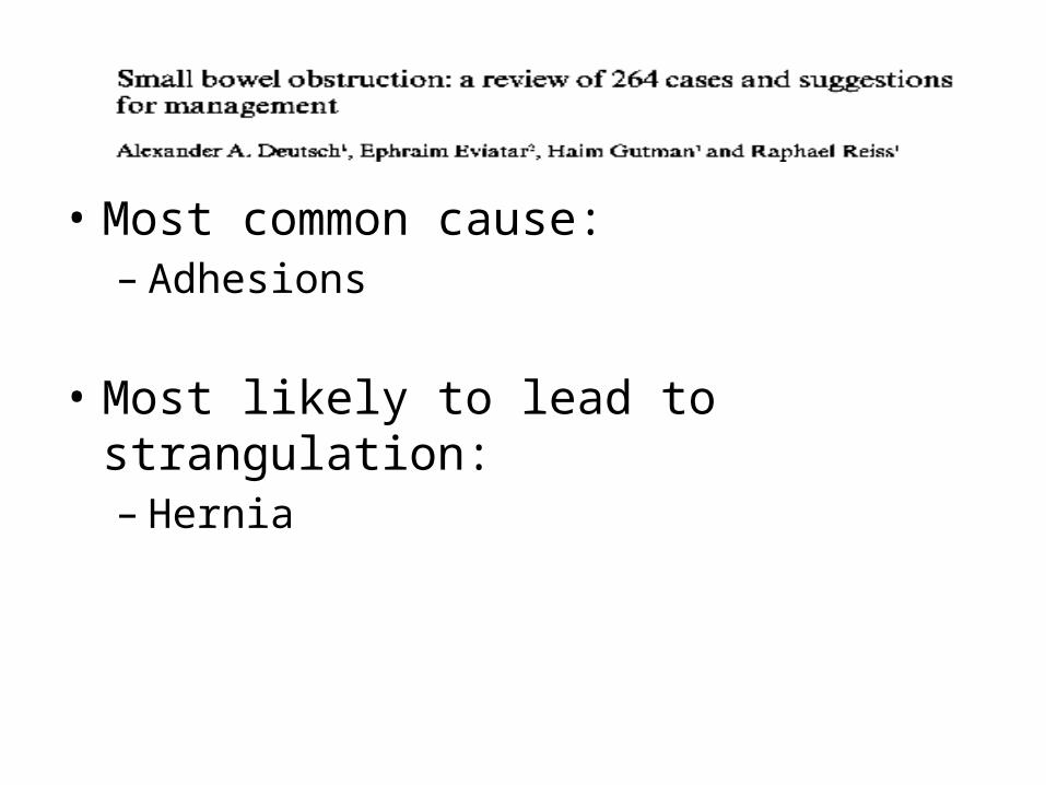
• Most common cause: – Adhesions
• Most likely to lead to strangulation: – Hernia

Pathophysiology

Clinically
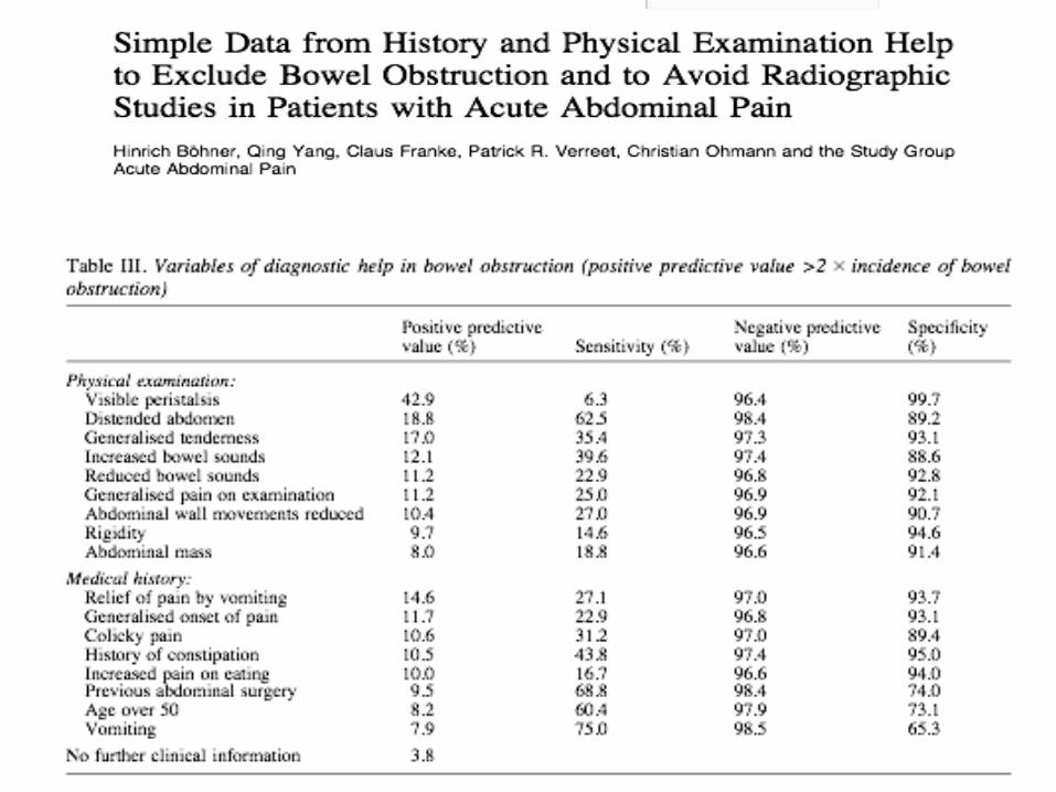
• History: – Colicky abdominal pain q4-5minutes– Abdominal pain is worse with a proximal
obstruction– Nausea and vomiting– Later: obstipation and constipation– Be aware of the pain that changes from
intermittent and colicky to constant and severe: intestinal ischemia and perforation

• Physical Exam: – Inspection: surgical scars, distended hernia,
distended abdomen, peristalsis– Auscultation: early: you may hear high pitched
bowel sounds, later you may hear no bowel sounds
– Percussion: Tympany– Palpation: Masses– Look for any peritoneal signs


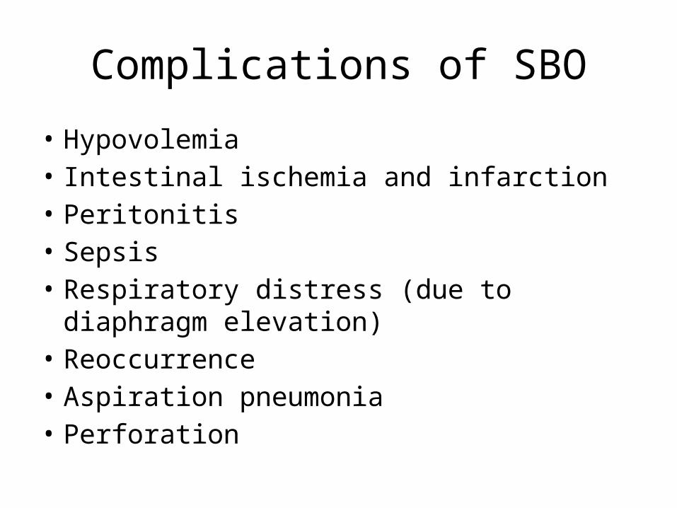
Complications of SBO
• Hypovolemia• Intestinal ischemia and infarction• Peritonitis• Sepsis• Respiratory distress (due to diaphragm elevation) • Reoccurrence• Aspiration pneumonia• Perforation

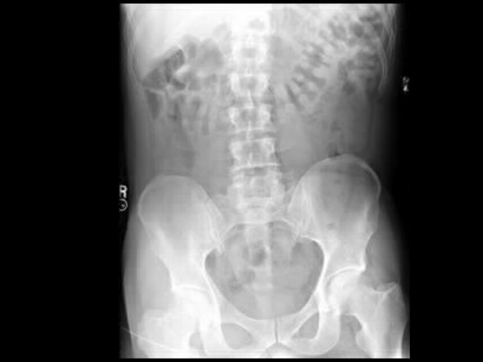
Imaging

Plain Films• 1. Normal small bowel gas pattern:
– Absence of small bowel gas or small amounts of gas with up to four variably shaped non-distended loops of small bowel (less than 2.5 cm in diameter)
• 2. Abnormal but non-specific gas: – One loop of borderline or mildly distended small bowel (2.5 to 3 cm), with
three or more air-fluid levels.– Normal colonic gas pattern
• 3. Probable SBO: – Multiple gas or fluid filled loops of dilated small bowel with a moderate
amount of colonic gas
• 4. Definite SBO: – Dilated gas or fluid filled loops of small bowel in the setting of a gasless colon

Supine


Limitations to Abdominal Radiography
• Negative and non-specific illeus patterns do not exclude the diagnosis– Can be too early thus the colon size and small
bowel size are similar– Can be too proximal and thus only a small
segment is dilated– Can be too fluid filled to see dilation
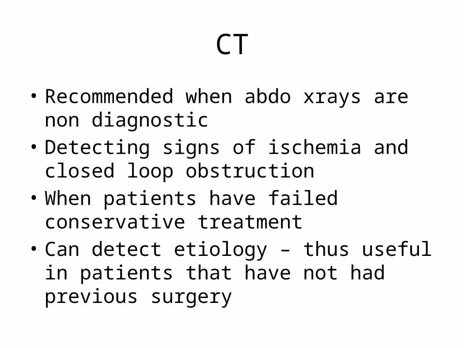
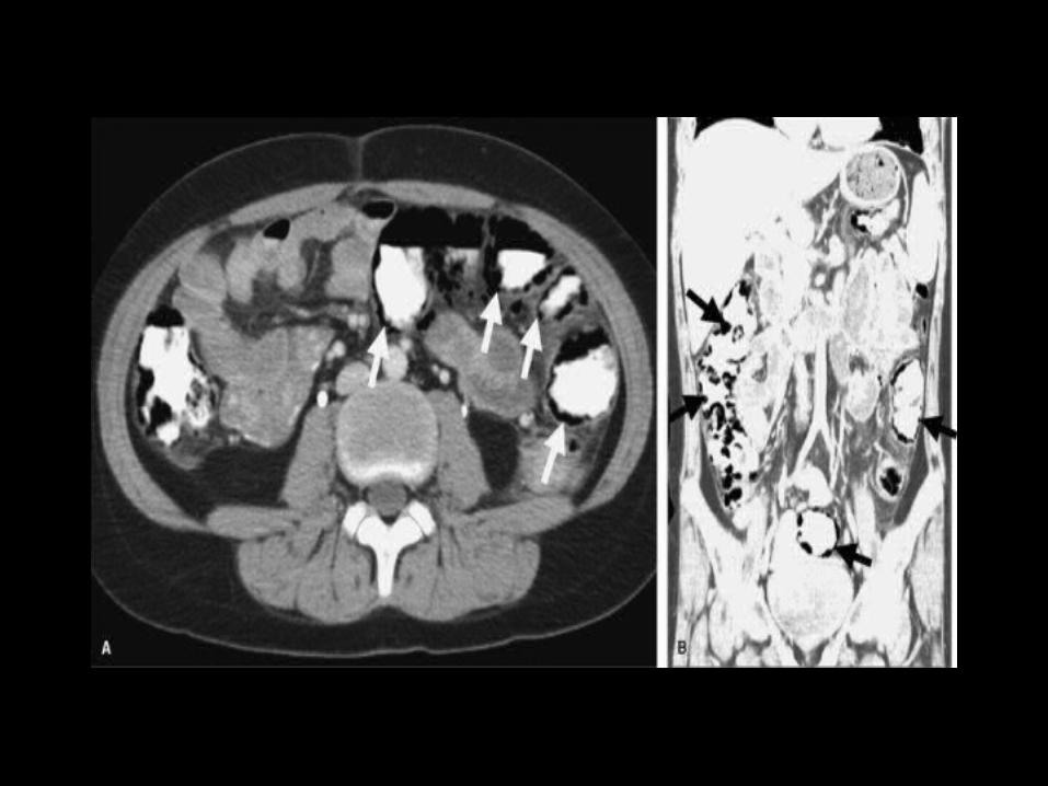
CT
• Recommended when abdo xrays are non diagnostic
• Detecting signs of ischemia and closed loop obstruction
• When patients have failed conservative treatment
• Can detect etiology – thus useful in patients that have not had previous surgery

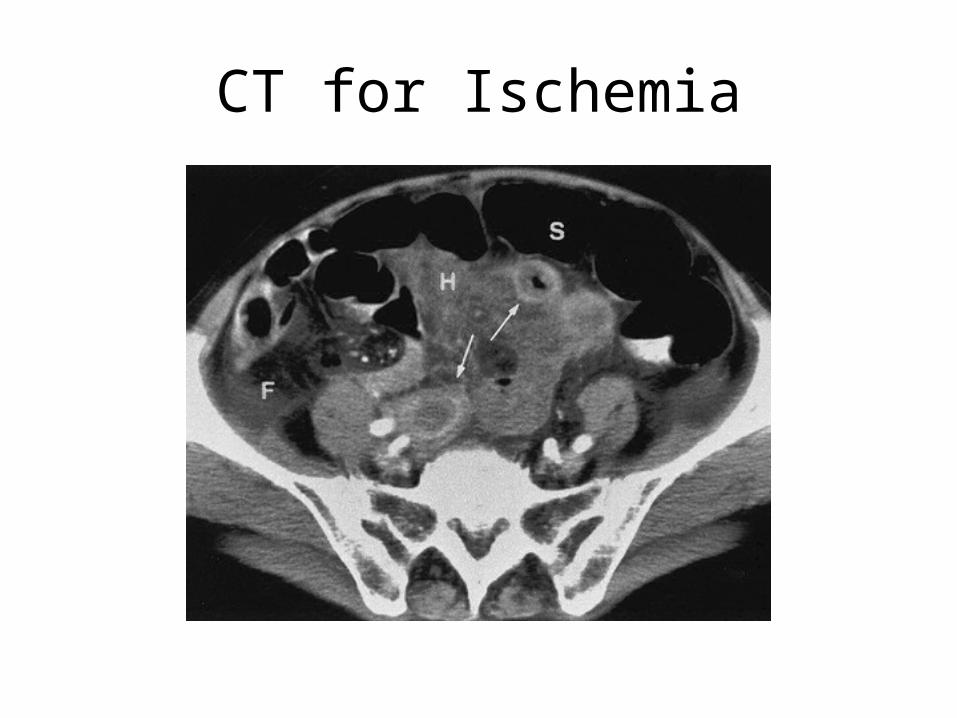
CT for Ischemia

Ultrasound
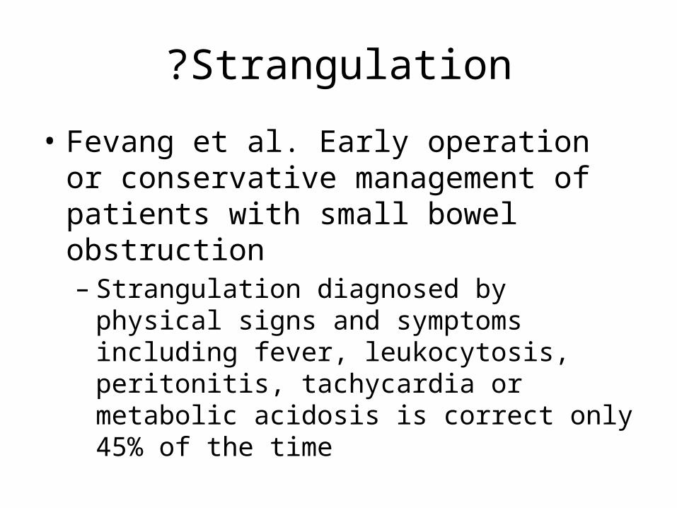
?Strangulation
• Fevang et al. Early operation or conservative management of patients with small bowel obstruction– Strangulation diagnosed by physical signs and
symptoms including fever, leukocytosis, peritonitis, tachycardia or metabolic acidosis is correct only 45% of the time
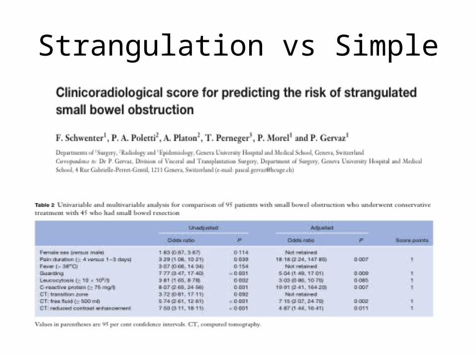
Strangulation vs Simple


• On univariate analysis other factors that made one think of strangulation were:
• Hypotension• Acidosis • Elevated BUN
• But when put in multivariate analysis this was not proven

Management

Reoccurrence
• There is about a 50% reoccurrence rate after the first small bowel obstruction– Gowen GF, 2003
• There is an 81% reoccurrence rate after 4 obstructive episodes– Fevang et al., 2004

Case 2


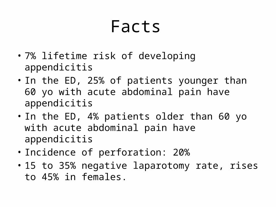
Facts
• 7% lifetime risk of developing appendicitis• In the ED, 25% of patients younger than 60 yo
with acute abdominal pain have appendicitis• In the ED, 4% patients older than 60 yo with
acute abdominal pain have appendicitis• Incidence of perforation: 20%• 15 to 35% negative laparotomy rate, rises to
45% in females.
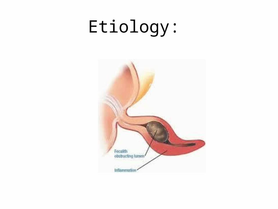
Etiology:

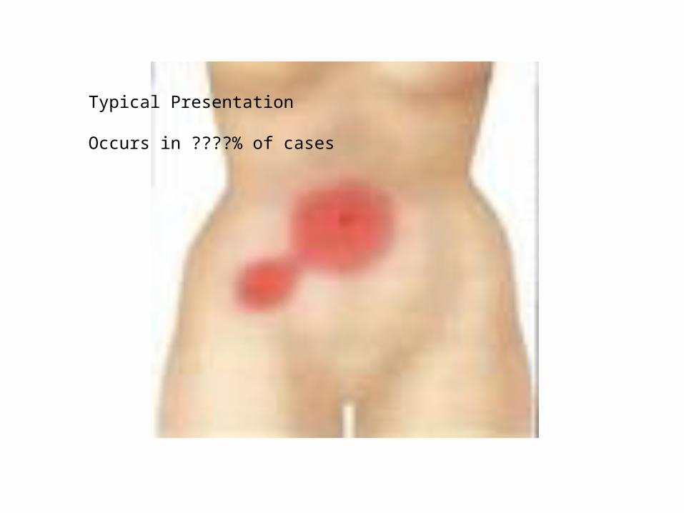
Typical Presentation
Occurs in ????% of cases

• Three Findings With a high positive likelihood ratio
– RLQ pain: • Sensitivity: 81%• Specificity: 53%• LR+: 7.31, LR-: 0.20
– Rigidity: • Sensitivity: 27%• Specificity: 83%• LR+: 3.76, LR-: 0.82
- Migration: Sensitivity: 64%Specificity: 82%LR+: 3.18, LR-: 0.50

…Ruling out appendicitis?
• Signs with Powerful Negative Likelihood Ratios: – Absence of RLQ pain
• LR-: 0.20
– Presence of similar previous pain• LR-: 0.50
– Lack of migration of pain• LR-: 0.50

Other SymptomsSymptom Sensitivity Specificity LR+
Pain before vomiting
100% 64% 2.76
Fever 67% 79% 1.94
Anorexia 68% 36% 1.27
Vomiting 51% 45% 0.92
Nausea 58% 37% 0.69-1.20

Other Signs…
Sign Sensitivity Specificity LR+
Rebound tenderness
63% 69% 1.10 to 6.30
Guarding 74% 57% 1.65 to 1.78
Rectal tenderness 41% 77% 0.83 to 5.34
Psoas sign 16% 95% 2.38

McBurney’s Point

Rovsing Sign
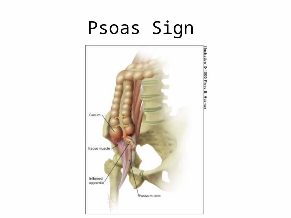
Psoas Sign

Obturator Sign


Suspected Appendicitis
1-4 5-6 7-10
Observation/ Investigation
Alvarado Score
Discharge Surgical consult
The use of the Alvarado score in the management of right lower quadrant abdominal pain in the adult Y. Pouget-Baudry et al. 2010
PPV = 61.82%, NPV = 79.21%;
PPV = 89.16%, NPV = 41.33%
Sensitivity: 92.77%Specificity: 58.18%

…but the WBC is normal…they can’t have appendicitis?

Imaging
• In 50 to 60% of patients the diagnosis of appendicitis can be made clinically
• Alvarado score 4-6 ….you can wait and watch, or image

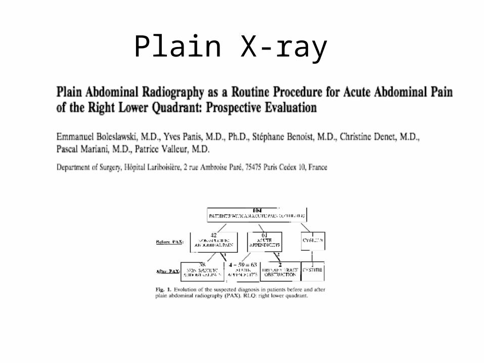
Plain X-ray
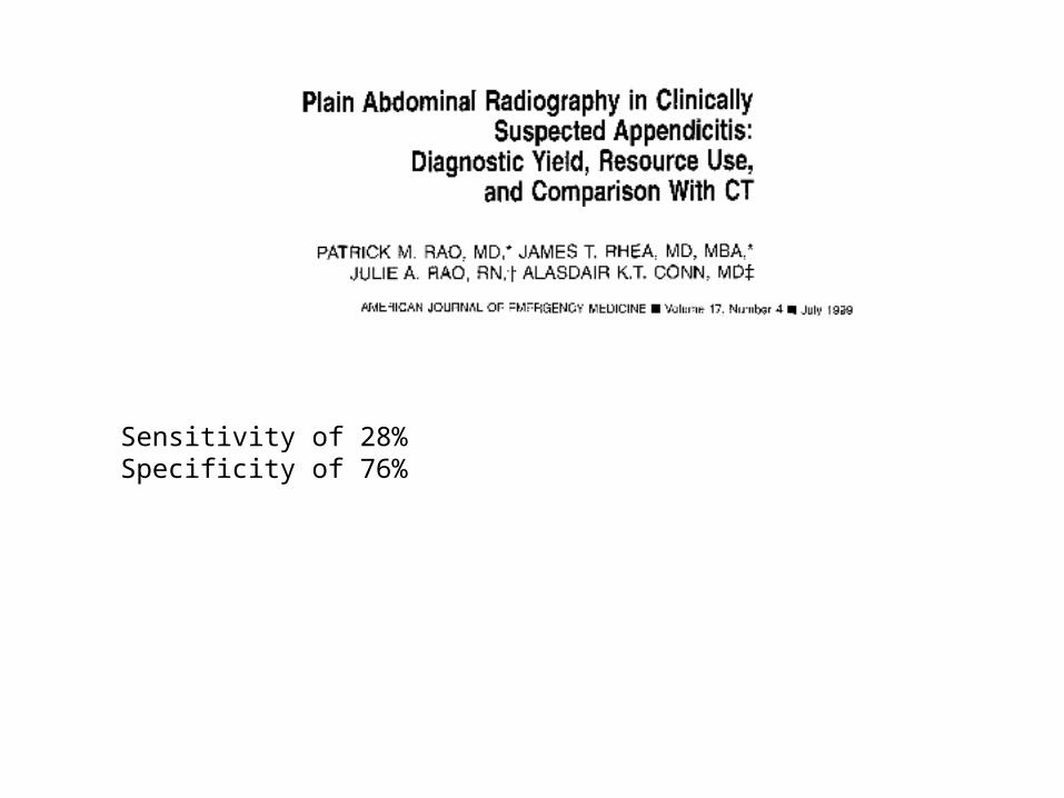
Sensitivity of 28%Specificity of 76%

Ultrasound
• Used to help confirm the diagnosis of suspected appendicitis
• Sensitivity: 86%• Specificity: 81%

CT
• Used to help confirm the diagnosis of suspected appendicitis
• Sensitivity: 95%• Specificity: 94%

Benefit of imaging

Perforation
Treatment
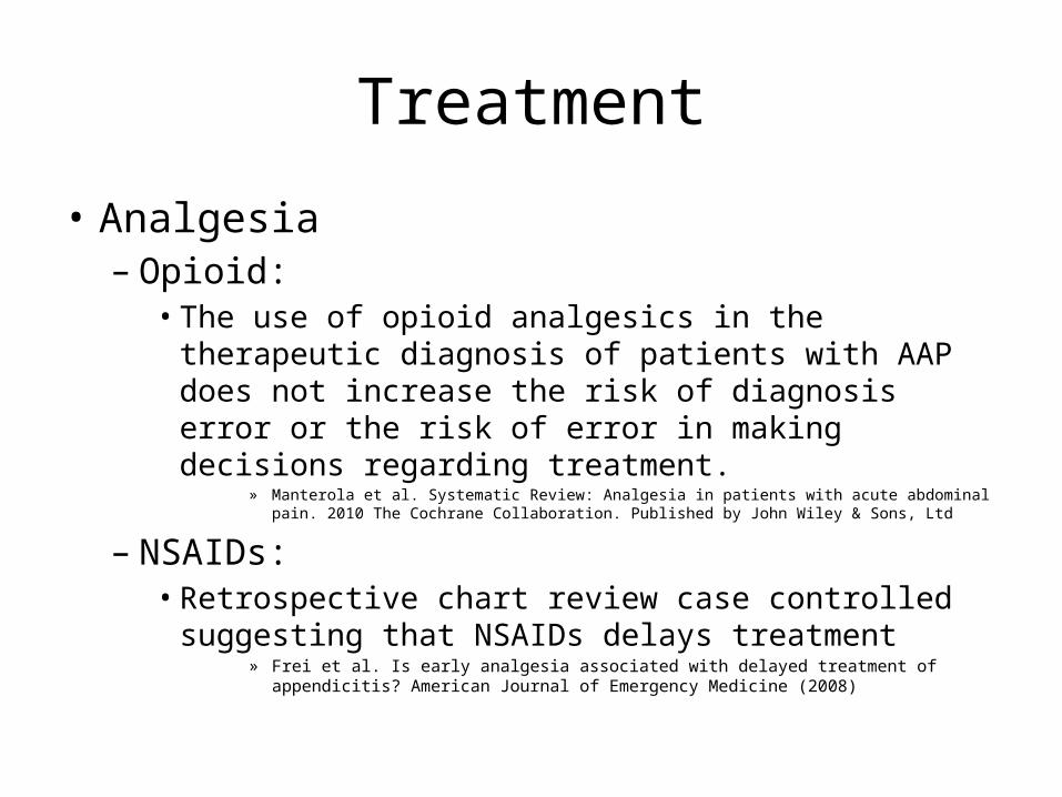
• Analgesia– Opioid:
• The use of opioid analgesics in the therapeutic diagnosis of patients with AAP does not increase the risk of diagnosis error or the risk of error in making decisions regarding treatment.
» Manterola et al. Systematic Review: Analgesia in patients with acute abdominal pain. 2010 The Cochrane Collaboration. Published by John Wiley & Sons, Ltd
– NSAIDs: • Retrospective chart review case controlled suggesting
that NSAIDs delays treatment» Frei et al. Is early analgesia associated with delayed treatment of appendicitis? American
Journal of Emergency Medicine (2008)

• IV Fluid• Perioperative Antibiotics:

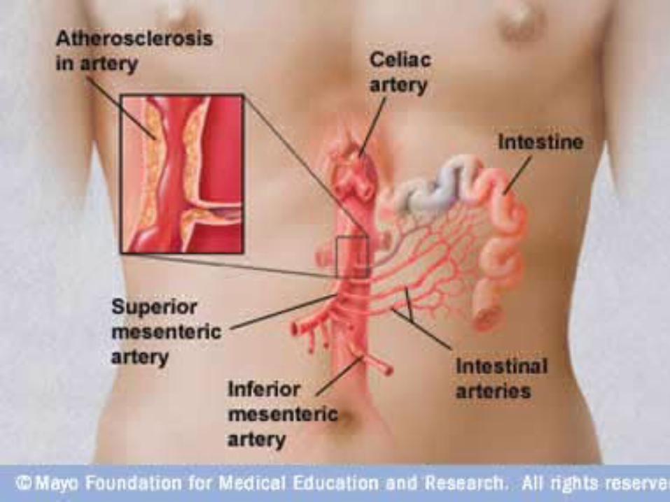


High Index of suspicion required

Clinical
• Need a high index of suspicion• Ischemia of the viscera: leading to pain out of proportion
with findings
– Abdominal pain: 83%– Vomiting: 44%– Diarrhea: 19.3%– GI bleeding: 20.1%
• Infarction
Huang et al. Clinical Factors and Outcomes in Patients with Acute Mesenteric Ischemia in the Emergency Department. July 2003: Acad Emerg Med.
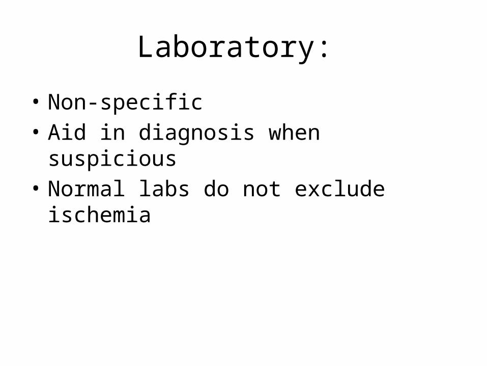
Laboratory:
• Non-specific• Aid in diagnosis when suspicious• Normal labs do not exclude ischemia


Other Tests…
• LDH– Sensitivity 70%, Specificity 42%, LR+ 1.2, LR- 0.7
• Lactate– Sensitivity 90%, Specificity 44%, NPV 96%, PPV 70
• Alpha-GST– Sensitivity 72%, Specificity 77%, NPV 86%, PPV 58%
• ALP (marker of intestinal mucosal ischemia)– Sensitivity: 80%, Specificity: 64%, LR+ 2.2, LR – 0.3

Predictors of mortality
• Bandemia 68.9% sensitive, 74.2% specific• Elevated AST 62.1% sensitive, 78.9.% specific• Elevated BUN 88.5% sensitive, 39.3% specific• Metabolic acidosis: 53.6% sensitive, 85.5.%
specific
Huang et al. Clinical Factors and Outcomes in Patients with Acute Mesenteric Ischemia in the Emergency Department. July 2003: Acad Emerg Med.


Management
• 1. Stabilize the patient• 2. Antibiotics
– Evidence that survival improved • 3. Heparin• 4. Vasodilators
– ? Glucagon• 5. Papaverine• 6. Surgery

Glucagon
• Vasodilator• Intestinal vasodilator and hypotonicity to
reduce oxygen demand• Used if no evidence of peritonitis
• Studies in rat’s and dogs have shown improved survival
• No studies in humans

Papaverine
• Phosphodiesterase inhibitor• Improves mesenteric blood flow• Arterial embolic disease or non-occlusive
disease• Intra-arterial (60 mg bolus and then 60mg/h
infusion) • Survival improvement by 20 to 50%


Asymptomatic Diverticulosis
• CT scan finds incidental diverculosis
• Should we do anything? – Inverse association between dietary fiber intake
and the risk of subsequently developing clinically evident diverticular disease


Symptomatic Uncomplicated Diverticulitis
• History: – LLQ abdominal pain– Better with defecation– Worse with eating– No rebound– No guarding

Symptomatic Diverticuli
• History: – Low grade fever– Left lower quadrant pain– Colonic dysfunction (bloating, constipation, diarrhea, mucous per
rectum) – Signs of obstruction– Signs of colovescial fistula
• Physical Exam: – Localized tenderness in the LLQ – Guarding and reboud– Palpable mss
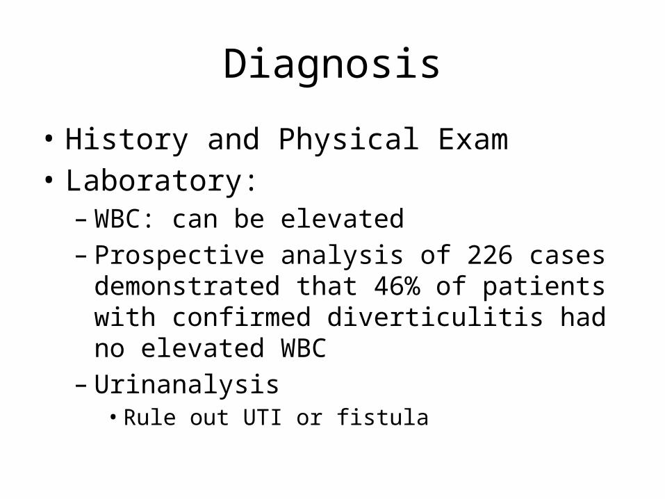
Diagnosis
• History and Physical Exam• Laboratory:
– WBC: can be elevated– Prospective analysis of 226 cases demonstrated
that 46% of patients with confirmed diverticulitis had no elevated WBC
– Urinanalysis• Rule out UTI or fistula

To image or not to image
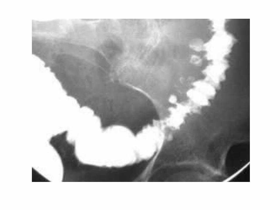

CT
• Sensitivity: 93-98%• Specificity: 77%• Water soluble contrast orally and IV
• Pro’s:– Therapeutic: percutaneous drainage of abscess (if >4cm)– Determine alternate pathology – Identify complicated diverticulitis

Ultrasonography
• Sensitivity: 84% to 98%• Specificity: 90 to 93%
• Pros: – Avoids radiation– Gyne structures are seen
• Cons: – Patients often acutely tender here compression by probe is
uncomfortable– Cannot identify perforation/air– Obese patient or overlying gas

Endoscopy and MRI:
• Not in the ER

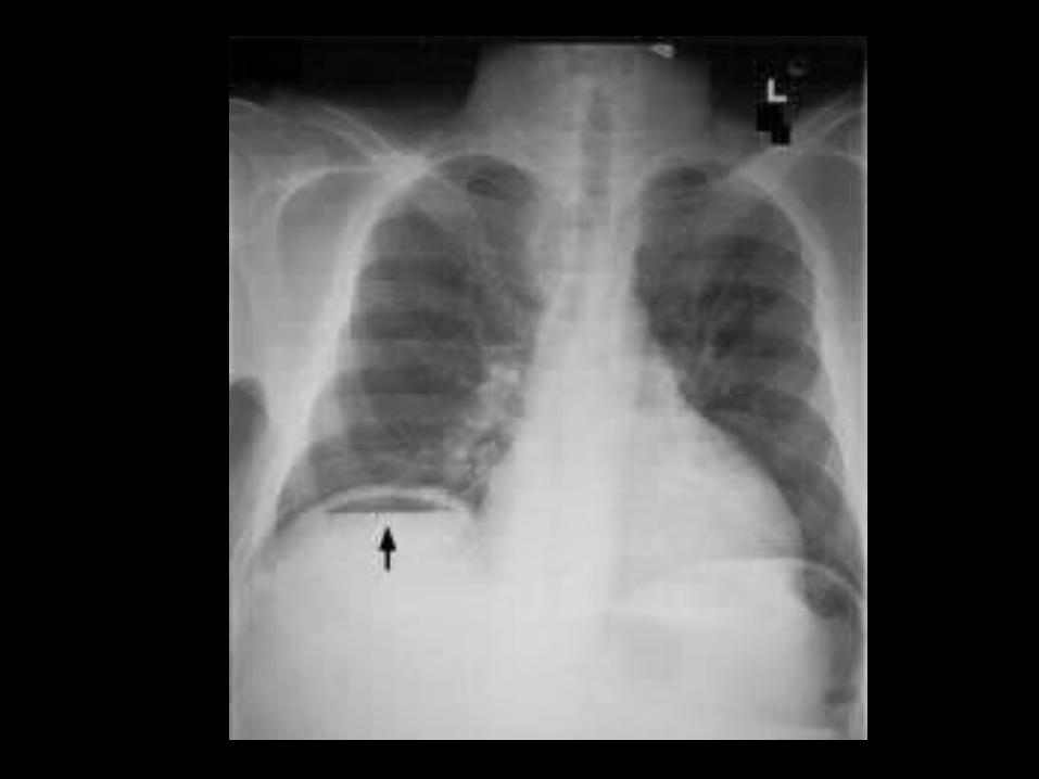
Complicated Diverticulitis
• Abscess (15%)• Obstruction (10%)• Free Perforation (1%)• Fistula (2%)
• Diverticular hemorrhage

Management
• Symptomatic uncomplicated diverticulitis

Versus…no antibiotics
• Controversy• Most studies showing symptomatic and
complication rates benefit from antibiotics versus just bran

Acute Diverticulitis
• Outpatient– Mild symptoms– No peritonitis– Able to tolerate a clear liquid diet – Close follow-up– Return to ED: increasing pain, fever, inability to
tolerate oral fluids
Acute Diverticulitis
• Inpatients: – Elderly– Immunocompromised– Severe comorbidities– High fever– Significant leukocytosis

AntibioticsRosen’s BOX 93-5 INTRAVENOUS ANTIBIOTIC COVERAGE FOR BOWEL FLORA
Mild to Moderate Infection– Ticarcillin-clavulanate, 3.1 g IV q6h – Ampicillin-sulbactam, 3 g IV q6h or– Ciprofloxacin, 400 mg IV q12h, and metronidazole, 1 g IV q12h
Severe Infection • Ampicillin, 2 g IV q6h, and metronidazole, 500 mg IV q6h, and
(gentamicin, 7 mg/kg q24h, or ciprofloxacin) 400 mg IV q12h or • Imipenem, 500 mg IV q6h
Surgery
• Emergency: perforation with peritonitis• Non-emergency: fistula, stricture, • Elective:
– Recurrent episodes (greater than 2) – Younger than 40 yo (more likely to have severe
disease) – Initial attack and immunocompromised

Reoccurrence Risk
• Reoccurrence rate varies from 7 to 45% and reoccur within a year


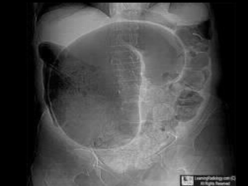
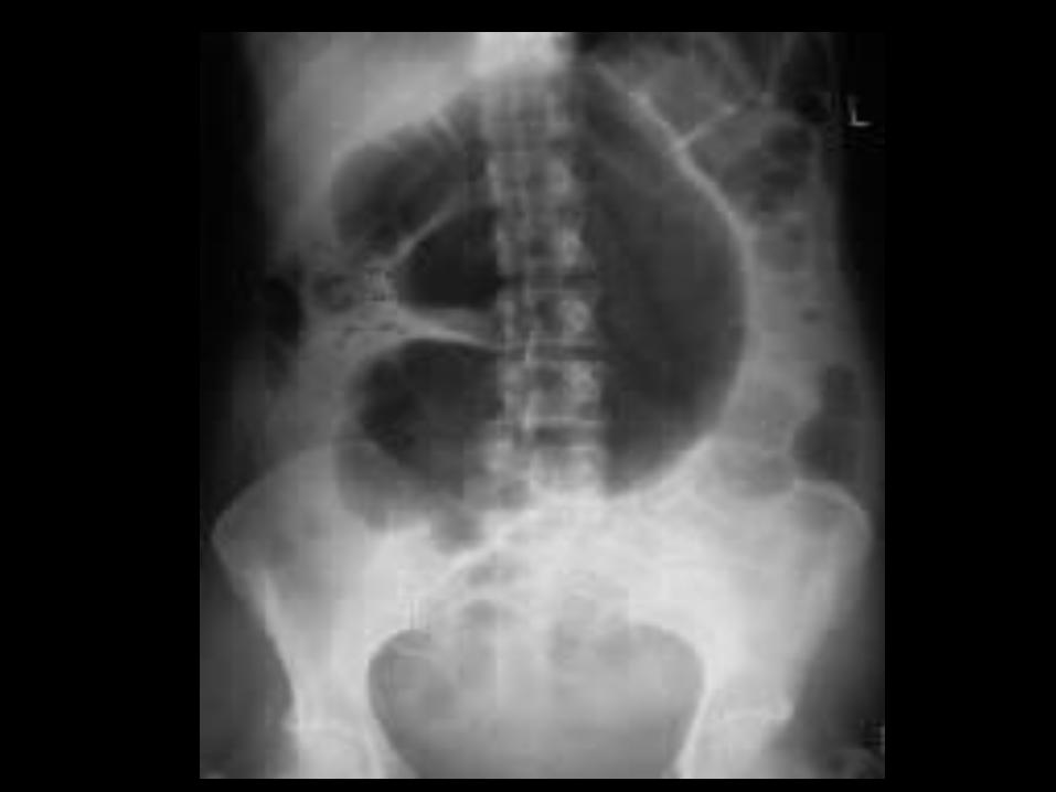
Sigmoid Volvulus• 859 patients with sigmoid volvulus
– 83% were males– 17% were females (of which 6.3% were pregnant) – Mean symptom duration of 39.4 hours– Clinical Triad highly suggestive of SV: Abdominal pain
(98.7%), asymmetric abdominal distention (96%), obstipation (92.3%)


![Welcome [weillcornellbrainandspine.org] · Maricruz Rivera, MD, PhD PGY-3. Neurological Surgery Residents. Evan Bander, MD PGY-5 Alexander D. Ramos, MD, PhD PGY-5 Joseph Carnevale,](https://static.fdocuments.net/doc/165x107/5f7167444c714e55d46f024a/welcome-weill-maricruz-rivera-md-phd-pgy-3-neurological-surgery-residents.jpg)
