Bowel Obstruction - GI Diseases, Complications and … · Bowel Obstruction Faculty of Medicine ,...
61
Bowel Obstruction Faculty of Medicine , University of British Columbia. Department of Surgery Division of General Surgery Photography and text by D.B. Allardyce MD, FRCS. Research and technical assistance by Ryan Janicki Medicine 2006
Transcript of Bowel Obstruction - GI Diseases, Complications and … · Bowel Obstruction Faculty of Medicine ,...

Bowel Obstruction
Faculty of Medicine , University of British Columbia.Department of Surgery
Division of General Surgery
Photography and text by D.B. Allardyce MD, FRCS.
Research and technical assistance by Ryan Janicki Medicine 2006

Objectives
Presumed background knowledge
Able to describe the gross anatomy of the abdominal wall andintrabdominal viscera.
Able to describe the motility, absorptive and secretoryfunctions of the small and large bowel.
Able to describe the gross and microscopic appearance of [a]adenocarcinoma of the colon [b]Crohns disease of the smallbowel.
Knowledge to be acquired
Able to classify causes of bowel obstruction under thesubheadings of “acute onset” and ” gradual onset”
Define the terms incarceration and strangulation
Have a knowledge of the usual history of these conditions; takea history from the patient, using direct questions whereappropriate. The questions should reflect an awareness of otherpossible diagnoses.
Examine a patient with a possible bowel obstruction, identifyingthe signs volume depletion when present. Examine theabdomen with a focus which reflects knowledge of the possiblecauses of the bowel obstruction, the complications ofobstruction, and the alternative diagnoses.
Be able to write orders for the management of a patient withrecent onset obstruction.
Describe, in general terms, how you would approach apatientwith the following problems.[include a basic description of theoperation or procedure] Use proper names for the operativeprocedure eg. ”Right Hemicolectomy” not “take out the rightcolon.
2

a. complete sbo due to adhesionsb. complete sbo due to incarcerated groin hernia.c. advanced Crohns stricture of the ileum, with malnutrition and
anemia.d. a sigmoid volvulus, with marked abdominal distention, pain,
and repiratory distress.e. complete rectosigmoid obstruction by a carcinoma
3

Introduction – Bowel Obstruction
This presentation is confined to the topic of intestinal obstruction inadults. It reviews the etiology and management of obstructionarising between the duodenal-jejunal flexure (ligament of Trietz) andthe rectum. Obstructions in the duodenum or more proximal are notaddressed.From the standpoint of etiology and management strategies smallbowel obstruction and large bowel obstruction require quitedistinctive and separate approaches in most instances. It istherefore useful to distinguish if the obstruction is in the small bowelor large bowel when approaching a patient who appears on clinicalgrounds to possibly have a bowel obstruction.
Background Information – Bowel Obstruction
I. Introduction to Small and Large Bowel Obstruction
4

Small bowel obstruction (SBO) is more common than obstructionin the large bowel largely because of the great frequency ofobstruction caused by postoperative adhesions and external hernias.The latter are rarely a cause of obstruction in the large bowel. Mostreviews would indicate SBO makes up about 80% of the cases ofintestinal obstruction between the ligament of Trietz and the rectum.The most common cause of SBO in the industrialized world is postoperative adhesions. All patients undergoing open abdominalsurgery will develop some degree of an adhesive process early in thepost operative period. The extensiveness of this process is relatedto the degree of peritoneal contamination, extent of dissection andother factors. Peritoneal adhesions develop quickly following surgeryand become very intense in the following few weeks. Gradually overa period of 2 to 3 months the process softens and reverses to someextent as the adhesions are remodeled.
SBO is a challenging clinical problem. Herniation and strangulationof intestine is often involved in the obstructive process.Strangulation is usually the result of either internal hernia through adefect in the peritoneal cavity or external herniation through one ofmany common sites. External herniation with incarceration ofviscera is usually readily identified. Internal herniation, however, ismuch less easily detected. Delay in interventions with eventualinfarction of the involved bowel necessitating resection forms thebasis for most mortalities related to SBO. If an operativeintervention with SBO occurs early enough to avoid resection,morality rates are less than 5%.
Large bowel obstruction (LBO) is less common than SBO.Adhesive bands and external hernias rarely capture the colon. Thecolon, in a significant portion of its course is retroperitoneal andtherefore seldom accessible to external hernia or any internal orificecreated by adhesive bands. The transverse colon is suspended by amesentery fused to the greater curve of the stomach by thegastrocolic omentum and is not likely to be captured by adhesivebands. Rarely, the sigmoid colon or transverse colon maybe caughtin an inguinal or umbilical hernia.
5

Mortality due to LBO is often related to intra-abdominal and woundsepsis. Carcinoma of the colon is the commonest cause of LBO andthe lesions causing this usually arise in the sigmoid or rectosigmoidarea. In this area of the colon tumors are sclerotic and stricturing.The increased consistency of the fecal stream and the narrowerlumen makes this site the most common point for colonicobstruction.
Although strangulation is a great risk in complete SBO, it is seldom afeature of colonic obstruction except for rare cases of cecal andsigmoidal volvulus.
Fluid and electrolyte disorder with sequestration of significantcomponents of the extracellular fluid is not usually a feature of LBO.Gaseous distention of the colon with marked colonic distention andrisk of cecal rupture is the usual clinical concern . Septiccomplications may follow operative interventions with attempts atanastomosis, stoma formation or occasionally the preoperativeoccurrence of colonic rupture due to distention. In contrast to the surgical approach to the small bowel it is seldomacceptable to resect obstructing lesions or segments which haveundergone volvulus and do a primary anastomosis. This isparticularly true when the lesion is encountered in the left side of thecolon. Many patients who develop LBO are elderly and suffer fromcomorbid illness. Surgical approaches that feature a stagedapproach may have a better outcome.
II. Acute onset SBO
Adhesive Bands
An acute onset SBO is one of the most common reasons foremergency admissions for abdominal pain. Many of theseobstructions are of sudden onset and most are on the basis ofprevious operative interventions and residual adhesive bands.Adhesive bands that are in the lower abdomen, in the infracoliccompartment, and are solitary pose the greatest risk for SBO.Passages between loops of bowel or a loop of bowel and theomentum form sites for potential internal hernias. Obstruction
6
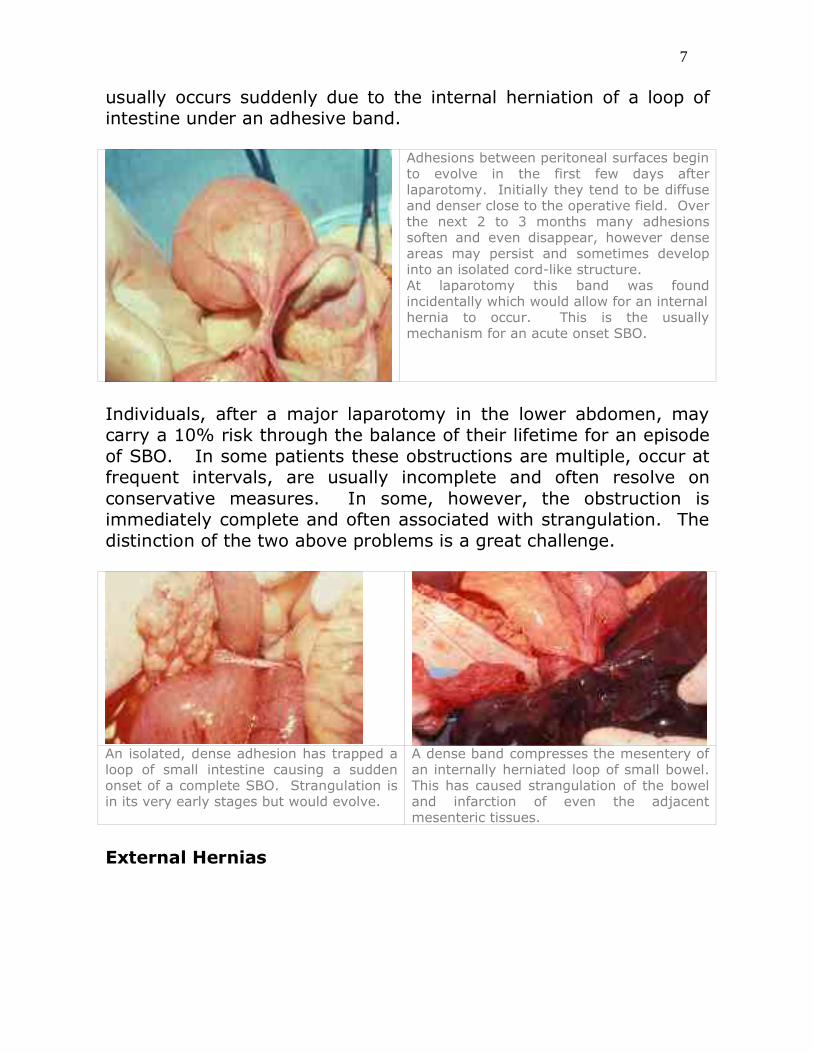
usually occurs suddenly due to the internal herniation of a loop ofintestine under an adhesive band.
Adhesions between peritoneal surfaces beginto evolve in the first few days afterlaparotomy. Initially they tend to be diffuseand denser close to the operative field. Overthe next 2 to 3 months many adhesionssoften and even disappear, however denseareas may persist and sometimes developinto an isolated cord-like structure.At laparotomy this band was foundincidentally which would allow for an internalhernia to occur. This is the usuallymechanism for an acute onset SBO.
Individuals, after a major laparotomy in the lower abdomen, maycarry a 10% risk through the balance of their lifetime for an episodeof SBO. In some patients these obstructions are multiple, occur atfrequent intervals, are usually incomplete and often resolve onconservative measures. In some, however, the obstruction isimmediately complete and often associated with strangulation. Thedistinction of the two above problems is a great challenge.
An isolated, dense adhesion has trapped aloop of small intestine causing a suddenonset of a complete SBO. Strangulation isin its very early stages but would evolve.
A dense band compresses the mesentery ofan internally herniated loop of small bowel.This has caused strangulation of the boweland infarction of even the adjacentmesenteric tissues.
External Hernias
7

An external hernia causing acute SBO often provides a greatopportunity for a prompt diagnosis of a complete obstruction,strangulation and an early operative intervention. Unlike internalherniation due to adhesions in the abdomen, the external hernia isusually quite evident on the physical examination. Sometimes anincarcerated external hernia maybe quite small and carefulexamination of the umbilical, femoral or inguinal areas are requiredto detect it. This is especially true in obese individuals.
In this patient with a history of recent onset ofabdominal cramps and vomiting, a tender massappeared in the right groin area. Onexamination the mass is very tense, wide basedand fixated. On gentle pressure there is noindication that it will reduce. It can bedemonstrated that the mass in fact below andlateral to the right pubic tubercle suggesting thatthis is in fact an incarcerated right femoralhernia.
If a hernia is trapped in an orifice and cannot be easily reduced intothe abdominal cavity on recumbency and analgesia then it isincarcerated. Most incarcerated hernias involving the intestinaltract contain the small intestine and in most instances a degree ofstrangulation is involved. The strangulation will begin to evolve andpursue an unpredictable course to infarction. It is rare for a loop ofintestine to be incarcerated in an external hernia and not exhibitsome degree of ischemia. This would be particularly true when theincarceration is of recent onset.
In this incarcerated hernia, which was operatedon electively, there was a mass of tissue, whichwas tender and caused periumbilical pain. Itcould not be reduced but had been present for alengthy period of time. When a hernia isincarcerated and cannot be reduced and thereare no symptoms or indications of bowelobstruction, the incarcerated tissue is usuallygreater omentum. The sac has been opened and viable omentumis caught in the hernia effectively plugging theopening and preventing access to the sac byany of the hollow viscera. Because of thecosmetic issues and localized pain,repair of thisincarcerated umbilical hernia was indicated.
8

Inguinal hernias are probably the most common external hernia totrap intestine. This is partly because inguinal hernias are the mostcommon hernia, by some margin. Umbilical hernias are probablysecond in frequency as a site of incarceration and femoral herniasthe least common site. The femoral orifice features rigid marginssuch as the lacunar ligament, Cooper’s ligament and the inguinalligament. Incarceration here is commonly associated withstrangulation.
Obturation by food, stones, foreign objects, etc.
Obstruction of the bowel lumen by a swallowed object, food materialor a gallstone should be suspected in patients with acute SBO and nohistory previous of abdominal surgery or identifiable externalhernias. In these patients there is almost never strangulation sincethe process involves a simple internal plugging of the bowel lumen.
When an acute onset SBO develops and there isno history of pervious surgery or external herniaunusual causes of SBO should be expected.Obturation by swallowed or other foreign bodiessuch as gallstones are one of the diagnosis thatshould be considered under these circumstances.In this patient there was an idiopathic stricture ofthe small intestine which was previouslyasymptomatic. Unfortunately the patient ingesteda large amount of vegetable matter with highroughage content, which was poorly masticatedand digested. This food bolus was held up at theprevious stricture point causing complete SBO.
Occasionally these obstructions may resolve if the obstructingmaterial is digestible. In many instances there is such aconsiderable mass of fibrous vegetable matter or the object isinorganic and too large to pass through the distal small bowel. Manyof these obstructions occur in the distal ileum where the caliber ofthe small bowel narrows.
9

This mentally handicapped individual unfortunatelyswallowed a small rubber ball; know popularly as a“squeeze ball”. He was not seen doing this and hisabdominal pain and repeated vomiting was ascribed toother causes. Eventually he aspirated and developedpneumonia and renal failure. Even though operationwas performed and the impacted ball removed, thepatient subsequently succumbed to the aspirationpneumonia.Aspiration pneumonia is a constant hazard in patientswith bowel obstruction and they are particularlyvulnerable during the perioperative period.
In elderly patients, who are known to have gallstone disease, astone obstructing the intestine should be considered. The finding ofair in the biliary tree on plain abdominal films or the demonstrationof an opaque gallstone usually in the right iliac fossa near theterminal ileum usually gives the diagnosis.
Obstruction of the small intestine due toswallowed food material is more likely to occurin the elderly, the edentulous and patients withprevious gastric surgery. In this elderly man,who was edentulous, complete bowelobstruction evolved for which there was noapparent cause. It would not resolve and atoperation a mass off impacted food material wasfound in the mid small intestine. This could notbe dislodged and the bowel was opened as seenin the lower picture. The obstruction was to dueto the swallowing of an intact shitakemushroom. Foreign bodies in the intestine should bedislodged form the site of impaction and moved,if possible, more proximally, where the intestineis more resilient with higher circulation, allowingfor safer enterotomy.
Small Bowel Volvulus
A volvulus occurs in the small bowel when the mesentery is long andattenuated. It is actually a rare and uncommon cause of SBO inadults. Strangulation usually evolves very quickly and the bowel willbe irreversibly infarcted soon after onset. Large sections of theintestine may be lost in the process.
10
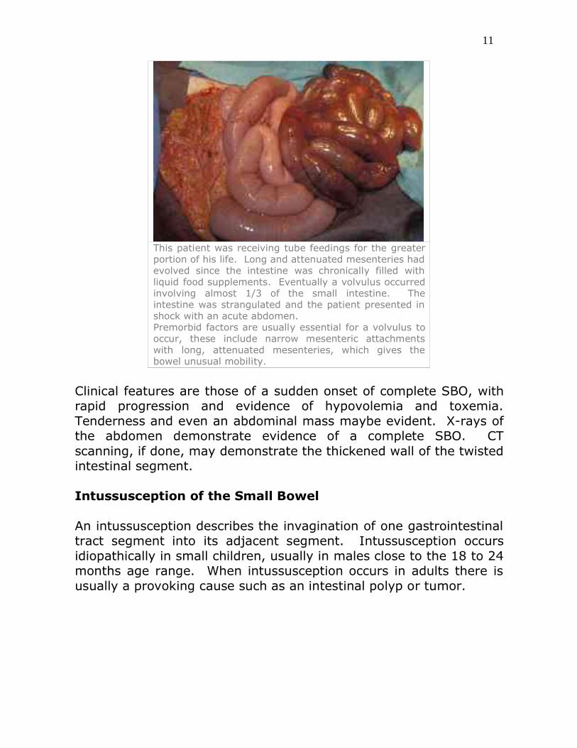
This patient was receiving tube feedings for the greaterportion of his life. Long and attenuated mesenteries hadevolved since the intestine was chronically filled withliquid food supplements. Eventually a volvulus occurredinvolving almost 1/3 of the small intestine. Theintestine was strangulated and the patient presented inshock with an acute abdomen. Premorbid factors are usually essential for a volvulus tooccur, these include narrow mesenteric attachmentswith long, attenuated mesenteries, which gives thebowel unusual mobility.
Clinical features are those of a sudden onset of complete SBO, withrapid progression and evidence of hypovolemia and toxemia.Tenderness and even an abdominal mass maybe evident. X-rays ofthe abdomen demonstrate evidence of a complete SBO. CTscanning, if done, may demonstrate the thickened wall of the twistedintestinal segment.
Intussusception of the Small Bowel
An intussusception describes the invagination of one gastrointestinaltract segment into its adjacent segment. Intussusception occursidiopathically in small children, usually in males close to the 18 to 24months age range. When intussusception occurs in adults there isusually a provoking cause such as an intestinal polyp or tumor.
11

In this patient who had AIDSand was known to have alymphoma, an acute SBOevolved. Ultrasoundexamination revealed acharacteristic double walledstructure suggestive ofintussusception. This wasindeed found during operation.Strangulation can occur duringintussusception due to thedragging of the mesentery intothe distal bowel
The outer layer of an intussusception is described as theintussuscipiens and the bowel to which it is passing in to it isdescribed as the intussusceptum. Often the intussusceptingintestine with its accompanying mesentery becomes ischemic andwill eventually infarct if it is not reduced. An intussusception canoccur in adults at almost any level of the intestinal tract and in someinstances it can be occurring at multiple levels simultaneously.
In this unusual case the patient had had alaparotomy and surgery for an in injury tothe liver due to blunt trauma. A bowelobstruction evolved postoperatively thatwas thought to be due to adhesions. Itbecame complete requiring re-operation atwhich time intussusception was found. It israre for an adult to develop intussusceptionwithout a precipitating tumor or otherreason. The intussusception was difficult toreduce due to thickening and scarring of theadjacent bowel. A short resection wasrequired.
A tumor of the small bowel that has a polypoid structure and growsinto the lumen of the bowel is most likely to cause intussusception.Carcinoid tumors because of their inflammatory reaction andstiffening of the surrounding bowel will seldom cause anintussusception. Adenomatous polyps however, whether benign ormalignant, will readily cause intussusception. Metastatic melanomaand metastatic cancer from undifferentiated lung lesions may alsoprovoke intussusceptions.
12

Unlike children, a underlying intestinal polyp ortumor almost always provokes intussusceptionin adults. In the patient with AIDS, shownabove, the provoking factor was a localizedlymphoma in the small bowel that acted as thehead of the intussusception. In this casestrangulation did not occur. Although thepatient was receiving chemotherapy, thissegment of small bowel was resected to avoidearly recurrence of the intussusception.
III. Gradual Onset SBO
Crohn’s disease
Patients with gradual onset SBO are usually encountered in adifferent setting than the recent onset obstruction patient. Oftenthey give histories, going back for weeks or months and present inambulatory care areas. There is often an opportunity forconsiderable investigation in order to sort out the diagnoses.
The most common cause of gradual onset obstruction of the smallbowel is Crohn’s disease. Although multiple levels of the intestinaltract may be involved, its propensity for involvement of the terminalileum usually leads to obstructive symptoms at this level.Symptoms of gradual SBO are very slow and progressive resulting ingradual weight loss, intermittent episodes of crampy abdominal pain,occasional vomiting and episodes of diarrhea. When the diseaseinvolves the terminal ileum an inflammatory mass may be palpablein right iliac fossa.
13

Crohn’s disease is the commonestcause of gradual onset SBO. It wouldbe suspected in a young patient with along history of weight loss, abdominalcramps, intermittent diarrhea,occasional distention and vomiting. Inthis patient a laparotomy revealed adense strictured area near theterminal ileum. Fat and inflammatorychanges can be seen under theserosa. The bowel is rubbery andfirm, much like a garden hose. In theupper right corner of the picture,normal proximal small intestine isevident.
The stricturing lesion of Crohn’s disease seldom ever progresses tocompletion and most episodes of high-grade obstruction can bemanaged conservatively at least for a short period of time.
Crohn’s disease almost never causes a complete SBO andalthough the lumen may be severely strictured there is always apassage. Conservative measures with intravenous therapy andoccasionally nasogastric decompression will usually result inresolution of Crohn’s obstructions. Impaction with food materialand even medications may occasionally provoke completeobstruction that resolves on conservative management.
Frequently, contrast studies, small bowel follow through, will show anarrowed segment in the ileum (“string sign”). Occasionally skiplesions may be seen with obstruction at a variety of levels.
Neoplasms of the Small Bowel
Tumors are increasing as an explanation for gradual onset SBO.
14

Primary tumors of the small bowel causing obstruction are quiterare. However, metastatic cancer in the peritoneal cavity spreadingfrom lesions in other levels of the intestinal tract or from the ovariesis commonly encountered.
In the older patient group, slowly evolvingsmall bowel obstruction suggests thepossibility of a neoplastic etiology. Primaryneoplasms of the small bowel are quite rare.Metastatic neoplasm to the small bowel;hematogenously are also rare. Secondaryspread of neoplasm in the peritoneal cavity iscommon however. In this picture anothercommon reason for a distal SBO of gradualonset is shown. A carcinoma of the cecum hasoccurred right near the ileocecal valve causingmassive distention and hypertrophy of theimmediate proximal ileum. The tumor istethered into the ileocecal valve; thedescending colon is collapsed and empty.
Transperitoneal metastases may implant on the serosa of the smallbowel and cause an obstructing, contracting, tumor mass thateventually results in obstruction. Occasionally pelvic tumor masses,as in ovarian cancer, may completely engulf the small bowel andcause obstruction by extrinsic pressure.
Patients presenting with episodes of cramping abdominal pain,abdominal distention, loud borborygmi, weight loss and who areunlikely to have inflammatory bowel disease should be suspected tohave a small bowel tumor causing there obstructive symptoms. .Anemia and occult blood in the stool may be present in thesepatients. Occasionally a palpable abdominal mass is found, buttumors of the small bowel are extremely mobile and particularlydifficult to identify consistently on physical examination. If there isextensive metastatic disease in the abdomen, ascites may developand a pelvic masses may be palpable on digital rectal examination.
Contrast studies and follow through of the small bowel maydemonstrate an obstructing lesion. CT scanning may identify anintra-abdominal mass.
Tumors of the small bowel are more likely to bleed or perforate thanthey are to obstruct. The bowel content is extremely fluid and many
15

of the lesions grow extrinsically outside the intestine rather thancompromising the lumen. Occasionally tumors of extraordinary sizemay arise in the small bowel and simply displace the intestinal tractinto the corners of the peritoneal cavity.
Smooth muscle tumors and hematogenous metastases to the small bowel frequentlywill not cause obstruction even though they reach very significant size. They tend togrow in the bowel wall, extending in a dumbbell fashion into peritoneum withoutencroaching significantly on the small bowel lumen. Very large masses can evolvewithout bowel obstruction. In this patient a metastatic lesion to the small bowel canbe seen, the bowel is collapsed and there is no bowel obstruction. The lesion has beencausing a GI bleed. A closer view of the tumor shows how the lumen of the bowel is preserved. The tumorhas ulcerated into the lumen of the bowel causing the lower GI bleed. A limitedresection was required to stop the continued loss of blood.
Of the primary tumors of the small bowel, adenocarcinomas arethe most likely to cause an obstructive pattern. If the lesion ispolypoid, intussusception may occur. Perforation and bleeding arealso more likely and commonly are features of metastases to thesmall bowel from lung cancer or melanoma.
16

This massive malignant tumor of the small boweldisplaced the intestine but did not cause obstruction.
Carcinoid tumors, when they are malignant, incite a surroundinginflammatory response with desmoplasia that may result in anobstructing lesion.
Of the primary small bowel tumors the carcinoid tumor ismost likely to cause obstruction due to encroachment on thelumen. As seen in this picture they cause a fibrous reactionsomewhat similar to an adenocarcinoma of the colon.Carcinoid tumors cause a localized stricturing of the bowelwall. GI bleeding from these lesions is uncommon.
Very rarely other benign and malignant lesions are located either inthe small bowel mesenteries or arise within the small bowel.Although obstruction may rarely occur, the vast majority of thesetumors are rapidly expanding and simply displace the bowel withoutcausing obstruction.
17
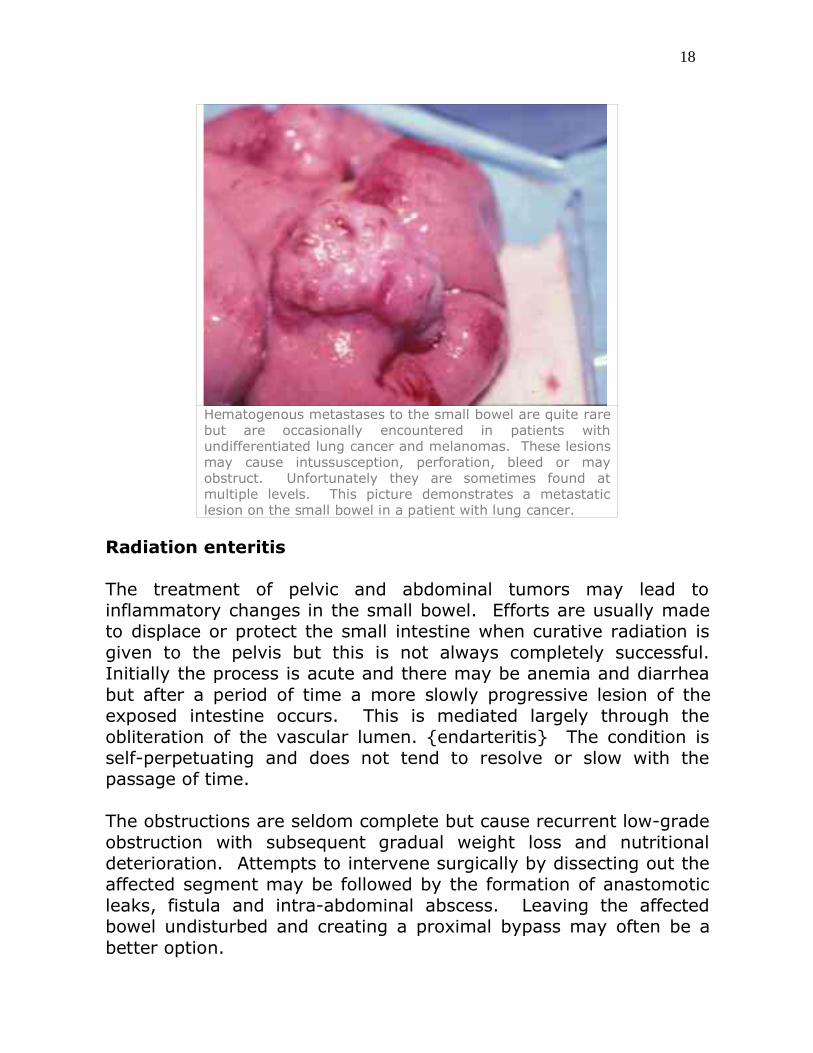
Hematogenous metastases to the small bowel are quite rarebut are occasionally encountered in patients withundifferentiated lung cancer and melanomas. These lesionsmay cause intussusception, perforation, bleed or mayobstruct. Unfortunately they are sometimes found atmultiple levels. This picture demonstrates a metastaticlesion on the small bowel in a patient with lung cancer.
Radiation enteritis
The treatment of pelvic and abdominal tumors may lead toinflammatory changes in the small bowel. Efforts are usually madeto displace or protect the small intestine when curative radiation isgiven to the pelvis but this is not always completely successful.Initially the process is acute and there may be anemia and diarrheabut after a period of time a more slowly progressive lesion of theexposed intestine occurs. This is mediated largely through theobliteration of the vascular lumen. {endarteritis} The condition isself-perpetuating and does not tend to resolve or slow with thepassage of time.
The obstructions are seldom complete but cause recurrent low-gradeobstruction with subsequent gradual weight loss and nutritionaldeterioration. Attempts to intervene surgically by dissecting out theaffected segment may be followed by the formation of anastomoticleaks, fistula and intra-abdominal abscess. Leaving the affectedbowel undisturbed and creating a proximal bypass may often be abetter option.
18

Endometriosis
Although endometriosis more commonly involves the rectum andsigmoid colon, the deposits can affect the distal ileum. Theinflammatory reaction and subsequent stricturing of the bowel wherethe deposits are located are more intense at the time of themenstrual period. The cyclical nature of these symptoms may berecognized raising suspicions of this relatively uncommon condition.
If the lesion is encountered during laparotomy the deposits andpuckered and stricture bowel around it simulate a malignancy.
Female patients with symptoms of SBO obstruction thatseem to occur in a cyclical manner should be consideredto have endometriosis if they are in the proper age group.In this patient there were recurring episodes of abdominalpain and partial obstruction thought to be anintraperitoneal malignancy. At laparotomy these smallhemorrhagic deposits surrounded by inflammatoryreaction and scarring, puckering the intestine were found.The obstruction resolved after the conservative treatmentfor endometriosis.
III. Large Bowel Obstruction (LBO)
19

Volvulus (sigmoid, cecal)
The colon is very rarely involved in internal herniation throughadhesions or external entrapment in hernial orifices. Most acuteonset LBO is secondary to volvulus. Sigmoid is the most commonsite for volvulus followed by cecal.
In this operative photograph there is acecal volvulus. The bowel is dusky incoloration due to venous obstruction butis likely viable. This cecum can be easilyand completely delivered from theabdomen due to its poor peritonealattachment. Poor attachments are thebasis for volvulus formation. It is notpossible to decompress a cecal volvulusnon-operatively. Operation is indicateddue to the high incidence of cecalnecrosis and perforation. A righthemicolectomy is the best management.
In many third world countries volvulus of the colon is thecommonest cause of obstruction. This is thought to be due to theintake of a high fiber diets causing the colon and mesentery tobecome redundant and attenuated. As a cause of obstruction, inNorth America and Western European countries, volvulus of thecolon is a distant third in frequency to cancer of colon anddiverticular disease.
Patients suffering form colonic volvulus are frequently very elderlyand many have longstanding problems with constipation. Theseindividuals are often institutionalized and at times on chronicpsychiatric medications. They are frequently prohibitive candidatesfor major surgical intervention. In some cases, sigmoid volvulus maybe managed by endoscopy and decompression, on a recurring basis.Occasionally a sigmoid colon volvulus will relentlessly recur or willnot be decompressed by endoscopy. Distention, to the point ofinterfering with ventilation, may necessitate surgical intervention.
20

This is a massive sigmoid volvulus that wasimpossible to decompress despite severalattempts. The patient was in respiratorydistress due to the marked abdominaldistention. A rectal tube was placed andthe abdomen opened. The massivelydistended colon was delivered from theabdomen and then decompressed out of theoperative field through the rectal tube. Itwas then possible to carry out a resection ofthe twisted sigmoid colon and form atemporary colostomy. The patient waseventually re-anastomosed.
External Hernias
Obstruction of the colon by adhesive bands is extremely rare.Similarly the colon is rarely trapped in external hernias. Occasionallythe ileocecal area or sigmoid may enter a groin hernia sac becomingobstructed. The transverse colon may also become trapped in anumbilical or incisional hernia.
Carcinoma of Colon
55% of colonic obstruction is due to carcinoma. Anywhere from 6%to 26% of patients with colorectal cancer develop obstruction. Ofthis acute obstruction occurs approximately 10% of the time.
Tumor incidence by site; rectum (50%), sigmoid (20%), left colon(15%), transverse (10%) and descending (5%). Rectal tumorsrarely progress to obstruction due to the considerable capacity of therectum. Large tumors evolving in the rectum will usually causetenesmus and rectal bleeding and be addressed long beforeobstruction occurs.
21

Carcinoma of the colonusually causesobstruction in thesigmoid and rectosigmoidareas althoughobstruction may occurmore proximally. Thegrowth pattern of thesetumors is annular andconstricting. Thepathological term is“napkin-ring”.
Obstruction in the colon of gradual onset is most frequently due tocarcinoma. Tumors in the rectosigmoid area commonly have anannular growth pattern that is constricting. Their maybe preliminarysymptoms with altered bowel habits and crampy abdominal pains.Infrequently the onset of obstruction may seem to be fairly suddenand likely is the result of fecal impaction in a narrowed lumen.
This photograph shows aresected carcinoma of thesigmoid colon that to thispoint was non-obstructingbut the same annular growthpattern is seen. If a patientcan be diagnosed and thebowel prepared beforeobstruction intervenes then asingle stage resection can becarried out.
Diverticular Disease
Diverticular disease is extremely common in the elderly population.It most frequently presents with episodes of diverticulitis andlocalized perforation or occasional abscess formation. Obstruction ofthe colon is probably the least common complication. Occasionallythe inflammatory process may be intense with considerable fibrosisand constriction of the area of involvement. Fecal impaction in anarrow channel may often lead to near complete obstruction of thecolon. The course may be slightly more acute but simulates thecourse of malignant obstruction very closely.
22

This patient has an inflammatory mass at the rectosigmoid junction withcomplete colonic obstruction. The x-rays show a markedly dilated transversecolon to splenic flexure. The contour and the location of the splenic flexureidentify this segment of bowel as the transverse colon. It would be necessary toperform a hypaque enema to confirm the point of distal obstruction.
Assessment – Bowel Obstruction
I. Assessment of the Patient with SBO
Symptoms of Small Bowel Obstruction
Four major symptoms are characteristic of SBO. These areabdominal pain, vomiting, abdominal distention and cessationof flatus passed per rectum. These symptoms vary depending onthe anatomical location of the obstruction, the degree of obstructionand the presence or absence of strangulation.
The abdominal pain is characteristically generalized and colicky.Patients often appear restless and uncomfortable. Features of wave-like accentuations in pain may or may not be present. Pain isseldom localized to either side of the abdomen or epigastrium. As arule pain is generalized and periumbilical. The onset of pain isusually quite sudden and relates closely to the moment of internal
23
herniation. Although an adhesion may have been present in apatients abdomen for months or many years the instant of internalherniation probably requires only a few minutes. Swallowed gas andupper GI secretions quickly distend the proximal bowel and hyper-peristalsis with increasing tension in the bowel wall ensues causingthe colicky pain.
The degree of distention may be greater if the obstruction is distalrather than proximal in the jejunum. Vomiting, however, is likelyto be more frequent and profuse when the obstruction is proximalrather than distal. The bowel may occasionally move following theonset of a complete obstruction but soon thereafter the passage offlatus in particular comes to a complete halt. The absence of flatusbeing passed per rectum is a more consistent symptom relating tothe completeness of bowel obstruction.
The intensity of the above symptoms is related to the completenessof the obstruction. Partial obstructions are less likely to causerepeated vomiting and marked abdominal distention. Smallamounts of flatus may continue to be passed per rectum and theremay be a passage of liquid stool with partial obstruction.
Physical Signs of Acute Onset Small Bowel Obstruction
General physical signs are related to the degree of third spacing andsubsequent intravascular fluid losses. Theses signs may not beevident early in the obstruction. A postural drop in blood pressure,detecting a diminished quality in the pulse and noting a collapse inperipheral veins and poor filling in the jugular veins are all consistentwith decreased intravascular volume.
On inspection a degree of abdominal distention, usuallysymmetrical, maybe appreciated. Diffuse tenderness of a mild tomoderate degree is usually evident but true rigidity and guarding isunusual. Occasionally in thin patients dilated loops of small bowelcan be visualized and seen in a peristaltic motion. Hernia orificesshould be searched carefully for evidence of incarceration. Inpatients who have had previous surgery, abdominal scars will beevident. These scars should be palpated looking for signs ofincarceration. In obese patients detecting small incarceratedincisional hernias is not easily demonstrated on palpation.
24

Abdominal mass may be occasionally felt with acute onsetobstructions where there is a volvulus or an internal herniation. Theloops of strangulated bowel are usually quite turgid and filled withfluid under considerable tension giving rise to a significant abdominalmass.
The abdomen is usually tympanic to percussion due to thesignificant amount of gas retained in the intestine proximal to theobstruction. Loud bowel sounds are sometimes present inincomplete obstructions but soon disappear in complete obstructionsand the abdomen may be silent by the time the patient presents.
Digital rectal examination (DRE) should always be performed.Peritoneal metastases may be evident by the finding of a rectalshelf. Hemoccult positive testing of the stool may indicateIntraluminal tumors.
Identifying patients with complete SBO
Skilled clinicians have great difficulty in distinguishing patients withcomplete bowel obstruction and strangulation for those withincomplete obstruction. In approximately one third of patients thediagnosis of incomplete obstruction is readily made on the basis ofthe, rare or occasional vomiting, lesser degree of pain, passage ofsmall amounts of flatus continuing, and a degree of softness andlack of serious distention to the abdomen.
Some of patients have a history of repeated presentation to hospitalwith partial obstructions that are resolved on many occasions in thepast with conservative measures. Some of the patients have hadmultiple surgeries and are known to have extensive intra-abdominaladhesions.
Another third of patients present with a degree of acuteness anddemonstrate physical signs that strongly suggest completeobstruction that is easily confirmed by plain x-rays. These patientshave a sudden and abrupt onset of severe generalized colic, vomitrepeatedly and the abdomen becomes very distended and tensequickly. No flatus is passed per rectum. On physical examinationsigns of hypovolemia evolve early. The patient’s abdomen is tense,
25
tender and silent.
There are a significant number of individuals however who presentwith a spectrum of symptoms and physical signs in a mid zonebetween complete and incomplete obstruction. The decision onwhether this is an early complete obstruction or partial obstructionmay be extremely difficult on clinical grounds alone. X-rays of theabdomen are not always definitive in these cases.
Identifying Patients with Strangulating SBO
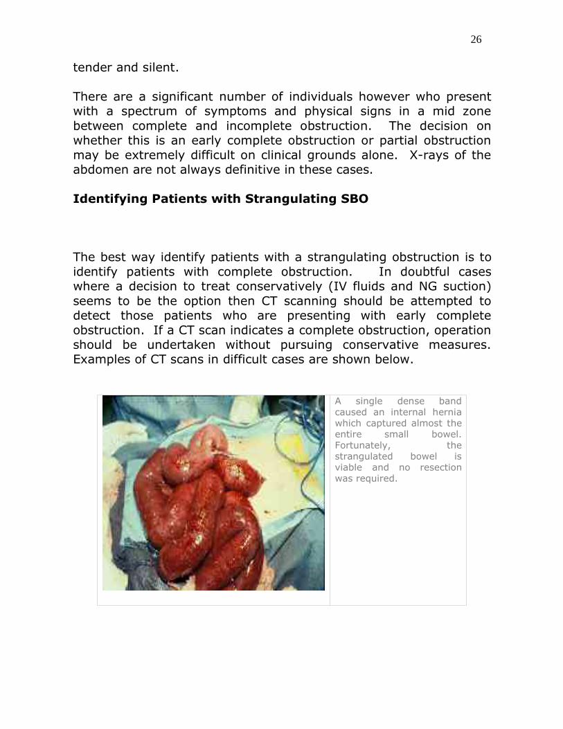
The best way identify patients with a strangulating obstruction is toidentify patients with complete obstruction. In doubtful caseswhere a decision to treat conservatively (IV fluids and NG suction)seems to be the option then CT scanning should be attempted todetect those patients who are presenting with early completeobstruction. If a CT scan indicates a complete obstruction, operationshould be undertaken without pursuing conservative measures.Examples of CT scans in difficult cases are shown below.
A single dense bandcaused an internal herniawhich captured almost theentire small bowel.Fortunately, thestrangulated bowel isviable and no resectionwas required.
26

In this case a shortsegment of the smallbowel was strangulatedand unfortunately will notsurvive. A short resectionand anastomosis will berequired.
Again, operation is toolate. If the patient delayedcoming to hospital and theoperation was immediatethen that is not your fault.However, if the patientwas treated conservativelyfor 24hours or longer andthen operated upon withthese findings, you shouldget a substandard grade.
27
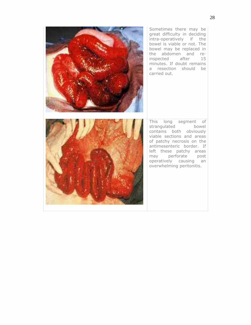
Sometimes there may begreat difficulty in decidingintra-operatively if thebowel is viable or not. Thebowel may be replaced inthe abdomen and re-inspected after 15minutes. If doubt remainsa resection should becarried out.
This long segment ofstrangulated bowelcontains both obviouslyviable sections and areasof patchy necrosis on theantimesenteric border. Ifleft these patchy areasmay perforate postoperatively causing anoverwhelming peritonitis.
28

An internal hernia of ashort segment of smallbowel resulted instrangulation and necrosis.The segment has notperforated and there is nota large enough mass ofdead tissue to incite aclinically identifiableinflammatory response.
Lesson: Identify acomplete obstruction andoperate for this indication.
In this patient there is anadhesive band causing aninternal herniation ofapproximately 20 cm ofsmall bowel that is in anadvanced stage ofstrangulation.
The length of intestine that is involvedin a strangulation is variable and canoccasionally involve almost the entireintestine. In this patient there was anadhesive band in the left upperquadrant that was causing an internalhernia of almost the entire small bowel.Fortunately, surgery was carried outearly enough. The band was dividedsaving the patient from the loss of themajority of their intestine. Some patchyareas of hemorrhage and ischemia areseen but none of these where felt torequire resection.
Identifying patients with acute non-obstructive abdominalconditions
29

This intraoperative photography demonstratesgangrene of small bowel due to a superior mesentericartery thrombosis. These patients are vasculopathsand may have a background of myocardial infarction orarrhythmia. Generalized abdominal pain is severe.Acidosis and shock develops early. Because of theabsence of peritonitis in the early stages rigidity andguarding of the abdominal wall may not be a feature.X-rays of the abdomen do not show proximal dilationand distal collapse.
This intraoperative photography demonstrates peritonitisdue to colonic perforation. In these patients abdominaldistention and vomiting may suggest obstruction.However, there is generalized rigidity and reboundtenderness (signs of peritonitis). X-rays may showdilated bowel, but again there is no proximal dilation ordistal collapse.
30

A segment of intestine may become infarcted due todistal embolus or thrombosis. This may produce a partialobstruction even though there is no actual occlusion. Asnecrosis progresses, peritonitis will develop. Earlydiagnosis is difficult but operation and resection before ageneralized spill of intestinal content occurs will reducemorbidity and mortality.
Acute appendicitis initially presents with a centralabdominal pain, and may be associated with vomiting.However, pain will soon shift to the RLQ and localizedtenderness will develop there. X-rays usually show only anon-specific picture.
Incarcerated External Hernias causing SBO
31

In third world countries and as little as a hundred years ago inindustrialized western countries incarceration of the bowel inexternal hernias was the leading cause of SBO. Due to thewidespread repair of external hernias early after their presentationthe numbers of patients presenting with incarcerated externalhernias and SBO has diminished significantly. This is a progressivetrend because strangulation of bowel in external hernias is asignificant cause of mortality.
The small bowel and the omentum are the most likely structures tobecome incarcerated within an external hernia, given their mobility. Once small bowel becomes entrapped in an external hernia it quicklybecome congested due to the obstruction of venous return.Congestion leads to edema and intramural hemorrhage and soon thestrangulated loop becomes irreducible. Signs of intestinalobstruction generally appear thereafter with generalized crampingpains, vomiting and distention.
Patients presenting with the above symptoms and an identifiablemass at a hernia orifice need to be quickly evaluated andresuscitated and operated upon. If at emergency surgery theincarcerated bowel is found to be viable and can be released andreduced into the peritoneal cavity and repair of the hernia carriedout then the mortality risk quite low, in the margin of 1 or 2%. Ifhowever the bowel is non-viable and a resection is required mortalityrates increase significantly into the area of 5 to 10%. Needless tosay every effort should be made to quickly evaluate, resuscitate andoperate on these patients without unnecessary delay.
In this photo an incarcerated inguinal hernia withbowel obstruction is being explored. When the sac wasexposed its contents appeared hemorrhagic. The sacwas opened, bloody and foul smelling fluid aspiratedand a strangulated non-viable loop of small bowel wasdemonstrated. The internal ring was enlarged laterally,carefully controlling the ischemic bowel. It waspossible to deliver the bowel through the inguinalwound and perform a short resection. The hernia wasthen repaired. If appropriate mesh plugs or patchescan be used.
Incarcerated hernias at the umbilicus, inguinal and femoral orprevious abdominal incisions should be identified as the cause of
32

obstruction if at all possible. Many of the procedures in the inguinaland femoral area in particular can be carried out through a groinincision, significantly reducing the patient’s morbidity. Strangulationof bowel in unusual hernia sites such as obturator and sciatic areseldom diagnosed preoperatively and is usually discovered at fulllaparotomy.
Umbilical defects approaching 2 cm in diameter andwith a rigid margin, pose a risk of incarceration andstrangulation. The patient in this picture had anumbilical protrusion recognized for many years. Itwas easily reducible and surgical management wasnot entertained. After spinal surgery, in hospital,the hernia incarcerated and a bowel obstructiondeveloped. Emergency surgery revealedstrangulated intestine and omentum. A resectionwas required.
ECF deficits in Patients with Bowel Obstruction
In patients with high grade or complete SBO significant amount ofextracellular fluid become sequestered into the bowel lumen and inthe peritoneal cavity. This may accumulate to several liters andsignificantly diminish the intravascular volume giving rise tosymptoms of hypovolemia. In patients who are not appropriatelyresuscitated the administration of analgesics or in particularanesthesia may cause them to decompensate and become "shocky".
Patients giving a history of significant duration (>24 hrs)characterized by severe pain, vomiting and absence of flatus perrectum with x-rays demonstrating significant fluid within the bowellumen as evidenced by numerous, long, fluid levels or filling of thelumen on CT scanning should be anticipated to be hypovolemic.Often there is supportive laboratory evidence in the form of a highhemoglobin indicating a reduced intravascular volume and elevatedcreatinine indicating a degree of pre-renal failure. Abnormalities insodium and potassium levels are inconsistent and should notnecessarily be anticipated.
Signs of hypovolemia should be searched for early in themanagement of patients with SBO. If the history and physical signsindicate hypovolemia then peripheral venous access should be
33
established quickly. A balanced salt solution should be infused usinga bolus technique. A Foley catheter should be inserted early on tomonitor urine output. Repeat physical examination looking for fillingof the jugular veins, reversal of the orthostatic hypotension andimprovement in pulse quality are helpful in establishing adequateresuscitation.
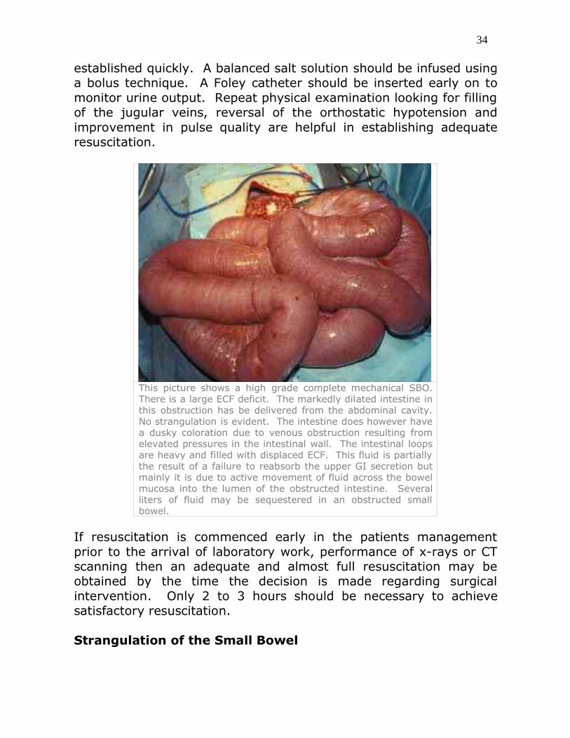
This picture shows a high grade complete mechanical SBO.There is a large ECF deficit. The markedly dilated intestine inthis obstruction has be delivered from the abdominal cavity.No strangulation is evident. The intestine does however havea dusky coloration due to venous obstruction resulting fromelevated pressures in the intestinal wall. The intestinal loopsare heavy and filled with displaced ECF. This fluid is partiallythe result of a failure to reabsorb the upper GI secretion butmainly it is due to active movement of fluid across the bowelmucosa into the lumen of the obstructed intestine. Severalliters of fluid may be sequestered in an obstructed smallbowel.
If resuscitation is commenced early in the patients managementprior to the arrival of laboratory work, performance of x-rays or CTscanning then an adequate and almost full resuscitation may beobtained by the time the decision is made regarding surgicalintervention. Only 2 to 3 hours should be necessary to achievesatisfactory resuscitation.
Strangulation of the Small Bowel
34

Strangulation by definition is the development of vascularcompromise of the incarcerated bowel whether this be an internalhernia within the peritoneal cavity or in the sac of an externalhernia. Strangulation means vascular compromise and does notnecessarily infer the involved intestine is necrotic, requiringresection.
If the bowel is entrapped in an external hernia, and is obstructed, adegree of strangulation is certain to be present. The diagnosis ofstrangulation is relatively easily made in these circumstances whichshould lead to prompt surgical exploration.
When the obstruction and strangulation is within the abdominalcavity and caused by an internal hernia, the identification ofstrangulation becomes difficult. If the loop of intestine involved islong then fever, severe tenderness and a palpable mass may bepresent. If the loop of bowel involved is a short knuckle then theseindicators may not be detectable even by the most astute clinician.It follows that the search for a complete obstruction should beundertaken; if found, operation should soon follow.
The evolution of strangulation in a herniated loop of intestine isunpredictable. Occlusion of venous return quickly leads toengorgement of the bowel leading to interstitial and luminalhemorrhage. The subsequent edema and venous stasis results inthe elevation of vascular pressures and eventual absence of arterialflow.
35

If a extended length of small bowel is strangulated the subsequenthypovolemia and toxemia will likely be detected on clinical grounds.There may be a palpable mass and a considerable increase inabdominal findings. However if the intestinal loop that is internallyherniated and strangulated is very short the systemic effects of thisdying intestine may be minimal and impossible to detect. A completeintestinal obstruction should be evident by imaging studies. It iseasier to detect the completeness of obstruction than thestrangulation. Complete obstruction should be an indication forsurgical intervention due to the high incidence of strangulationassociated with complete obstruction.
Radiographic Imaging patients with Acute SBO
Patients presenting with acute abdominal pain, as a general rule,should have supine and upright views of the abdomen and a chest x-ray performed as part of the initial assessment. The assessment ofthe patient should be well underway as should the resuscitation ofthe patient before such radiographs are sought. The patient’s vitalsigns including their ventilatory reserves, oxygenation andcirculatory status should be assessed and correct to as near normalas possible before the patient departs the emergency department forthe x-ray department.
Patients with fluid volume deficits with postural hypotension and pre-renal failure tolerate transport poorly. The patient’s intravenousresuscitation should be well underway and if there has been vomitingor abdominal distention a NG tube should be in position andfunctioning. Aspiration of vomitus is a serious complication of
36

intestinal obstruction. This is particularly dangerous if it occursoutside the emergency department in the darkness of a x-ray suite.
X-rays of the abdomen should first be scrutinized with a view todetermine whether there is in fact an obstruction and if so, where itis located.
2 patients with complete SBO. Dilated, centrally locatedsmall bowel loops with no colonic gas identifiable. Historystrongly supported the conclusion that the obstruction wascomplete. Further imaging is unnecessary.
Acute abdomens with peritonitis, retroperitoneal trauma or bleedingand even remote injuries such as rib fractures, spinal injury andpelvic injury may cause a degree of dilatation of the intestinal tractand even fluid level formation. X-rays of patients with truemechanical obstruction should show true proximal dilation of bowelwith distal collapse.
X-rays that demonstrate SBO are usually readily identified by theappearance of centrally located loops of dilated intestine oftenforming a “ladder-like” or clustered pattern in the central abdomen.Multiple fluid levels are often seen in the upright view. If theobstruction is in the small intestine and complete, absence of gas inthe cecum and transverse colon can usually be confirmed.
In cases of colonic obstruction the cecum and transverse colon areusually readily identified. Occasionally loops of small intestine mayalso be distended if there is incompetence of the ileocecal valve. Ifcolonic obstruction is suspected then the site of obstruction can be
37

confirmed by a contrast study given per rectum.
As a general rule, contrast given by mouth or NG tube to attempt todemonstrate points of obstruction in cases of SBO are unsuccessfulas the contrast dilutes out in the intestinal fluids. It becomes verydifficult to demonstrate a point of obstruction or assess thecompleteness of obstruction.
In any case of acute abdomen or in particular suspected SBO, x-rays of the abdomen in supine andupright positions should be obtained. Many times a plain film of the abdomen will be sufficient todistinguish between high grade complete obstructions and partial obstructions. These series ofradiographs show cases of SBO which from a clinical standpoint where complete. Radiographs shouldbe searched for evidence of gas in the cecum and other identifiable areas of the colon. In the middleradiograph (upright abdomen) the amount of dilated small bowel is difficult to appreciate. In anycase where the clinical picture suggests bowel obstruction, but the radiographs are inconclusive thenCT scanning is indicated. The degree of dilation, the numbers of dilated loops or the length of their fluid levels are notnecessarily an indication of the completeness or incompleteness of obstruction. In the radiograph onthe right (upright abdomen) there is clear identification of complete obstruction showing only 3 shortfluid levels in the small intestine. There is no gas located in colon or cecum; the obstruction isradiographically complete. An early operation revealed a strangulating obstruction.
CT scanning patients with acute SBO
The CT scan may be a very useful tool in the assessment of patientswith SBO. Neither intravenous nor oral contrast are absolutelynecessary if the patient requires continuous NG suction or renalfunction is impaired. Fluid filled loops of bowel, sometimes difficultto discriminate on plain upright films of the abdomen, may bereadily seen on CT. When all of the images are present they can betraced through and a point of obstruction, if there is one, can usuallybe discriminated. Warning signs of strangulation including free fluidwithin the peritoneum, thickened loops of bowel or even gas in the
38

bowel wall may be identified.
Patients with incomplete obstruction, where there is a compatiblehistory and physical exam, and x-rays show nonspecific patternswithout proximal dilation, may not need CT scanning.
Some other patients with a very clear history of sudden onset ofcomplete obstruction, concerning physical signs and a set of x-raysshowing dilated bowel loops and no visible colon do not require CTscanning; they should be resuscitated and proceed to operation.*A CT scan may be a very significant and discriminating test to applyto at least a third or more of patients with acute onset SBO.
39

CT scanning in patients with suspect SBO can be more successful inshowing complete points of obstruction as well as indicating ischemicor strangulated loops of intestine. Intestinal obstruction in whichthere are large amounts of fluid but little gas entrapped within theobstructed loops, are identified more readily by CT scanning thanplain radiographs. Some selected panels of patients with smallbowel obstruction are shown. There are considerable limitations inviewing a single panel of a study. That said, the panel in the top left corner demonstrates markedlydilated small intestine; most loops of bowel are completely filled withsequestered ECF. The descending colon is a tiny gas filled structurein the left paracolic gutter. The obstruction is very high grade andessentially complete. In the right upper quadrant (different patient), dilated loops ofintestine are seen in a pelvic panel. There is thickening of the bowelwall. The rectum is seen as a tiny collapsed gas filled structure justin front of the sacrum. The panel in the lower left corner demonstrates many dilated loopsof small bowel in the pelvis and again the rectum is a tiny structurewith a small amount of gas in it just in front of the sacrum. The panel in the lower right corner demonstrates contrast migratingthrough distended and obstructed loops of small bowel. A transitionpoint is noted just the midline to the right, just deep to abdominalwall. The contrast within the lumen demonstrates intestine proximalto the obstruction. Contrast does not reach the colon The obstructedbowel is not particularly dilated, there is no free fluid, no thickenedbowel wall. In this case, a period of observation would be safe, withrepeat of the CT scan..
Symptoms and Physical examination of Gradual onset SBO
Patients with a gradual evolving obstruction of the small bowelusually present in an ambulatory care setting. Often their historywill span many weeks and sometimes months to years. The basiccomplaints are similar to those of acute SBO; however, they are lesssustained and less severe. The distribution of the pain is similar inthe sense that it is located in the periumbilical area. The pain has acolicky nature that causes restlessness. The pain may have a wave-like character and is often accompanied by loud borborygmi. Inbetween episodes of obstruction there may be, paradoxically,periods of diarrhea. Usually symptoms are increased with eatingand the more solid and amount food the more intense thesymptoms. A degree of abdominal distention is usually evident. Thepatient may occasionally have nausea and rarely vomiting. Becauseof the relationship between pain and food, patients often develop agradual weight loss due to self imposed dietary limitations.
40

An enquiry should be made into the previous medical and surgicalhistory. Previous abdominal surgery for malignancy would suggestthe symptoms represent a recurrence with peritoneal metastases.Radiation therapy to the pelvis may induce radiation enteritis andobstruction may develop years after treatment is completed.
Younger patients with weight loss, intermittent diarrhea, crampyabdominal pain and anemia should be suspected of havinginflammatory bowel disease. Older patients with serious vasculardisease should be assessed for mesenteric vascular occlusion andintestinal ischemia. The symptoms of this condition may closelymimic those of gradual onset SBO.{intestinal angina}
On physical examination a careful search of the abdomen for apalpable mass should be made. Tumor masses on the small bowelor omentum are frequently mobile. Any previous incisions and theumbilicus should be carefully searched for tumor implantation. ADRE may reveal tumor implantation in the pouch of Douglas or therectovesicalar pouch. Auscultation of the abdomen will frequentlyreveal loud, high pitched bowel sounds that occur in waves, if theyare not audible at the bedside.
Investigations of Gradual Onset SBO
In such cases there is usually a significant time frame available tocarry out investigations. Hematological studies may demonstrateanemia if there is an ulcerative lesion in the small bowel or ileocecalarea. Stool may be positive for occult blood.
Plain x-rays of the abdomen may demonstrate dilated loops of smallbowel, particularly during times of increased symptoms. Films takenduring asymptomatic periods may fail to show any abnormalities.Most small bowel tumors are beyond the reach of conventionalendoscopic studies, although a lesion at the ileocecal valve may bevisualized by colonoscopy. Enteroclysis, which is the injection ofbarium into the duodenum by a nasoenteric tube, may be successfulin demonstrating one or more points of obstruction in the smallbowel. In patients whom mesenteric vascular occlusion is thesuspected diagnosis angiography may be indicated.
CT scanning may be successful in demonstrating tumor masses in
41

the omentum, bowel or mesenteries and even the peritoneum.Retroperitoneal nodal disease and liver metastases may also beshown.
II. Assessment of the Patient with LBO
Presentation of Acute Onset LBO due to Volvulus
Volvulus of the colon occurs relatively rarely North America. In othercountries, presumable on the basis of a bulky high fiber diet, theincidence of volvulus causing obstruction is much higher (Iran 33%,North India 30%, Uganda 20%). Many individuals have a pattern ofmultiple recurrence of volvulus.
Volvulus of the colon usually has a short history (for that particularepisode), often less than 24 hours. There is rapid and progressiveabdominal distention accompanied by some degree of generalizedabdominal pain. At times the distention is so severe it compromisesventilation.
This photo from the operating room shows themarkedly distended, tympanic abdomen that isa feature of colon obstruction.
Patients often have a long history of irregular bowel motion andlaxative abuse. The patient may be using antipsychotic medicationsthat have effects on bowel motility. Many of the individuals arefrail, demented and institutionalized making them unfavorable forsurgical intervention. Occasionally patients decompressintermittently resulting in the discharge of large amounts of
42

“mucusy” secretions. Frequently individuals become well known toemergency departments due to multiple visits.
Investigation of LBO Caused by Volvulus
In the case of sigmoid volvulus a “coffee bean sign” may be seen onplain abdominal films. The massively distended sigmoid colon isorientated form the left iliac fossa to the right upper quadrant withthe intervening mesentery separating the large loop. At times theradiological picture is confusing only showing dilated loops of colon.When there is some uncertainty of cause or level of obstruction acontrast study instilled per rectum may be helpful.
When LBO is suspected onclinical grounds and plain x-rays of the abdomen appear toconfirm the suspicion, watersoluble contrast medium maybe delivered per rectum toclarify both the nature andlevel of obstruction. In thispatient with a confusing plainradiograph, but massivelydilated loops of bowel, ahypaque enema has beenperformed which demonstratesthe twisting at therectosigmoid junction. Thetwisting produces thecharacteristic “beak” at therectal end of the obstruction.
43

The point obstruction in this radiograph is inthe right colon suggesting this is a cecalvolvulus. The typical “beak-like”appearance of the water soluble contrast isshown.
Symptoms and Physical signs of LBO due to Colon Cancer
Many patients with LBO due to colonic cancer have a history of lowerabdominal cramps, periodic episodes of rectal bleeding andalterations in bowel habits. These symptoms may have been in placefor weeks; hopefully, if the patient seeks medical attention,appropriate investigations [barium enema, endoscopy] and anelective surgery, on prepared bowel, will follow. The colon can squeeze stool through a 1cm neoplastic stricturefor an astonishing time, but a complete occlusion will inevitablyoccur, usually by impaction of a fecal pellet. Patients presenting with complete obstruction of the left colonhave significant abdominal distention and tympany. A palpable massis rarely felt because of the small and sclerotic nature of thesetumors. Frequently the tumor is beyond the reach of DRE.
Identifying the Site of LBO due to Colon Cancer
Patients with suspicious symptomatology, in particular crampinglower abdominal pain with alterations in bowel habit should haveendoscopic and contrast studies carried out. If a DRE is negativethen at least a rigid sigmoidoscopy of 20 cm should be performed toexclude an obvious lesion within this area of the bowel. If this isnegative a barium enema would be the most appropriate test in anambulatory setting. Frequently an “apple core” or a narrowedsegment of the rectosigmoid area will be demonstrated. Theresidual lumen is often surprisingly narrow and attempts to fill theproximal colon with contrast should be resisted by the radiologist. Acolonoscopic examination to obtain a biopsy and image the lesion isappropriate, but not absolutely necessary in typical cases. A scanfor metastatic disease is often performed in these patients in theform of US or CT of the abdomen.
44
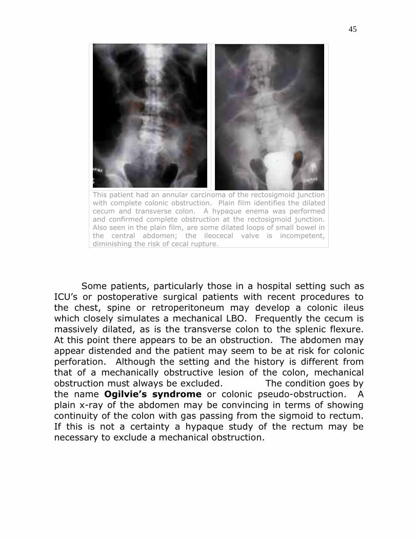
This patient had an annular carcinoma of the rectosigmoid junctionwith complete colonic obstruction. Plain film identifies the dilatedcecum and transverse colon. A hypaque enema was performedand confirmed complete obstruction at the rectosigmoid junction.Also seen in the plain film, are some dilated loops of small bowel inthe central abdomen; the ileocecal valve is incompetent,diminishing the risk of cecal rupture.
Some patients, particularly those in a hospital setting such asICU’s or postoperative surgical patients with recent procedures tothe chest, spine or retroperitoneum may develop a colonic ileuswhich closely simulates a mechanical LBO. Frequently the cecum ismassively dilated, as is the transverse colon to the splenic flexure.At this point there appears to be an obstruction. The abdomen mayappear distended and the patient may seem to be at risk for colonicperforation. Although the setting and the history is different fromthat of a mechanically obstructive lesion of the colon, mechanicalobstruction must always be excluded. The condition goes bythe name Ogilvie’s syndrome or colonic pseudo-obstruction. Aplain x-ray of the abdomen may be convincing in terms of showingcontinuity of the colon with gas passing from the sigmoid to rectum.If this is not a certainty a hypaque study of the rectum may benecessary to exclude a mechanical obstruction.
45

Further value of a water soluble construct study of apatient with suspect colonic obstruction is the rulingout of colonic ileus (Ogilvie’s syndrome). Somepatients with adynamic ileus present with a pictureconsistent with mechanical colonic obstruction. Inthis radiograph contrast has be instilled per rectumand can be seen flowing through a patent butmarkedly dilated colon to the cecum. A level ofmechanical obstruction is ruled out.
Occasionally the degree of distention from a pseudo-obstruction maybe so significant that colonoscopic decompression may be required.A significant success rate has been obtained by intravenousadministration of a cholinergic stimulus in the form of Neostigme.
Besides colonic pseudo-obstruction other conditions are known tocause colonic dilatation that may simulate a mechanical obstruction.Toxic megacolon may develop in the course of ulcerative colitis.Usually this condition can be discriminated by the chronic history ofulcerative colitis, usually spanning years. The toxic megacolonusually develops during the course of a fulminating episode of colitis.
46

This photograph demonstrates the operative appearance of apatient with toxic megacolon due to colitis. The omentum isshown draped over the massively dilated colon. Someportions of the colon appear somewhat dusky and thinwalled. Megacolon and ulcerative colitis should be readilydistinguished from mechanical obstruction by the history ofthe patient with colitis. Often the diagnosis of toxicmegacolon needs to be made on clinical grounds andcontrast and endoscopic studies may be extensively risky.
C. difficile colitis often occurs on surgical wards. Although most areheralded by the onset of diarrhea, in some instances, dilation of thecolon without significant diarrhea occurs..
Management – Bowel Obstruction
I. Management of Patients with SBO
Operative Strategies for Adhesive Bands
Patients coming to the operating room for complete SBO suspectedto be due to adhesive bands should be fully resuscitated and haveestablished a good hourly urine volume with no evidence ofhypovolemia. A NG tube should be in place and the stomach shouldbe empty prior to anesthetic induction.Intravenous broad spectrum antibiotic should be given before theincision is made.
47

Vertical scars are often present in patients who have adhesiveobstructions. Often this scar is orientated to the lower abdomensince surgery in this zone is more often complicated by adhesiveobstructions.
The abdomen needs to be opened very cautiously because thedistended bowel is often adhered to back of the incision. The patientshould have received IV antibiotics to cover both anaerobes andaerobic coliforms. These drugs should be given just prior to surgeryto assure adequate blood and tissue levels.
The vertical incision is then extended cephalad or distal in order toenter the peritoneal cavity at an area where bowel is less likely to beadherent to the back of the abdominal wall. Once a safe entry ismade into the peritoneal cavity the incision should then be openedfurther to provide adequate exposure.
The inadvertent entry into a distended, obstructed bowel withspillage of contents into the wound and peritoneum is a significantnegative event. Perforation and spillage of the bowel greatlyincreases the chances of post operative morbidity and mortality.Great caution is required in the initial stages to lessen the chances ofsuch a catastrophe.
Once a free entry into the peritoneal cavity is obtained the point ofobstruction should be very carefully sought out. The distended loopsof bowel may be delivered from the abdomen to provide bettervisualization and access. This should be done without twisting orkinking of the mesentery or traction on the delicate loops ofintestine. Often the obstruction is due to a single band. If the bandcan be identified early, the band divided and the obstructionreleased then the procedure may be over.. Identifying collapsedsmall intestine is also helpful since it may be traced proximally toidentify the point of the obstruction.
The mechanism of obstruction is usually an internal herniation.Once the adhesive band creating the internal defect is divided it isoften necessary to assess the viability of the loop of bowel that hasbeen caught. At times a viable loop must be resected because theadhesion has caused a band of pressure necrosis.
48

Loops of bowel that have been strangulated may be observed over aperiod of 10 or 15 minutes after restoring the bowel to the abdomenand covering the exposed bowel with warm sponges. Sometimesquite congested intestine may clearly improve in terms of colorationobtaining a more hemorrhagic and pinker color rather than purple.Peristalsis can often be seen passing through viable areas andpulsation of small vessels close to the bowel wall may be observed.In any case if doubt remains it is usually the wiser choose to resectthe involved segment of small bowel.
If resection is required, spillage from the obstructed intestine isagain a risk.. Carefully blocking off the strangulated segment withsponges and controlling the proximal bowel with a non-crushingintestinal clamp is often effective.
Bowel stomas are seldom necessary in SBO except under veryunusual circumstances. Once the strangulated bowel is resected aprimary anastomosis is usually performed. The risk of anastomoticleak is about 2% but this is considered reasonable. Staged surgeriesin small bowel obstruction are often overly morbid for the patientand fluid losses from stomas are excessive. Eventually, difficultywith late reconnection is usually encountered.
Operative Strategies for SBO caused by External Hernias
It is of great advantage for a patient to have an external herniaidentified as the cause of their SBO. Occasionally in obeseindividuals or when the hernia is particularly small the externalhernia is missed on inspection and a vertical laparotomy incision ismade only to find that the bowel is incarcerated in a inguinal orfemoral hernia sac. It is preferable to avoid this latter course.
A standard skin crease incision in the groin should be made in casesof an incarcerated hernia. The sac should be carefully dissected.The constricting fascia or peritoneal ring should be left in placecontrolling the incarcerated bowel as long as possible. If the bowelis clearly strangulated by its appearance it may be preferable toleave the sac in place and perform a resection of the sac andstrangulated loop in continuity.
In cases where the viability of the bowel is in doubt the hernia sac
49

can be carefully surrounded with operative sponges and then openedand its contents aspirated. If the bowel is strangulated and requiresresection this can usually be achieved by lengthening the herniaincision laterally through the oblique muscles of the abdominal walland delivering further intestine into the groin. Once the anastomosisis performed it can be reduced back into the peritoneal cavitythrough the groin incision and followed by a standard repair of thehernial defect.
This patient had a SBO due to an incarcerated femoral hernia. Theobstruction had been in place for almost two days and strangulation of theincarcerated bowel was strongly suspected. In the first operativephotograph it is shown the peritoneal sac with its entrapped bowel has beendelivered from under the inguinal ligament without opening the sac. In thesecond photography, after careful protection of the wound the sac has beenopened which revealed necrosis at the point of compression. The Cooper’sligament, lancunar ligament and inguinal ligaments of femoral hernias arevery rigid and unforgiving. This segment of bowel required resectionbecause of the compression necrosis. The resection was carried out easilythrough the groin incision.
Operative Strategies for SBO caused by Obturation
Patients with intra-luminal obstructions are not threatened bystrangulation however their courses are usually longer than thosewith an adhesive or hernial obstruction. Often their fluid, electrolyteand renal problems are more pronounced. If obturation is suspected
50

due to the absence of incisions or an identifiable external hernia orthere is a clear history of swallowing a potential obstructing body therelative urgency of the surgical procedure is diminished. Fullevaluation and resuscitation of the patient is possible.
If preoperative radiology reveals air in the biliary tree then acholecystoduodenal fistula with a gallstone in the intestinal tract islikely the cause of SBO. Often a stone can be visualized in the rightiliac fossa. The stone is usually solitary and large. Access to the GItract is usually by virtue of a cholecystoduodenal fistula caused byrecurrent inflammation with erosion of a large calculus.
The stone can usually be dislodged form the point of impaction in theterminal ileum and gentle milked proximally up the small bowel. Thestone should be reversed to the jejunum where the bowel is lessdistended and more resilient. A careful search of the intestinal tractshould be made for other potential obstructing stones. Anenterotomy can then be performed and the stone removed. As arule no effort is made to close the cholecystoduodenal fistula or toremove the gallbladder.
The management of the other forms of obturation is similar.Although in some instances of phytobezoars causing SBO it ispossible to gently breakdown the impacted material and to displaceit through into the cecum to be passed uneventfully. This ispreferable to enterotomy.
Indications for Surgical Intervention in Gradual Onset SBO
The approach to this group of patients is quite variable given thedifferent time course and prognosis of the underlying conditions.Crohn’s disease is a common cause of incomplete SBO. If there isprogressive nutritional deficits evolving and the patient can beshown to have localized disease of a relatively short segment of theintestinal tract (frequently ileocecal) then a resection is likelyindicated. The outcome in the case will likely be satisfactory overtime allowing for a return of nutritional vigor and the withdrawalfrom debilitating medications. Close cooperation between surgeon
51
and gastroenterologist is clearly a favorable scenario.
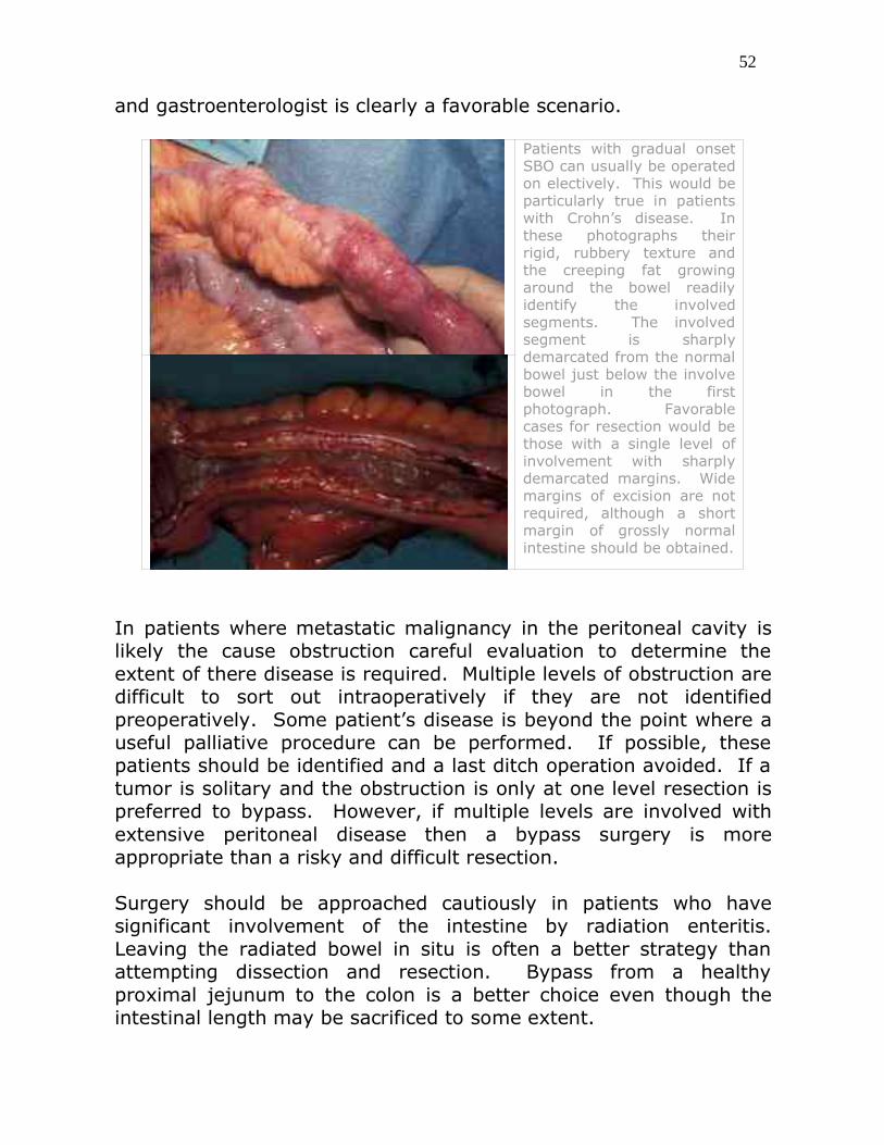
Patients with gradual onsetSBO can usually be operatedon electively. This would beparticularly true in patientswith Crohn’s disease. Inthese photographs theirrigid, rubbery texture andthe creeping fat growingaround the bowel readilyidentify the involvedsegments. The involvedsegment is sharplydemarcated from the normalbowel just below the involvebowel in the firstphotograph. Favorablecases for resection would bethose with a single level ofinvolvement with sharplydemarcated margins. Widemargins of excision are notrequired, although a shortmargin of grossly normalintestine should be obtained.
In patients where metastatic malignancy in the peritoneal cavity islikely the cause obstruction careful evaluation to determine theextent of there disease is required. Multiple levels of obstruction aredifficult to sort out intraoperatively if they are not identifiedpreoperatively. Some patient’s disease is beyond the point where auseful palliative procedure can be performed. If possible, thesepatients should be identified and a last ditch operation avoided. If atumor is solitary and the obstruction is only at one level resection ispreferred to bypass. However, if multiple levels are involved withextensive peritoneal disease then a bypass surgery is moreappropriate than a risky and difficult resection.
Surgery should be approached cautiously in patients who havesignificant involvement of the intestine by radiation enteritis.Leaving the radiated bowel in situ is often a better strategy thanattempting dissection and resection. Bypass from a healthyproximal jejunum to the colon is a better choice even though theintestinal length may be sacrificed to some extent.
52

Management Strategies for LBO caused by Volvulus
As mentioned above many of these individuals are chronically ill anddebilitated making surgical intervention unpalatable. Unfortunatelytheir condition is often relentless and recurrence becomes sofrequent that intervention must be considered. Occasionally anoperative intervention is forced in the acute situation because of theinability to endoscopically decompress a sigmoid volvulus and attimes out of concern for the patient’s ventilatory status.
Volvulus of the cecum is at higher risk for ischemia and eventualinfarction or perforation of the massively distended cecum. Anoperative intervention may be indicated in the first instant for theproblem. Endoscopic decompression of cecal volvulus is probablynot neither safe nor possible.
If an operative intervention for sigmoid volvulus is required then arectal tube should be positioned prior to preparing and draping theabdomen. The twisted sigmoid may then be immediatelydecompressed intraoperatively and exteriorized. A sigmoidcolostomy and closure of the rectal stump is likely the best option.In the majority of patients re-anastomosis will not be attempted.
When the indication for surgery is a cecal volvulus then the bestprocedure is a right hemicolectomy with a primary anastomosis, ifthe patient remains stable. Attempts to pexy the cecum to theparietal peritoneum or to drain the cecum (cecostomy) are usuallyill-fated.
Approach To The Patient With Colon Obstruction Likely DueTo Carcinoma
Obstructing colon cancers usually occur in the sigmoid colon. Moreproximal lesions may also cause obstruction however, and themanagement may be different.
X-rays of the abdomen will usually show dilation of the cecum if theobstruction is in the colon, and then variable lengths of more distalcolon dilation. The precise point of obstruction may be uncertain.Contrast studies from “below” are indicated to confirm obstruction
53

and show the level.
Obstruction is confirmed. It is total.There is no need to make a tissuediagnosis. This can be established later.
There are several approaches to a completely obstructing left colonlesion. In the presence of a massively dilated colon, loaded withfeces, with attenuated and fragile wall, attempts to resect the lesionshould be resisted (in the author’s opinion)
The colon will be very distended. It canbe decompressed with a 18 gauge needleand gently lifted out on the abdominalwall. A plastic rod is through themesentery and prevents retraction. Thebowel is opened on the antemesentericside.
A proximal diverting colostomy (loop, with transverse colon) is thesafest approach, to be followed in about 2 weeks by resection of theidentified lesion. The colostomy should be left to protect thisanastomosis, and then closed several months later.
54

Diverticulitis may closely mimiccarcinoma. A tight stricture may beformed which will not resolve, and willstill need resection.
This completely obstructing cancer wasresected 2 weeks after an emergencyloop colostomy. The bowel is collapsedand empty, very easy to dissect andhandle without risk.
55
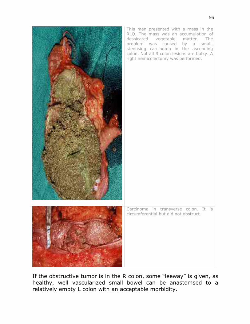
This man presented with a mass in theRLQ. The mass was an accumulation ofdessicated vegetable matter. Theproblem was caused by a small,stenosing carcinoma in the ascendingcolon. Not all R colon lesions are bulky. Aright hemicolectomy was performed.
Carcinoma in transverse colon. It iscircumferential but did not obstruct.
If the obstructive tumor is in the R colon, some “leeway” is given, ashealthy, well vascularized small bowel can be anastomsed to arelatively empty L colon with an acceptable morbidity.
56

Short Cases with Questions for You
1.
A inguinal hernia was incarcerated. The sacwas exposed and contained bloody fluid. Thesac was opened and bowel of questionableviability was seen. During dissection, thebowel slipped into the abdominal cavity andcould not be retrieved. What would you do?
2.
A tender mass appeared in the right inguinalarea. It was painful and the patientpresented two days later. Temperature andvital signs were normal. The abdomen wassoft with no localized tenderness. Anoperation was undertaken.
Strangulated bowel was found. Why wasthere no obstruction?
What would have happened if surgery wasfurther delayed?
3.
57
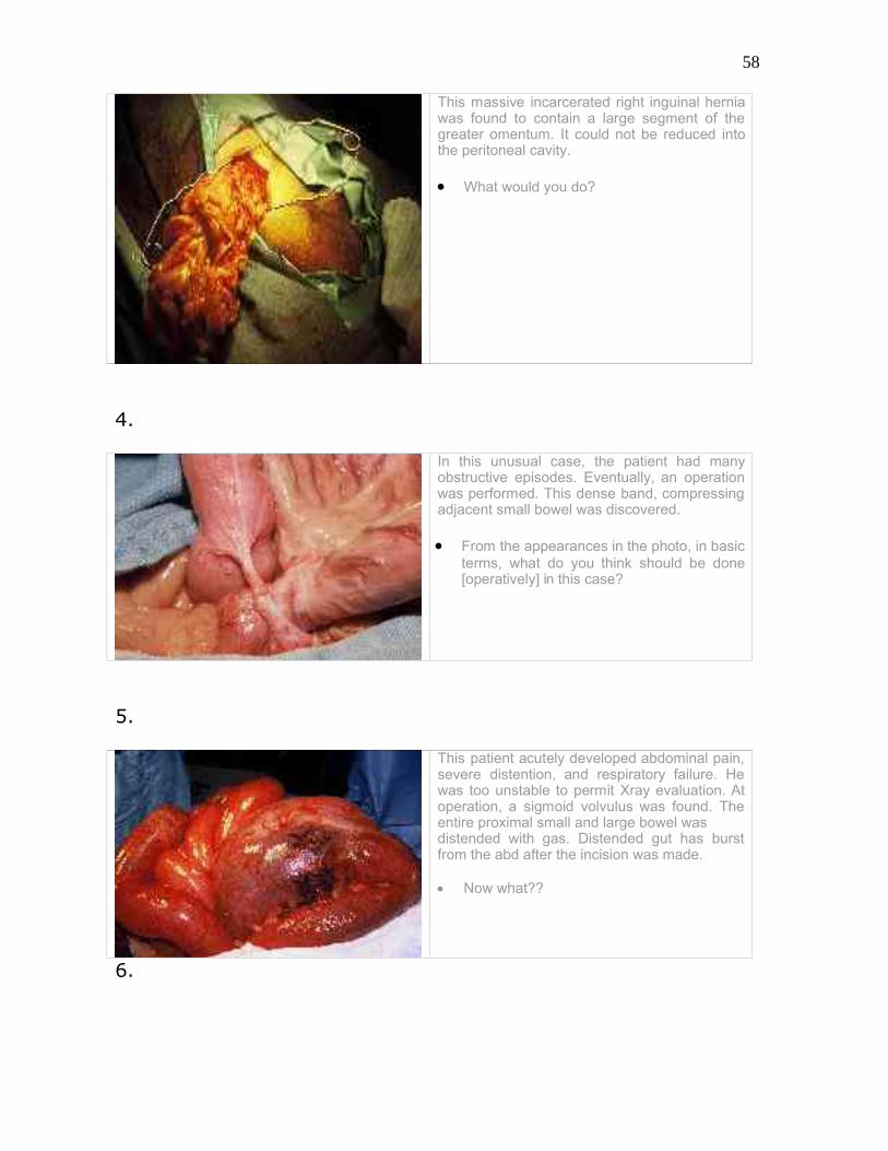
This massive incarcerated right inguinal herniawas found to contain a large segment of thegreater omentum. It could not be reduced intothe peritoneal cavity.
What would you do?
4.
In this unusual case, the patient had manyobstructive episodes. Eventually, an operationwas performed. This dense band, compressingadjacent small bowel was discovered.
From the appearances in the photo, in basicterms, what do you think should be done[operatively] in this case?
5.
This patient acutely developed abdominal pain,severe distention, and respiratory failure. Hewas too unstable to permit Xray evaluation. Atoperation, a sigmoid volvulus was found. Theentire proximal small and large bowel was distended with gas. Distended gut has burstfrom the abd after the incision was made.
Now what??
6.
58

Xrays demonstrated acomplete rectosigmoidobstruction. The colon was massivelydistended. The patient had bone marrowtransplant previously, for AML. He is onimmunosuppressive drugs. He is in the OR,ready for prep and drape.
What procedure do you think would bethe best option in this situation?
7.
The operative photo shows a fatty bandcompressing the terminal ileum. The patientgave a history of several months duration.
Develop what you think would have beenthe History of Present Illness.
8.
This patient had a large, reducible umbilicalhernia for many years. There were fewsymptoms. He was advised, by MDs, to leave itbe. Finally, it suddenly became incarcerated. Atoperation, the sac was opened, revealing acongested, hemorrhagic loop of bowel. It willnot reduce.
What to do??
For feedback on your answers to the above problems look below thesection on references.
59

References – Bowel Obstruction
Lawrence, Bell and Dayton, Essentials of General Surgery ThirdEdition
Chapter 14: Small Intestine and Appendix (pp. 255-259) Chapter 15: Colon, Rectum, and Anus (pp. 284286) Chapter 11: Abdominal Wall, Including Hernia
European Association of Radiology E-Learning Initiative http://www.eurorad.org/browse_sections.cfm?sectionid=5
University of Iowa Family Practice Handbook, Fourth Edition, Chapter15http://www.vh.org/adult/provider/familymedicine/FPHandbook/Chapter15/12-15.html
MDChoice.com: the ultimate medical information finderhttp://mdchoice.com/xray/xr.asp - abdomen
Oregon Health Sciences University: Radiology on Call (CT SBO)http://www.ohsu.edu/ps-DiagRadiol/kojima/ctobstr.htm
FamilyPracticeNotebook.com (point form review of bowelobstruction)http://www.fpnotebook.com/SUR56.htm
Memorial University of Newfoundland, Faculty of Medicine http://www.med.mun.ca/ugme/Pediatric Radiology/bowel troublepage 2.htm
Indian Journal of Radiology and Imaging: Case of Gallstone Ileushttp://www.ijri.org/articles/archives/2002-12-3/abdominal_349.htm
Emedicine: Small Bowel Obstructionhttp://www.emedicine.com/EMERG/topic66.htm
homeearthlink.net: CT images and case of closed loop bowelobstructionhttp://www.emedicine.com/EMERG/topic66.htm
University Hospitals of Cleveland: case of radiation enteritis with CT
60

scanshttp://www.uhrad.com/ctarc/ct176.htm
Feedback on short questions
1. Unfortunately, this adverse event will make it necessary to open theabdomen through a lower midline incision. This may increase the elderlypatients morbidity, but the strangulated section of bowel absolutely must belocated and its viability assessed.
2. This strangulated femoral hernia was not obstructing, as only theantemesenteric portion of the bowel was involved [ Richters hernia] If thestrangulated bowel became necrotic a fasciitis of the proximal thigh and lowerabdomen would commence-likely to a fatal conclusion.
3. No amount of pushing, poking and prodding will reduce this mass ofedematous and congested omentum, Resect it, being careful to ligate allvessels securely; do not take too much tissue in each tie. 4. Of course, divide the band. The bowel has been compressed by it for solong it is very scarred and strictured. The symptoms of obstruction may not berelieved unless a resection is performed.
5. There is patchy gangrene of the sigmoid; a sigmoid resection and endcolostomy, with closure of the rectal stump [Hartman procedure] is theprocedure. Closure of the incision will be a struggle. Opening the bowel in theoperative field to ”suck it out” may cause disastrous soilage; having had theforsight to place a rectal tube would allow some decompression of the colonbefore resecting the sigmoid. 6. Loop colostomy in the tranverse colon!
7. Long history of repeated episodes of cramping pain, nausea, distention; loudborborygmi. Maybe gradual weight loss or multiple ER visits? X-rays nothelpful unless done during an acute episode.
8. Hernias with large sacs and small, rigid abdominal defects are “high risk”;the patient received poor advice. To free the bowel, carefully slip the end of acurved forcep [Lauer]under the edge of the defect and incise the lineainferiorally. Once the defect is large enough, the mesentery and gut should bedecompressed-check the bowel wall where it caught in the fascial ring-necrosisdue to direct pressure can occur.
61