


Bovine viral diarrhoea virus: Prevention of persistent...
48
1 Bovine viral diarrhoea virus: Prevention of persistent foetal infection by a combination of two mutations affecting the E rns RNase and the N pro protease 5 Gregor Meyers*, Andreas Ege, Christiane Fetzer 1 , Martina von Freyburg 2 , Knut Elbers 3 , Veronica Carr 4 , Helen Prentice 4 , Bryan Charleston 4 , and Eva-Maria Schürmann Institut für Immunologie, Friedrich-Loeffler-Institut, D-72001 Tübingen 10 1 Present address: BioScreen European Veterinary Disease Management Center GmbH, Mendelstr. 11, Build. Ll, D-48149 Münster, Germany 2 Present address: Boehringer Ingelheim Vetmedica GmbH, D-55216, Ingelheim am Rhein, 15 Germany 3 Boehringer Ingelheim Vetmedica GmbH, D-55216, Ingelheim am Rhein, Germany 4 Institute for Animal Health, Compton, Newbury, Berkshire, RG20 7NN, UK *Corresponding author: 20 Institut für Immunologie, Friedrich-Loeffler-Institut, Paul-Ehrlich-Str. 28, D-72076 Tübingen, Germany Phone: +49 7071-9670 Fax.: +49 7071-967303 e-mail: [email protected] Running title: prevention of BVDV persistence by a double deletion 25 ACCEPTED Copyright © 2007, American Society for Microbiology and/or the Listed Authors/Institutions. All Rights Reserved. J. Virol. doi:10.1128/JVI.02372-06 JVI Accepts, published online ahead of print on 10 January 2007 on May 22, 2018 by guest http://jvi.asm.org/ Downloaded from
Transcript of Bovine viral diarrhoea virus: Prevention of persistent...
1
Bovine viral diarrhoea virus: Prevention of persistent foetal infection by a combination
of two mutations affecting the Erns RNase and the Npro protease
5
Gregor Meyers*, Andreas Ege, Christiane Fetzer1, Martina von Freyburg
2, Knut Elbers
3,
Veronica Carr4, Helen Prentice
4, Bryan Charleston
4, and Eva-Maria Schürmann
Institut für Immunologie, Friedrich-Loeffler-Institut, D-72001 Tübingen 10
1Present address: BioScreen European Veterinary Disease Management Center GmbH,
Mendelstr. 11, Build. Ll, D-48149 Münster, Germany
2 Present address: Boehringer Ingelheim Vetmedica GmbH, D-55216, Ingelheim am Rhein, 15
Germany
3 Boehringer Ingelheim Vetmedica GmbH, D-55216, Ingelheim am Rhein, Germany
4Institute for Animal Health, Compton, Newbury, Berkshire, RG20 7NN, UK
*Corresponding author: 20
Institut für Immunologie, Friedrich-Loeffler-Institut, Paul-Ehrlich-Str. 28, D-72076 Tübingen,
Germany
Phone: +49 7071-9670 Fax.: +49 7071-967303
e-mail: [email protected]
Running title: prevention of BVDV persistence by a double deletion 25
ACCEPTED
Copyright © 2007, American Society for Microbiology and/or the Listed Authors/Institutions. All Rights Reserved.J. Virol. doi:10.1128/JVI.02372-06 JVI Accepts, published online ahead of print on 10 January 2007
on May 22, 2018 by guest
http://jvi.asm.org/
Dow
nloaded from

2
ABSTRACT
Different genetically engineered mutants of bovine viral diarrhea virus (BVDV) were
analyzed for their ability to establish infection in the foetuses of pregnant heifers. The
virus mutants exhibited either a deletion of the overwhelming part of the genomic
region coding for the N-terminal protease Npro, a deletion of codon 349 which 5
abrogates the RNase activity of the structural glycoprotein Erns, or a combination of
both mutations. Two months after infection of pregnant cattle with wild type virus or
either of the single mutants, the majority of the foetuses contained virus or were
aborted or found dead in the uterus. In contrast, the double mutant was not recovered
from foetal tissues after a similar challenge and no dead foetuses were found. This 10
result was verified with a non-related BVDV containing similar mutations. After intra-
uterine challenge with wild type, mutated viruses and cytopathogenic BVDV, all
viruses could be detected in foetal tissue after 5, 7 and 14 days. Type-1 interferon
could be detected in foetal serum after challenge, except with wild type non-
cytopathogenic BVDV. On day 7 and 14 after challenge the largest quantities of IFN 15
were induced in foetal serum by the Npro and RNase negative double mutant virus.
Longer duration of foetal infection with the double mutant resulted in abortion.
Therefore, for the first time we have demonstrated the essential role of both Npro and
Erns RNase in blocking interferon induction and establishing persistent infection by a
pestivirus in the natural host. 20
Introduction
Bovine viral diarrhea virus (BVDV) is a member of the genus Pestivirus within the family
Flaviviridae that also contains the genera Flavivirus and Hepacivirus (19). Other members of
the genus are the important animal pathogens Classical swine fever virus (CSFV) and 25
Border disease virus (BDV) of sheep. Pestiviruses are single-stranded, positive-sense RNA
viruses with genomes of ~ 12,3 kB length that contain one long open reading frame coding
for a polyprotein of about 4,000 amino acids, which is co- and posttranslationally processed
ACCEPTED
on May 22, 2018 by guest
http://jvi.asm.org/
Dow
nloaded from

3
into at least 12 mature proteins (31,32). The four proteins C, Erns, E1 and E2 are structural
components of the virion (56,63). Both Erns and E2 induce neutralizing antibodies in infected
animals (61,62) and elicit protective immunity (25,27,49,59).
Cytopathogenic (cp) and noncytopathogenic (ncp) biotypes of all pestivirus species can be
differentiated during replication in tissue culture cells (28,32,40). According to recent 5
publications the cp phenotype is characterized by a loss of control of genome replication and
reduced ability of the infected cell to prevent a type I interferon (IFN) response to double
stranded RNA (dsRNA) (2,30,31,53).
With regard to genome organisation, strategy of gene expression, biochemical properties
and functions of viral proteins pestiviruses exhibit striking similarity with human hepatitis C 10
virus (32). The most obvious difference between the two viruses at the genome level is the
presence of two additional protein-coding regions in the pestivirus RNA. These sequences
code for the non-structural protein Npro and the viral envelope protein Erns. Npro represents the
first protein encoded by the long pestivirus ORF. It exhibits protease activity and is not
essential for virus replication in tissue culture cells (16,48,57). Npro has been reported to 15
interfere with the host cellular IFN response to different stimuli, for example, infection with
different viruses or extracellular dsRNA (16,46,47). Deletion of the complete Npro-coding
sequence from the genomes of CSFV resulted in reduced growth rates and attenuation in the
natural host (33,57).
The Erns protein represents an essential component of the pestivirus particle. Deletion of the 20
Erns-coding region from the viral genome resulted in replicons capable of autonomous RNA
replication but unable to produce infectious virus particles (36,64). In addition to its function
as a structural protein, Erns has the unique feature of containing an intrinsic RNase activity
(18,22,51,65), whose active site exhibits sequence homology with ribonuclease Rh, a
member of the T2/S RNase superfamily (20,22). The protein forms a disulfide-linked 25
homodimer of about 90 kDa, nearly half of which is due to glycosylation (27,50). Erns lacks a
typical transmembrane region and accomplishes its association with the viral envelope by a
yet unknown mechanism dependent on its utmost C-terminal region (14,23,50). The protein
ACCEPTED
on May 22, 2018 by guest
http://jvi.asm.org/
Dow
nloaded from

4
is not only part of the viral envelope, but is also secreted in considerable amounts into the
extracellular space (14,50). A role of Erns for virulence and pathogenicity is strongly
suggested by the fact that recombinant pestiviruses, in which the RNase activity of Erns is
knocked out, are clinically attenuated (35,38). A role of Erns and its RNase in the interaction
of the virus and the immune system of the host or the host cell has been proposed 5
(26,26,35,38). Recently, Erns was shown to interfere with the type I IFN response of cells to
dsRNA and this activity was dependent on the RNase activity and a recently described
capacity of the protein to bind dsRNA (26).
The definitive functions of the Npro protein and the Erns RNase activity have so far not been
clarified. Deletion of Npro or abrogation of the RNase activity results in viable viruses that are 10
able to replicate in tissue culture cells and in the natural hosts (16,24,33,35,38,57).
Accordingly, the necessity for conservation of these two features during evolution is not well
defined.
In the field, pestiviruses establish persistent infections in their natural hosts (42,55). This is
best understood for BVDV, where a prerequisite for virus persistence is the infection of 15
pregnant cows with a ncpBVDV during the first ca. 3 months of gestation (4,7,40). In contrast
to ncpBVDV, cp viruses are not able to establish persistent infection (6,8), and this
difference has been hypothesized to because cp viruses induce a solid type I IFN response
in the foetus whereas ncp viruses do not (9).
Both Npro and the Erns RNase have been reported to interfere with a type I IFN response in 20
cells infected with different viruses or treated with dsRNA (16,26,29,46). It was therefore
tempting to analyze whether one of these proteins is involved in the establishment of
persistent infections. To this end, we generated different BVDV mutants, used these viruses
for infection of pregnant heifers and analyzed whether the viruses induced IFN and
established infection in the foetuses. 25
ACCEPTED
on May 22, 2018 by guest
http://jvi.asm.org/
Dow
nloaded from

5
MATERIALS AND METHODS
Cells and viruses. MDBK cells were obtained from the American Type Culture Collection
(Rockville, Md.). Individual cell clones were prepared from these cells by end point dilution
and clone B2 was selected for further work because of its superior properties in transfection
experiments. MDBK-B2 cells were shown to mount a type I IFN response after e.g. infection 5
with cytopathogenic BVDV (not shown). Cells were grown in Dulbecco’s modified Eagle’s
medium supplemented with 10% foetal calf serum (FCS; tested for the absence of pestivirus
and antibodies against pestiviruses) and nonessential amino acids.
BVDV-1 strain KE9 was isolated during a routine screening of german BVDV field strains. It
was selected for further work because it is efficiently transmitted to the foetuses in pregnant 10
animals. Viruses XIKE-A and XIKE-B derived from BVDV New York ‘93 (field isolate
VLS#399) are described before (35). Cytopathogenic BVDV Pec515 has been described
previously (7)
Infection of cells and immunofluorescence assay. 15
Since pestiviruses tend to be associated with their host cells, lysates of infected cells were
used for infection. Lysates were prepared by freezing and thawing cells 3 to 5 days after
infection and were stored at –70°C. Unless indicated otherwise in the text, a multiplicity of
infection (m.o.i.) of 0.1 was used for infection of culture cells.
BVDV infection in MDBK-B2 cells was monitored by indirect immunofluorescence (IF) 20
analysis with monoclonal antibody (MAb) 8.12.7 directed against pestivirus NS3 (11). The
cultures were washed twice with phosphate-buffered saline (PBS), fixed with 4%
paraformaldehyde-2% glutaraldehyde in PBS for 20 min at 4°C, and then washed again with
PBS. Permeabilization of the cells was achieved by the addition of 0.1% Saponin in PBS for
5 min at 4°C. After three washes with PBS, bound antibodies were detected with an FITC-25
conjugated goat anti mouse serum (Dianova, Hamburg, Germany).
ACCEPTED
on May 22, 2018 by guest
http://jvi.asm.org/
Dow
nloaded from

6
PCR and RT-PCR
PCR was carried out either with Pfu-Polymerase (Promega, Mannheim, Germany) or with
Taq-Polymerase (Appligene, Heidelberg, Germany) following the manufacturer’s
recommendations and using ca. 50-100 ng of DNA template and 25 pmol of each primer.
Reverse transcription (RT) PCR was done with the One Step RT-PCR System (Qiagen, 5
Hilden, Germany), using 2 µg of total RNA as a template, following the manufacturer’s
instructions.
The amplified PCR products were purified by preparative agarose gel electrophoresis and
elution with the Nucleospin II kit (Macherey-Nagel, Düren, Germany) as recommended by
the manufacturer. 10
Construction of BVDV-2 Npro deletion mutants
In PCR reactions with primers CM172, CM173, CM174 and CM189 (each together with
CM175) cDNA fragments containing Npro deletions with the 5’ terminal 1, 3, 4, 6 codons of
the Npro gene preserved were generated and inserted into pKANE22A, a cDNA clone 15
corresponding to the 5’ terminal part of the BVDV New York’93 genome. After sequencing,
the mutated cDNA fragments were inserted into full length construct pKANE40A via EagI and
PmlI resulting in the constructs pK87A, B, C and D respectively. Similarly, the full length
construct pK88C that carries the same Npro deletion as pK87C together with the RNase
inactivating mutation H349∆, was established by insertion of the respective Npro deleted 20
fragment via the same enzymes into pKANE40B (35). Basic features of the cDNA constructs
and the resulting viruses are presented in Tab. 1. Further details of the cloning procedures
are available on request.
cDNA cloning and nucleotide sequencing 25
5 µg of RNA from MDBK-B2 cells infected with BVDV KE9 (passage No. 8) were used for
cDNA synthesis as described before (39). For second strand synthesis the Time Saver cDNA
Synthesis Kit (GE Healthcare Europe, Freiburg, Germany) was used as recommended by
ACCEPTED
on May 22, 2018 by guest
http://jvi.asm.org/
Dow
nloaded from

7
the supplier. After ligation of EcoRI/NotI adaptors (Time Saver kit) the cDNA was selected for
fragments with sizes above 3 kb by preparative gel elctrophoresis as described before (37).
Ligation with phage ZAPII DNA (EcoRI/CIP treated, Stratagene), packaging of phage DNA
into particles (Gigapack Gold, Stratagene) and plating of phages was done as described by
the supplier. Screening of the library was done as described before using the complete insert 5
of full length clone pA/BVDV (39) as a probe. Positive phage clones were isolated and
plasmids were recovered by in vivo excision as recommended (Stratagene).
RT-PCR was used to obtain cDNA fragments downstream of nucleotide position 10,000 in
the viral genome. RT-PCR was conducted with primers: PCR1: Ol-KE9/4 and Ol-KE9/4R;
PCR2: Ol-KE9/4 and Ol-KE9/5R; PCR3: Ol-KE9/5 and Ol-KE9/6R; PCR4: Ol-KE9/5 and Ol-10
KE9/7R; PCR6: Ol-KE9/8 and Ol-KE9/11R; PCR7: Ol-KE9/8 and Ol-KE9/12R: PCR12: Ol-
KE9/10 and Ol-KE9/11R (primer sequences available on request).
Determination of the 3’ terminal sequence via ligation of a DNA oligonucleotide to the RNA
was done as described before (35). RT-PCR was conducted with primers KE9/3’P and
KE9/3’R, and 25% of the product of the ligation reaction. Thereafter, a nested PCR was 15
conducted with primers KE9/3’ Pn + KE9/3'PrN. Sequencing of the PCR product revealed a
3’ terminal sequence ending with 4 C residues. This result fits well with the published data
according to which the pestivirus genomic 3’ end is somewhat variable with 3 to 5 terminal C
residues (5,12,41,43,45).
Nucleotide sequencing was done with a primer walking strategy (primer sequences are 20
available on request) using the Big Dye Terminator Cycle Sequencing Kit (Perkin Elmer,
Applied Biosystems, Weiterstadt, Germany) and an ABI Prism 377 DNA Sequencer (Perkin
Elmer Applied Biosystems). Sequence analysis and alignments were done with Genetics
Computer Group software (13). The BVDV KE9 sequence has been deposited at the
GenEMBL data library under accession number EF101530. 25
ACCEPTED
on May 22, 2018 by guest
http://jvi.asm.org/
Dow
nloaded from

8
Construction of the BVDV-1 KE9 full-length cDNA clone and establishment of mutants
thereof
Starting with the cloned cDNA and the PCR fragments, the full length cDNA clone pKE9 was
established according to standard procedures. The 5’ and 3’ terminal sequences were
established in PCR reactions. For the 5’ end, a first PCR was conducted with primers Ol-5
KE9/5’voll and Ol-KE9/1R using cDNA clone pKE9-11/8 as a template. The 5’ terminal
sequence contained in Ol-KE9/5’voll represents the sequence of the most closely related
sequence identified in sequence comparison studies with published BVDV sequences.
Subsequently, a T7 RNA polymerase promoter was introduced via further PCR procedures.
The 3’ end was established with a PCR using the product of the successful 3’ end RT-PCR 10
described above as template together with primers Ol-KE9/3Pn and Ol-KE9/3’Srf. The
resulting fragment contains an SrfI restriction site for linearization of the full length clone prior
to ‘run off’ transcription. Cloning was done via AatII and an XbaI site that was also introduced
by the Ol-KE9/3’Srf primer.
An RNase negative mutant of the full length clone with a deletion of codon 349 was 15
established via a PCR based approach with primers Ol-KE9/11 and Ol-KE9d349R. Similarly,
a Npro deletion construct corresponding to pK87C and a double mutant equivalent to that in
pK88C with a deletion in the Npro coding region and the above described RNase inactivating
mutation were generated using PCR and standard cloning procedures. The basic
characteristics of the constructs and the names of the recovered viruses are presented in 20
Tab. 1. All primer sequences and additional details of the cloning procedures are available
from the authors on request.
Maternal infection experiments with pregnant heifers
Animal experiments with infection of pregnant animals were conducted according to the 25
authors protocols in the animal experiment facility Bár, Mohács, Hungary by staff of the
Veterinary Medical Research Institute (VMRI), Budapest, Hungary in a facility where the
animals were kept strictly isolated. Briefly, pregnant heifers from a herd selected for this sudy
ACCEPTED
on May 22, 2018 by guest
http://jvi.asm.org/
Dow
nloaded from

9
because of the lack of a history of abortive diseases and tested free of BVDV and BVDV
antibodies were inoculated between days 60 and 90 of gestation either intranasally or
intramuscularly with the indicated amounts of the different viruses. During the first 14 days
p.i. the animals were examined daily for signs of disease [especially raised body
temperature, respiratory abnormalities (respiratory rate, nasal or ocular discharge, 5
conjunctivitis, sneezing, coughing), reduced appetite and diarrhoea]. Blood was taken for
leukocyte counts and buffy coat preparation. Buffy coats were used for detection of viremia
as described before with the exception that one blind passage was carried out before
detection of viral antigen (35). On the day of challenge, study termination and at days 14, 28
and 42 p.i. serum was prepared and tested for the presence of BVDV neutralizing antibodies 10
(35).
The animals were euthanased 2 months p.i. according to currently practised routine
procedures. Foetuses and foetal material was extracted immediately. Foetuses were
necropsied by an experienced veterinary pathologist, findings recorded and the following
panel of samples collected: exudate from the peritoneal cavity or thorax (if present), spleen, 15
piece of small intestine (corresponding to the area of the peyers patches), distal illeo-
caecum, mesenteric lymph nodes if found enlarged, kidney, thymus, bone marrow from the
sternum, cerebellum, and a sample from the placenta (if available).
Tissue suspensions were made in a mortar using sterile sea sand and 2 ml of ice-cold PBS-
A (PBS without calcium or magnesium). Mortars were rinsed with 1 ml ice-cold PBS-A. The 20
suspensions were centrifuged for 10 min at 2000 x g at 4 °C. The supernatant was cleared
by passage through a disposable 0.45µm filter holder, followed by passage through a 0.2 µm
filter. 200µl of the supernatant were used for virus isolation, which was carried out in
duplicate on a monolayer of MDBK-B2 cells in a 24 well tissue culture plate. After one hour
incubation with the inoculum, supernatants were replaced by fresh culture medium. Tissue 25
cultures were checked daily for CPE or microbial contamination. After an incubation time of 5
days, plates were frozen and thawed twice and 100 µl of tissue culture suspensions were
ACCEPTED
on May 22, 2018 by guest
http://jvi.asm.org/
Dow
nloaded from

10
passaged on MBDK-B2 cells. After an additional 5 day incubation, the virus was detected by
indirect immunofluorescence staining.
In utero infection.
Eleven BVDV antibody-negative cows were presented for in utero infection at approximately 5
60 days of pregnancy (range 58–70 days). A laparotomy was performed on each of the cows
as described previously (9) and 10 ml of amniotic fluid was removed by aspiration using an
18 gauge needle. Five ml of the appropriate challenge material was injected directly into the
amniotic fluid. Three animals were injected with 5x10⁶ TCID50 of XIKE-A-NdN (Npro deleted),
three with 5x10⁶ TCID50 of XIKE-B (Erns RNase negative), three with 5x10⁶ TCID50 of XIKE-B-10
NdN (Npro deleted/RNase negative), one with XIKE-A (wild type ncpBVDV) and one with
Pec515 cpBVDV. One animal from each group of the XIKE-A-NdN, XIKE-B and XIKE-B-
NdN infected animals was killed on days 5, 7 and 14 post-challenge. The animals
challenged with XIKE-A and cpBVDV were killed on day 7. Samples of amniotic fluid, foetal
spleen, foetal liver and foetal blood were harvested at post-mortem examination and stored 15
at -70 C. Maternal blood samples were collected daily pre and post challenge.
Type I IFN assay.
Levels of biologically active type-I IFN were assayed in triplicate in samples of serum and
amniotic fluid using a Mx promoter chloramphenicol acetyltransferase (CAT) reporter assay 20
(15).
Western blot.
The levels of the IFN induced Mx protein in foetal liver samples were determined as an
alternative indicator of IFN induction. Homogenates of foetal livers were prepared and the 25
protein concentration of each sample determined (BCA protein assay kit; Pierce). An
equivalent quantity of protein from each sample was suspended in 15µl of electrophoresis
sample buffer, resolved under reducing conditions, and transferred to ECL-nitrocellulose
ACCEPTED
on May 22, 2018 by guest
http://jvi.asm.org/
Dow
nloaded from

11
membrane as described previously for the preparation of nitrocellulose-bound antigen (Collen
et al., 2000). After blocking the membrane with 5% (w/v) semi-skimmed milk in PBS
containing 0·1% (v/v) NP40, the membranes were probed and processed for ECL
visualization of proteins according to the manufacturer’s instructions (Amersham). Mx protein
was detected using a rabbit antiserum raised against human MxA (Serum no. 49; a gift from 5
P. Staeheli, Freiburg, Germany) at a dilution of 1:800, and horseradish peroxidase-
conjugated anti-rabbit IgG (Aldrich–Sigma) at a dilution of 1:2500.
ACCEPTED
on May 22, 2018 by guest
http://jvi.asm.org/
Dow
nloaded from

12
Results
Wild type BVDV XIKE-A and RNase negative mutant XIKE-B infect the foetus at high
frequency
BVDV-2 recombinants XIKE-A (wild type) and XIKE-B (RNase negative) were recovered
from infectious cDNA clones derived from the highly pathogenic BVDV-2 strain New York’93. 5
XIKE-A was shown to be highly pathogenic for calves whereas the mutant XIKE-B induced
only mild signs of disease although it showed wild type-like growth in tissue culture (35). To
test whether these viruses cross the placenta and are able to establish persistent foetal
infection, two groups of 5 pregnant heifers were inoculated intranasally with 105 TCID50 of
either XIKE-A (group 1) or XIKE-B (group 2). The animals in group 1 showed clear signs of 10
disease, for example, fever, leucopoenia, loss of appetite, respiratory symptoms and
diarrhoea (data not shown). One animal was euthanased on day 12 p.i. and the foetus was
recovered for further analysis. One animal aborted 6 weeks p.i. The remaining animals were
euthanased 8 weeks p.i. Post mortem examination showed that one animal (No. 526)
contained no foetus; only remains of autolysed placenta were found indicating either 15
undetected abortion or reabsorbtion of the foetus. In the other two animals (No. 598, No.
618) the foetuses were dead and showed general autolysis indicating that they died ca. 2 to
3 weeks before post-mortem, respectively. Samples from different tissues of the extracted or
aborted foetuses were recovered and analyzed for the presence of BVDV (Tab. 2A). The
foetuses from animals No. 626 and No. 598 were virus positive, whereas the samples 20
obtained from No. 615 and No. 618 were negative. The latter result does not necessarily
mean that the foetuses did not contain virus at the time point of their death since the degree
of autolysis was so high that inactivation of virus could be expected. Since death of the
foetuses, abortion and/or reabsorbtion are most likely due to BVDV infection, even when
virus was not detected in the samples obtained from the animals, it can be concluded that in 25
all 5 animals transplacental infection of the foetuses occurred and caused extensive damage
and/or persistent virus infection. Thus, XIKE-A represents a good starting point for testing
mutations with regard to their influence on establishment of foetal infection.
ACCEPTED
on May 22, 2018 by guest
http://jvi.asm.org/
Dow
nloaded from

13
As expected, only mild signs of disease were recorded for the animals in group 2 challenged
with XIKE-B (RNase negative). No animal died but again one animal (No. 469) aborted 6
weeks p.i. The foetuses of the other 4 animals were recovered 8 weeks p.i. and analyzed for
virus. One foetus (No. 619) was dead with autolysis indicating that it died 3 to 6 weeks before
post-mortem. Two other foetuses showed abnormalities, for example, perirenal oedema (No. 5
608), ascites, degeneration of the liver and general tissue oedema (No. 565), whereas one
appeared normal (No. 588). Virus was identified in the foetuses from animals No. 565 and
No. 608 (Tab. 2B). Once again the high degree of autolysis in No. 619 and No. 469 is most
likely responsible for the difficulty detecting virus. Taken together, in 4 out of 5 infected
animals, the foetuses contained virus and/or died or were aborted, most likely as a 10
consequence of BVDV infection. It therefore can be concluded that destruction of the RNase
activity of Erns does not prevent establishment of foetal infection and damage of the foetus
even though abrogation of RNase activity leads to strong attenuation in the adult host animal
(35,38).
15
Deletion of most of the Npro coding region from the genome of XIKE-A results in
recovery of viable viruses
Npro represents a nonessential protein that because of its published role in interference with
the innate immune system of the host cell could have an important function in establishment
of persistent pestivirus infections (16,46,47,57). To be able to determine the effects of Npro 20
deletion on the outcome of infections of pregnant animals Npro gene deletion mutants were
established starting with the full length plasmid pK40A. It has been reported that the
sequences downstream of the translation initiation codon of the pestivirus ORF are important
for translation efficiency and viability of the viruses (44,54). Thus, deletion of the entire Npro
coding sequence could have deleterious effects for the viruses or at least reduce their 25
replication efficiency. However, preservation of a considerable number of Npro gene codons
would result in capsid proteins with aminoterminal extensions that once again could have a
major impact on virus viability. We therefore established several full length constructs with
ACCEPTED
on May 22, 2018 by guest
http://jvi.asm.org/
Dow
nloaded from

14
different deletions and tested whether the corresponding virus mutants could be recovered.
Viable viruses were obtained from the mutant plasmids containing 3, 4 and 6 5’ terminal
codons of the Npro coding sequence. Initial tests showed that the virus with the 3 residual Npro
codons did grow to only low titers (not shown) and we therefore decided to continue the
studies with the virus recovered from pK87C (XIKE-A-NdN, Tab.1), since it represented the 5
virus with the least number of residual Npro residues (4 residues preserved) that showed
acceptable growth characteristics with only slight retardation compared to the wild type virus
(Fig. 1).
XIKE-A-NdN establishes persistent foetal infection despite Npro gene deletion 10
Five pregnant animals were infected with the Npro deletion mutant XIKE-A-NdN. For control
purposes, 3 animals were infected with XIKE-B (RNase negative) since this virus showed
efficient foetal infection and did not produce severe clinical signs in the dam. Infection was
carried out with 105 TCID50 of virus by intramuscular application. In both groups only some
animals showed mild clinical signs and leucopoenia with a reduction of white blood cell 15
counts of more than 40% of the reference values determined before infection. Clinical signs
seemed to be more frequent in those animals infected with XIKE-B, but because of the low
number of animals this result was not statistically significant. Viremia was not detected in the
infected animals. Two months post infection, the animals were euthanased. In all three
animals infected with XIKE-B, a foetus was present at post-mortem, one of which (animal No. 20
1438) looked normal and did not contain infectious virus. The other two showed lesions, for
example, serous fluid in the abdominal cavity accompanied with general oedema of the
tissues. All of the tissues tested, except placenta, were virus positive in these foetuses (Tab.
3A).
One of the animals infected with the Npro deletion mutant XIKE-A-NdN did not contain a 25
foetus and no reason for the terminated pregnancy could be established (heifer No. 1637).
One of the remaining 4 foetuses was normal and virus was not detected in any of the tissues
ACCEPTED
on May 22, 2018 by guest
http://jvi.asm.org/
Dow
nloaded from

15
examined, whereas the other 3 showed abnormalities similar to those described above and
were found to be virus positive in all tissues examined, except placenta (Tab. 3B).
The identity of the recovered viruses was verified by RT-PCR and nucleotide sequencing for
at least one of the virus positive samples from each of the foetuses. Thus, it can be
concluded that similar to the RNase inactivation the deletion of most of the Npro-coding 5
sequence does not prevent the establishment of persistent infection in the foetus although
both mutations lead to considerable attenuation in the adult animals.
BVDV-2 double mutant XIKE-B-NdN with inactivated Erns RNase and deleted Npro-
coding sequences does not result in foetal infection at 2 months p.i. 10
Since the Erns RNase and the Npro protein have both been reported as antagonists of the
innate immune response in pestivirus-infected host cells (16,26,46,47), the results of the
above described experiments supported the hypothesis that a certain redundancy of the
systems could compensate for the loss of either function and thereby allow establishment of
foetal infection in our experimental system in the absence of either the RNase activity or Npro. 15
To test this hypothesis, we combined both of the above described mutations in the full length
plasmid pK88C. A virus mutant was recovered from this construct that exhibited similar
growth characteristics to the Npro deletion mutant XIKE-A-NdN and thus showed some slight
growth retardation compared to a virus containing the Npro-coding sequence in its genome
(Fig. 1). This virus mutant was tested in two animal experiments. In a first experiment 5 20
pregnant heifers were infected intranasally with 105 TCID50 of the mutant. In a second study
9 pregnant animals were inoculated via the intramuscular route with 106 TCID50. No clinical
signs and no leucopoenia were observed in the dams after challenge. Viraemia was found in
3 animals of the second study (No. 4388, No. 4493, No. 4559), but only at one day each and
only in one of the duplicate samples (not shown). Thus, it could be hypothesized that the 25
double mutant replicated only inefficiently in the heifers. However, all heifers seroconverted
and developed significant neutralizing antibody titers (Tab. 4).
ACCEPTED
on May 22, 2018 by guest
http://jvi.asm.org/
Dow
nloaded from
16
When the animals were euthanased 2 months post infection, all were found to contain
foetuses with no signs of abnormal development. Analysis of foetal tissues revealed that all
foetuses were free of virus. It therefore could be concluded that the double mutant was not
able to establish persistent foetal infection upon maternal challenge.
5
Establishment of a BVDV-1 double mutant equivalent to XIKE-B-NdN
The above described results could be specific for the mutant based on the BVDV-2 strain
NewYork’93. We therefore wanted to establish an equivalent double mutant for another
BVDV strain that did not produce overt clinical signs and was only distantly related to BVDV-
2 New York’93. We therefore generated a new infectious full length clone for the German 10
BVDV-1 field isolate KE9 that exhibited only 70% of sequence homology to the NewYork’93
RNA. We used the KE9 strain before as a challenge virus in experiments with pregnant
heifers. In 2 independent experiments, a total of 6 out of 6 foetuses extracted from the
heifers infected with KE9 were persistently infected (not shown).
A full length cDNA clone was established with selected cDNA fragments (Fig. 2). The 5’ and 15
3’ terminal sequences were generated in PCR reactions which also served for the
introduction of a T7 RNA polymerase promoter at the 5’ end and a unique SrfI site at the 3’
end. Infectious BVDV was recovered from the resulting full length construct pKE9 that
showed growth characteristics very similar to those of the parental virus (Fig. 3).
In a second step a BVDV KE9 genome with a deletion of most of the Npro coding sequence 20
was established. We decided to generate a mutant plasmid with a deletion preserving only
the four 5’ terminal codons of the Npro gene. This configuration is equivalent to the one in
constructs pK87C and pK88C that allowed the recovery of the viruses XIKE-A-NdN and
XIKE-B-NdN. Because of sequence variation the mutant BVDV KE9 polyprotein derived from
the resulting plasmid pKE9/N- starts with MELI whereas the pK87C/pK88C encoded proteins 25
start with the residues MELF (Tab. 1). The resulting virus was named BVDV KE9-A-NdN. In
addition, a construct with an RNase inactivating mutation (pKE9/R- with deletion of codon
ACCEPTED
on May 22, 2018 by guest
http://jvi.asm.org/
Dow
nloaded from

17
349) and a double mutant equivalent to the pK88C construct was established (pKE9/N-/R-
with deletion of the histidine codon 349 of the ORF and the Npro coding sequence) (Tab. 1).
The mutant viruses were recovered from the cDNA constructs after in vitro transcription and
RNA transfection. The genome of the recovered virus contained 6 deviations from the
expected sequence resulting from changes in the cloned sequences or variation in cDNA 5
fragments [CA2560/2561TC, A9054T, A9728G, C10192T (silent) and T10645C (silent)]. The
first of these mutations leads to a Gln to Ser change in a variable region of the E2 protein.
The respective position is occupied by Val, Asn and Glu in other pestivirus sequences. The
second mutation results in a Asn to Ile change in the NS5A protein at a position where all the
analyzed pestivirus sequences display Ile. The third mutation leads to a Ile to Val exchange 10
at a position in NS5A where also Leu is found in other pestivirus proteins. Since the
recovered virus KE9-A showed growth characteristics very similar to those of the parental
virus (Fig. 3) a significant impact of these mutations on the viral phenotype is highly unlikely.
All the recovered viruses including those with the deletions in the Npro coding region showed
similar growth rates (Fig. 4). 15
BVDV-1 KE9-B-NdN does not establish persistent foetal infection
Ten pregnant heifers were infected with 106 TCID50 BVDV KE9-B-NdN via the intramuscular
route. The infection did not result in signs of disease, fever or significant leucopoenia.
Analysis of buffy coat samples did not result in detection of virus but, once again, the 20
development of neutralizing antibody responses in all 10 heifers showed that infection was
productive (Tab. 5). At study termination 2 months p.i. normal foetuses were found in all 10
heifers. Virus could not be detected in any of the analyzed foetal tissues. It therefore can be
concluded that infection of pregnant heifers with double mutants lacking most of the Npro-
coding sequence and expressing an RNase negative Erns protein does not lead to 25
establishment of persistent foetal infection, regardless whether the infection is performed
intranasally or intramuscularly.
ACCEPTED
on May 22, 2018 by guest
http://jvi.asm.org/
Dow
nloaded from

18
Infection of foetuses by intra-uterine challenge
The results of the experiments described above can be explained either by the fact that the
virus mutants do not get into the foetus because of a lack of systemic spread, or that the
mutations prevent the establishment or maintenance of persistent infection in the foetus. To
discriminate between these two possibilities further studies were conducted, in which viruses 5
were injected directly into the amniotic fluid of pregnant animals. Earlier experiments with cp
and ncpBVDV indicated that the ability of the viruses to prevent a type I IFN response to viral
infection represents a prerequisite for persistent infection (9). We therefore monitored IFN
production in samples harvested at post-mortem in addition to detection of virus infection in
the foetuses. As a control for the IFN analysis, we infected the foetus in another animal with 10
the same amount of a cpBVDV that was shown before to induce IFN production and not
establish persistent infection. The foetus in a further animal was infected with the same dose
of wild type ncp virus XIKE-A which was shown to infect the foetus at high frequency. These
two animals were euthanased on day 7 p.i., a time point that was identified in earlier
experiments as optimal for IFN detection. For each of the mutants XIKE-B, XIKE-A-NdN and 15
XIKE-B-NdN three animals were infected that were euthanased on days 5, 7 and 14 p.i.
At study termination, samples of foetal tissues and body fluids were removed. Type-1 IFN
was measured in foetal serum and maternal serum samples. Foetal liver samples were
collected to detect the presence of Mx protein by western blot analysis. A second set of 20
foetal samples including buffy coats, amnion and allantois fluid, and tissue material from
liver, spleen and kidney were tested for the presence of virus by co-cultivation of sterile
filtrated fluids or tissue extracts with MDBK-B2 cells in duplicate. After one passage, virus
infection was demonstrated by indirect immunofluorescence. Negative results were verified
by repetition of the analysis starting with a second aliquot of the original material. In all the 25
foetuses, expect one, virus could be detected in several samples. Only in animal No. 6 that
had been infected with the Npro negative mutant XIKE-A-NdN just one virus positive sample
was found (Tab. 6). The positive material was amniotic fluid but only 1 well of 2 independent
ACCEPTED
on May 22, 2018 by guest
http://jvi.asm.org/
Dow
nloaded from

19
duplicate tests contained virus. This result might be due to technical problems or a failure to
establish infection in this animal, because the samples obtained from the other animals
infected with the same virus were positive. Thus, animal No. 6 has to be regarded with
caution when conclusions are drawn.
Virus titrations were performed on samples positive by virus isolation. The sample extracts 5
were serially diluted before inoculation of the cells in two wells of 24-well plates for each
sample. There was variation in the virus titers between different tissues of each foetus (Fig.
5). Interestingly, the most reliable source of tissue for virus detection seemed to be the liver
that was positive in all cases and contained high titers of virus except for foetus No. 6.
Spleen was also positive in most cases, but contained approximately 102 to 103 fewer 10
infectious virus particles per gram of tissue. Virus isolations from buffy coat samples were
variable, therefore titrations were not performed.
A more thorough evaluation of the virus titers in liver samples showed that the highest
amounts of virus were found in the foetus infected with the ncp wild type control whereas the
titer of the cp virus was approximately 103 TCID50/g lower. This finding fits very well with the 15
fact that the cp virus in contrast to the ncp virus is not able to establish persistent infection in
the foetus which is thought to be due to the activation of innate immune responses by the cp
virus (9). For all the mutant viruses, titers were in between the values determined for cp and
ncp virus. On day 14 p.i. the Npro deletion single mutant XIKE-A-NdN reached the highest
titer among the 3 mutants (Fig. 5). 20
Analysis of IFN biological activity induced in the foetuses after infection with the cp and ncp
controls reproduced the results obtained in earlier experiments with no detection of IFN in the
ncp XIKE-A infected foetus and detection of IFN after cp virus infection. Interestingly, all
three mutants induced IFN (except Npro negative virus in animal No. 6, 7 days after
challenge, see above). The levels determined for the single mutants were comparable to 25
those in cpBVDV infected foetuses whereas the double mutant induced much higher
amounts of IFN (Fig. 6).
ACCEPTED
on May 22, 2018 by guest
http://jvi.asm.org/
Dow
nloaded from

20
The expression of the IFN inducible Mx protein in the foetal liver samples largely
corroborated the foetal serum IFN results (Fig. 6). On day 5 p.i., similar quantities of IFN
were detectable in each foetus and equivalent expression of Mx was detected in the liver
samples. Similarly, on day 14 the double mutant virus induced the highest quantity of IFN
and the highest level of Mx expression. However, there were some inconsistencies in the 5
correlation between IFN production and Mx expression in the foetuses extracted on day 7.
The quantity of Mx expression in the liver of the cpBVDV infected animal was greater or
equivalent to the Mx expression in the liver of the double mutant infected foetuses, despite
the double mutant virus stimulating approximately 6 fold more IFN. Also, ncp virus is clearly
inducing Mx expression, despite the absence of any IFN production. This is consistent with 10
previous studies (9). In contrast, Mx protein is not induced in vitro in response to ncpBVDV
(52). Therefore, Mx induction in response to ncpBVDV in the early foetus may be via an IFN
independent pathway [reviewed in (52)].
No Mx expression was detectable in animal No. 6 samples, consistent with the inability to
detect IFN in the serum of this foetus and a failure to detect significant viral growth. 15
The I/U infection experiment showed that all viruses including the double mutant infected and
replicated in the foetus. Since the double mutants were not detected in the foetus after
parental challenge it was important to analyze whether this virus is eliminated after longer
incubation following I/U challenge. Three pregnant heifers were infected with XIKE-B-NdN at 20
day 74 of gestation according to the procedure described above. We intended to analyze
foetuses for virus infection at about 2 months p.i. However, this experiment was interrupted
because we observed abortion in all three heifers. The first abortion was observed in animal
No. 2 on day 23 p.i.. The other two heifers aborted on days 32 (animal No. 3) and 37 (animal
No. 1) p.i. The aborted fetuses showed extensive autolysis. Attempts to detect virus via 25
inoculation of tissue cultures with organ suspensions (same organs and same procedure as
described above) or via nested RT-PCR failed.
ACCEPTED
on May 22, 2018 by guest
http://jvi.asm.org/
Dow
nloaded from

21
Discussion
Infection with ncpBVDV is common in the cattle populations of most countries worldwide (21).
Crucial for the maintenance of the virus within herds is the ability of ncpBVDV to establish
lifelong persistent infection following exposure to virus in utero. Persistent infection occurs
only if the foetus is exposed to virus prior to the onset of immune competence, at about 120 5
days of gestation; as a result persistently infected animals do not exhibit detectable antibody
or T cell responses to the virus (10). Challenge with BVDV at later stages of pregnancy and
post-natally, when the adaptive immune response has developed, usually results in transient
infection.
Previous experimental studies have shown that in contrast to ncpBVDV, cpBVDV does not 10
establish a persistent infection after foetal challenge during the first trimester of pregnancy
(6). Further studies (9) showed infection with cpBVDV Pec515 was associated with a strong
type I IFN response, as indicated by the presence of biological activity in amniotic fluid. By
contrast, there was no detectable IFN activity in the amniotic fluid of ncpBVDV-challenged
animals, despite the finding that ncp virus replicated to higher levels than cp virus. It was 15
proposed the failure of cpBVDV to establish persistent infections was due to the induction of
IFN production.
This difference in the capacity of ncp and cp viruses to induce an IFN response can also be
demonstrated in tissue culture experiments and is apparently due to the ability of the ncp
virus to block the dsRNA induced IFN response (1,2,53). The fact that cp viruses cannot 20
block the dsRNA induced IFN response is most likely at least in part a consequence of their
enhanced rate of RNA replication (3,30,31,34,60). Natural isolates of ncp viruses were
shown to produce less RNA in infected cells and this seems to be a consequence of their
absolute dependence on a cellular chaperon that is only present in limited amounts in the cell
(30,31). Both cp and ncpBVDV need to replicate their RNA presumably via a double 25
stranded so-called replicative form and would therefore stimulate an antiviral response.
However, for the ncp virus the initial stimulating effect can be expected to be only temporary
and weak because of the low level of RNA replication that occurs only during the first ca. 10
ACCEPTED
on May 22, 2018 by guest
http://jvi.asm.org/
Dow
nloaded from

22
hours p.i. (31). The predicted low quantities of dsRNA in the ncp virus infected cells should
be an inefficient trigger for induction of an IFN response, which is obviously efficiently
blocked by the viral antagonistic activities.
The nature of the mechanisms responsible for blocking the IFN response is still a matter of
debate. Both the Npro protein and the RNase activity residing in Erns have been proposed to 5
be involved (26,46,47). Recombinant Erns protein has been shown to block dsRNA induced
IFN production in tissue culture cells. Also, the capacity of Erns to block IFN production was
abrogated by mutating the active site of the enzyme, thus inactivating the RNase activity
(26). In addition, deletion of the majority of the Npro coding sequence in different pestiviruses
has been reported to prevent dsRNA induced IFN production in tissue culture cells 10
(17,29,46). Similarly, expression of Npro alone can block IFN induction by dsRNA (46).
Experiments with virus mutants in the natural hosts show both alterations lead to virus
attenuation (33,35,38). In the case of the Npro deletion, the connection between attenuation
and the Npro function is difficult to prove since the complete deletions of the coding regions
often have a significant impact on virus growth (16,33,36,57). So far, the role of the two 15
activities during infection of the adult host is still obscure.
Since Npro and/or the Erns RNase have been shown to block IFN induction, we investigated
whether these proteins had a role in the establishment of persistent infection in the foetus. In
the first series of experiments, parental challenge of pregnant animals was conducted and
showed that wild type ncp virus and Npro negative and RNase negative single mutants could 20
be found in foetuses 2 months p.i. In contrast, two different double mutant viruses could not
be detected in foetuses after parental challenge. Interpreting the results of these studies is
complicated because the virus mutants may have restricted growth in the dam, thus limiting
the exposure of the foetus to viral challenge. The single mutant viruses may be less
attenuated so that foetal infection may occur and persistent infection established. In contrast, 25
the double mutant virus is severely attenuated, probably due to the high levels of IFN
induced at the site of inoculation. Hence, the pregnant uterus is possibly only exposed to
very low titers of virus and infection of the foetus does not readily occur. Interestingly,
ACCEPTED
on May 22, 2018 by guest
http://jvi.asm.org/
Dow
nloaded from

23
viraemia was not routinely detected in the blood of the heifers after challenge with either the
single or double mutants (data not shown). Thus, detectable maternal viraemia is obviously
not a prerequisite for foetal infection. Taken together, the failure to establish persistent
infection after parental challenge may be due to critical attenuation of the virus, resulting in a
failure to infect the foetus. Alternatively, the foetus may become infected but persistent 5
infection does not occur due to the induction of IFN and elimination of the virus.
To discriminate between the two alternatives mentioned above we conducted foetal
challenge experiments and analyzed virus infection and IFN response. These I/U challenges
clearly show knocking out either Npro or the RNase activity of Erns results in IFN production in
the early foetus. The double mutant virus, with Npro and Erns RNase activity deleted, induced 10
IFN production in the early foetus and strikingly at day 7 post-challenge there was greatly
enhanced IFN production compared to either of the single mutant viruses or cpBVDV virus.
Also on day 14 p.i. the IFN production was higher in the foetus infected with the double
mutant compared to the single mutant infected foetuses. Interestingly, in the present study
there wasn’t a strict correlation between the capacity to establish persistent infection and the 15
ability to prevent IFN induction. Npro and Erns mutated viruses and cpBVDV induced similar
quantities of IFN in the early foetus; however, the mutated viruses are able to persist for at
least two months in the foetus.
Importantly, all mutant viruses replicated in the foetus. In contrast to the single mutants, the
double mutants were not detected in the foetus after maternal challenge. We therefore 20
analyzed whether these viruses are eliminated after longer incubation following I/U challenge
in three pregnant heifers. Abortion was observed in all three heifers 3 to 5 weeks p.i. These
results indicate that the absence of the double mutants from the foetuses after maternal
infection is more likely due to prevention of foetal infection by enhanced control of the virus in
the adult animal than to its elimination from the foetus. 25
Abortion after I/U challenge with the double mutant may be a consequence of the very strong
induction of innate immune responses in the foetuses by this virus. Even though XIKE-A, the
parental virus of the double mutant, is a highly pathogenic virus (35) and induces abortion
ACCEPTED
on May 22, 2018 by guest
http://jvi.asm.org/
Dow
nloaded from

24
(this study), we have shown this virus does not induce IFN in the early foetus. Therefore, wild
type XIKE-A has the capacity to induce abortion in the absence of IFN induction. In contrast,
the double mutant is highly attenuated in the adult host (no leucopoenia, no fever or clinical
signs), but consistently induced abortion after intra-uterine challenge. These results either
suggest the intrinsic capacity of XIKE-A to induce abortion is preserved or even enhanced in 5
the double mutant virus or the abortion induced by the double mutant results from a different
mechanism most likely connected with the induction of high levels of IFN. Foetal pathology
associated with the induction of high levels of IFN and other proinflammatory cytokines has
been suggested for influenza virus infection in humans (58). Death of embryos in utero in
connection with the induction of high levels of IFN-β was also reported for a mouse system 10
(66).
The idea that the double mutant virus has an enhanced capacity to induce foetal death
compared to wild type virus, due to strong IFN production, is supported by the results of the
single mutant parental challenge studies. We have shown the single mutants can persist for
at least two months in foetuses without causing abortion or death of the foetus, especially the 15
Npro single mutants where no abortion was detected in any of the challenged animals. Thus,
the double mutant is apparently more pathogenic for the foetus than the single mutants.
These pathological observations are consistent with the double mutant virus inducing higher
concentrations of IFN than the single mutants.
Variation in the capacity of different virus isolates to cause abortion has been described 20
previously. Abortion and foetal death was observed after I/U challenge with cpBVDV strain
NADL at approximately 70 days of gestation (8). In contrast, the study by Brownlie et al (6)
showed I/U challenge between 79 and 90 days of gestation with cpBVDV Pec515 did not
result in abortion and virus negative calves were born. Our own unpublished observations
agreed with these findings, cpBVDV Pec515 I/U challenge resulted in the birth of normal 25
virus negative calves indicating that differences in the outcome of I/U challenge with cpBVDV
might depend on the strain used for challenge. However, in the present study we compare
viruses that all derive from one parental strain and only differ by defined mutations. It
ACCEPTED
on May 22, 2018 by guest
http://jvi.asm.org/
Dow
nloaded from

25
therefore seems justified to conclude that the early abortion observed for the foetuses after
I/U challenge with the double deletion mutant is a specific effect due to loss of both the Npro
and Erns RNase function.
The results of our experiments indicate there is redundancy in the capacity of Npro and Erns to
enable establishment of foetal infection after maternal challenge since the absence of one of 5
these functions alone was not sufficient to prevent the establishment of persistence. The
analysis of the IFN response of infected foetuses indicates that the two functions are additive
which fits into a concept that both functions contribute to the observed blockage of type I IFN
induction and in the end control of innate immune reactions against the virus.
Establishing persistent infections is an essential component of the life-cycle of BVDV in cattle 10
populations. To enable a persistent infection, a foetus has to be infected in utero. We show
here that viruses deficient in both the Npro protein and the Erns RNase activity are not able to
achieve transplacental infection in a frequency detectable in our experimental setups despite
normal growth in tissue culture cells. Since both single mutants reach the foetus without
showing significantly higher viremia, our results might reflect a loss of specific functions 15
necessary for transplacental infection that most likely concern the inability of the double
mutant to inhibit a strong local IFN induction.
As a further prerequisite for persistence, foetal challenge has to occur prior to the
development of the adaptive immune response, resulting in tolerance to viral proteins.
Furthermore, the virus has to prevent induction of the innate immune response; published 20
observations suggest this is achieved by two mechanisms (16,26,46). Firstly, controlling viral
RNA replication reduces the expression of dsRNA, a ‘pathogen associated molecular pattern’
(PAMP), thereby preventing a burst of triggers for an immune response that would be difficult
to control early in infection by viral proteins. However, the expression of PAMPs cannot be
suppressed completely, so viral proteins are employed to block the induction of innate 25
immune responses. In the current study we demonstrate for the first time both the Erns and
Npro BVDV proteins are required to block IFN induction in the early foetus, hence, enabling
establishment of persistent infection. Since Npro was shown to block IFN induction within the
ACCEPTED
on May 22, 2018 by guest
http://jvi.asm.org/
Dow
nloaded from

26
infected cell the secreted protein Erns could be responsible to counteract crosspriming of an
IFN response by viral RNA. The complex model outlined here will not lead to very high
incidences of persistent infection and epidemiological data indicate that less than 1% of the
host population are usually persistently infected. However, the individual virus carriers can
live for years during which they continuously shed huge amounts of virus so that this strategy 5
has to be regarded as highly successful.
ACCEPTED
on May 22, 2018 by guest
http://jvi.asm.org/
Dow
nloaded from

27
Acknowledgments
The authors thank Maren Ziegler, Petra Wulle and Janett Wieseler for excellent technical
assistance and Ferenc Kovács and Tibor Magyar for conducting animal experiments. This
study was supported by grants from Boehringer Ingelheim Vetmedica GmbH.
5
REFERENCES
1. Baigent, S. J., S. Goodbourn, and J. W. McCauley. 2004. Differential activation of interferon regulatory factors-3 and -7 by non-cytopathogenic and cytopathogenic bovine viral diarrhoea virus. Vet. Immunol. Immunopathol. 100:135-144. 10
2. Baigent, S. J., G. Zhang, M. D. Fray, H. Flick-Smith, S. Goodbourn, and J. W. McCauley. 2002. Inhibition of beta interferon transcription by noncytopathogenic bovine viral diarrhea virus is through an interferon regulatory factor 3-dependent mechanism. J. Virol. 76:8979-8988.
3. Becher, P., M. Orlich, and H. J. Thiel. 2001. RNA recombination between persisting 15 pestivirus and a vaccine strain: generation of cytopathogenic virus and induction of lethal disease. J Virol 75:6256-64.
4. Bolin, S. R., A. W. McClurkin, R. C. Cutlip, and M. F. Coria. 1985. Severe clinical disease induced in cattle persistently infected with noncytopathogenic bovine viral diarrhea virus by superinfection with cytopathogenic bovine viral diarrhea virus. Am J 20 Vet Res 46:573-576.
5. Brock, K. V., R. Deng, and S. M. Riblet. 1992. Nucleotide sequencing of 5' and 3' termini of bovine viral diarrhea virus by RNA ligation and PCR. J. Virol. Methods 38:39-46.
6. Brownlie, J., M. C. Clarke, and C. J. Howard. 1989. Experimental infection of cattle 25 in early pregnancy with a cytopathic strain of bovine virus diarrhoea virus. Res. Vet. Sci. 46:307-311.
7. Brownlie, J., M. C. Clarke, and C. J. Howard. 1984. Experimental production of fatal mucosal disease in cattle. Vet Rec 114:535-536.
8. Casaro, A. P., J. W. Kendrick, and P. C. Kennedy. 1971. Response of the bovine 30 fetus to bovine viral diarrhea-mucosal disease virus. Am. J. Vet. Res. 32:1543-1562.
9. Charleston, B., M. D. Fray, S. Baigent, B. V. Carr, and W. I. Morrison. 2001. Establishment of persistent infection with non-cytopathic bovine viral diarrhoea virus in cattle is associated with a failure to induce type I interferon. J. Gen. Virol. 82:1893-1897. 35
10. Collen, T. and W. I. Morrison. 2000. CD4(+) T-cell responses to bovine viral diarrhoea virus in cattle. Virus Res. 67:67-80.
11. Corapi, W. V., R. O. Donis, and E. J. Dubovi. 1990. Characterization of a panel of monoclonal antibodies and their use in the study of the antigenic diversity of bovine viral diarrhea virus. Am J Vet Res 51:1388-1394. 40
ACCEPTED
on May 22, 2018 by guest
http://jvi.asm.org/
Dow
nloaded from

28
12. Deng, R. and K. V. Brock. 1992. Molecular cloning and nucleotide sequence of a pestivirus genome, noncytopathogenic bovine viral diarrhea virus strain SD-1. Virology 191:867-879.
13. Devereux, J., P. Haeberli, and O. A. Smithies. 1984. A comprehensive set of sequence analysis programs for the VAX. Nucleic Acids Res 12:387-395. 5
14. Fetzer, C., B. A. Tews, and G. Meyers. 2005. The carboxy-terminal sequence of the pestivirus glycoprotein E(rns) represents an unusual type of membrane anchor. J. Virol. 79:11901-11913.
15. Fray, M. D., G. E. Mann, and B. Charleston. 2001. Validation of an Mx/CAT reporter gene assay for the quantification of bovine type-I interferon. J. Immunol. Methods 10 249:235-244.
16. Gil, L. H., I. H. Ansari, V. Vassilev, D. Liang, V. C. Lai, W. Zhong, Z. Hong, E. J. Dubovi, and R. O. Donis. 2006. The amino-terminal domain of bovine viral diarrhea virus Npro protein is necessary for alpha/beta interferon antagonism. J. Virol. 80:900-911. 15
17. Gil, L. H., A. L. van Olphen, S. K. Mittal, and R. O. Donis. 2006. Modulation of PKR activity in cells infected by bovine viral diarrhea virus. Virus Res. 116:69-77.
18. Hausmann, Y., G. Roman-Sosa, H. J. Thiel, and T. Rumenapf. 2004. Classical swine fever virus glycoprotein E rns is an endoribonuclease with an unusual base specificity. J. Virol. 78:5507-5512. 20
19. Heinz, F. X., M. S. Collett, R. H. Purcell, E. A. Gould, C. R. Howard, M. Houghton, J. M. Moormann, C. M. Rice, and H.-J. Thiel. 2000. Family Flaviviridae, p. 859-878. In R. v. M.H.V., C. M. Fauquet, D. H. L. Bishop, E. B. Carstens, M. K. Estes, S. M. Lemon, J. Maniloff, M. A. Mayo, D. J. McGeoch, C. R. Pringle, and R. B. Wickner (ed.), Virus Taxonomy. Seventh Report of the International Committee on Taxonomy 25 of Viruses. Academic Press, San Diego, USA.
20. Horiuchi, H., K. Yanai, M. Takagi, K. Yano, E. Wakabayashi, A. Sanda, S. Mine, K. Ohgi, and M. Irie. 1988. Primary structure of a base non-specific ribonuclease from Rhizopus niveus. J. Biochem. (Tokyo) 103:408-418.
21. Houe, H. 2003. Economic impact of BVDV infection in dairies. Biologicals 31:137-30 143.
22. Hulst, M. M., G. Himes, E. Newbigin, and R. J. M. Moormann. 1994. Glycoprotein E2 of classical swine fever virus: expression in insect cells and identification as a ribonuclease. Virology 200:558-565.
23. Hulst, M. M. and R. J. Moormann. 2001. Erns protein of pestiviruses. Methods 35 Enzymol. 342:431-440.
24. Hulst, M. M., F. E. Panoto, A. Hoekman, H. G. van Gennip, and R. J. Moormann. 1998. Inactivation of the RNase activity of glycoprotein Erns of classical swine fever virus results in a cytopathogenic virus. J Virol 72:151-7.
25. Hulst, M. M., D. F. Westra, G. Wensvoort, and R. J. Moormann. 1993. 40 Glycoprotein E1 of hog cholera virus expressed in insect cells protects swine from hog cholera. J. Virol. 67:5435-5442.
ACCEPTED
on May 22, 2018 by guest
http://jvi.asm.org/
Dow
nloaded from
29
26. Iqbal, M., E. Poole, S. Goodbourn, and J. W. McCauley. 2004. Role for bovine viral diarrhea virus Erns glycoprotein in the control of activation of beta interferon by double-stranded RNA. J. Virol. 78:136-145.
27. König, M., T. Lengsfeld, T. Pauly, R. Stark, and H.-J. Thiel. 1995. Classical swine fever virus: independent induction of protective immunity by two structural 5 glycoproteins. J Virol 69:6479-6486.
28. Kümmerer, B. M., N. Tautz, P. Becher, H. Thiel, and G. Meyers. 2000. The genetic basis for cytopathogenicity of pestiviruses. Vet Microbiol 77:117-28.
29. La Rocca, S. A., R. J. Herbert, H. Crooke, T. W. Drew, T. E. Wileman, and P. P. Powell. 2005. Loss of interferon regulatory factor 3 in cells infected with classical 10 swine fever virus involves the N-terminal protease, Npro. J. Virol. 79:7239-7247.
30. Lackner, T., A. Müller, M. Konig, H. J. Thiel, and N. Tautz. 2005. Persistence of bovine viral diarrhea virus is determined by a cellular cofactor of a viral autoprotease. J. Virol. 79:9746-9755.
31. Lackner, T., A. Müller, A. Pankraz, P. Becher, H. J. Thiel, A. E. Gorbalenya, and 15 N. Tautz. 2004. Temporal modulation of an autoprotease is crucial for replication and pathogenicity of an RNA virus. J. Virol. 78:10765-10775.
32. Lindenbach, B. D. and C. M. Rice. 2001. Flaviviridae: The Viruses and Their Replication, p. 991-1042. In D. M. Knipe and P. M. Howley (ed.), Fields Virology, vol. 1. Lippincott - Raven Publishers, Philadelphia, New York. 20
33. Mayer, D., M. A. Hofmann, and J. D. Tratschin. 2004. Attenuation of classical swine fever virus by deletion of the viral N(pro) gene. Vaccine 22:317-328.
34. Mendez, E., N. Rüggli, M. S. Collett, and C. M. Rice. 1998. Infectious bovine viral diarrhea virus (strain NADL) RNA from stable cDNA clones: a cellular insert determines NS3 production and viral cytopathogenicity. J Virol 72:4737-45. 25
35. Meyer, C., M. Von Freyburg, K. Elbers, and G. Meyers. 2002. Recovery of virulent and RNase-negative attenuated type 2 bovine viral diarrhea viruses from infectious cDNA clones. J. Virol. 76:8494-8503.
36. Meyers, G. unpublished results. 30
37. Meyers, G., T. Rümenapf, and H.-J. Thiel. 1989. Molecular cloning and nucleotide sequence of the genome of hog cholera virus. Virology 171:555-567.
38. Meyers, G., A. Saalmüller, and M. Büttner. 1999. Mutations abrogating the RNase activity in glycoprotein e(rns) of the pestivirus classical swine fever virus lead to virus attenuation. J Virol 73:10224-35. 35
39. Meyers, G., N. Tautz, P. Becher, H.-J. Thiel, and B. Kümmerer. 1996. Recovery of cytopathogenic and noncytopathogenic bovine viral diarrhea viruses from cDNA constructs. J Virol 70:8606-8613.
40. Meyers, G. and H.-J. Thiel. 1996. Molecular characterization of pestiviruses. Advances in virus research 47:53-117. 40
ACCEPTED
on May 22, 2018 by guest
http://jvi.asm.org/
Dow
nloaded from

30
41. Meyers, G., H.-J. Thiel, and T. Rümenapf. 1996. Classical swine fever virus: Recovery of infectious viruses from cDNA constructs and generation of recombinant cytopathogenic defective interfering particles. J Virol 70:1588-1595.
42. Moennig, V. and P. G. W. Plagemann. 1992. The pestiviruses. Adv Vir Res 41:53-98. 5
43. Moormann, R. J. M., H. G. P. van Gennip, G. K. W. Miedema, M. M. Hulst, and P. A. van Rijn. 1996. Infectious RNA transcribed from an engineered full-length cDNA template of the genome of a pestivirus. J Virol 70:763-770.
44. Myers, T. M., V. G. Kolupaeva, E. Mendez, S. G. Baginski, I. Frolov, C. U. Hellen, and C. M. Rice. 2001. Efficient translation initiation is required for replication of 10 bovine viral diarrhea virus subgenomic replicons. J Virol 75:4226-38.
45. Ridpath, J. F. and S. R. Bolin. 1995. The genomic sequence of a virulent bovine viral diarrhea virus (BVDV) from the type 2 genotype: Detection of a large genomic insertion in a noncytopathic BVDV. Virology 212:39-46.
46. Rüggli, N., B. H. Bird, L. Liu, O. Bauhofer, J. D. Tratschin, and M. A. Hofmann. 15 2005. N(pro) of classical swine fever virus is an antagonist of double-stranded RNA-mediated apoptosis and IFN-alpha/beta induction. Virology 340:265-276.
47. Rüggli, N., J. D. Tratschin, M. Schweizer, K. C. McCullough, M. A. Hofmann, and A. Summerfield. 2003. Classical swine fever virus interferes with cellular antiviral defense: evidence for a novel function of N(pro). J. Virol. 77:7645-7654. 20
48. Rümenapf, T., R. Stark, M. Heimann, and H. J. Thiel. 1998. N-terminal protease of pestiviruses: identification of putative catalytic residues by site-directed mutagenesis. J Virol 72:2544-7.
49. Rümenapf, T., R. Stark, G. Meyers, and H.-J. Thiel. 1991. Structural proteins of hog cholera virus expressed by vaccinia virus: further characterization and induction of 25 protective immunity. J Virol 65:589-597.
50. Rümenapf, T., G. Unger, J. H. Strauss, and H.-J. Thiel. 1993. Processing of the envelope glycoproteins of pestiviruses. J Virol 67:3288-3295.
51. Schneider, R., G. Unger, R. Stark, E. Schneider-Scherzer, and H.-J. Thiel. 1993. Identification of a structural glycoprotein of an RNA virus as a ribonuclease. Science 30 261:1169-1171.
52. Schweizer, M., P. Matzener, G. Pfaffen, H. Stalder, and E. Peterhans. 2006. "Self" and "nonself" manipulation of interferon defense during persistent infection: bovine viral diarrhea virus resists alpha/beta interferon without blocking antiviral activity against unrelated viruses replicating in its host cells. J. Virol. 80:6926-6935. 35
53. Schweizer, M. and E. Peterhans. 2001. Noncytopathic bovine viral diarrhea virus inhibits double-stranded RNA-induced apoptosis and interferon synthesis. J Virol 75:4692-8.
54. Tautz, N., T. Harada, A. Kaiser, G. Rinck, S. E. Behrens, and H.-J. Thiel. 1999. Establishment and characterization of cytopathogenic and noncytopathogenic 40 pestivirus replicons. J Virol 73:9422-9432.
ACCEPTED
on May 22, 2018 by guest
http://jvi.asm.org/
Dow
nloaded from

31
55. Thiel, H.-J., P. G. W. Plagemann, and V. Moennig. 1996. Pestiviruses, p. 1059-1073. In B. N. Fields, D. M. Knipe, and P. M. Howley (ed.), Fields Virology, vol. 1. Lippincott - Raven Publishers, Philadelphia, New York.
56. Thiel, H.-J., R. Stark, E. Weiland, T. Rümenapf, and G. Meyers. 1991. Hog cholera virus: molecular composition of virions from a pestivirus. J Virol 65:4705-4712. 5
57. Tratschin, J. D., C. Moser, N. Rüggli, and M. A. Hofmann. 1998. Classical swine fever virus leader proteinase Npro is not required for viral replication in cell culture. J Virol 72:7681-4.
58. Uchide, N., K. Ohyama, T. Bessho, and H. Toyoda. 2005. Induction of pro-inflammatory cytokine gene expression and apoptosis in human chorion cells of fetal 10 membranes by influenza virus infection: possible implications for maintenance and interruption of pregnancy during infection. Med. Sci. Monit. 11:RA7-16.
59. van Zijl, M., G. Wensvoort, E. de Kluyver, M. Hulst, H. van der Gulden, A. Gielkens, A. Berns, and R. Moormann. 1991. Live attenuated pseudorabies virus expressing envelope glycoprotein E1 of hog cholera virus protects swine against both 15 pseudorabies and hog cholera. J Virol 65:2761-2765.
60. Vassilev, V. B. and R. O. Donis. 2000. Bovine viral diarrhea virus induced apoptosis correlates with increased intracellular viral RNA accumulation. Virus Res 69:95-107.
61. Weiland, E., R. Ahl, R. Stark, F. Weiland, and H.-J. Thiel. 1992. A second envelope glycoprotein mediates neutralization of a pestivirus, hog cholera virus. J Virol 20 66:3677-3682.
62. Weiland, E., R. Stark, B. Haas, T. Rümenapf, G. Meyers, and H.-J. Thiel. 1990. Pestivirus glycoprotein which induces neutralizing antibodies forms part of a disulfide linked heterodimer. J Virol 64:3563-3569.
63. Weiland, F., E. Weiland, G. Unger, A. Saalmüller, and H. J. Thiel. 1999. 25 Localization of pestiviral envelope proteins E(rns) and E2 at the cell surface and on isolated particles. J. Gen. Virol. 80 ( Pt 5):1157-1165.
64. Widjojoatmodjo, M. N., H. G. van Gennip, A. Bouma, P. A. van Rijn, and R. J. Moormann. 2000. Classical swine fever virus E(rns) deletion mutants: trans-complementation and potential use as nontransmissible, modified, live-attenuated 30 marker vaccines. J. Virol. 74:2973-2980.
65. Windisch, J. M., R. Schneider, R. Stark, E. Weiland, G. Meyers, and H.-J. Thiel. 1996. RNase of Classical swine fever virus: Biochemical characterization and inhibition by virus-neutralizing monoclonal antibodies. J Virol 70:352-358.
66. Yoshida, H., Y. Okabe, K. Kawane, H. Fukuyama, and S. Nagata. 2005. Lethal 35 anemia caused by interferon-beta produced in mouse embryos carrying undigested DNA. Nat. Immunol. 6:49-56.
ACCEPTED
on May 22, 2018 by guest
http://jvi.asm.org/
Dow
nloaded from

32
FIGURE LEGENDS
Fig. 1
Growth curves of the recombinant viruses XIKE-B, XIKE-A-NdN (A-NdN) and XIKE-B-NdN
(B-NdN). XIKE-B represents an Erns RNase negative mutant of BVDV-2 New York’93 that 5
was shown before to exhibit wild type like growth characteristics (35). MDBK-B2 cells were
infected with the viruses at an m.o.i of 0.1 and harvested by freezing and thawing at the
indicated time points. Titers were determined after infection of MDBK-B2 cells by
immunofluorescence staining 72h p.i. The curves show the mean values of 3 independent
experiments with standard deviations given for each time point. 10
Fig. 2
Representation of the cDNA fragments used for construction of the infectious cDNA clone
pKE9. The upper part scetches a BVDV genome and the encoded polyprotein [grey bars:
coding for non-structural proteins; white bars: coding for structural proteins; black bars: 15
notranslated regions (NTR)]. The middle part shows the cDNA clones (white), and the RT-
PCR products (grey) used for engineering the infectious cDNA clone. Restriction sites used
for cloning: X: XhoI; K: KpnI; S: SalI; N: NotI; Ag: AgeI; Ei: EcoRI; Xb: XbaI: Ev: EcoRV; A:
AatII. XhoI, KpnI, SalI, EcoRI, XbaI and EcoRV cut more than once in the full length clone.
20
Fig. 3
Growth curves of wild type BVDV-1 KE9 and the recombinant virus KE9-A. Infection was
done with an m.o.i. of 1. Equivalent results were obtained with a total of three independent
viruses recovered from plasmids isolated from three different bacterial clones. See also
legend to Fig. 1. 25
ACCEPTED
on May 22, 2018 by guest
http://jvi.asm.org/
Dow
nloaded from

33
Fig. 4
Growth curves of the recombinant viruses KE9-B, KE9-A-NdN and KE9-B-NdN. The original
wild type virus KE9-A recovered from the infectious clone served as a control. KE9-B
represents an Erns RNase negative mutant of KE9-A and the ‘NdN’ versions contain the Npro
deletion either in context with the wild type Erns (KE9-A-NdN) or the RNase negative Erns 5
(KE9-B-NdN). Infection with an m.o.i. of 1. See also legend to Fig. 1.
Fig. 5
BVDV viral titers (TCID50) in 0.1 g of foetal liver (grey bars) and spleen (black bars) samples
at 7 and 14 days post-challenge. Tissue samples were obtained from foetuses challenged 10
with wild type cytopathogenic BVDV (CP), wild type non-cytopathogenic BVDV XIKE-A, Npro
negative non-cytopathogenic BVDV XIKE-A-NdN, Erns RNase negative non-cytopathogenic
BVDV XIKE-B, Npro negative/ Erns RNase negative non-cytopathogenic BVDV XIKE-B-NdN.
Virus was detected in at least one sample from each animal (except Npro negative 7 days
post-challenge, animal No. 6, see text for details). 15
Fig. 6
Interferon (IFN) biological activity [(given a international units per ml (iu/ml)] was measured
using MDBK cells transfected with a Mx promoter chloramphenicol acetyltransferase reporter
assay (15), in foetal serum samples at time points after intra-uterine BVDV challenge. Foetal 20
serum samples were obtained from animals challenged with wild type cytopathogenic BVDV
(CP), wild type non-cytopathogenic BVDV XIKE-A, Npro negative non-cytopathogenic BVDV
XIKE-A-NdN, Erns RNase negative non-cytopathogenic BVDV XIKE-B and Npro negative/Erns
RNase negative non-cytopathogenic BVDV XIKE- B-NdN. Each sample was analysed in
triplicate. Error bars are standard error of the mean. 25
ACCEPTED
on May 22, 2018 by guest
http://jvi.asm.org/
Dow
nloaded from

34
Fig. 7
Expression of IFN induced protein Mx in the foetal liver samples after intra-uterine challenge
with BVDV (5 x 10⁶ TCID50). Mx protein was detected by western blot analysis of
homogenates of foetal liver homogenates from animals challenged with wild type
cytopathogenic BVDV (CP), wild type non-cytopathogenic BVDV XIKE-A, Npro negative non-5
cytopathogenic BVDV XIKE-A-NdN, Erns RNAse negative non-cytopathogenic BVDV XIKE-B
and Npro negative/ Erns RNAse negative non-cytopathogenic BVDV XIKE-B-NdN. Mx protein
was detected as the higher molecular weight band of an approximately 75 kD doublet. Equal
quantities of total protein were loaded in each well; equal sample loading was confirmed by
reprobing the blot with anti-tubulin antibody. 10
ACCEPTED
on May 22, 2018 by guest
http://jvi.asm.org/
Dow
nloaded from

35
Tab. 1 Designations and properties of the viruses recovered from the full length cDNA
constructs
Name of virus mutant
Name of cDNA construct
Parental virus
RNase Npro res. Npro sequencea
XIKE-A pK40A BVDV-2/NY + + full length XIKE-A-NdN pK87C BVDV-2/NY + - MELF
XIKE-B pK40B BVDV-2/NY - + full length XIKE-B-NdN pK88C BVDV-2/NY - - MELF
KE9-A pKE9 BVDV-1/KE9 + + full length
KE9-A-NdN pKE9/N- BVDV-1/KE9 + - MELI KE9-B pKE9/R- BVDV-1/KE9 - + full length
KE9-B-NdN pKE9/N-/R- BVDV-1/KE9 - - MELI
Introduced mutations: - in column “RNase”: RNase negative by deletion of codon 349; - in
column “Npro”: presence of an Npro deletion. a residual (res.) Npro amino acid sequence is
indicated using the one letter code (full length: no deletion).
ACCEPTED
on May 22, 2018 by guest
http://jvi.asm.org/
Dow
nloaded from

37
Tab. 2A Detection of virus in the foetuses from heifers infected with BVDV XIKE-A
Animal Abdominal Small Spleen Thymus Kidney Bone Cerebellum Placenta
No. fluid intestine marrow
(sternum)
615**§ NA neg. neg. neg. neg. neg. neg. neg.
526* NA NA NA NA NA NA NA NA
626§ (++) neg. neg. (++) (++) neg. (++) neg.
618**§ neg. neg. neg. neg. neg. neg. neg. neg.
598**§ (++) NA (++) (++) (++) (++) neg. neg.
Tab. 2B Detection of viruses in the foetuses from heifers infected with BVDV XIKE-B
Animal Abdominal Small Spleen Thymus Kidney Bone Cerebellum Placenta
No. fluid intestine marrow
(sternum)
565** (++) (++) (++) (++) (++) (++) (++) neg.
588 neg. NA neg. neg. neg. neg. NA NA
608 NA NA (++) (++) (++) (++) (++) neg.
469**§ neg. neg. neg. neg. neg. neg. neg. neg.
619**§ NA neg. neg. neg. neg. neg. neg. neg.
Each of the samples obtained from foetuses extracted 2 month p.i. was tested twice. (+ +) virus detected in
both of the duplicate wells; neg: no virus detected; NA: not available; * no foetus found; ** abortion, foetus
found; § foetus showed autolysis. ACCEPTED
on May 22, 2018 by guest
http://jvi.asm.org/
Dow
nloaded from

38
Tab. 3A Detection of virus in the foetuses from heifers infected with BVDV XIKE-B
Animal Abdominal Small Spleen Thymus Kidney Bone Cerebellum Placenta
No. fluid intestine marrow
(sternum)
1438 NA NA neg. neg. neg. neg. neg. neg.
1464 (++) (++) (++) (++) (++) (++) (++) neg.
1585 (++) (++) (++) (++) (++) (++) (++) neg.
Tab. 3B Detection of virus in the foetuses from heifers infected with BVDV XIKE-A-NdN
Animal Abdominal Small Spleen Thymus Kidney Bone Cerebellum Placenta
No. fluid intestine marrow
(sternum)
1562 NA NA neg. neg. neg. neg. neg. neg.
1583 (++) neg. (++) (++) (++) (+ -) (++) neg.
1596 (++) (++) (++) (++) (++) (++) (++) neg.
1621 (++) (++) (++) (++) (++) (++) (++) neg.
1637* NA NA NA NA NA NA NA neg.
Each of the samples obtained from foetuses extracted 2 month p.i. was tested twice. (+ +) virus detected in
both of the duplicate wells; neg: no virus detected; NA: not available; * no foetus found.
ACCEPTED
on May 22, 2018 by guest
http://jvi.asm.org/
Dow
nloaded from

39
Tab. 4 Titers of neutralizing antibodies in heifers infected with XIKE-B-NdN
Animal
BVDV type-2 reciprocal titers (NY93/C)
ID No. 0 dpi 14 dpi 28 dpi 42 dpi Termination
921 neg. <2 161 323 256
1013 neg. <2 5 323 256
1015 neg. 5 161 323 323
1055 neg. 6 40 323 406
1075 neg. <2 128 203 406
4387* neg. 8 51 161 101
4388* neg. 64 32 64 128
4467* neg. 51 256 405 256
4491* neg. 20 32 101 80
4493* neg. 25 322 256 128
4499* neg. 80 161 405 256
4518* neg. 3 64 256 161
4543* neg. 80 128 256 202
4559* neg. 3 128 512 1024
Results are expressed as the reciprocal of the serum BVDV-specific neutralizing antibody
titers against ca. 102 TCID50 of the heterologous BVDV-2 isolate KE13. *: numbers of animals
from the second independent study; dpi: days post infection.
ACCEPTED
on May 22, 2018 by guest
http://jvi.asm.org/
Dow
nloaded from

40
Tab. 5 Titers of neutralizing antibodies in heifers infected with BVDV KE9-B-NdN
Animal BVDV type-1 reciprocal
titers (KE-9) ID No. 0 dpi 14 dpi 28 dpi 42 dpi Termination
4384 neg. 13 80 256 202
4412 neg. 4 64 64 202
4454 neg. 40 202 512 405
4465 neg. 5 202 256 405
4500 neg. 5 161 1024 405
4546 neg. 25 80 161 202
4555 neg. 20 256 809 644
4564 neg. 13 64 405 512
4570 neg. 25 405 1024 405
4572 neg. 3 128 202 161
Results are expressed as the reciprocal of the serum BVDV-specific neutralizing antibody
titers against ca. 102 TCID50 of BVDV KE9. dpi: days post infection
ACCEPTED
on May 22, 2018 by guest
http://jvi.asm.org/
Dow
nloaded from
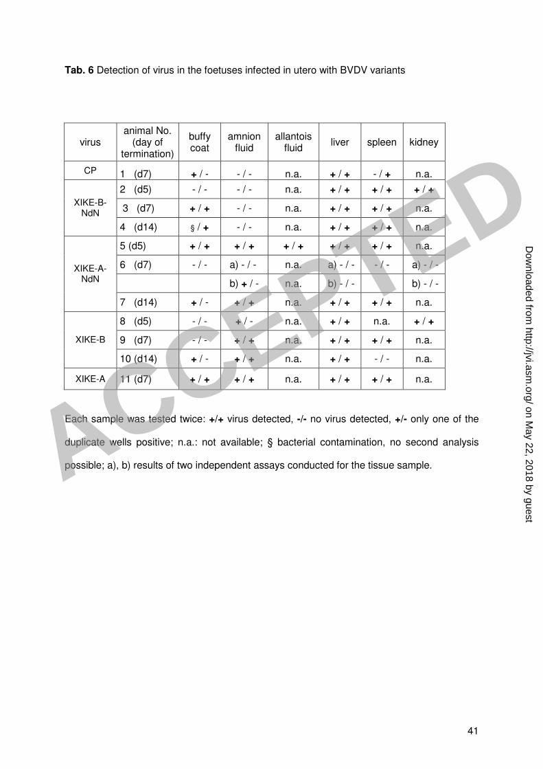
41
Tab. 6 Detection of virus in the foetuses infected in utero with BVDV variants
virus animal No.
(day of termination)
buffy coat
amnion fluid
allantois fluid
liver spleen kidney
CP 1 (d7) + / - - / - n.a. + / + - / + n.a.
2 (d5) - / - - / - n.a. + / + + / + + / +
3 (d7) + / + - / - n.a. + / + + / + n.a. XIKE-B-
NdN
4 (d14) § / + - / - n.a. + / + + / + n.a.
5 (d5) + / + + / + + / + + / + + / + n.a.
6 (d7) - / - a) - / - n.a. a) - / - - / - a) - / -
b) + / - n.a. b) - / - b) - / -
XIKE-A-NdN
7 (d14) + / - + / + n.a. + / + + / + n.a.
8 (d5) - / - + / - n.a. + / + n.a. + / +
9 (d7) - / - + / + n.a. + / + + / + n.a. XIKE-B
10 (d14) + / - + / + n.a. + / + - / - n.a.
XIKE-A 11 (d7) + / + + / + n.a. + / + + / + n.a.
Each sample was tested twice: +/+ virus detected, -/- no virus detected, +/- only one of the
duplicate wells positive; n.a.: not available; § bacterial contamination, no second analysis
possible; a), b) results of two independent assays conducted for the tissue sample.
ACCEPTED
on May 22, 2018 by guest
http://jvi.asm.org/
Dow
nloaded from