Bovine origin Staphylococcus aureus: A new zoonotic … – 625 021, Tamil Nadu, India; 2. Vascular...
6
Veterinary World, EISSN: 2231-0916 1275 Veterinary World, EISSN: 2231-0916 Available at www.veterinaryworld.org/Vol.10/October-2017/18.pdf RESEARCH ARTICLE Open Access Bovine origin Staphylococcus aureus: A new zoonotic agent? Relangi Tulasi Rao 1 , Kannan Jayakumar 1 and Pavitra Kumar 2 1. Department of Animal Behaviour & Physiology, School of Biological Sciences, Madurai Kamaraj University, Madurai – 625 021, Tamil Nadu, India; 2. Vascular Biology Laboratory, AU-KBC Research Centre, Anna University, Chennai – 600 044, Tamil Nadu, India. Corresponding author: Kannan Jayakumar, e-mail: [email protected] Co-authors: RTR: [email protected], PK: [email protected] Received: 07-07-2017, Accepted: 25-09-2017, Published online: 26-10-2017 doi: 10.14202/vetworld.2017.1275-1280 How to cite this article: Rao RT, Jayakumar K, Kumar P (2017) Bovine origin Staphylococcus aureus: A new zoonotic agent? Veterinary World, 10(10): 1275-1280. Abstract Aim: The study aimed to assess the nature of animal origin Staphylococcus aureus strains. The study has zoonotic importance and aimed to compare virulence between two different hosts, i.e., bovine and ovine origin. Materials and Methods: Conventional polymerase chain reaction-based methods used for the characterization of S. aureus strains and chick embryo model employed for the assessment of virulence capacity of strains. All statistical tests carried on R program, version 3.0.4. Results: After initial screening and molecular characterization of the prevalence of S. aureus found to be 42.62% in bovine origin samples and 28.35% among ovine origin samples. Meanwhile, the methicillin-resistant S. aureus prevalence is found to be meager in both the hosts. Among the samples, only 6.8% isolates tested positive for methicillin resistance. The biofilm formation quantified and the variation compared among the host. A Welch two-sample t-test found to be statistically significant, t=2.3179, df=28.103, and p=0.02795. Chicken embryo model found effective to test the pathogenicity of the strains. Conclusion: The study helped to conclude healthy bovines can act as S. aureus reservoirs. Bovine origin S. aureus strains are more virulent than ovine origin strains. Bovine origin strains have high probability to become zoonotic pathogen. Further, gene knock out studies may be conducted to conclude zoonocity of the bovine origin strains. Keywords: chicken embryo model, Staphylococcus aureus, virulence, zoonotic agent. Introduction Staphylococcus aureus is a major opportunis- tic pathogen responsible for wide range of infec- tions. The ability of S. aureus to cause diseases in humans and animals during immune compromised times has been attributed to its capacity to produce a variety of virulence factors [1]. The epidemiolog- ical circumstances changed mainly with the advent and spread of community-acquired methicillin-re- sistant S. aureus (CA-MRSA) strains, appearing in individuals without healthcare-associated risk fac- tors and also in animals. Nevertheless, even where household animals and horses were known to be colonized by MRSA strains, animal-to-human (and vice versa) transmission marked rare evolutionary accidents rather than being the rule. In the new mil- lennium, a novel MRSA lineage was born: Sequence type ST398 MRSA can colonize a broad spectrum of animals and has been shown to be transmitted to humans, raising the question of the advent of a new emerging zoonotic agent [2]. India is primarily based on agriculture, and most of the rural population in India actively depend on livestock for their economic growth and sustainabil- ity. In such situation, there is a significant probability for a spread of zoonotic S. aureus. In the dawn of this century, the evolution of multidrug resistance (MDR) S. aureus strains gain the attention of bacteriologists and molecular biologists. The epidemiological situa- tions differed from the previous ages with the advent and spread of CA-MRSA strains, appearing in indi- viduals without healthcare-associated risk factors and also in animals [2]. In this context, this study has been designed to understand the nature of S. aureus strains of animal origin in Madurai district, Tamil Nadu. The study pri- marily is focused on the characterization of animal origin strains and virulence capacity variations in strains isolated from bovine and ovine hosts. In this context, this study has been designed to understand the nature of S. aureus strains in Madurai district and Tamil Nadu. The study primarily is focused on the characterization of zoonotic strains and virulence capacity variations in zoonotic strains isolated from bovine and ovine hosts. Materials and Methods Ethics approval The authors declare that the research was conducted with prior approval from Institutional Copyright: Rao, et al. Open Access. This article is distributed under the terms of the Creative Commons Attribution 4.0 International License (http://creativecommons.org/licenses/by/4.0/), which permits unrestricted use, distribution, and reproduction in any medium, provided you give appropriate credit to the original author(s) and the source, provide a link to the Creative Commons license, and indicate if changes were made. The Creative Commons Public Domain Dedication waiver (http://creativecommons.org/ publicdomain/zero/1.0/) applies to the data made available in this article, unless otherwise stated.
Transcript of Bovine origin Staphylococcus aureus: A new zoonotic … – 625 021, Tamil Nadu, India; 2. Vascular...
Veterinary World, EISSN: 2231-0916 1275
Veterinary World, EISSN: 2231-0916Available at www.veterinaryworld.org/Vol.10/October-2017/18.pdf
RESEARCH ARTICLEOpen Access
Bovine origin Staphylococcus aureus: A new zoonotic agent?Relangi Tulasi Rao1, Kannan Jayakumar1 and Pavitra Kumar2
1. Department of Animal Behaviour & Physiology, School of Biological Sciences, Madurai Kamaraj University, Madurai – 625 021, Tamil Nadu, India; 2. Vascular Biology Laboratory, AU-KBC Research Centre, Anna University,
Chennai – 600 044, Tamil Nadu, India.Corresponding author: Kannan Jayakumar, e-mail: [email protected]
Co-authors: RTR: [email protected], PK: [email protected]: 07-07-2017, Accepted: 25-09-2017, Published online: 26-10-2017
doi: 10.14202/vetworld.2017.1275-1280 How to cite this article: Rao RT, Jayakumar K, Kumar P (2017) Bovine origin Staphylococcus aureus: A new zoonotic agent? Veterinary World, 10(10): 1275-1280.
AbstractAim: The study aimed to assess the nature of animal origin Staphylococcus aureus strains. The study has zoonotic importance and aimed to compare virulence between two different hosts, i.e., bovine and ovine origin.
Materials and Methods: Conventional polymerase chain reaction-based methods used for the characterization of S. aureus strains and chick embryo model employed for the assessment of virulence capacity of strains. All statistical tests carried on R program, version 3.0.4.
Results: After initial screening and molecular characterization of the prevalence of S. aureus found to be 42.62% in bovine origin samples and 28.35% among ovine origin samples. Meanwhile, the methicillin-resistant S. aureus prevalence is found to be meager in both the hosts. Among the samples, only 6.8% isolates tested positive for methicillin resistance. The biofilm formation quantified and the variation compared among the host. A Welch two-sample t-test found to be statistically significant, t=2.3179, df=28.103, and p=0.02795. Chicken embryo model found effective to test the pathogenicity of the strains.
Conclusion: The study helped to conclude healthy bovines can act as S. aureus reservoirs. Bovine origin S. aureus strains are more virulent than ovine origin strains. Bovine origin strains have high probability to become zoonotic pathogen. Further, gene knock out studies may be conducted to conclude zoonocity of the bovine origin strains.
Keywords: chicken embryo model, Staphylococcus aureus, virulence, zoonotic agent.
Introduction
Staphylococcus aureus is a major opportunis-tic pathogen responsible for wide range of infec-tions. The ability of S. aureus to cause diseases in humans and animals during immune compromised times has been attributed to its capacity to produce a variety of virulence factors [1]. The epidemiolog-ical circumstances changed mainly with the advent and spread of community-acquired methicillin-re-sistant S. aureus (CA-MRSA) strains, appearing in individuals without healthcare-associated risk fac-tors and also in animals. Nevertheless, even where household animals and horses were known to be colonized by MRSA strains, animal-to-human (and vice versa) transmission marked rare evolutionary accidents rather than being the rule. In the new mil-lennium, a novel MRSA lineage was born: Sequence type ST398 MRSA can colonize a broad spectrum of animals and has been shown to be transmitted to humans, raising the question of the advent of a new emerging zoonotic agent [2].
India is primarily based on agriculture, and most of the rural population in India actively depend on livestock for their economic growth and sustainabil-ity. In such situation, there is a significant probability for a spread of zoonotic S. aureus. In the dawn of this century, the evolution of multidrug resistance (MDR) S. aureus strains gain the attention of bacteriologists and molecular biologists. The epidemiological situa-tions differed from the previous ages with the advent and spread of CA-MRSA strains, appearing in indi-viduals without healthcare-associated risk factors and also in animals [2].
In this context, this study has been designed to understand the nature of S. aureus strains of animal origin in Madurai district, Tamil Nadu. The study pri-marily is focused on the characterization of animal origin strains and virulence capacity variations in strains isolated from bovine and ovine hosts. In this context, this study has been designed to understand the nature of S. aureus strains in Madurai district and Tamil Nadu. The study primarily is focused on the characterization of zoonotic strains and virulence capacity variations in zoonotic strains isolated from bovine and ovine hosts.Materials and MethodsEthics approval
The authors declare that the research was conducted with prior approval from Institutional
Copyright: Rao, et al. Open Access. This article is distributed under the terms of the Creative Commons Attribution 4.0 International License (http://creativecommons.org/licenses/by/4.0/), which permits unrestricted use, distribution, and reproduction in any medium, provided you give appropriate credit to the original author(s) and the source, provide a link to the Creative Commons license, and indicate if changes were made. The Creative Commons Public Domain Dedication waiver (http://creativecommons.org/publicdomain/zero/1.0/) applies to the data made available in this article, unless otherwise stated.
Veterinary World, EISSN: 2231-0916 1276
Available at www.veterinaryworld.org/Vol.10/October-2017/18.pdf
Biosafety and Ethical Committee (IBEC) of the School of Biological Sciences, Madurai Kamaraj University(Dated 14 Nov 2014).Sample collection
Nasal swabs collected from healthy adult ani-mals include cows (Bovines) and sheep (Ovine) with sterile cotton swabs (HiMedia Sterile culture collect-ing device) in Madurai district during October 2015-March 2016. Collected samples transported to the laboratory in Cary-Blair transport media. The sam-ples processed on the day of sample collection itself to ensure the maximum recovery of bacteria. Samples not more than five were collected to provide heteroge-neity from any herd or farm.Initial screening for S. aureus
The nasal swabs collected initially spread on Mannitol salt agar (MSA) and Baird-Parker Agar, a selective media for S. aureus. Further Gram staining performed for positive isolates on agar plates. Plates incubated at 37°C for 24-48 h.Multiplex polymerase chain reaction (PCR) amplification
For molecular characterization, PCR-based methods were used. DNA isolation performed with the aid of Favorogen Tissue DNA extraction kit, Taiwan. DNA extracted according to the manufactur-er’s protocol. Lysostaphin (Sigma, USA) is used for better lysis of the cell walls as per manufacturer’s rec-ommendation. The PCR conditions and primers used as described in the previous literature. Two different multiplex PCR reactions have been carried out based on standard methods [3-5] with little modifications, to confirm S. aureus at species level and methicillin resistance. One multiplex PCR reactions with the Staphylococcus-specific 16s rRNA, nuc, and sdrE and another set comprised Staphylococcus-specific 16s rRNA, mecA, and sdrE. PCR products were resolved on agarose (1% w/v) gel electrophoresis.Virulence analysis carried out with following experiments
Assessment of biofilm formationBiofilm quantification was carried out accord-
ing to the protocol of O’Toole [6]. Absorbance quan-tified using microtiter plate reader (Spectramax M2, Molecular Devices, USA). Cells were grown on 96-well costar plates and incubated at 37°C for 24 h for visualization of biofilm development. After the incubation, the wells were washed with sterile phos-phate-buffered saline and stained with acridine orange (0.1% w/v), and the biofilms were imaged using high-content screening (HCS) using Operetta imaging system (Perkin Elmer, Germany). The Z-stack analy-sis performed using Harmony software 2.0.1.In vivo estimation of virulence - chicken embryo infection model
The estimation of the virulence capacity of strain was performed using chicken embryo as a model.
The experiment conducted for few selected strains, whereas MRSA- MTCC1430 served as the positive control and saline (0.7% NaCl) as a negative con-trol. The experiment carried according to the proto-col developed earlier with the hatching eggs obtained from commercial hatcheries and ensured feed does not contain any antibiotic [7]. Briefly, the eggs were incu-bated at 37°C and a relative humidity of 50-60% in a laboratory incubator. On the 10th day of development, the eggs candled and dead and unfertilized embryos removed. A small hole was aseptically drilled in the eggshell using a needle, and a suspension (50 µl, ~ 106 colony-forming units [CFUs]) of the S. aureus strain being tested was inoculated intraallantoically, and 20 eggs per group challenged with bacterial strain. The liver damage further confirmed by histopathological analysis.Statistical analyses
All statistical analyses executed on R program (Welch two sample t-test), version 3.0.4 on local Linux machine.Results
Samples collected in Madurai district from adult healthy animals only. The sampling details found in Figure-1. A total of 128 samples collected in Madurai district mostly with the rural backdrop.Initial screening for S. aureus
The nasal swabs collected were initially spread on MSA and Baird-Parker Agar, a selective media for S. aureus. Further, Gram staining performed for pos-itive isolates on agar plates. After incubation at 37°C for 24 h, the agar plates observed for the growth of bacterial colonies. The colonies with yellow to golden yellow color on MSA plates and black colonies on Baird-Parker agar plates selected for further screen-ing. 30 samples from the bovine origin and 20 sam-ples from ovine origin were positive on both selective media agar plates. Gram staining performed for further confirmation. False-positive samples eliminated with Gram staining. After Gram staining, 32 samples from bovine origin exhibited spherical-shaped morphology and Gram-positive and 25 samples from ovine origin positive for S. aureus morphology.Molecular characterization
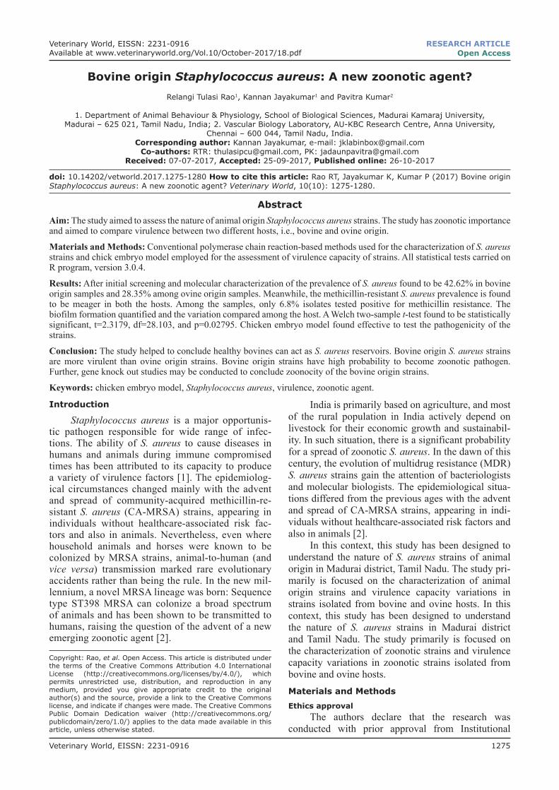
Two multiplex PCR reactions were carried out for further confirmation. Multiplex PCR performed with reported species-specific primers. In this study, one multiplex PCR carried out with Staphylococcus-specific 16s rRNA, nuc, and sdrE gene primers to confirm isolated strain as S. aureus at molecular level. Another multiplex carried out with a little modifica-tion to assess the strain’s methicillin resistance at the molecular level. In this set, nuc gene primer replaced with mecA primer. Figure-1a and b illustrated the multiplex PCR results. After PCR confirmation, 26 isolates from the bovine origin and 19 samples from ovine origin confirmed as S. aureus. Among these
Veterinary World, EISSN: 2231-0916 1277
Available at www.veterinaryworld.org/Vol.10/October-2017/18.pdf
isolates, 10 isolates confirmed as MRSA based on the presence of mecA gene in their genome.Prevalence analysis
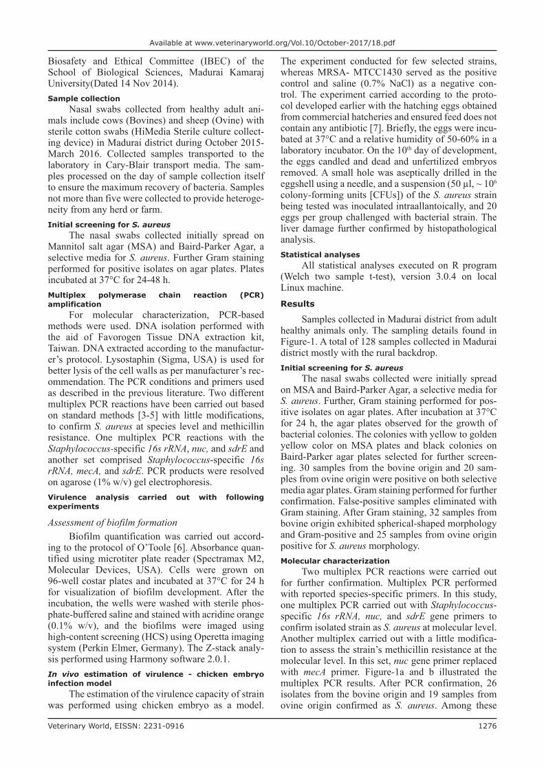
After molecular confirmation, the prevalence rate of S. aureus in the study population was analyzed. It has been observed that the prevalence rate in bovine origin samples was around 42.62%, while it was low in the case of ovine origin samples with 28.35% only. The prevalence rates can be seen in a bar diagram with MRSA rates as well (Figure-2). The prevalence rate of MRSA found to be very meager to an extent of 6.8%.Virulence analysis
Different experiments including in vivo virulence analysis performed to estimate the virulence capacity of the positive S. aureus isolates of this study and to compare the host-based virulence assessment.Biofilm formation capacity
Biofilm formation is a potential virulence factor in S. aureus isolated from clinical mastitis [8]. Hence, to have an understanding of the disease establishment by S. aureus strains from healthy animals, the biofilm formation assessment was carried out. In Table-1, the mean and standard deviation values of biofilm of each strain of this study enlisted. The results showed that the strains were weak to high capacity of biofilm for-mation. A parametric statistic test, Welch two sample t-test used to detect the variation of biofilm forma-tion among the host-based specificity. This test found to be statistically significant, t=2.3179, df=28.103, and p=0.02795. This result suggests bovine origin S. aureus strains has the better capacity of biofilm for-mation than the ovine origin strains.Visualization of biofilm
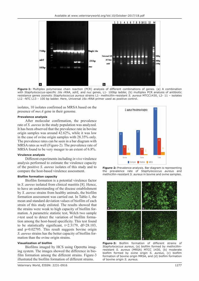
Biofilms imaged by HCS using Operetta imag-ing system. The images showed the difference in bio-film formation among the different strains. Figure-3 illustrated the biofilm formation of different strains.
Figure-2: Prevalence analysis. Bar diagram is representing the prevalence rate of Staphylococcus aureus and methicillin-resistant S. aureus in bovine and ovine samples.
Figure-1: Multiplex polymerase chain reaction (PCR) analysis of different combinations of genes. (a) A combination with Staphylococcus-specific 16s rRNA, sdrE, and nuc genes, L1- 100bp ladder, (b) multiplex PCR analysis of antibiotic resistance genes zoonotic Staphylococcus aureus strains L1- methicillin-resistant S. aureus MTCC1430, L2- 11 – isolates L12 –NTC L13 – 100 bp ladder. Here, Universal 16s rRNA primer used as positive control.
ba
Figure-3: Biofilm formation of different strains of Staphylococcus aureus, (a) biofilm formed by methicillin-resistant S. aureus (MRSA) MTCC 1430, (b) moderate biofilm formed by ovine origin S. aureus, (c) biofilm formation of bovine origin MRSA, and (d) biofilm formation of bovine origin S. aureus.
dc
ba

Veterinary World, EISSN: 2231-0916 1278
Available at www.veterinaryworld.org/Vol.10/October-2017/18.pdf
Chicken embryo assaySince the biofilm formation is the key criteria
for initial adherence and disease establishment, and again, it is also acts as a virulence factor in disease establishment [8] the strains which are capable of more biofilm (Table-1) along with which positive for toxic shock syndrome toxin gene (tsst-1, data not shown) selected for in vivo analysis. The mean sur-vival rates noted till 7th day after the commencement of the experiment. On the 7th day, liver dissected from euthanized embryos and tested for the confirmation of pathogen with counted conventional serial dilu-tion method. The results showed the presence of S. aureus in liver samples and evident for bacterial col-onization. CFU counted conventionally. MANOVA analysis of CFUs and liver weight for different groups with respect to control yielded a significant differ-ence among groups after challenging with pathogen. Results from this MANOVA demonstrated a signifi-cant multivariate effect for the relationship, Wilkin’s Λ (7, 21) = 0.011, p<0.0001. The survival analysis of embryos also performed till the end of the experiment and results illustrated in Figure-4.Histopathological studies
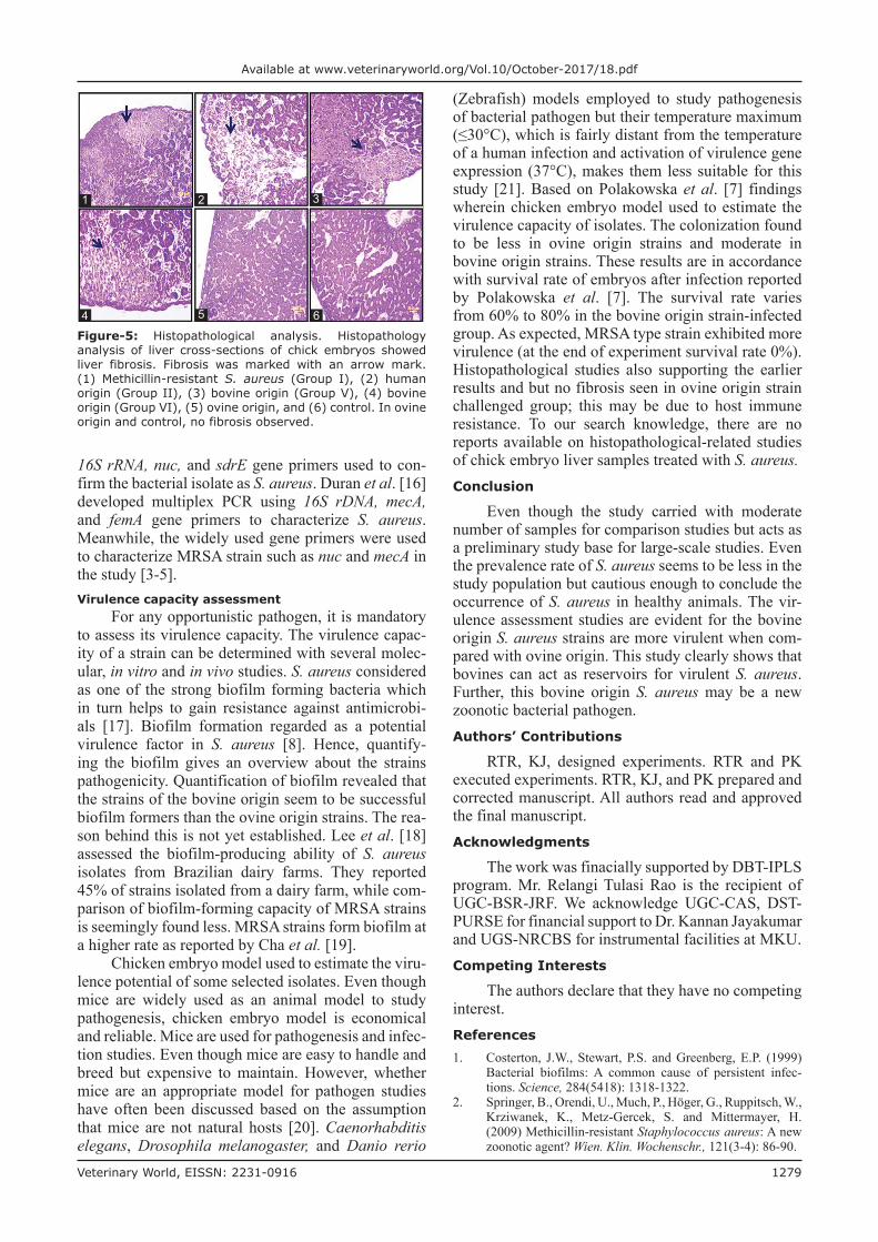
Histopathological studies carried to check liver damage by pathogen (Figure-5). As expected with respect to control fibrosis noticed in the liver samples challenged with pathogen but no fibrosis observed liver samples which challenged with ovine origin strains.Discussion
S. aureus being an opportunistic pathogen and MDR superbugs have been reported recently, and hence, the scientific community interested in studying the evolutionary trends of this bacterium. The pres-ent study focused on the prevalence and pathogen potentiality of zoonotic origin strains. Nasal swabs used as samples since it is the prescribed sampling method [9]. After initial screening and molecular characterization of the prevalence of S. aureus found to be 42.62% in bovine origin samples and 28.35% among ovine origin samples. Meanwhile, the MRSA prevalence is found to be meager in both the hosts. Among the samples, only 6.8% isolates tested positive for methicillin resistance. Despite the prevalence rate seems to be very less, the hosts still acts as reservoirs. However, the reason for the prevalence of S. aureus found more in bovines (cows) while compared to that of ovine samples is yet too established.
The majority of the prevailing reports focused mainly on human origin and bovine mastitis S. aureus [10,11]. MRSA prevalence results can be comparable with Haran et al.’s [12], wherein they are reported only as 4% of MRSA in herd animals. In India, Kumar et al. [13] also reported that preva-lence of 13.1% MRSA in Sahiwal cattle. In contrast to animal sample, milk carries less S. aureus as it was noticed in the reports of Fagundes et al. [14] and
Thaker et al. [15] which revealed the presence as 7.3% and 6.25%, respectively. Increasing number of sam-ples may alter the prevalence rate and helps to obtain the approximate prevalence in the study population.
The prevalence of any pathogen and its epidemi-ological importance can be confirmed only after con-firming the strain with the aid of molecular tools only. Molecular characterization of S. aureus performed with established protocols in accordance to the pre-vious studies. In this study, Staphylococcus-specific
Table-1: Biofilm quantification of S. aureus strains.
Biofilm quantification
Bovine origin strains Ovine origin strains
Sample ID
Mean±SD Sample ID
Mean±SD
A1 0.0384±0.007635 C1 0.0402±0.011212A2 0.0386±0.028086 C2 0.0734±0.01491A3 0.0434±0.009529 C9 0.0478±0.012988A5 0.1228±0.0561 C12 0.0472±0.008438A6 0.0694±0.038188 C13 0.0768±0.018089A7 0.1254±0.043362 C15 0.144±0.06804A8 0.1186±0.023158 C16 0.1026±0.020888A9 0.0322±0.0113 C18 0.0884±0.02446A10 0.0654±0.009864 C19 0.0636±0.019008A12 0.0314±0.00929 C22 0.0606±0.030648A13 0.0796±0.007301 C25 0.052±0.007106A15 0.0476±0.0177 C29 0.0616±0.015192A19 0.0506±0.010262 C30 0.0928±0.016634A20 0.0412±0.02955 C31 0.0434±0.013975A21 0.0368±0.017635 C32 0.049±0.011895A22 0.055±0.015427 AG7 0.0378±0.012637A24 0.0508±0.011389 AG9 0.0434±0.023533A25 0.2442±0.010918 CS5 0.0382±0.008438A27 0.0432±0.009121 CS7 0.0638±0.008468O1 0.0842±0.033641B1 0.18625±0.017443B2 0.176±0.049221B3 0.30375±0.082553B8 0.255±0.02735
S. aureus=Staphylococcus aureus, SD=Standard deviation
Figure-4: Survival analysis curve. The curves are representing the survival rates of chicken embryos from day 1 to day 7 after challenging with Staphylococcus aureus. Control: 0.7% NaCl, Group I - methicillin-resistant S. aureus, Group II - human origin S. aureus, Group - III and IV - ovine origin S. aureus (Sample ID C15 and C30), and Group V – VII – bovine origin S. aureus strains (Sample IDs- A25, B1, and B2, respectively).
Veterinary World, EISSN: 2231-0916 1279
Available at www.veterinaryworld.org/Vol.10/October-2017/18.pdf
16S rRNA, nuc, and sdrE gene primers used to con-firm the bacterial isolate as S. aureus. Duran et al. [16] developed multiplex PCR using 16S rDNA, mecA, and femA gene primers to characterize S. aureus. Meanwhile, the widely used gene primers were used to characterize MRSA strain such as nuc and mecA in the study [3-5].Virulence capacity assessment
For any opportunistic pathogen, it is mandatory to assess its virulence capacity. The virulence capac-ity of a strain can be determined with several molec-ular, in vitro and in vivo studies. S. aureus considered as one of the strong biofilm forming bacteria which in turn helps to gain resistance against antimicrobi-als [17]. Biofilm formation regarded as a potential virulence factor in S. aureus [8]. Hence, quantify-ing the biofilm gives an overview about the strains pathogenicity. Quantification of biofilm revealed that the strains of the bovine origin seem to be successful biofilm formers than the ovine origin strains. The rea-son behind this is not yet established. Lee et al. [18] assessed the biofilm-producing ability of S. aureus isolates from Brazilian dairy farms. They reported 45% of strains isolated from a dairy farm, while com-parison of biofilm-forming capacity of MRSA strains is seemingly found less. MRSA strains form biofilm at a higher rate as reported by Cha et al. [19].
Chicken embryo model used to estimate the viru-lence potential of some selected isolates. Even though mice are widely used as an animal model to study pathogenesis, chicken embryo model is economical and reliable. Mice are used for pathogenesis and infec-tion studies. Even though mice are easy to handle and breed but expensive to maintain. However, whether mice are an appropriate model for pathogen studies have often been discussed based on the assumption that mice are not natural hosts [20]. Caenorhabditis elegans, Drosophila melanogaster, and Danio rerio
(Zebrafish) models employed to study pathogenesis of bacterial pathogen but their temperature maximum (≤30°C), which is fairly distant from the temperature of a human infection and activation of virulence gene expression (37°C), makes them less suitable for this study [21]. Based on Polakowska et al. [7] findings wherein chicken embryo model used to estimate the virulence capacity of isolates. The colonization found to be less in ovine origin strains and moderate in bovine origin strains. These results are in accordance with survival rate of embryos after infection reported by Polakowska et al. [7]. The survival rate varies from 60% to 80% in the bovine origin strain-infected group. As expected, MRSA type strain exhibited more virulence (at the end of experiment survival rate 0%). Histopathological studies also supporting the earlier results and but no fibrosis seen in ovine origin strain challenged group; this may be due to host immune resistance. To our search knowledge, there are no reports available on histopathological-related studies of chick embryo liver samples treated with S. aureus.Conclusion
Even though the study carried with moderate number of samples for comparison studies but acts as a preliminary study base for large-scale studies. Even the prevalence rate of S. aureus seems to be less in the study population but cautious enough to conclude the occurrence of S. aureus in healthy animals. The vir-ulence assessment studies are evident for the bovine origin S. aureus strains are more virulent when com-pared with ovine origin. This study clearly shows that bovines can act as reservoirs for virulent S. aureus. Further, this bovine origin S. aureus may be a new zoonotic bacterial pathogen.Authors’ Contributions
RTR, KJ, designed experiments. RTR and PK executed experiments. RTR, KJ, and PK prepared and corrected manuscript. All authors read and approved the final manuscript.Acknowledgments
The work was finacially supported by DBT-IPLS program. Mr. Relangi Tulasi Rao is the recipient of UGC-BSR-JRF. We acknowledge UGC-CAS, DST-PURSE for financial support to Dr. Kannan Jayakumar and UGS-NRCBS for instrumental facilities at MKU.Competing Interests
The authors declare that they have no competing interest.References1. Costerton, J.W., Stewart, P.S. and Greenberg, E.P. (1999)
Bacterial biofilms: A common cause of persistent infec-tions. Science, 284(5418): 1318-1322.
2. Springer, B., Orendi, U., Much, P., Höger, G., Ruppitsch, W., Krziwanek, K., Metz-Gercek, S. and Mittermayer, H. (2009) Methicillin-resistant Staphylococcus aureus: A new zoonotic agent? Wien. Klin. Wochenschr., 121(3-4): 86-90.
Figure-5: Histopathological analysis. Histopathology analysis of liver cross-sections of chick embryos showed liver fibrosis. Fibrosis was marked with an arrow mark. (1) Methicillin-resistant S. aureus (Group I), (2) human origin (Group II), (3) bovine origin (Group V), (4) bovine origin (Group VI), (5) ovine origin, and (6) control. In ovine origin and control, no fibrosis observed.
4
32
6
1
5
Veterinary World, EISSN: 2231-0916 1280
Available at www.veterinaryworld.org/Vol.10/October-2017/18.pdf
3. Molla, B., Byrne, M., Abley, M., Mathews, J., Jackson, C.R., Fedorka-Cray, P., Sreevatsan, S., Wang, P. and Gebreyes, W.A. (2012) Epidemiology and genotypic characteristics of methicillin-resistant Staphylococcus aureus strains of porcine origin. J. Clin. Microbiol., 50(11): 3687-3693.
4. Frana, T.S., Beahm, A.R., Hanson, B.M., Kinyon, J.M., Layman, L.L., Karriker, L.A., Ramirez, A. and Smith, T.C. (2013) Isolation and characterization of methicillin-resis-tant Staphylococcus aureus from pork farms and visiting veterinary students. PLoS One, 8(1): e53738.
5. Rahimi, H., Saei, H.D. and Ahmadi, M. (2015) Nasal car-riage of Staphylococcus aureus: Frequency and antibiotic resistance in healthy ruminants. Jundishapur. J. Microbiol., 8(10): e22413.
6. O’Toole, G.A. (2011) Microtiter dish biofilm formation assay. J. Vis. Exp., 47: 2437.
7. Polakowska, K., Lis, M.W., Helbin, W.M., Dubin, G., Dubin, A., Niedziolka, J.W., Miedzobrodzki, J. and Wladyka, B. (2012) The virulence of Staphylococcus aureus correlates with strain genotype in a chicken embryo model but not a nematode model. Microb. Infect., 14(14): 1352-1362.
8. Hensen, S.M., Pavičić, M.J.A., Lohuis, J.A.C., de Hoog, J.A.M. and Poutrel, B. (2000) Location of Staphylococcus aureus with in the experimentally infected bovine udder and the expression of capsular polysaccharide Type 5 in situ. J. Dairy Sci., 83(9): 1966-1975.
9. Mørk, T., Kvitle, B., Mathisen, T. and Jørgensen, H.J. (2010) Bacteriological and molecular investigations of Staphylococcus aureus in dairy goats. Vet. Microbiol., 141(1-2): 134-141.
10. Dubey, D., Rath, S., Sahu, M.C., Pattnaik, L., Debata, N.K. and Padhy, R.N. (2013) Surveillance of infection status of drug resistant Staphylococcus aureus in an Indian teaching hospital. Asian Pac. J. Trop. Dis., 3(2): 133-142.
11. Gharsa, H., Slama, K.B., Gómez-Sanz, E., Lozano, C., Zarazaga, M., Messadi, L., Boudabous, A. and Torres, C. (2015) Molecular characterization of Staphylococcus aureus from nasal samples of healthy farm animals and pets in Tunisia. Vector Borne Zoonotic Dis., 15(2): 109-115.
12. Haran, K.P., Godden, S.M., Boxrud, D., Jawahir, S., Bender, J.B. and Sreevatsan, S. (2012) Prevalence and
characterization of Staphylococcus aureus, including meth-icillin-resistant Staphylococcus aureus, isolated from bulk tank milk from Minnesota dairy farms. J. Clin. Microbiol., 50(3): 688-695.
13. Kumar, R., Yadav, B.R. and Singh, R.S. (2011) Antibiotic resistance and pathogenicity factors in Staphylococcus aureus isolated from mastitic Sahiwal cattle. J. Biosci., 36(1): 175-188.
14. Fagundes, H., Barchesi, L., Filho, A.N., Ferreira, L.M. and Oliveira, C.A.F. (2010) Occurrence of Staphylococcus aureus in raw milk produced in dairy farms in São Paulo state, Brazil. Braz. J. Microbiol., 41(2): 376-380.
15. Thaker, H.C., Brahmbhatt, M.N. and Nayak, J.B. (2013) Isolation and identification of Staphylococcus aureus from milk and milk products and their drug resistance patterns in Anand, Gujarat. Vet. World, 6(1): 10-13.
16. Duran, N., Ozer, B., Duran, G.G., Onlen, Y. and Demir, C. (2012) Antibiotic resistance genes and susceptibility pat-terns in staphylococci. Indian J. Med. Res., 135(3): 389-396.
17. Gefen, O. and Balaban, N.Q. (2009) The importance of being persistent: heterogeneity of bacterial populations under antibiotic stress. FEMS Microbiol. Rev., 33(4): 704-717.
18. Lee, S.H.I., Mangolin, B.L.C., Gonçalves, J.L., Neeff, D.V., Silva, M.P., Cruz, A.G. and Oliveira, C.A.F. (2014) Biofilm producing ability of Staphylococcus aureus isolates from Brazilian dairy farms. J. Dairy Sci., 97(3): 1812-1816.
19. Cha, J.O., Yoo, J.I., Yoo, J.S., Chung, H.S., Park, S.H., Kim, H.S., Lee, Y.S. and Chung, G.T. (2013) Corrigendum to “investigation of biofilm formation and its association with the molecular and clinical characteristics of methicil-lin-resistant Staphylococcus aureus”. Osong Public Health Res. Perspect., 4(5): 225-232.
20. Mulcahy, M.E., Geoghegan, J.A., Monk, I.R., O’Keeffe, K.M., Walsh, E.J., Foster, T.J. and McLoughlin, R.M. (2012) Nasal colonisation by Staphylococcus aureus depends upon clumping factor B binding to the squamous epithelial cell envelope protein loricrin. PLoS Pathog., 8(12): e1003092.
21. Anderson, C., Gripenland, J. and Johansson, J. (2015) Using the chicken embryo to assess virulence of Listeria monocytogenes and to model other microbial infections. Nat. Protoc., 10(8): 1155-1164.
********