fruiting bodies and mycelia obtained by in vitro culture ...

BONIT® SURFACE COATING FOR IMPLANTS

BONIT® INTRODUCTIONThe ideal surgical outcome of a dental implant is stability and a sustained connection to surrounding bone. Achieving this outcome relies on a number of factors, not the least of which is the formation of a tight, sealed bond between implant and bone. Implant design, and specifically the surface properties of the implant, have a profound impact on osseointegration. Surface modification of implants facilitates biocompatibility and long-term stability {1, 2}.
An ideal implant offers a macroporous surface and is biocompatible with bone, supporting osseointegration. Typically, implant surfaces have combined a porous titanium coating with a bioactive calcium phosphate coating to achieve a biocompatible finish. Calcium phosphates are commonly used in medical coating technology as they support accelerated growth of bone tissue and promote a strong bond between the implant and surrounding tissue, optimizing healing {3}. For over 30 years, the
combination of sprayed titanium and the poorly-soluble calcium phosphate hydoroxyapatite (HA) was considered the industry standard. This coating was applied to the implant surface at a thickness of >50-200 μm, with the porous titanium coating responsible for mechanical anchorage of the bone, and the HA coating responsible for the promotion of osteogenesis. Despite its many positive properties, this type of coating presents a range of surgical challenges, including thermal decomposition of the HA powder during spraying (which may cause infiltration or separation of the coating), and coating delamination and flaking caused by the high solubility in vivo of amorphous calcium phosphate. The consequences of coating deteriorations such as these include the potential formation of a connective tissue failure in the resulting gap, causing an aseptic loosening of the implant {4}. Furthermore, the “line-of-sight” spraying process does not take into account the porous surfaces and complex geometries of some implants.
surgikorimplants.com | 2

Thus, implant coatings have been approached with renewed interest, and current thinking holds that bioactive coatings are required on the implant surface only until the implant has successfully osseointegrated {5, 6}. Recent scientific literature supports this theory and shows a shift from the standard HA coating to other calcium phosphates such as brushite, monetite, OCP or TCP, which offer controlled solubility and subsequent bone growth directly on the implant as the surface coating dissolves {7, 8, 9, 10, 11, 12}. To achieve controlled resorption of the calcium phosphate, a thin coating with a fine, crystalline structure is required, which is generally not possible with conventional spraying technology.
The BONIT® coating, developed by DOT, meets the necessary requirements for accelerated formation
of new bone. The coating is a thin, bioactive calcium phosphate surface that is applied to the implant by electrochemical deposition, resulting in a fine, crystalline structure and improved solubility and resorption. This process eliminates hard particles and subsequent flaking, and the coating process from a liquid phase ensures consistent, thin (20±10μm) coverage of complex implant architectures. BONIT® coating is a composite of brushite, an easily-soluble calcium phosphate, and HA. Brushite is known for its excellent biocompatibility {13}, and biologically, brushite forms a reservoir for calcium and phosphate ions that support bone apposition. During the process of mineralization, an easily soluble modification like brushite is converted into less-soluble HA, supporting the natural osseointegration process {14, 15}.
BONIT® FEATURESI. Shape and Structure
Macroscopically, the BONIT® coating presents as a light-gray, finely-structured surface (Fig. 1).
II. Phase Composition and Ca:P Ratio
The phase composition of BONIT® coating is the mass-fraction-weighted total of the Ca:P values of the brushite and HA components. The calcium phosphate of the BONIT® coating is a composite (Fig. 2a), the majority of which is a calcium phosphate brushite phase [CaH(PO4) x 2 H2O] and the remainder a less-
soluble hydroxyapatite phase [Ca5(PO4)3OH] {16}. The brushite and HA are both naturally-occurring, inorganic compounds.
Fig. 2a
surgikorimplants.com | 3
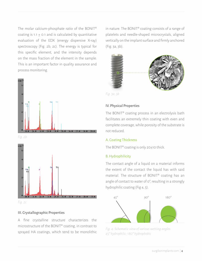
The molar calcium-phosphate ratio of the BONIT® coating is 1.1 ± 0.1 and is calculated by quantitative evaluation of the EDX (energy dispersive X-ray) spectroscopy (Fig. 2b, 2c). The energy is typical for this specific element, and the intensity depends on the mass fraction of the element in the sample. This is an important factor in quality assurance and process monitoring.
III. Crystallographic Properties
A fine crystalline structure characterizes the microstructure of the BONIT® coating, in contrast to sprayed HA coatings, which tend to be monolithic
in nature. The BONIT® coating consists of a range of platelets and needle-shaped microcrystals, aligned vertically on the implant surface and firmly anchored (Fig. 3a, 3b).
IV. Physical Properties
The BONIT® coating process in an electrolysis bath facilitates an extremely thin coating with even and complete coverage, while porosity of the substrate is not reduced.
A. Coating Thickness
The BONIT® coating is only 20±10 thick.
B. Hydrophilicity
The contact angle of a liquid on a material informs the extent of the contact the liquid has with said material. The structure of BONIT® coating has an angle of contact to water of 0°, resulting in a strongly hydrophilic coating (Fig 4, 5).
Fig. 2b
Fig. 3a, 3b
Fig. 2c45° 90° 180°
Fig. 4: Schematic view of various wetting angles45° hydrophilic; 180° hydrophobic
surgikorimplants.com | 4

C. Capillarity
The vertically-aligned, closely packed calcium phosphate crystals and associated large surface area result in high capillarity on the implant surface, ensuring a complete wetting of the implant surface at the slightest contact with fluids (Fig. 6a, 6b). This in turn transports growth factors from blood to the coating and the metallic implant surface where they are immobilized. As a result of the bone-like chemical environment, stem cells are stimulated to form osteoblasts and new bone tissue, forming a critical basis for successful osseointegration of the implant.
D. Adhesion Strength
The fine, even crystalline surface properties result in adhesion strength greater than or equal to 15 MPa. A screw test in cortical swine bone demonstrated a very minor shearing on the outer thread flanks. While the crystals were partially compressed or laterally displaced (Fig. 7a, 7b), they still formed a strongly adhesive film on the implant surface, and delamination and cracking were not observed. This confirmed that the coating adhesion was retained despite severe torsional loading, demonstrating that the BONIT® coating could retain its osteogenetic function{17}. Adhesion strength testing was conducted in accordance with the ASTM F1147 standard.
Fig. 5: Water-contact angle of BONIT®, drops aspirated, contact angle = 0°
ANGLE OF CONTACT TO WATER 0°
Fig. 6a-b: Capillary effect of BONIT®-coated dental im-plants in vivo
Fig. 7a-b: BONIT®-coated implant after screw test and BONIT® coating after screw test in swine bonein swine bone
V. Biological Properties
The BONIT® coating is a bioactive calcium phosphate surface that supports the adhesion of osteoblasts and promotes cell proliferation. The cells demonstrate good adhesion on the BONIT® surface and a typical morphology for osteoblasts (Fig. 8a, 8b). Under a scanning electron microscope, the integration of cells into the BONIT® material is clearly visible (Fig. 8c).
surgikorimplants.com | 5

BONIT® coating consists of two calcium phosphate phases. The more easily-soluble outer calcium phosphate phase, brushite, occurs in natural bone as an intermediate stage during calcification of new bone tissue {18}. When brushite dissolves, high concentrations of calcium and phosphate ions are released, resulting in fast-contact osteogenesis and high mineralization rate {19}. Brushite stimulates bone synthesis in the short term, and accelerates the osseointegration of the implants, particularly in the primary phase.
The inner calcium phosphate phase HA is resorbed more slowly and releases ions that promote formation of new bone over a longer period. Consequently, the BONIT® coating is fully resorbed over a period of 6-12 weeks after implant placement, and simultaneously replaced by newly formed bone tissue. The result is an optimal bond between bone and implant. The osteoinductive properties and controlled resorption are the primary advantages of the BONIT® coating.
A. Differentiation of Cells In Vitro with BONIT®
The influence of the BONIT® coating on cell differentiation was examined by a Co culture of the hFOB1.19 osteoblast cell line with TPS/BONIT®-coated platelets of TiAI6V4. The osteoblast-specific collagen synthesis was analyzed at various points
during incubation. The result after 6 days and after 10 days of incubation showed increased collagen synthesis on the BONIT®-coated test bodies (Fig. 9).
Fig. 8a: Bone tissue formation on BONIT® in vivo
Fig. 8b: Human osteoblasts on BONIT® in vitro
Fig. 8c: MG 63 osteoblast cells on BONIT®, Side view
Fig. 9: Influence of the BONIT® coating on the collagen synthesis in vitro
200
150
100
50
06.-8. day
CICP
(ng/
ml) Control
(uncoated)
BONIT®-coated
8.-10. day
B. Mineralization In Vitro with BONIT®
The influence of the BONIT® coating on mineralization was analyzed by incubating test bodies coated with BONIT® in cell culture medium (DMEM) for 7 days. The extract was added to a confluent cell layer and the mineralization was confirmed by van Kossa staining. With van Kossa staining, mineralized areas appear black. Figure 10 shows the difference between
Fig. 10: Mineralization pattern of osteoblasts under vari-ous culture conditions (van Kossa staining)
surgikorimplants.com | 6

the control medium and the BONIT® extract. While slight mineralization could be confirmed in the cells in the control medium, strong mineralization could be confirmed with the BONIT® extract. This indicates that the calcium phosphate phases in BONIT® coating stimulate the mineralization of human osteoblasts.
C. Immunological Reactivity In Vitro with BONIT®
Immunological reactivity to the BONIT® coating was analyzed via its interaction with interleukin 1ß (IL-1ß). IL-1ß is a typical enzyme that is released during the early inflammation phase, and influences bone resorption. The test was conducted with monocytes and macrophages of the mouse cell line J-774A.1, which were cultured either with control bodies (TiAI6V4/TPS) or with BONIT®-coated test bodies. After three days of culture, the expression of IL-1ß was analyzed. The BONIT®-coated samples in comparison with the uncoated control demonstrated a significant reduction of IL-1ß release (Fig. 11). This demonstrates that the BONIT® coating triggers virtually no inflammation parameters, and may be classified as very biocompatible.
Fig. 11: Release of IL-1ß under various culture conditions
Fig. 12: Influence if the BONIT® coating on the protein adsorption in vitro
4
3
2
1
0
uncoated
IL-1ß
(pg/
ml) Control(uncoated)
BONIT®-coated
BONIT®
4
3
2
1
0
1h
Prot
ein ad
sorp
tion Control
(uncoated)
BONIT®-coated
4h
D. Analysis of Protein Absorption In Vitro with BONIT®The protein absorption or immobilization of proteins at the implant surface is, clinically viewed, an important step in the osseointegration of implants. To determine protein absorption, BONIT®-coated test bodies and uncoated control bodies were incubated in fetal calf serum for several hours. After various incubation times of 1 hour and 4 hours, the protein absorption on the different test bodies was analyzed. As demonstrated in Figure 12, the test bodies with BONIT® coating significantly increased protein absorption in comparison with the uncoated test bodies.
E. In Vitro Precipitation Tests with BONIT®
For in vitro tests, BONIT®-coated test bodies were colonized with osteoblast cells of the cell line MG-63 and cultured in cell culture medium for 48 hours. After 30 hours in the culture, the fine crystalline precipitation could be observed on the coating surface. The cells on the BONIT® surface were partially covered with precipitate. As shown by EDX analysis, the precipitate was also a calcium-phosphate
surgikorimplants.com | 7
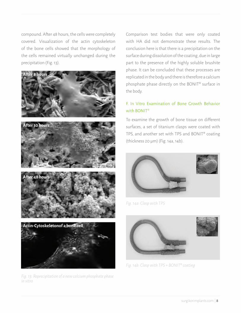
compound. After 48 hours, the cells were completely covered. Visualization of the actin cytoskeleton of the bone cells showed that the morphology of the cells remained virtually unchanged during the precipitation (Fig. 13).
Fig. 13: Reprecipitation of a new calcium phosphate phase in vitro
Fig. 14a: Clasp with TPS
Fig. 14b: Clasp with TPS + BONIT® coating
Comparison test bodies that were only coated with HA did not demonstrate these results. The conclusion here is that there is a precipitation on the surface during dissolution of the coating, due in large part to the presence of the highly soluble brushite phase. It can be concluded that these processes are replicated in the body and there is therefore a calcium phosphate phase directly on the BONIT® surface in the body.
F. In Vitro Examination of Bone Growth Behavior with BONIT®
To examine the growth of bone tissue on different surfaces, a set of titanium clasps were coated with TPS, and another set with TPS and BONIT® coating (thickness 20 μm) (Fig. 14a, 14b).
After 8 hours
After 30 hours
After 48 hours
Actin-Cytoskeleton of a bone cell
surgikorimplants.com | 8

Fig. 15: Growth behavior of cells on BONIT® in vitro
Human bone tissue was clamped between the clasps and the test setup was transferred to a tissue culture slide. After an incubation period of 10 days, the contact area between the human bone and the implant surface was examined, and the spread and growth of the osteoblasts was examined under an electron microscope. After 10 days, the clasps with the BONIT® coating showed widespread osteoblasts on the implant surface (Fig. 15). In comparison, the clasps with only TPS coating showed only marginal bone growth. This demonstrated that the biomimetic calcium phosphate coating forms an ideal temporary matrix for bone regeneration and the osseointegration of implants{20}.
surgikorimplants.com | 9
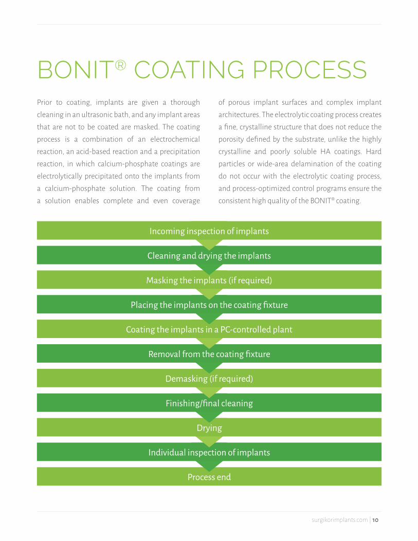
BONIT® COATING PROCESSPrior to coating, implants are given a thorough cleaning in an ultrasonic bath, and any implant areas that are not to be coated are masked. The coating process is a combination of an electrochemical reaction, an acid-based reaction and a precipitation reaction, in which calcium-phosphate coatings are electrolytically precipitated onto the implants from a calcium-phosphate solution. The coating from a solution enables complete and even coverage
of porous implant surfaces and complex implant architectures. The electrolytic coating process creates a fine, crystalline structure that does not reduce the porosity defined by the substrate, unlike the highly crystalline and poorly soluble HA coatings. Hard particles or wide-area delamination of the coating do not occur with the electrolytic coating process, and process-optimized control programs ensure the consistent high quality of the BONIT® coating.
Incoming inspection of implants
Cleaning and drying the implants
Masking the implants (if required)
Placing the implants on the coating fixture
Coating the implants in a PC-controlled plant
Removal from the coating fixture
Demasking (if required)
Finishing/final cleaning
Drying
Individual inspection of implants
Process end
surgikorimplants.com | 10

BUFFER CONCENTRATION WEIGHT LOSS OF BONIT® COATING (7D)
Duchenne 0,05M Tris/HCl 31,4%
Gomori 0,2M Tris/HCl 18,3%
TESTINGI. Chemical Testing
A. Analysis of Starting Materials
The BONIT® coating consists of two calcium phosphate components, brushite and hydroxyapatite. The proportion of heavy metals (Cd, Hg, As, Pb) is determined by the quality of the starting materials. Compliance with defined maximum concentrations of the aforementioned heavy metals is guaranteed. This is below the requirements of the U.S. standards ASTM F 1185 and ASTM F 1609. Every batch of starting materials is analyzed for purity before it is utilized in the coating process, and the proportion of heavy metals is also analyzed directly in the completed BONIT® coating.
B. Phase Composition and Ca:P Ratio
The molar calcium-phosphate ratio of the BONIT® coating is determined by the quantitative evaluation of the EDX spectroscopy. The X-ray radiation is measured by the electron beam excited by a semiconductor detector. The energy is typical for the specific element, and the intensity depends on the mass fraction of the element in the sample. The molar ratio of calcium to phosphate is 1.1±0.1 and identified the phase composition of the BONIT® coating.
C. Solubility
To determine the solubility of the BONIT® coating, different buffers with different compositions and ion concentrations can be used. The first is the buffer solution after Ducheyne{21}, which comprises a 0.05M Tris/CHI solution with pH=7.3 used at 37° C. This buffer is physiologically similar to the solution recommended by the FDA{22} for testing the solubility of Ca-P coatings. The usage of the Ducheyne 0.05M Tris/HCI buffer resulted in a weight loss of the BONIT® coating of 31.4% after 7 days. The second buffer used to determine solubility of BONIT® coating was the 0.2M Tris/HCI buffer after Gomori with pH=7.3 at 37° C{23}. The use of the Gomori buffer resulted in a weight loss of 18.3% after 7 days (Table 1), during which the dissolution rate was the highest within the first six hours after placement in the fresh buffer solution. After the initial dissolution and the associated presence of calcium and phosphate ions in the solution, the solution process becomes significantly slower. This state corresponds to the physiological processes in the body after placement of an implant.
Table 1: Solubility of BONIT®-coating as a function of the buffer solution
surgikorimplants.com | 11

D. Shelf Life
The BONIT® coating may be stored for at least five years in dry conditions at normal storage temperature. The qualitative XRD tests have not shown any phase-dependent change in the coating after the gamma-radiation sterilization.
II. Physical Testing
A. Adhesion Strength
The adhesion strength of the BONIT® coating was calculated in accordance with ASTM F 1147 and is in the range of ≥15MPa.
B. Coating Thickness
The thickness of the BONIT® coating is established via the eddy current test method in accordance with EN ISO 2360 and is an inductive, non-destructive test procedure. The coating process yields a thin, even coating with a thickness of 20±10μm.
III. Biological Testing
BONIT® is a coating for dental implants that are in contact with soft tissue and bone. Because the duration of contact is over 30 days, testing for cytotoxicity, sensitization, irritation and acute systemic toxicity are all conducted in accordance with the applicable standards.
A. Cytotoxicity
The cytotoxicity test for BONIT® coating was conducted in 2001 by an accredited laboratory in accordance with DIN EN ISO 10993-5. The test was conducted on a murine fibroblast cell line by analysis
of the inhibition of the mitochondrial activity. The results demonstrated that mitochondrial activity was not inhibited on the BONIT®-coated test bodies. This indicates there was no indication of any relevant cytotoxic effect, and that the tested material was not cytotoxic{24}. The result was confirmed by additional tests in 2004{25}, 2005{26} and 2010{27}.
B. Sensitization
The test for sensitization is recommended in the standard 10993-1 to enable an assessment of allergic and sensitization reactions triggered by the soluble components of the material. In the test for sensitization in accordance with DIN EN ISO 10993-10 the BONIT® coating gave no indication of any sensitizing properties{28}.
C. Irritation
The test for irritation was conducted in accordance with standard 10993-1 to discover any irritation effects caused by the product. BONIT®-coated samples were subjected to an irritation test under GLP conditions (intracutaneous reactivity) with polar and non-polar extraction agents as specified by DIN EN ISO 10993-10. The test report confirmed that the tested BONIT® coating does not have an irritant effect{29}.
D. Acute Systemic Toxicity
The test for acute systemic toxicity was conducted in 2010 by an accredited and GLP-certified laboratory. The result of the test showed that BONIT®-coated samples have no detectable acute systemic toxicological properties{30}.
surgikorimplants.com | 12

TEST CRITERION RESULT
Color Dark grey
Roughness Ra = 1.1 ± 0.5 μm
Residue analysis Acid residues below the limit of detection
EDX analysis No contamination, no acid residual ions
Cytotoxicity Not cytotoxic (in accordance with DIN EN ISO 10993-5)
Surface structure Uniform structured in the SEM image
Durability 5 years
IV. Summary of Tests
CLINICAL DATAI. Chemical Use
BONIT® coating has been used in medical applications since 1995, and since its introduction to the market it has been applied to more than 400,000 dental implants with no evidence of any adverse results (Fig. 16).
Fig. 16: Number of BONIT®-coated dental implants
20.000
40.000
60.000
80.000
100.000
0
2005 2006 2007 2008 2009 2010
Table 2: Summary of tests
surgikorimplants.com | 13
B. Effectiveness of The BONIT® Coating in the Swine Model
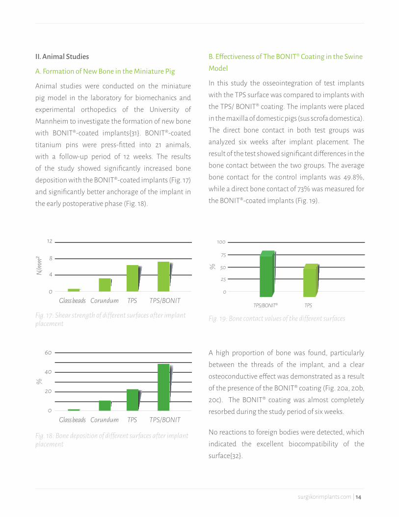
In this study the osseointegration of test implants with the TPS surface was compared to implants with the TPS/ BONIT® coating. The implants were placed in the maxilla of domestic pigs (sus scrofa domestica). The direct bone contact in both test groups was analyzed six weeks after implant placement. The result of the test showed significant differences in the bone contact between the two groups. The average bone contact for the control implants was 49.8%, while a direct bone contact of 73% was measured for the BONIT®-coated implants (Fig. 19).
II. Animal Studies
A. Formation of New Bone in the Miniature Pig
Animal studies were conducted on the miniature pig model in the laboratory for biomechanics and experimental orthopedics of the University of Mannheim to investigate the formation of new bone with BONIT®-coated implants{31}. BONIT®-coated titanium pins were press-fitted into 21 animals, with a follow-up period of 12 weeks. The results of the study showed significantly increased bone deposition with the BONIT®-coated implants (Fig. 17) and significantly better anchorage of the implant in the early postoperative phase (Fig. 18).
12
8
4
0Glass beads
N/m
m²
Corundum TPS TPS/BONIT
100
50
25
75
0
TPS/BONIT®
%
TPS
60
40
20
0Glass beads
%
Corundum TPS TPS/BONIT
Fig. 17: Shear strength of different surfaces after implant placement
Fig. 19: Bone contact values of the different surfaces
Fig. 18: Bone deposition of different surfaces after implant placement
A high proportion of bone was found, particularly between the threads of the implant, and a clear osteoconductive effect was demonstrated as a result of the presence of the BONIT® coating (Fig. 20a, 20b, 20c). The BONIT® coating was almost completely resorbed during the study period of six weeks.
No reactions to foreign bodies were detected, which indicated the excellent biocompatibility of the surface{32}.
surgikorimplants.com | 14

C. Study of BONIT®-Coated Implants in the Canine Model
The goal of this study was to determine the effect of BONIT® coating on osseointegration over extended periods with immediate loading. Implants with three different surfaces, TPS, plasma-sprayed HA surface and TPS/ BONIT® surface, were placed in the mandibles of dogs (beagles). The implants were immediately restored with a crown and placed under immediate loading. The crowns were not in contact with neighboring teeth or other implants. The follow-up period was seven months. The results
of the trial showed that the BONIT® coating was fully resorbed after seven months and had been replaced by newly formed bone tissue (Fig. 21, 22). In contrast, fragmentation of the coating and unhomogenous resorption could be observed with the plasma-sprayed HA surface. Isolated HA particles were also found.
The BONIT®-coated implants also demonstrated the highest bone density (Fig. 23). However, the difference between the surfaces decreased with increasing implant placement time{33}.
Fig. 20a: Implant with TPS surface (control group)
Fig. 20b: Implant with TPS/BONIT® surface (test group)
Fig. 20c: Formation of new bone by BONIT®
Fig. 21: Inserted dental implant with restoration under immediate loading
Fig. 22: Bone and implant interface (TPS/BONIT)
65
55
60
50
TPS TPS/BONIT®
%
HA
Fig. 23: BIC values of the different surfaces under immedi-ate loading
surgikorimplants.com | 15

D. Effect of Differential Application of CaP Coatings on Osseointegration
The effect of differential applications of CaP coatings on the osseointegration of titanium implants was investigated in the animal model. The study included three groups with different surface modifications. Group 1 had a rough surface, group 2 had a biomimetic CaP coating and group 3 had an electrochemically-deposited CaP coating. A total of 36 implants were placed in the tibias of 18 rabbits. The study period was 6 and 12 weeks.
On the biomimetically-deposited CaP coating, the crystals were arranged as flakes, while the electrochemically-deposited CaP coating had rod-shaped crystals with a hexagonal cross section. The histological analyses after six weeks showed bone growth along the surfaces. On the electrochemically-deposited CaP coating, the larger BIC values were measured and compared with the rough and biomimetically-deposited CaP surfaces. The study showed that the electrochemically-deposited CaP coating appears to improve osseointegration, and as a result can ensure a long-term and stable fixation of the implants in bone tissue{34}.
III. Clinical Results
A. Multicentric Study on Immediate Loading of CaP-Coated Dental Implant
This study investigated Pitt-Easy implants (Oraltronics) with a CaP coating (FBR surface on a porous TPS surface). The implants were placed in the maxilla and the mandible. The study protocol included immediate loading. A total of 156 implants were placed in 62 patients, with 40 implants placed in
fresh extraction alveoli. After six months, 8 implants in 6 patients had been lost, 6 in the mandible and 2 in the maxilla. After 6 months under load 94.4% of the implants were osseointegrated and functional{35}.
B. Early Loading of Endosseous Implants with BONIT® Coating
Fifty-five patients received 159 BONIT®-coated (FBR surface) Pitt-Easy implants. The average patient age was 55.6 years. The healing phase in the mandible was 7 weeks and in the maxilla 12 weeks. The healing phase was extended to 18 weeks in poor bone quality (D4 bone) and in combination with a sinus floor elevation. At the time of exposure, three implants were poorly osseointegrated. The cumulative success rate of the remaining implants after 30 months was 98.11%. Coating the implants with the electrochemical calcium-phosphate surface reduced the healing phase by half{36}.
surgikorimplants.com | 16
ADVANTAGES AT A GLANCE• Fine crystalline structure with large free surface• Complete, controlled resorption and replacement
by autogenous bone• Microporosity with high capillary effect • Outstanding biocompatibility• Improved osseointegration• Optimal solubility • Complete and even coverage of porous surface
and complex implant architecture• Ideal proliferation conditions for osteoblasts due
to large, free calcium and phosphate reservoir on the implant surface after surgery
• Faster and better healing• No mechanical release of particles or coating
1. Buratti C.A., D‘Arrigo C., Guido G., Lenzi F., Logroscino G. D., Maglioccetti G., Mannocci C., Patella S., Patella V., Salvi V., Speranza A., Speciale D., Spinarelli A., Topa A.: Assessment of the initial stability of the Symax femoral stem with EBRA-FCA: a multicentric study of 85 cases. Hip international Vol. 19 no. 1/2009.
2. Reno Barth: 5-Jahres-Ergebnisse mit einer modernen Pressfit-Pfanne. Größere Stabilität und bessere Verbindung zum Knochen. JATROS Unfallchirurgie & Sporttraumatologie 4/2010.S27-28.
3. Wintermantel E., Suk-Woo Ha: Medizintechnik mit biokompatiblen Werkstoffen und Verfahren. 3. Auflage, Berlin, Heidelberg, New York: Springer Verlag, S. 119-120, 216-227.
4. Heimann R.: Entwicklung biokeramischer Beschichtungen für Hüftendoprothesen Teil 2; www.tu- clausthal.de/presse/tucontact/2005/Mai/pdf
5. de Groot K et al., State of the Art: Hydroxyapatite Coatings for Dental Implants, J Oral Implantology 1994; 20 :232
6. Maxian SH et al., Mechanical and histological evaluation of amorphous calcium phosphate and poorly crystallized hydroxyapatite coatings on titanium implants, J. Biomed. Mater. Res. 1993; 27 : 717
7. Redepenning J, McIsaac JP, Electrocrystallization of Brushite Coatings on Prosthetic Alloys, Chemistry of Mater. 1990; 2: 625
8. Johnsson MS, Nancollas GH., The role of brushite and octacalcium phosphate in apatite formation, Critical Review in Oral Biology & Medicine 1992; 3 : 61
9. Redepenning J et al., Characterization of electrolytically prepared brushite and hydroxyapatite coatings on orthopedic alloys, J Biomed Mat Res 1996; 30 : 287
10. Neumann H-G et al., Multilayer systems for corrosion protection of stainless steel implants, Surface and Coatings Technology 1998; 98 : 1157
11. Kumar M et al., Electrodeposition of brushite coatings and their transformation to hydroxyapatite in aqueous solutions, J Biomed Mat Res 1999; 45 : 302
LITERATURE
surgikorimplants.com | 17

12. Campbell AA., Bioceramics for implant coatings, materials today, November 2003; 26
13. Szmukler- Moncler S., Zeggel P., Perrin D., Bernard JP, Neumann HG (2002). Actualites en Biomateriaux 6:185-203.
14. Wen HB et al., Microstructural Investigation of the Early external callus after Diaphyseal Fractures of Human Long Bone, J Structural Biology 1995; 114 : 115
15. Cui FZ et al., Microstructures of External Periosteal Callus of Repaired Femoral Fracture in Children, J Structural Biology 1996; 117 : 204
16. Becker P et al., Resorbable calcium phosphate composite coatings, Key Engineering Materials 2002; 218-220 : 653
17. Hegenbarth A.: Modifizierung der Oberfläche von Implantaten zur Optimierung der Osseointegration – Charakterisierung von Herstellungsverfahren und Schichteigenschaften. Bachelorarbeit Fachhochschule Osnabrück. Fakultät Ingenieurwissenschaften und Informatik. 2009.
18. Dorozkhin and Eple: Angewandte Chemie 114, 3260.
19. Becker P et al., Cellular investigation on electrochemically deposited calcium phosphate composites, J Mater Science: Mater in Medicine 2004; 15 : 437
20. Lüdtke B., Bader R., Erdmann C., Hansmann D., Fulda G., Neumann H.-G.: Scanning electron microscopy investigations of the effects of bioactive surfaces on human cancellous bone. Poster. 2nd International Symposium Interface Biology of Implants. Rostock.2006.
21. Ducheyne, P. , Radin, S., Heughebaert, M. and Heughebaert, J.C.:Calcium Phosphate Ceramic Coatings on Porous Titanium: Effect of Structure and Composition on Electrophoretic deposition, Vacuum Sintering and in vitro Dissolution Biomaterials, Vol. 11, No. 4 (1990), 244-254
22. FDA, Division of General and Restorative Divices. Calcium Phosphate (Ca-P) Coating Draft Guidance for Preparation of FDA Submissions for Orthopedic and Dental Endosseous Implants. November 11, 1992 (reformatted 2/21/97)
23. Gomori, G.: Preparation of Buffers for Use in Enzyme Studies. Methods in Enzymology, Vol. 1 (1955) 138- 146.1955.
24. Bioservice Scientific Laboratories GmbH.2001
25. DMT GmbH, Prüfbericht 4031/04.2004
26. BMP Prüfbericht B0067/05.2005
27. Bioservice Scientific Laboratories GmbH, Study no.101977.2010
28. Bioservice Scientific Laboratories GmbH, Study no. 101980.2010
29. Bioservice Scientific Laboratories GmbH, Study no. 101979.2010
30. Bioservice Scientific Laboratories GmbH, Study no.101981.2010
31. Schwarz M.L. et.al., 49th ORS, 2003, Poster1378, New Orleans.
32. Szmukler-Moncler S.,Perrin , Ahossi V, Pointaire P. : Evaluation of BONIT, a fully resorbable CaP coating obtained by electrochemical deposition after 6 w of healing : A pilot study in the pig maxillae. Key Engineering Materials, Vol 192-195, 2001, Trans Tech Publications, Switzerland, 395-398, Bio-ceramics 13.2001.
33. Szmukler-Moncler S, Figue-reido F, Trisi P, Legrand R : Immediate loading of single crowns retained by short implants. A histo-logic study with various surfaces in the canine mandible. Clinical Oral Implants Research, 11, 2000, 397 Abs
34. Guo-li Yang, Fu-ming He, Ji-an Hu, Xiao-xiang Wang, Shi-fang Zhao: Effects of biomimetrically and electrochemically deposited nano-hydroxyapatite coatings on osseointegration of porous titanium implants. Oral and Maxillofacial Implants. Vol.107, No. 6:782-789.2009
35. 35. Malchiodi L., Massei G., Turello C., Masotto P., Cassetta M., Bortoloni G., Cordioli G., Del Prete G., Della Bonna A., Casseler F., Masala P., Szmukler-Moncler S.: Immediate Loading of FBR-PITT-EASY Bio-Oss Implants. Preliminary results from a prospective multi-center study. Clinical Oral Implants Research 12(2001) 408.
36. 36. Böttcher R., Becker C., Semmler R., Barth T., Gross W., Henriot P., Stermann W., Szmukler-Moncler S.: Early loaded FBR-coated Pitt-Easy implants. A 30 month life-table-analysis. Clinical Oral Implants Research. 13(2002).
surgikorimplants.com | 18