Bones and Features of the Skull - Cranium and Face · Bones and Features of the Skull - Cranium and...
8
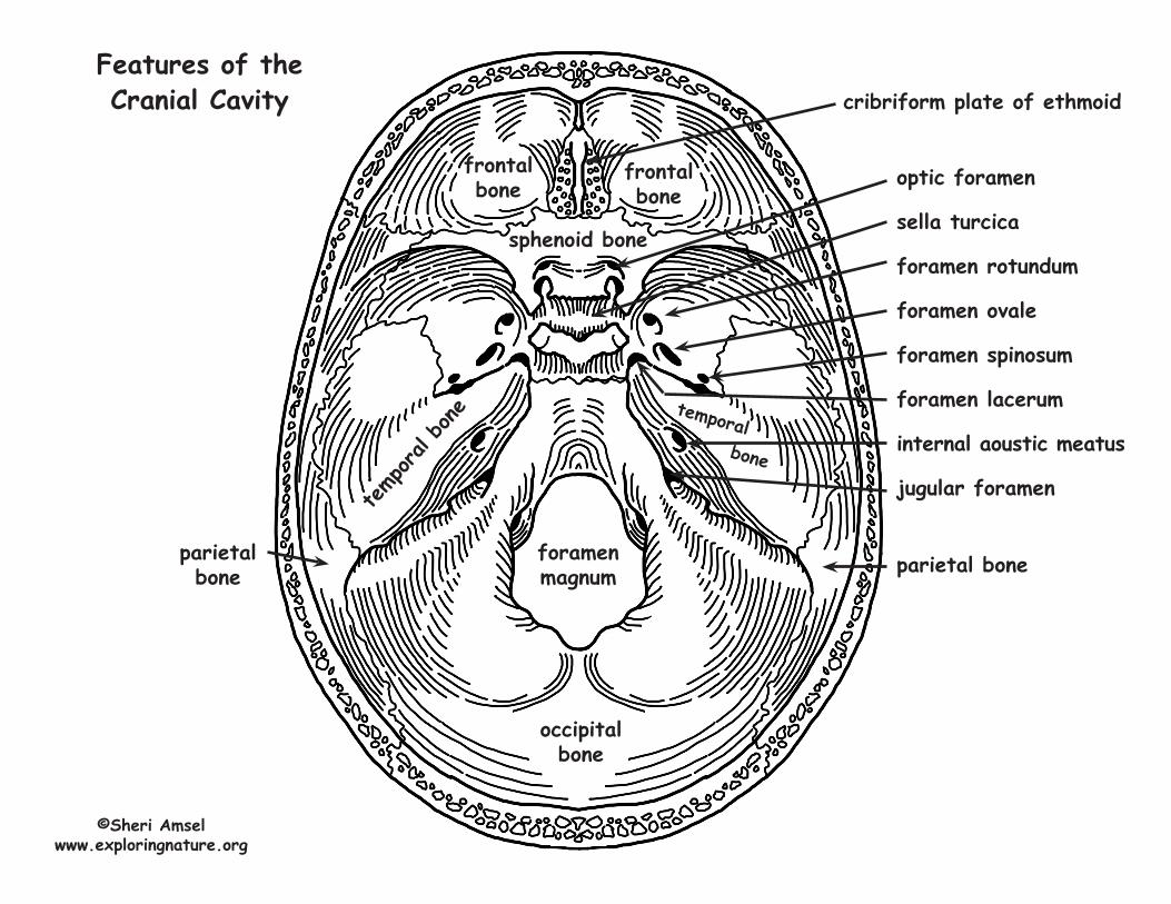
Bones and Features of the Skull - Cranium and Face ©Sheri Amsel • www.exploringnature.org Bones of the Cranium e cranium is made up of 8 bones: 2 (paired) parietal bones • 2 (paired) temporal bones • frontal bone • occipital bone • sphenoid bone • ethmoid bone e frontal bone is located on the anterior cranium and includes the following features: • It makes up the roof of the eye orbits. • Inside the skull, it forms the anterior cranial fossa, which contains the frontal lobes of the cerebrum. • It has the supraorbital foramen, where the supraorbital artery and nerve pass out of the skull onto the forehead. • It contains the frontal sinus (part of the paranasal sinuses). • It makes up (with the parietal bone) of the anterior fontanelle. e paired parietal bones make up the top and lateral aspects of the cranium. e occipital bone is located on the back of the cranium and includes the following features: • Internally, it forms the posterior cranial fossa where the occipital lobe of the brain sits. • It contains the foramen magnum – the passageway between the vertebral column and the cranial cavity. • It has the external occipital protuberance. e paired temporal bones are located on either side of the cranium, inferior to the parietal bones and contain the following features: • zygomatic process (posterior portion of the zygomatic arch). • mandibular fossa (underneath the zygomatic process where the jaw articulates with the cranium via the posterior condyle). is is the temporomandibular joint – TMJ. • external auditory meatus (external ear canal). • styloid process (a muscle attachment site). • mastoid process (the lump behind the ears where neck muscles are anchored). • stylomastoid foramen (between the styloid and mastoid processes, where the facial nerves leave the skull) • posterior part of the middle cranial fossa (where the temporal lobes of the brain sit). • jugular foramen (the most posterior foramen, where the internal jugular vein brain the brain). • carotid canal (opening where internal carotid artery enters. It passes so close to the inner ear, you can sometimes hear the blood thundering past). • foramen lacerum (a jagged opening allowing some nerve passage). • internal acoustic meatus (in the medial cranial fossa – which contains the internal auditory artery, facial nerve and the vestibulocochlear nerve).
Transcript of Bones and Features of the Skull - Cranium and Face · Bones and Features of the Skull - Cranium and...
Bones and Features of the Skull - Cranium and Face©Sheri Amsel • www.exploringnature.org
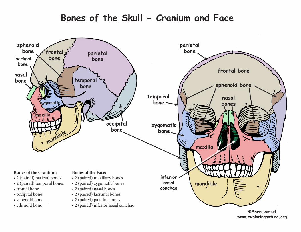
Bones of the CraniumThe cranium is made up of 8 bones: 2 (paired) parietal bones • 2 (paired) temporal bones • frontal bone • occipital bone • sphenoid bone • ethmoid bone
The frontal bone is located on the anterior cranium and includes the following features:• It makes up the roof of the eye orbits. • Inside the skull, it forms the anterior cranial fossa, which contains the frontal lobes of the cerebrum. • It has the supraorbital foramen, where the supraorbital artery and nerve pass out of the skull onto the forehead.• It contains the frontal sinus (part of the paranasal sinuses).• It makes up (with the parietal bone) of the anterior fontanelle.
The paired parietal bones make up the top and lateral aspects of the cranium.
The occipital bone is located on the back of the cranium and includes the following features:• Internally, it forms the posterior cranial fossa where the occipital lobe of the brain sits.• It contains the foramen magnum – the passageway between the vertebral column and the cranial cavity.• It has the external occipital protuberance.
The paired temporal bones are located on either side of the cranium, inferior to the parietal bones and contain the following features:• zygomatic process (posterior portion of the zygomatic arch).• mandibular fossa (underneath the zygomatic process where the jaw articulates with the cranium via the posterior condyle). This is the
temporomandibular joint – TMJ.• external auditory meatus (external ear canal).• styloid process (a muscle attachment site).• mastoid process (the lump behind the ears where neck muscles are anchored).• stylomastoid foramen (between the styloid and mastoid processes, where the facial nerves leave the skull)• posterior part of the middle cranial fossa (where the temporal lobes of the brain sit).• jugular foramen (the most posterior foramen, where the internal jugular vein brain the brain).• carotid canal (opening where internal carotid artery enters. It passes so close to the inner ear, you can sometimes hear the blood thundering past).• foramen lacerum (a jagged opening allowing some nerve passage).• internal acoustic meatus (in the medial cranial fossa – which contains the internal auditory artery, facial nerve and the vestibulocochlear nerve).
The sphenoid bone contains the following features:• It forms the paired sphenoid sinuses.• It forms the sella turcica (where the pituitary gland sits).• It forms the anterior portion of the middle cranial fossa.• It has the optic foramina (where the optic nerve and ophthalmic artery pass).• It has the superior orbital fissure (where the cranial nerve to the eye muscles pass).• It has the foramen rotundum (small round hole).• It has the foramen ovale (large oval hole).• It has the foramen spinosum (small hole).
The ethmoid bone is located between the nasal bones of the face and has the following features:• It forms the crista galli on top (superiorly) – the “cocks comb.”• It forms the cribriform plate (a horizontal plate beneath the crsita galli). It is full of holes (olfactory foramina) where the olfactory nerves pass to
pick up smell.• It forms the perpendicular plate (which projects doen to be part of the nasal septum).• It has the ethmoid sinuses (part of the paranasal sinuses)• It forms the superior and middle nasal conchae (extending laterally and inferiorly)
©Sheri Amsel www.exploringnature.org
Bones of the Skull - Cranium and Face
©Sheri Amselwww.exploringnature.org
frontal bone
frontal bone
parietal bone
parietal bone
temporal bone
temporal bone
occipital bone
zygomatic bone
mandible
mandible
maxilla
maxilla
nasal bone
zygomatic bone
nasal bones
sphenoid bone
sphenoid bone
lacrimalbone
Bones of the Cranium:• 2 (paired) parietal bones• 2 (paired) temporal bones• frontal bone• occipital bone• sphenoid bone• ethmoid bone
Bones of the Face:• 2 (paired) maxillary bones• 2 (paired) zygomatic bones• 2 (paired) nasal bones• 2 (paired) lacrimal bones• 2 (paired) palatine bones• 2 (paired) inferior nasal conchae
inferior nasal
conchae
Bones of the FaceThe face is made up of 14 bones: mandible • vomer • 2 (paired) maxillary bones • 2 (paired) zygomatic bones • 2 (paired) nasal bones • 2 (paired) lacrimal bones • 2 (paired) palatine bones • 2 (paired) inferior nasal conchae
The mandible (lower jaw) is the largest strongest bone of the face and contains the following features:• mandibular angle (angle of the jaw below the ear).• coronoid process (anterior process which is the attachment site for the temporalis muscle which elevates the jaw during chewing).• mandibular condyle (posterior process - part of the TMJ and articulates with the mandibular fossa on the zygomatic process of the temporal bone).• mandibular notch (between the two processes).• alveolar margin (where the lower teeth are inserted).• mandibular foramina (inside the ramus of the mandible) there the nerves to the jaw pass.• mental foramina (on the anterior and lateral (outside) mandible) where the blood vessels and nerves to the chin and upper lip pass.
The paired maxillary bones are fused medially and contain the following features:• They make up the upper jaw and central face.• They form the alveolar margin (where the upper teeth are inserted).• They include the palatine processes (the hard palate) that projects posteriorly.• They include the frontal processes (medial and inferior eye orbit by the bridge of the nose).• The form the maxillary sinuses (the largest part of the paranasal sinuses between the orbits and the upper teeth).• They form the zygomatic processes (the anterior part of the zygomatic arch).• They have the inferior orbital fissure (inside the orbit where the vagal nerve and zygomatic nerve and blood vessels pass).• They include the infraorbital foramen (where the infraorbital nerve and arteries to the face run).
The paired zygomatic bones (malar bones/cheek bones) sit between the zygomatic processes of the maxilla and temporal bone.
The paired nasal bones make up the bridge of the nose.
The paired lacrimal bones are made up of the medial walls of each orbit with a lateral “sulcus” through which tears can drain into the nasal cavities (which is why you get a runny nose when your eyes tear).
The paired palatine bones are inside the skull made up of the horizontal plate (which is the posterior portion of the hard palate) and the posterior, lateral wall of the nasal cavity.
The vomer is the plow-shaped bone within the nasal cavity that makes up part of the nasal septum.
The inferior nasal conchae form the lateral walls of the nasal cavity below the middle conchae of the ethmoid bone.
©Sheri Amsel www.exploringnature.org
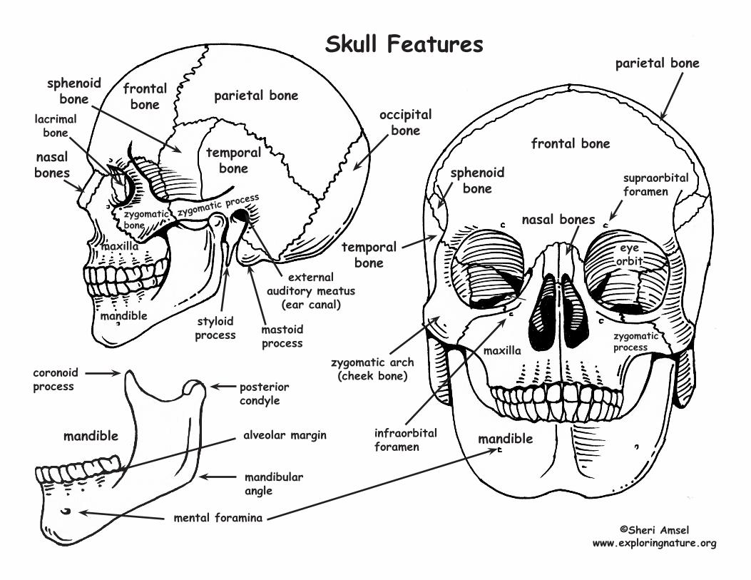
Skull Features
©Sheri Amselwww.exploringnature.org
frontal bone
nasal bones
temporal bone
zygomatic arch(cheek bone)
maxilla
mandible
external auditory meatus
(ear canal)
occipitalbone
frontalbone parietal bone
mandible
maxilla
nasal bones
temporal bone
styloidprocess mastoid
process
eyeorbit
supraorbital foramen
zygomatic process
posterior condyle
mandible
mandibular angle
coronoid process
mental foramina
alveolar margin
zygomaticprocess
infraorbital foramen
zygomaticbone
parietal bone
sphenoid bone
sphenoid bone
lacrimalbone
occipitalbone
©Sheri Amselwww.exploringnature.org
temp
oral
bone
frontal bone
frontal bone
temporal bone
sphenoid bone
foramenmagnum
cribriform plate of ethmoid
optic foramen
sella turcica
foramen rotundum
foramen ovale
foramen spinosum
foramen lacerum
internal aoustic meatus
jugular foramen
parietal boneparietal bone
Features of the Cranial Cavity
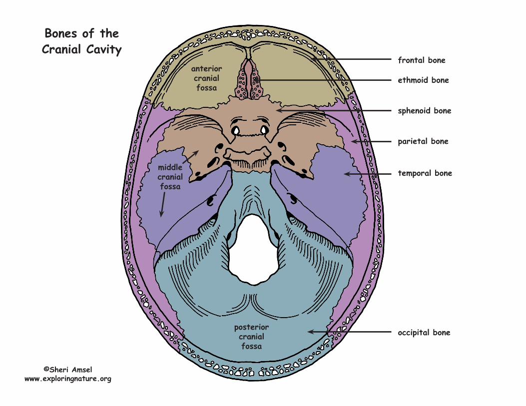
frontal bone
ethmoid bone
sphenoid bone
parietal bone
temporal bone
occipital bone
Bones of the Cranial Cavity
anterior cranial fossa
posterior cranial fossa
middle cranial fossa
©Sheri Amselwww.exploringnature.org
occipitalbone
©Sheri Amselwww.exploringnature.org
temp
oral
bone
frontal bone
frontal bone
temporal bone
sphenoid bone
Name the Features of the Cranial Cavity