Bone Physiology. Bones are Classified by shape and structure limbsCarpals tarsals vertebrae Skull...
38
Bone Physiology
-
Upload
gervais-simmons -
Category
Documents
-
view
227 -
download
2
Transcript of Bone Physiology. Bones are Classified by shape and structure limbsCarpals tarsals vertebrae Skull...
Bone Physiology
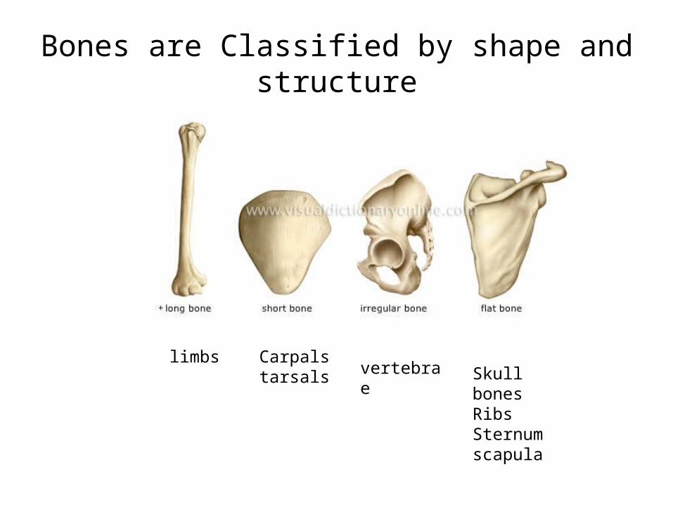
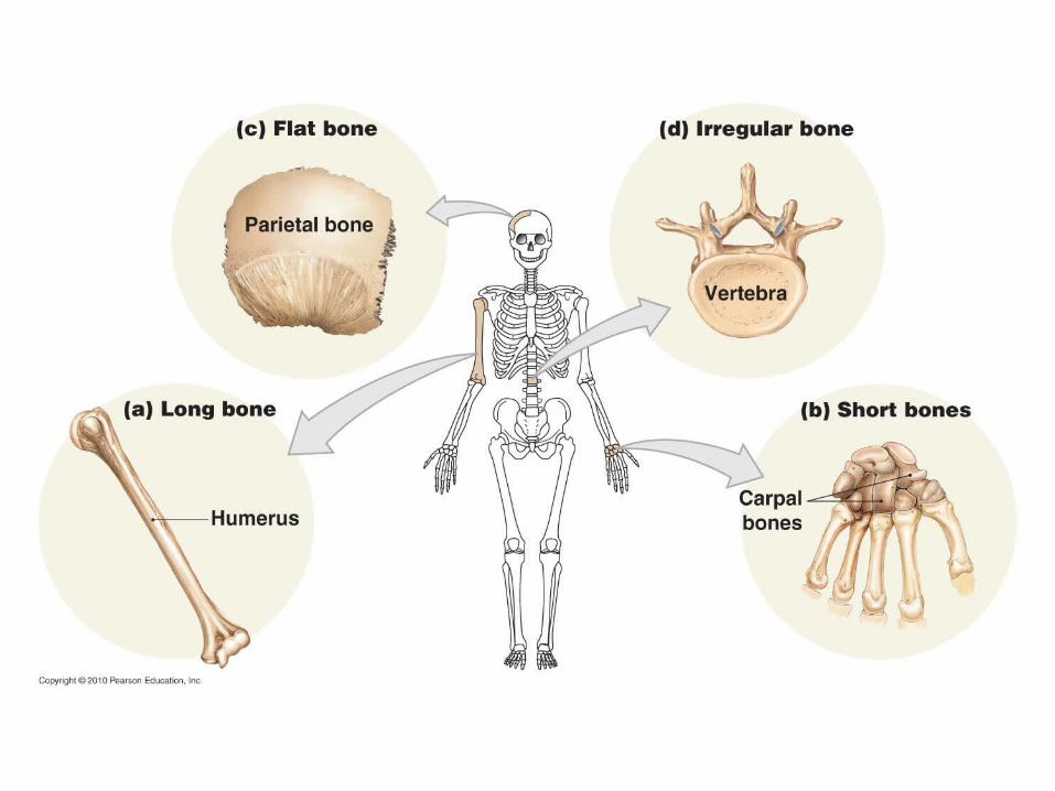
Bones are Classified by shape and structure
limbs Carpalstarsals vertebrae Skull bones
RibsSternumscapula

EpiphysisEpiphyseal LineDiaphysisPeriosteumArticulating Cartilage
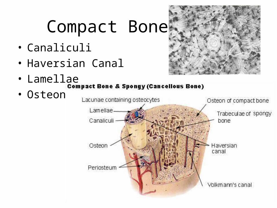
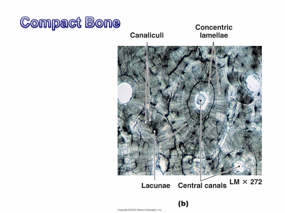
Compact Bone• Canaliculi• Haversian Canal• Lamellae• Osteon
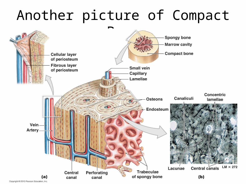
Another picture of Compact Bone
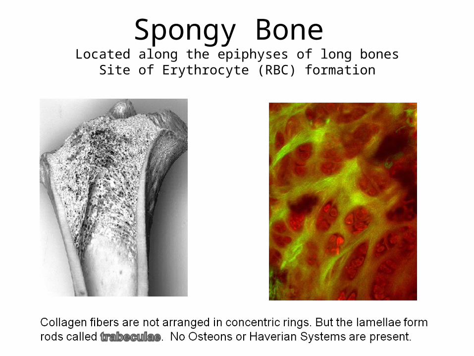
Spongy Bone Located along the epiphyses of long bones
Site of Erythrocyte (RBC) formation
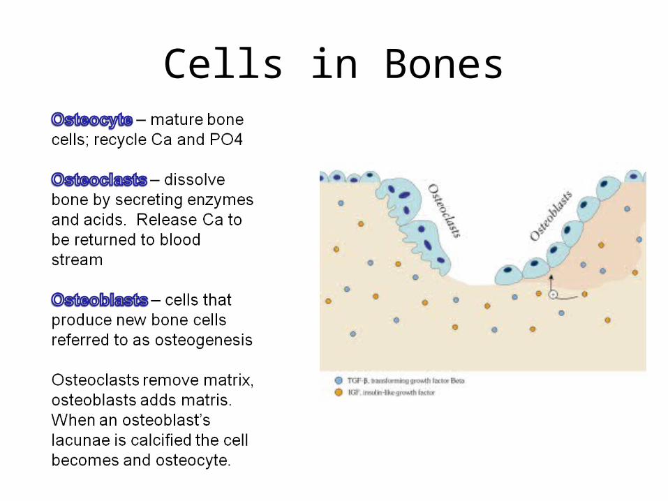
Cells in Bones
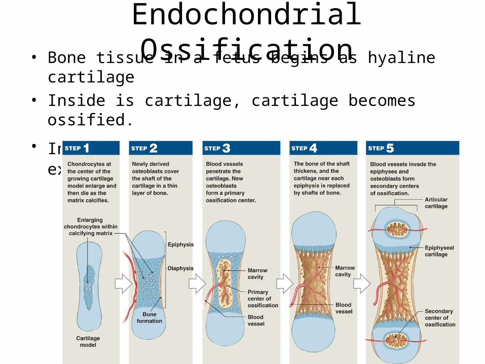
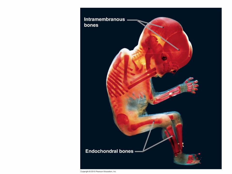
Endochondrial Ossification• Bone tissue in a fetus begins as hyaline cartilage• Inside is cartilage, cartilage becomes ossified.
• In 6 weeks cartilage is replaced to bone except at growth plates.
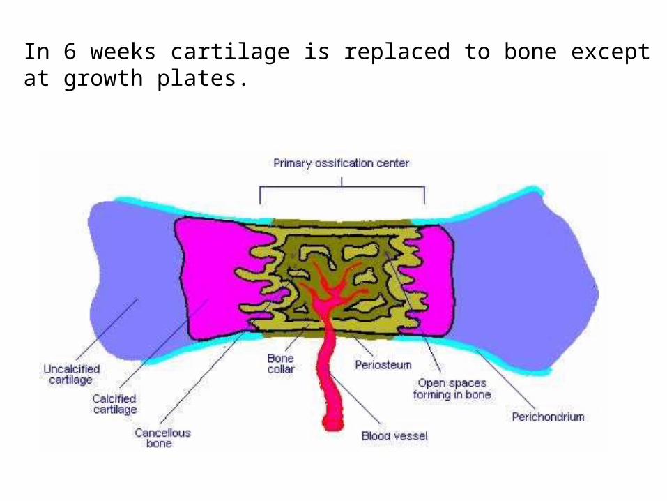
In 6 weeks cartilage is replaced to bone except at growth plates.
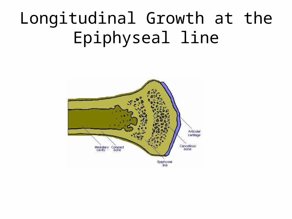
Longitudinal Growth at the Epiphyseal line
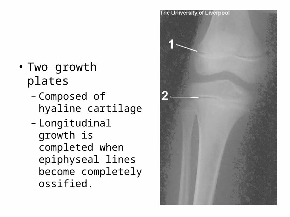
• Two growth plates – Composed of
hyaline cartilage– Longitudinal growth
is completed when epiphyseal lines become completely ossified.
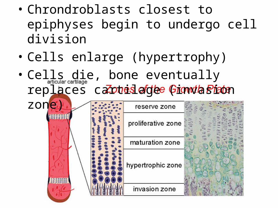
• Chrondroblasts closest to epiphyses begin to undergo cell division
• Cells enlarge (hypertrophy)
• Cells die, bone eventually replaces cartilage (invasion zone)
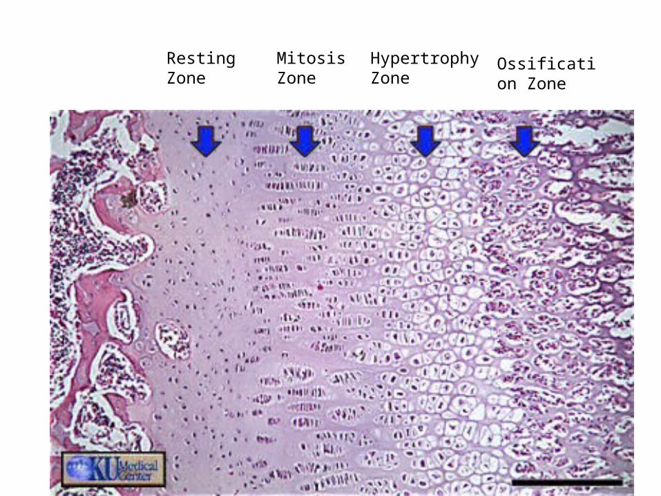
Resting Zone
Mitosis Zone
HypertrophyZone
Ossification Zone
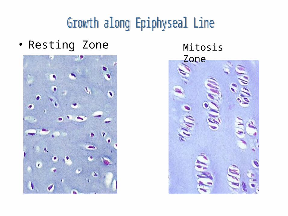
• Resting Zone Mitosis Zone
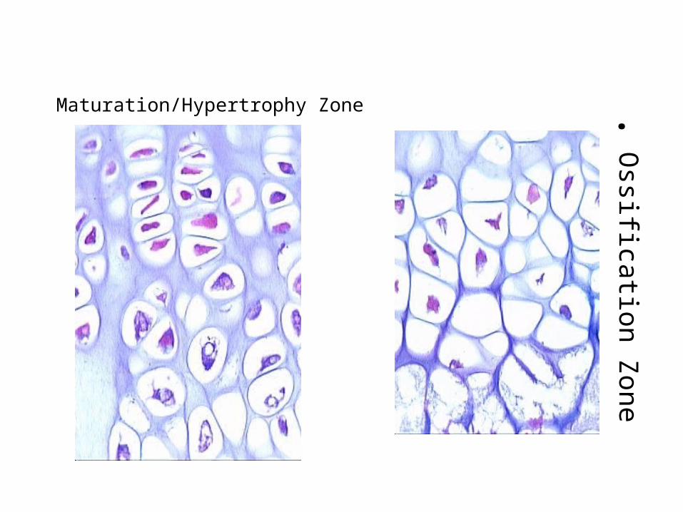
•O
ssification Zone
Maturation/Hypertrophy Zone
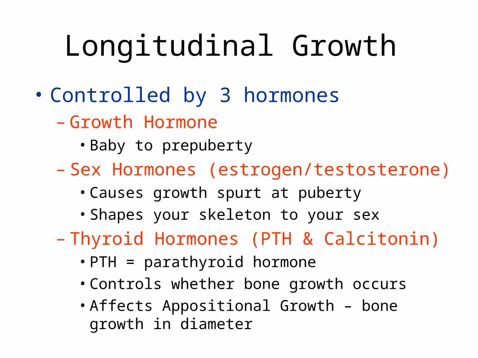
Longitudinal Growth
• Controlled by 3 hormones– Growth Hormone
• Baby to prepuberty
– Sex Hormones (estrogen/testosterone)• Causes growth spurt at puberty• Shapes your skeleton to your sex
– Thyroid Hormones (PTH & Calcitonin)• PTH = parathyroid hormone• Controls whether bone growth occurs• Affects Appositional Growth – bone growth in
diameter
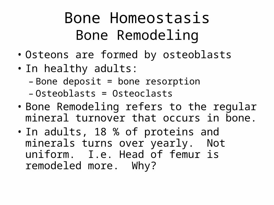
Bone HomeostasisBone Remodeling
• Osteons are formed by osteoblasts• In healthy adults:
– Bone deposit = bone resorption– Osteoblasts = Osteoclasts
• Bone Remodeling refers to the regular mineral turnover that occurs in bone.
• In adults, 18 % of proteins and minerals turns over yearly. Not uniform. I.e. Head of femur is remodeled more. Why?
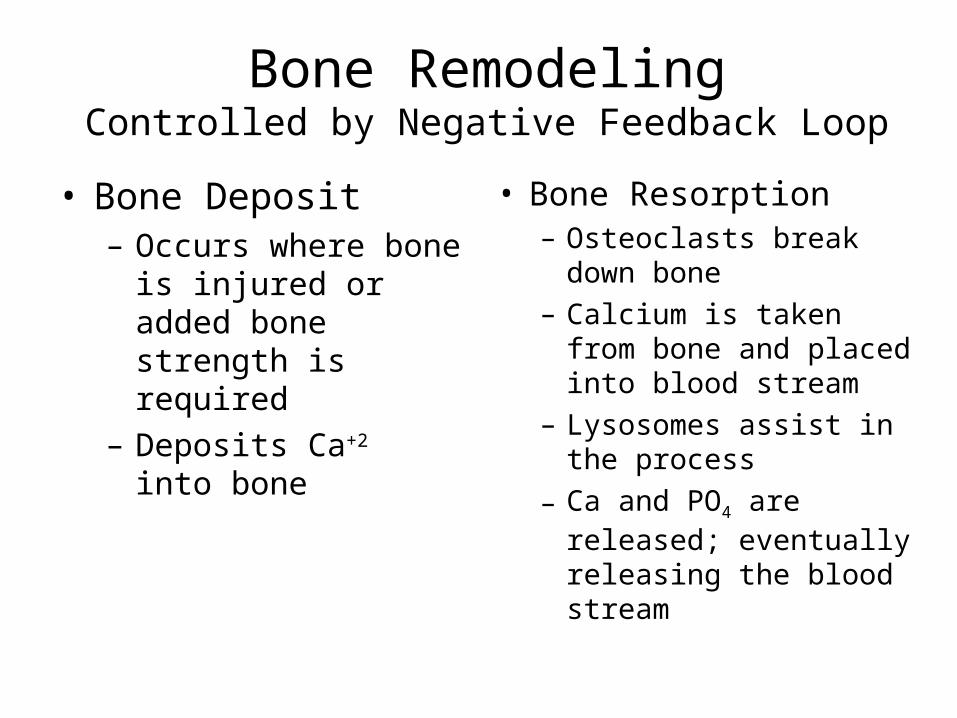
Bone RemodelingControlled by Negative Feedback Loop
• Bone Deposit– Occurs where bone is
injured or added bone strength is required
– Deposits Ca+2 into bone
• Bone Resorption– Osteoclasts break down
bone– Calcium is taken from
bone and placed into blood stream
– Lysosomes assist in the process
– Ca and PO4 are released; eventually releasing the blood stream
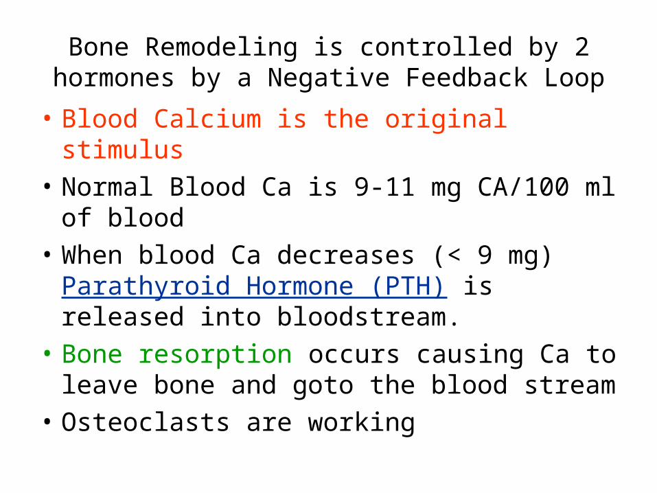
Bone Remodeling is controlled by 2 hormones by a Negative Feedback Loop
• Blood Calcium is the original stimulus
• Normal Blood Ca is 9-11 mg CA/100 ml of blood
• When blood Ca decreases (< 9 mg) Parathyroid Hormone (PTH) is released into bloodstream.
• Bone resorption occurs causing Ca to leave bone and goto the blood stream
• Osteoclasts are working
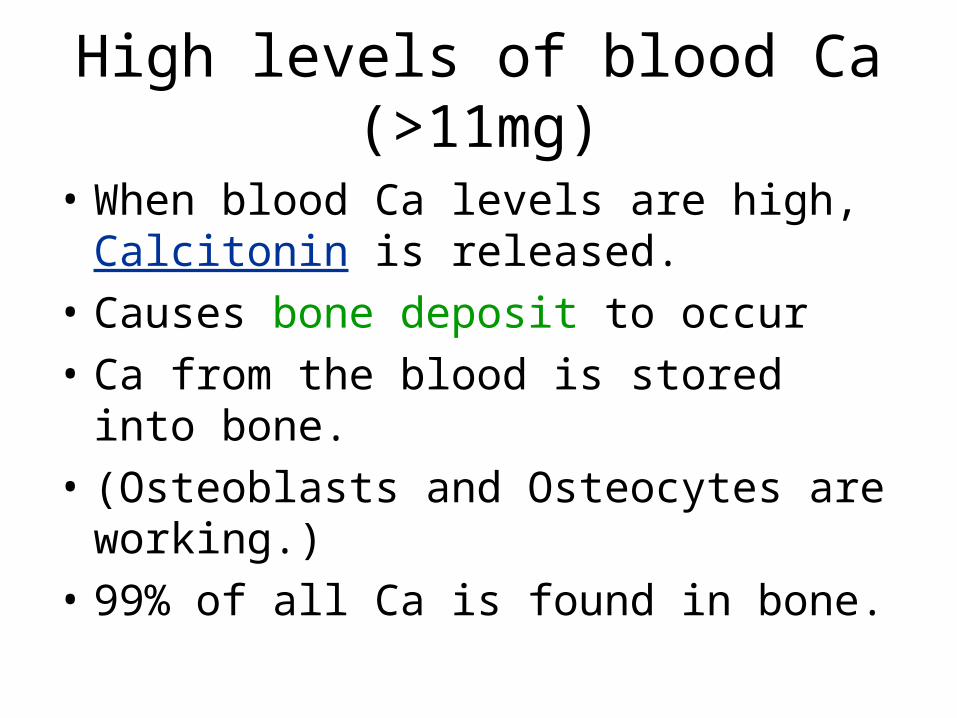
High levels of blood Ca (>11mg)
• When blood Ca levels are high, Calcitonin is released.
• Causes bone deposit to occur
• Ca from the blood is stored into bone.
• (Osteoblasts and Osteocytes are working.)
• 99% of all Ca is found in bone.
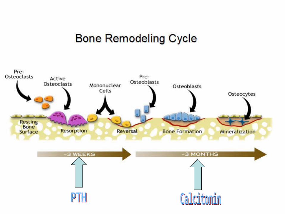
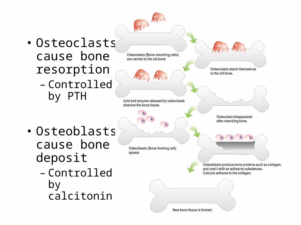
• Osteoclasts cause bone resorption– Controlled
by PTH
• Osteoblasts cause bone deposit– Controlled
by calcitonin
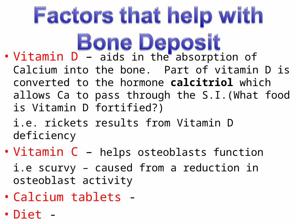
• Vitamin D – aids in the absorption of Calcium into the bone. Part of vitamin D is converted to the hormone calcitriol which allows Ca to pass through the S.I.(What food is Vitamin D fortified?)
i.e. rickets results from Vitamin D deficiency
• Vitamin C – helps osteoblasts function
i.e scurvy – caused from a reduction in osteoblast activity
• Calcium tablets -
• Diet -
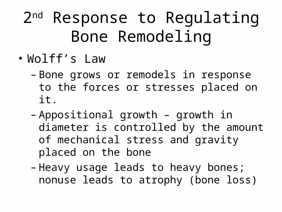
2nd Response to Regulating Bone Remodeling
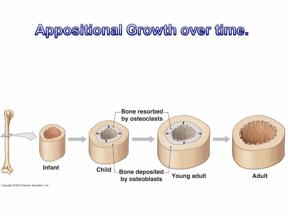
• Wolff’s Law– Bone grows or remodels in response to the
forces or stresses placed on it.– Appositional growth – growth in diameter is
controlled by the amount of mechanical stress and gravity placed on the bone
– Heavy usage leads to heavy bones; nonuse leads to atrophy (bone loss)
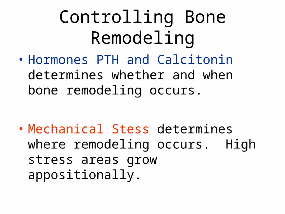
Controlling Bone Remodeling
• Hormones PTH and Calcitonin determines whether and when bone remodeling occurs.
• Mechanical Stess determines where remodeling occurs. High stress areas grow appositionally.
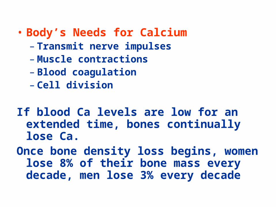
• Body’s Needs for Calcium– Transmit nerve impulses– Muscle contractions– Blood coagulation– Cell division
If blood Ca levels are low for an extended time, bones continually lose Ca.
Once bone density loss begins, women lose 8% of their bone mass every decade, men lose 3% every decade
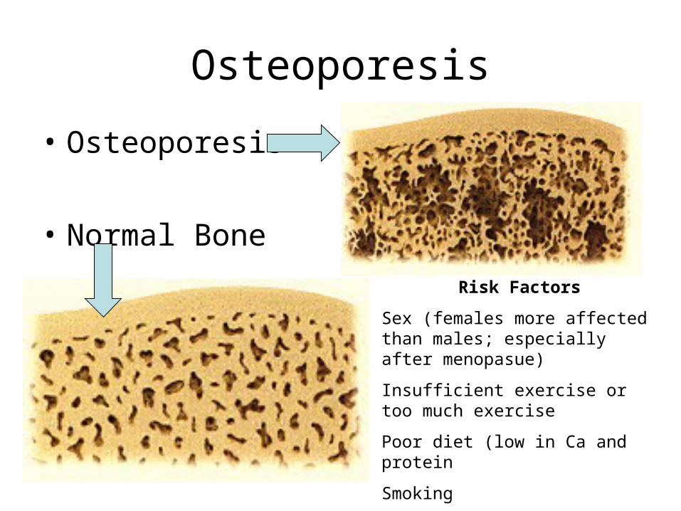
Osteoporesis
• Osteoporesis
• Normal Bone
Risk Factors
Sex (females more affected than males; especially after menopasue)
Insufficient exercise or too much exercise
Poor diet (low in Ca and protein
Smoking
Race: Black > bone density
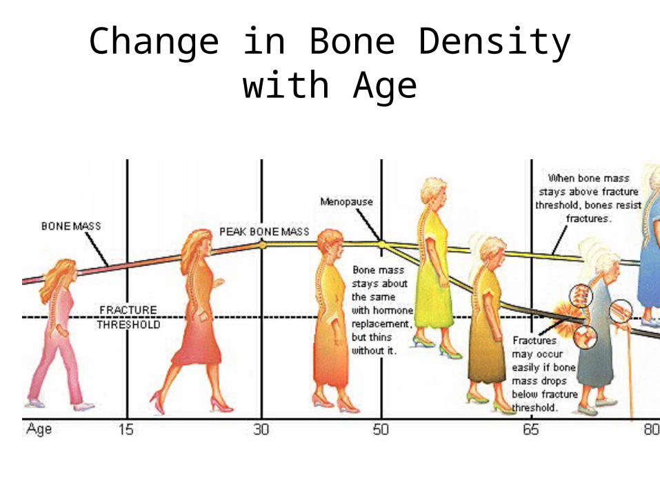
Change in Bone Density with Age
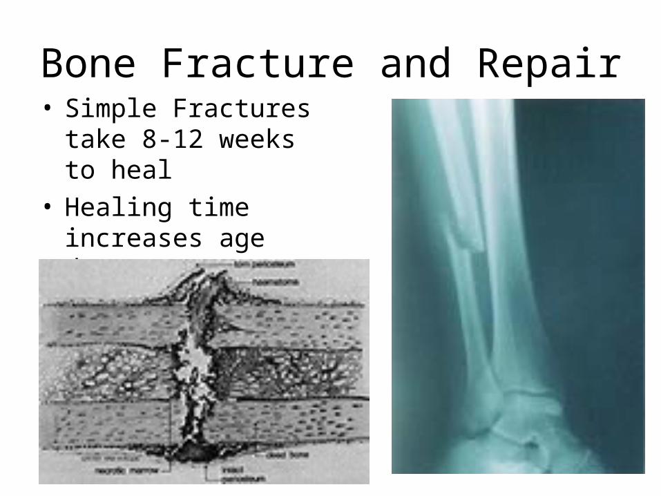
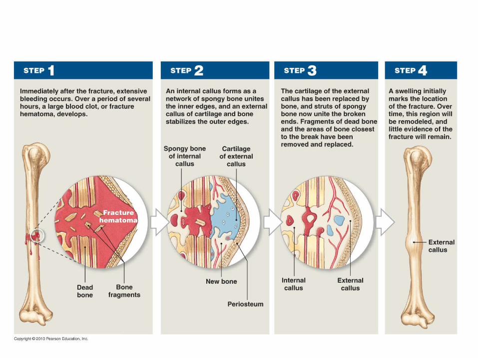
Bone Fracture and Repair• Simple Fractures take
8-12 weeks to heal• Healing time
increases age due to poor circulation
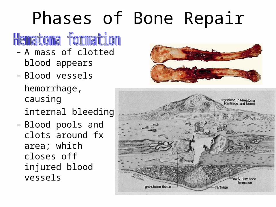
Phases of Bone Repair
– A mass of clotted blood appears
– Blood vessels
hemorrhage, causing
internal bleeding– Blood pools and clots
around fx area; which closes off injured blood vessels
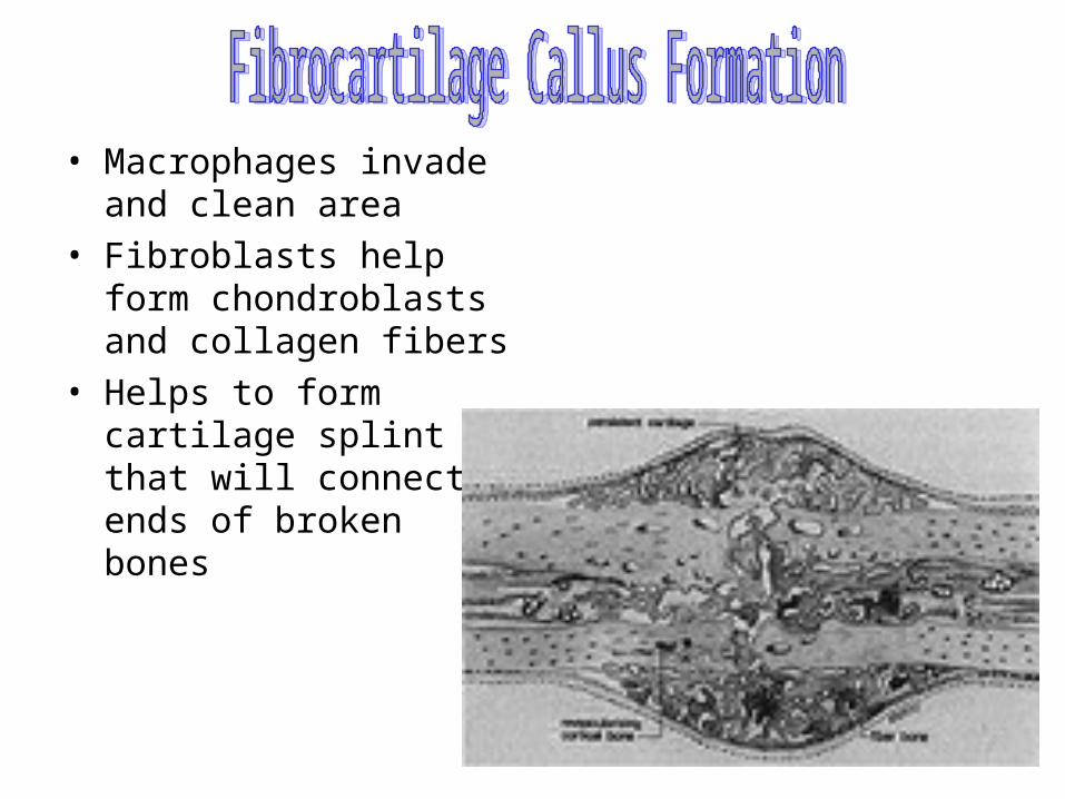
• Macrophages invade and clean area
• Fibroblasts help form chondroblasts and collagen fibers
• Helps to form cartilage splint that will connect ends of broken bones
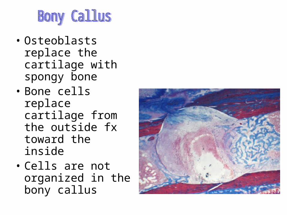
• Osteoblasts replace the cartilage with spongy bone
• Bone cells replace cartilage from the outside fx toward the inside
• Cells are not organized in the bony callus
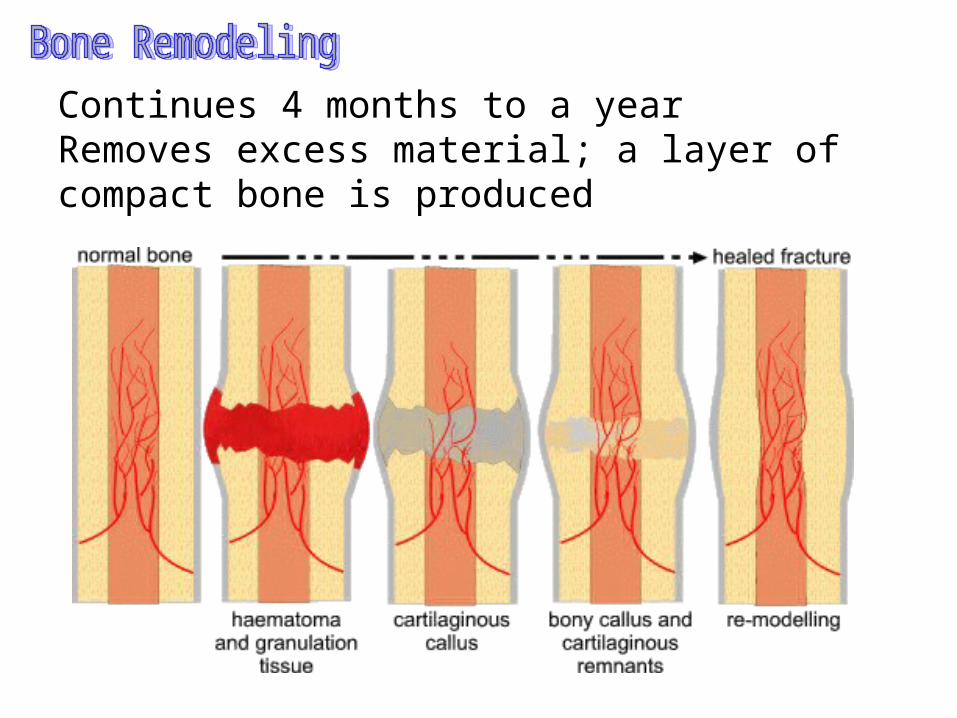
Continues 4 months to a yearRemoves excess material; a layer of compact bone is produced

![The Human Body The Skeleton provides … Support/ShapeMovement Produces [White blood cells]Protects Bones Upper Body: Clavicle, Scapula, Sternum, Ribs, Humerus,](https://static.fdocuments.net/doc/165x107/56649c755503460f9492861a/the-human-body-the-skeleton-provides-supportshapemovement-produces-white.jpg)