Bone morphogenic protein antagonist Drm/gremlin is a novel proangiogenic factor
8
CHEMOKINES, CYTOKINES, AND INTERLEUKINS Bone morphogenic protein antagonist Drm/gremlin is a novel proangiogenic factor Helena Stabile, 1 Stefania Mitola, 1 Emanuela Moroni, 1 Mirella Belleri, 1 Stefania Nicoli, 1 Daniela Coltrini, 2 Francesco Peri, 3 Antonello Pessi, 4 Laura Orsatti, 4 Fabio Talamo, 4 Vincent Castronovo, 5 David Waltregny, 6 Franco Cotelli, 7 Domenico Ribatti, 8 and Marco Presta 1 1 Unit of General Pathology and Immunology, Department of Biomedical Sciences and Biotechnology, University of Brescia, Italy; 2 Unit of Histology, Department of Biomedical Sciences and Biotechnology, University of Brescia, Italy; 3 Department of Biotechnology and Biosciences, University of Milan-Bicocca, Milan, Italy; 4 Istituto di Ricerche di Biologia Molecolare P. Angeletti, Pomezia, Rome, Italy; 5 Metastasis Research Laboratory, Center of Experimental Cancer Research, University of Lie ` ge, Belgium; 6 Department of Pathology, Center of Experimental Cancer Research, University of Lie ` ge, Belgium; 7 Department of Biology, University of Milan, Italy; 8 Department of Human Anatomy and Histology, University of Bari, Italy Angiogenesis plays a key role in various physiologic and pathologic conditions, including tumor growth. Drm/gremlin, a member the Dan family of bone morpho- genic protein (BMP) antagonists, is com- monly thought to affect different pro- cesses during growth, differentiation, and development by heterodimerizing vari- ous BMPs. Here, we identify Drm/gremlin as a novel proangiogenic factor expressed by endothelium. Indeed, Drm/gremlin was purified to homogeneity from the condi- tioned medium of transformed endothe- lial cells using an endothelial-cell sprout- ing assay to follow protein isolation. Accordingly, recombinant Drm/gremlin stimulates endothelial-cell migration and invasion in fibrin and collagen gels, binds with high affinity to various endothelial cell types, and triggers tyrosine phosphor- ylation of intracellular signaling proteins. Also, Drm/gremlin induces neovascular- ization in the chick embryo chorioallan- toic membrane. BMP4 does not affect Drm/gremlin interaction with endothe- lium, and both molecules exert a proan- giogenic activity in vitro and in vivo when administered alone or in combination. Finally, Drm/gremlin is produced by the stroma of human tumor xenografts in nude mice, and it is highly expressed in endothelial cells of human lung tumor vasculature when compared with non- neoplastic lung. Our observations point to a novel, previously unrecognized ca- pacity of Drm/gremlin to interact directly with target endothelial cells and to modu- late angiogenesis. (Blood. 2007;109: 1834-1840) © 2007 by The American Society of Hematology Introduction Gremlin, also known as Drm (Down-regulated by v-mos), belongs to the Dan family of cysteine knot-secreted proteins. 1,2 Drm/ gremlin exerts a potent bone morphogenic protein (BMP) antago- nist activity by binding BMP2, BMP4, and BMP7 and preventing their interaction with cell-surface receptors. 3 This capacity is thought to be responsible for the pattern-inducing activity of Drm/gremlin during embryonic development 4 and to play a role in human diseases. 5 However, intracellular, BMP-independent mecha- nism(s) of action 6 may mediate the ability of Drm/gremlin to suppress transformation and tumorigenesis under certain experimen- tal conditions. 7 Also, Drm/gremlin interacts with Slit proteins and acts as a negative regulator of monocyte chemotaxis, thus suggest- ing a role for this protein in inflammation and immunity. 8 Taken together these observations indicate that Drm/gremlin may exert multiple functions in different physiopathologic conditions via BMP-dependent and BMP-independent mechanisms of action. 1-5 Nevertheless, the possibility that Drm/gremlin may exert a direct effect on target cells has never been explored. Angiogenesis, the process of new blood vessel formation from preexisting ones, plays a key role in various physiologic and pathologic conditions, including inflammation and tumor growth. 9 The local, continuous release of angiogenic growth factors and/or alterations of the production of natural angiogenic inhibitors 10 are responsible for the uncontrolled endothelial-cell proliferation that takes place during tumor neovascularization and in angiogenesis- dependent diseases. 11 Numerous inducers of angiogenesis have been identified, including members of the vascular endothelial growth factor (VEGF) 12,13 and of the fibroblast growth factor (FGF) families. 14 These angiogenic growth factors induce a complex “proangiogenic phenotype” in endothelial cells that recapitulates several aspects of the in vivo angiogenesis process (summarized in Javerzat et al 15 ). To elucidate the molecular determinants of endothelial-cell activation during angiogenesis, we originated a stable mouse aortic endothelial (MAE) cell line transfected with a human FGF2 cDNA (FGF2-T-MAE cells). 16,17 Transfectants are characterized by the overexpression of numerous genes implicated in the modulation of cell growth, differentiation, cell adhesion, and stress/ survival. 18 FGF2-T-MAE cells are angiogenic and cause the formation of opportunistic vascular lesions by recruiting endo- thelial cells of the host. 16,19 Accordingly, FGF2-T-MAE cells release an endothelial-cell motogen that appears to be distinct from other well-characterized angiogenic growth factors, includ- ing FGF2 and VEGF. 19 Submitted June 28, 2006; accepted October 16, 2006. Prepublished online as Blood First Edition Paper, October 31, 2006; DOI 10.1182/blood-2006- 06-032276. The online version of this article contains a data supplement. The publication costs of this article were defrayed in part by page charge payment. Therefore, and solely to indicate this fact, this article is hereby marked ‘‘advertisement’’ in accordance with 18 USC section 1734. © 2007 by The American Society of Hematology 1834 BLOOD, 1 MARCH 2007 VOLUME 109, NUMBER 5
Transcript of Bone morphogenic protein antagonist Drm/gremlin is a novel proangiogenic factor
CHEMOKINES, CYTOKINES, AND INTERLEUKINS
Bone morphogenic protein antagonist Drm/gremlin is a novelproangiogenic factorHelena Stabile,1 Stefania Mitola,1 Emanuela Moroni,1 Mirella Belleri,1 Stefania Nicoli,1 Daniela Coltrini,2
Francesco Peri,3 Antonello Pessi,4 Laura Orsatti,4 Fabio Talamo,4 Vincent Castronovo,5 David Waltregny,6
Franco Cotelli,7 Domenico Ribatti,8 and Marco Presta1
1Unit of General Pathology and Immunology, Department of Biomedical Sciences and Biotechnology, University of Brescia, Italy; 2Unit of Histology, Departmentof Biomedical Sciences and Biotechnology, University of Brescia, Italy; 3Department of Biotechnology and Biosciences, University of Milan-Bicocca, Milan, Italy;4Istituto di Ricerche di Biologia Molecolare P. Angeletti, Pomezia, Rome, Italy; 5Metastasis Research Laboratory, Center of Experimental Cancer Research,University of Liege, Belgium; 6Department of Pathology, Center of Experimental Cancer Research, University of Liege, Belgium; 7Department of Biology,University of Milan, Italy; 8Department of Human Anatomy and Histology, University of Bari, Italy
Angiogenesis plays a key role in variousphysiologic and pathologic conditions,including tumor growth. Drm/gremlin, amember the Dan family of bone morpho-genic protein (BMP) antagonists, is com-monly thought to affect different pro-cesses during growth, differentiation, anddevelopment by heterodimerizing vari-ous BMPs. Here, we identify Drm/gremlinas a novel proangiogenic factor expressedby endothelium. Indeed, Drm/gremlin waspurified to homogeneity from the condi-tioned medium of transformed endothe-lial cells using an endothelial-cell sprout-
ing assay to follow protein isolation.Accordingly, recombinant Drm/gremlinstimulates endothelial-cell migration andinvasion in fibrin and collagen gels, bindswith high affinity to various endothelialcell types, and triggers tyrosine phosphor-ylation of intracellular signaling proteins.Also, Drm/gremlin induces neovascular-ization in the chick embryo chorioallan-toic membrane. BMP4 does not affectDrm/gremlin interaction with endothe-lium, and both molecules exert a proan-giogenic activity in vitro and in vivo whenadministered alone or in combination.
Finally, Drm/gremlin is produced by thestroma of human tumor xenografts innude mice, and it is highly expressed inendothelial cells of human lung tumorvasculature when compared with non-neoplastic lung. Our observations pointto a novel, previously unrecognized ca-pacity of Drm/gremlin to interact directlywith target endothelial cells and to modu-late angiogenesis. (Blood. 2007;109:1834-1840)
© 2007 by The American Society of Hematology
Introduction
Gremlin, also known as Drm (Down-regulated by v-mos), belongsto the Dan family of cysteine knot-secreted proteins.1,2 Drm/gremlin exerts a potent bone morphogenic protein (BMP) antago-nist activity by binding BMP2, BMP4, and BMP7 and preventingtheir interaction with cell-surface receptors.3 This capacity isthought to be responsible for the pattern-inducing activity ofDrm/gremlin during embryonic development4 and to play a role inhuman diseases.5 However, intracellular, BMP-independent mecha-nism(s) of action6 may mediate the ability of Drm/gremlin tosuppress transformation and tumorigenesis under certain experimen-tal conditions.7 Also, Drm/gremlin interacts with Slit proteins andacts as a negative regulator of monocyte chemotaxis, thus suggest-ing a role for this protein in inflammation and immunity.8 Takentogether these observations indicate that Drm/gremlin may exertmultiple functions in different physiopathologic conditions viaBMP-dependent and BMP-independent mechanisms of action.1-5
Nevertheless, the possibility that Drm/gremlin may exert a directeffect on target cells has never been explored.
Angiogenesis, the process of new blood vessel formation frompreexisting ones, plays a key role in various physiologic andpathologic conditions, including inflammation and tumor growth.9
The local, continuous release of angiogenic growth factors and/or
alterations of the production of natural angiogenic inhibitors10 areresponsible for the uncontrolled endothelial-cell proliferation thattakes place during tumor neovascularization and in angiogenesis-dependent diseases.11 Numerous inducers of angiogenesis havebeen identified, including members of the vascular endothelialgrowth factor (VEGF)12,13 and of the fibroblast growth factor(FGF) families.14 These angiogenic growth factors induce acomplex “proangiogenic phenotype” in endothelial cells thatrecapitulates several aspects of the in vivo angiogenesis process(summarized in Javerzat et al15).
To elucidate the molecular determinants of endothelial-cellactivation during angiogenesis, we originated a stable mouse aorticendothelial (MAE) cell line transfected with a human FGF2 cDNA(FGF2-T-MAE cells).16,17 Transfectants are characterized by theoverexpression of numerous genes implicated in the modulationof cell growth, differentiation, cell adhesion, and stress/survival.18 FGF2-T-MAE cells are angiogenic and cause theformation of opportunistic vascular lesions by recruiting endo-thelial cells of the host.16,19 Accordingly, FGF2-T-MAE cellsrelease an endothelial-cell motogen that appears to be distinctfrom other well-characterized angiogenic growth factors, includ-ing FGF2 and VEGF.19
Submitted June 28, 2006; accepted October 16, 2006. Prepublished online asBlood First Edition Paper, October 31, 2006; DOI 10.1182/blood-2006-06-032276.
The online version of this article contains a data supplement.
The publication costs of this article were defrayed in part by page chargepayment. Therefore, and solely to indicate this fact, this article is herebymarked ‘‘advertisement’’ in accordance with 18 USC section 1734.
© 2007 by The American Society of Hematology
1834 BLOOD, 1 MARCH 2007 � VOLUME 109, NUMBER 5
Here, we describe the purification of this factor from theconditioned medium (CM) of FGF2-T-MAE cells and its identifica-tion as the Drm/gremlin protein. Our data demonstrate for the firsttime that Drm/gremlin plays a BMP-independent role in theangiogenic process by binding the endothelial cell surface, thusactivating intracellular signaling and cell motility. Accordingly,Drm/gremlin induces new vessel growth in the chick embryochorioallantoic membrane. Also, the expression of Drm/gremlin inthe endothelium of human lung tumor specimens points to a rolefor this protein in blood vessel development in human cancers.
Materials and methods
Cell cultures
Immortalized Balb/c MAE cells were obtained from R. Auerbach (Univer-sity of Wisconsin, Madison) and grown in Dulbecco modified minimalessential medium (DMEM; Gibco Life Technologies, Rockville, MD)combined with 10% FCS (Gibco Life Technologies). FGF2-T-MAE cellswere grown in DMEM supplemented with 4 mM glutamine (Gibco LifeTechnologies) and 10% FCS. Bovine aortic endothelial (BAE) cells andnormal subcutaneous microvascular endothelial (SIE) cells20 (both pro-vided by A. Vecchi, Istituto Mario Negri, Milan, Italy) were cultured inDMEM supplemented with 10% heat-inactivated donor calf serum. Humanumbilical vein endothelial (HUVE) cells were cultured in EGM-2 medium(Clonetics, Palo Alto, CA).
Drm/gremlin purification
Conditioned medium (CM) was prepared by incubating confluent FGF2-T-MAE cell cultures grown in 10-cm dishes with 8 mL serum-free DMEM for2 to 3 days. CM (21 L) was precipitated with 70% of ammonium sulfate.Then, the protein precipitate was dissolved in 40 mL of 25 mM MES/NaOH(pH 6.5) plus 1.0 mM PMSF and dialyzed against the same buffer. Thedialyzed protein fraction (2 mg/mL) was applied onto a 80-mL Sp-Sepharose Fast Flow column (Amersham Bioscience, Uppsala, Sweden)pre-equilibrated with 50 mM MES/NaOH (pH 6.5). The column wasextensively washed with 50 mM MES/NaOH (pH 7.5) and eluted at a flowrate of 30 mL/hour with a linear 0 to 1.0 M NaCl gradient at 4°C. The eluatewas collected in 10-mL fractions, and aliquots from each fraction wereassayed for their ability to stimulate MAE-cell sprouting in 3-dimensionalfibrin gel.19 The biologically active fractions eluted at 0.8 to 0.9 M NaCl(see Figure 1B) were pooled together (10 mg total protein) and concentrated10 times with a 30-kDa cutoff ultrafiltration system (Amicon, Bedford,MA). Proteins were then loaded onto a 10-mL heparin-Sepharose columnequilibrated in 10 mM Mes/NaOH (pH 7.0) and eluted with a linear 0 to1.0 M NaCl gradient at 4°C at a flow rate of 1.0 mL/minute. The major peakof activity eluted at 1.0 M NaCl (Figure 1C). Pooled fractions, containing250 to 500 �g total proteins, were concentrated with a 50-kDa cutoffcentrifugal concentrator (Centriplus; Millipore, Bedford, MA) and freeze-dried. Next, the sample was dissolved in 1.0% trifluoroacetic acid andloaded onto a 1.0-mL C4 Symmetry 300 column (Waters, Milford, MA)equilibrated with 0.1% trifluoroacetic acid (1.0 mL/minute) for the initial 30minutes. Bound material was eluted at 1.0 mL/minute using a 40-minutelinear 0% to 50% acetonitrile gradient in 0.1% trifluoroacetic acid (Figure1D). Collected fractions were freeze-dried, resuspended in 25 mM Mes/NaOH (pH 7.0), and assessed for biologic activity and mass spectrometry(MS) identification.
Mass spectrometry
Peptide mixtures from tryptic or CNBr digestions of biologically activehigh-performance liquid chromatography (HPLC) fractions were desaltedand concentrated using C18 Zip Tips (Millipore) and analyzed by matrix-assisted laser desorption/ionization time of flight mass spectrometry(MALDI-ToF MS) (Voyager DE-sSTR; Applied Biosystems, Foster City,CA) using a 337-nm wavelength laser for desorption and the reflectron
mode of analysis. Mass spectra of digested peptides were searched againstthe FASTA database21 using the PROWL ProFound search engine22
(available at www.prowl.rockefeller.edu) using an unbiased all taxasearch. For amino acid sequencing, the peptide mixtures were alsoanalyzed by MS/MS (tandem mass spectrometry) using a Q-q-Tofhybrid system (Q-Star XL; Applied Biosystems) equipped with ananospray ion source. In particular, the doubly charged ion of thephosphopeptide (65-86) at m/z 1225.5287 was selected to sequence andidentify the Ser-77 phosphorylation site.
Three-dimensional gel and migration assays
MAE-cell aggregates were embedded in fibrin gel.17 Then, culture mediumcontaining the chromatographic fraction to be tested or murine rDrm (R&DSystems, Minneapolis, MN) was added on the top of the gel in the presenceof 10 �g/mL aprotinin to prevent the dissolution of the substrate. Formationof radially growing cell sprouts was observed during the next 24 to 72hours. Sprouts were photographed at � 40 magnification (Olympus[Melville, NY] IX51 inverted microscope equipped with a 4�/0.10numerical aperture [NA] objective and a Camedia C-4040 digital camera)and quantified by computerized analysis of the digitalized images. Three-dimensional gels of reconstituted rat tail tendon type I collagen fibrils(Boehringer Mannheim, Monza, Italy) were prepared as described.17 Then,BAE cells were seeded on the top of the collagen gel (80 000 cells/cm2) andallowed to reach confluence. Cell cultures were then treated with freshmedium containing rDrm plus 10% FCS. After 24 hours, cells werephotographed at � 100 magnification (Olympus IX51 inverted microscopeequipped with a CAch 10�/0.25 NA PhP objective), and endothelial cellsinvading the gel, in a plane of focus beneath the cell monolayer surface,were quantified by computerized analysis of the digitalized images. Tostudy migration, 50 000 MAE cells resuspended in DMEM plus 0.1%heat-inactivated FCS were seeded in the upper compartment of a Boydenchamber containing a gelatin-coated polycarbonate membrane filter (Nucleo-pore, 8-�m pores; Whatman, Mainstone, United Kingdom). rDrm and/orBMP4 (R&D Systems) were dissolved in the same medium and placed inthe lower chamber. Then, cells were allowed to migrate for 4 hours at 37°C.Migrated cells at the bottom surface of the filter were stained (Diff-Quick;DADE Behring, Marburg, Germany) and counted at � 250 magnification(5 fields/sample in triplicate) using a Dialux 20 EB microscope (Leitz,Wetzlar, Germany) equipped with an NPl 25�/0.50 NA objective.
125I-Drm binding and cross-linking to endothelial cells
rDrm (5 �g) was dissolved in 200 �L PBS, transferred into iodogen-coatedtubes (Pierce, Rockford, IL), and incubated for 5 minutes at 4°C with 0.2mCi [7.4 MBq] 125I (Amersham). The reaction products were separated on asize-exclusion Sephadex-G10 column. For 125I-rDrm binding experiments,SIE, MAE, and HUVE cells were plated in 24-well dishes at 70 000cells/cm2. After 24 hours, cells were washed 3 times with ice-cold PBS andincubated for 2 hours at 4°C in binding medium (serum-free mediumcontaining 0.15% gelatin, 20 mM HEPES, pH 7.5) with increasingconcentrations of 125I-Drm in the absence or in the presence of 100-foldexcess of unlabeled ligand. After a PBS wash, cells were washed twice with2.0 M NaCl in 20 mM HEPES buffer (pH 7.5) to elute 125I-rDrm bound tolow-affinity sites. Next, 125I-rDrm bound to high-affinity sites was elutedwith 2.0 M NaCl in 20 mM sodium acetate (pH 4.0).23 Low-affinity andhigh-affinity binding data were analyzed by Scatchard plot using Prism4software (GraphPad Software, San Diego, CA).
For cross-linking experiment, confluent SIE and HUVE cells wereincubated for 10 minutes at room temperature in binding medium with 5.0nM 125I-rDrm in the absence or presence of a 100-fold molar excess ofunlabeled rDrm or BMP4. Then, 1.0 mM bis[sulphosuccinamide]suberate(Pierce) was added. The cross-linking reaction was allowed to proceed at4°C for 2 hours. After a 2.0 M NaCl wash in 20 mM HEPES buffer (pH7.5), cells were lysed and proteins were separated by 6% sodium dodecylsulfate–polyacrylamide gel electrophoresis (SDS-PAGE) under reducingconditions. Complexes were visualized by autoradiography of the gel.
In a second set of experiments, 107 confluent SIE cells were incubatedunder the same experimental conditions with 5.0 nM unlabeled rDrm. After
DRM/GREMLIN AS AN ANGIOGENIC FACTOR 1835BLOOD, 1 MARCH 2007 � VOLUME 109, NUMBER 5
cross-linking, cells were lysed in lysis buffer [50 mM Tris-HCl buffer (pH7.4) containing 150 mM NaCl, 1% Triton X-100, and protease andphosphatase inhibitors (50 �g/mL pepstatin, 50 �g/mL leupeptin, 10 �g/mLaprotinin, 1.0 mM Na3VO4; all from Sigma, St Louis, MO)], and the wholesample (1.0 mg protein) was incubated with anti-Drm antibody (R&DSystems). Immunocomplexes were precipitated by overnight incubationat 4°C with Protein G-Sepharose beads and analyzed by Westernblotting under reducing conditions using the same anti-Drm antibody.The lysate of cells in which rDrm incubation was omitted was used as anegative control.
Intracellular signaling
Confluent SIE cells were made quiescent by a 20-hour starvation in 2%FCS. After stimulation with rDrm (50 ng/mL), cells were lysed, and 20-�galiquots were analyzed by 6% or 10% SDS-PAGE followed by Westernblotting with antibodies against pFAK, pPaxillin, pERK1/2, or Jak2 (SantaCruz Biotechnology, Santa Cruz, CA). For 2D-PAGE, samples weresolubilized in 8.0 M urea, 50 mM DTT, 4% CHAPS, 0.2% carrierampholytes (pH 3-10), 0.0002% bromophenol blue. Proteins were sepa-rated on pH 3-10 NL IPG strips (Bio-Rad, Munich, Germany) according tothe manufacturer’s instructions. The second dimension was performed on10% SDS-PAGE, and proteins were probed with anti-pTyr antibody (SantaCruz Biotechnology) in a Western blot. The same antibody was used todecorate rDrm-stimulated, paraformaldehyde-fixed SIE cells.
Drm/gremlin transfection in COS cells
COS cells were transfected with the pMEXneo expression vector harboringthe rat Drm cDNA (pMEX-DRM) or with the empty vector (both providedby D. G. Blair, National Cancer Institute [NCI]–Frederick, MD) to generatestable transfectants (mock-COS cells and Drm-COS cells, respectively)as described.24
Chicken embryo chorioallantoic membrane (CAM) assay
Gelatin sponges containing vehicle, 50 or 100 ng of rDrm or BMP4 or both(n � 10-20 eggs/group), were placed on the CAM of fertilized chicken eggsat day 8 of incubation.25 In a parallel experiment, mock-COS cells andDrm-COS cells were implanted via a gelatin sponge (18 000 cells/sponge)on the top of the CAM at day 8 (n � 20 eggs/group). At day 12, bloodvessels entering the sponge within the focal plane of the CAM werecounted at � 50 magnification using a STEMI SR stereomicroscopeequipped with an objective f equal to 100 mm with adapter ring 47 50 70(Carl Zeiss, Jena, Germany). When indicated, CAMs were processedfor light microscopy, and microvessel density was evaluated by a plani-metric method.25
Drm/gremlin expression in tumor xenografts
Drm/gremlin transcripts were evaluated by reverse-transcription–polymer-ase chain reaction (RT-PCR) analysis in human endometrial adenocarci-noma HEC-1-B–derived xenografts26 (provided by R. Giavazzi, MarioNegri Institute, Bergamo, Italy) using the following specie-specific primers:murine Drm/gremlin [(�)CTCAGCACAATGACTCCGAGC; (�)ATC-CAAGTCGATGGATATGCAA], human Drm/gremlin [(�)GTATGAGC-CGCACAGCCTACA; (�)CTCGCTTCAGGTATTTGCGCT]. After PCRreaction, 5-�L aliquots were separated on a 1.5% agarose gel andvisualized by ethidium bromide staining. Also, paraffin-embedded tumorsamples were stained with anti-Drm/gremlin antibody as detailed in thenext paragraph.
Human lung tumor collection and immunohistochemistry
Ten samples of formalin-fixed and paraffin-embedded human lung cancers(5 adenocarcinomas and 5 squamous-cell carcinomas) were obtained fromL. de Leval (Department of Pathology, Liege University Hospital, Bel-gium). Paraffin sections (5 �m) were stained with a goat polyclonalanti-Drm/gremlin antibody (R&D Systems) using the immunoperoxidaseABC Vectastain Kit (Vector Laboratories, Burlingame, CA). Antigen
retrieval was performed by heating slides in a water-bath at 95°C for 40minutes in 10 mM citrate buffer (pH 6.0). After blocking the endoge-nous peroxidase activity with 0.3% H2O2 in methanol for 30 minutes,slides were sequentially incubated with normal horse serum (1:20) for 30minutes and with the anti-gremlin antibody (1:30) overnight at 4°C. Slideswere then incubated with biotinylated antigoat antibody (Vector Laborato-ries) followed by the avidin-biotin-peroxidase complex. Hematoxylin-counterstained sections were reviewed by 2 independent observers, andimmunostaining intensity in cancer cells and in endothelial cells was scoredas follows: � indicates negative; �, weak staining; ��, strong staining.Analysis of tissue sections was performed using a Zeiss Axiovert 200Mmicroscope equipped with an Achroplan 40�/0.65 NA Ph2 or an Achroplan20�/0.45 NA objective.
Data representation
Data are expressed as mean � SD. Statistical analyses were done using theStudent t test. The significance level was set at P values less than .01.
Results
Drm/gremlin purification
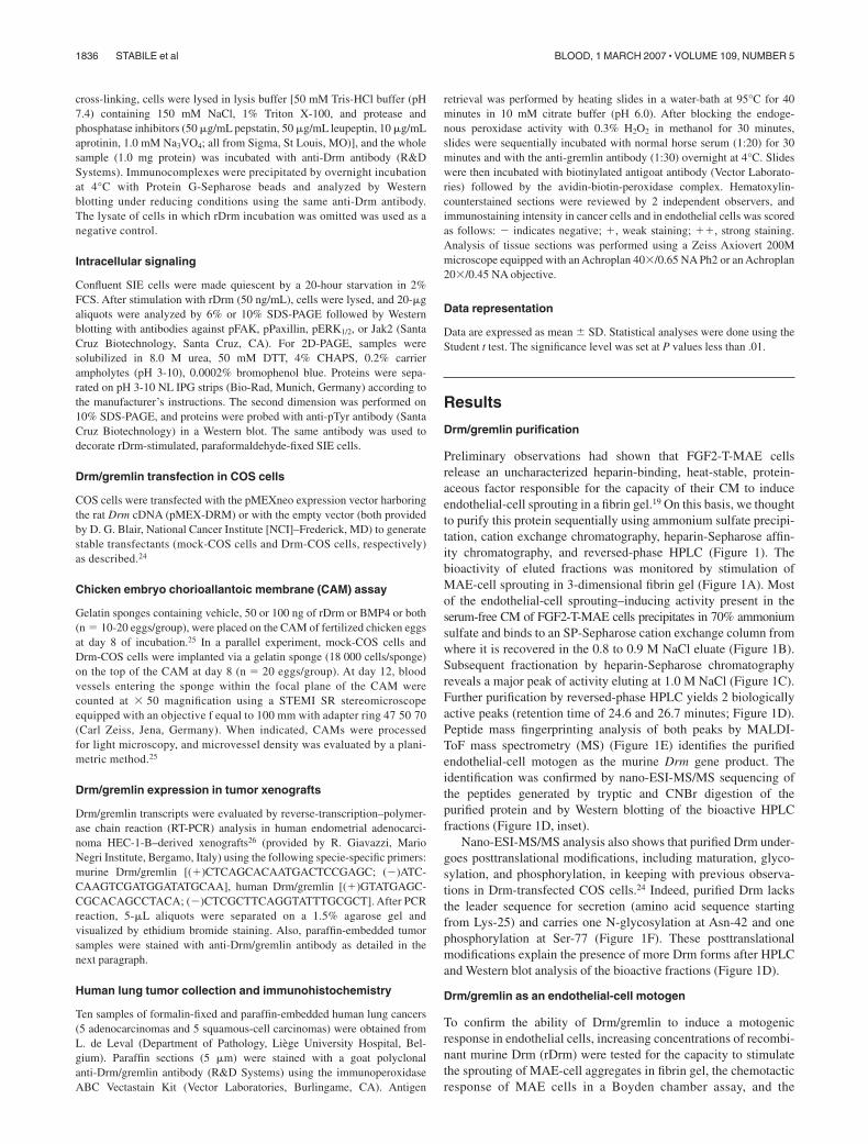
Preliminary observations had shown that FGF2-T-MAE cellsrelease an uncharacterized heparin-binding, heat-stable, protein-aceous factor responsible for the capacity of their CM to induceendothelial-cell sprouting in a fibrin gel.19 On this basis, we thoughtto purify this protein sequentially using ammonium sulfate precipi-tation, cation exchange chromatography, heparin-Sepharose affin-ity chromatography, and reversed-phase HPLC (Figure 1). Thebioactivity of eluted fractions was monitored by stimulation ofMAE-cell sprouting in 3-dimensional fibrin gel (Figure 1A). Mostof the endothelial-cell sprouting–inducing activity present in theserum-free CM of FGF2-T-MAE cells precipitates in 70% ammoniumsulfate and binds to an SP-Sepharose cation exchange column fromwhere it is recovered in the 0.8 to 0.9 M NaCl eluate (Figure 1B).Subsequent fractionation by heparin-Sepharose chromatographyreveals a major peak of activity eluting at 1.0 M NaCl (Figure 1C).Further purification by reversed-phase HPLC yields 2 biologicallyactive peaks (retention time of 24.6 and 26.7 minutes; Figure 1D).Peptide mass fingerprinting analysis of both peaks by MALDI-ToF mass spectrometry (MS) (Figure 1E) identifies the purifiedendothelial-cell motogen as the murine Drm gene product. Theidentification was confirmed by nano-ESI-MS/MS sequencing ofthe peptides generated by tryptic and CNBr digestion of thepurified protein and by Western blotting of the bioactive HPLCfractions (Figure 1D, inset).
Nano-ESI-MS/MS analysis also shows that purified Drm under-goes posttranslational modifications, including maturation, glyco-sylation, and phosphorylation, in keeping with previous observa-tions in Drm-transfected COS cells.24 Indeed, purified Drm lacksthe leader sequence for secretion (amino acid sequence startingfrom Lys-25) and carries one N-glycosylation at Asn-42 and onephosphorylation at Ser-77 (Figure 1F). These posttranslationalmodifications explain the presence of more Drm forms after HPLCand Western blot analysis of the bioactive fractions (Figure 1D).
Drm/gremlin as an endothelial-cell motogen
To confirm the ability of Drm/gremlin to induce a motogenicresponse in endothelial cells, increasing concentrations of recombi-nant murine Drm (rDrm) were tested for the capacity to stimulatethe sprouting of MAE-cell aggregates in fibrin gel, the chemotacticresponse of MAE cells in a Boyden chamber assay, and the
1836 STABILE et al BLOOD, 1 MARCH 2007 � VOLUME 109, NUMBER 5
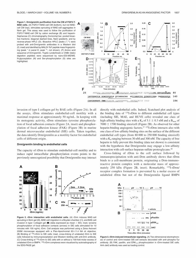
invasion of type I collagen gel by BAE cells (Figure 2A). In allthe assays, rDrm stimulates endothelial-cell motility with amaximal response at approximately 50 ng/mL. In keeping withits motogenic activity, rDrm stimulates tyrosine phosphoryla-tion of focal adhesion contacts (Figure 2A, inset) and phosphor-ylation of focal adhesion kinase (FAK) (Figure 3B) in murinedermal microvascular endothelial (SIE) cells. Taken together,the data identify Drm/gremlin as a motility factor for endothelialcells of different origin.
Drm/gremlin binding to endothelial cells
The capacity of rDrm to stimulate endothelial-cell motility and toinduce rapid intracellular phosphorylation events points to thepreviously unrecognized possibility that Drm/gremlin may interact
directly with endothelial cells. Indeed, Scatchard plot analysis ofthe binding data of 125I-rDrm to different endothelial cell types(including SIE, MAE, and HUVE cells) revealed one class ofhigh-affinity binding sites with a Kd of 5.1 � 0.3 nM and a Bmax of7000 � 1700 binding sites/cell (Figure 2B). As observed for otherheparin-binding angiogenic factors,27 125I-rDrm interacts also withone class of low-affinity binding sites on the surface of the differentendothelial cell types (from 80 000 to 250 000 binding sites/cell)with a Kd ranging between 30 nM and 300 nM. The capacity of freeheparin to fully prevent this binding (data not shown) is consistentwith the hypothesis that Drm/gremlin may engage a low-affinityinteraction with cell-surface heparan-sulfate proteoglycans.28
Cross-linking of rDrm to the cell surface followed byimmunoprecipitation with anti-Drm antibody shows that rDrmbinds to a cell-membrane protein, originating a Drm-immuno-reactive protein complex with a molecular mass of approxi-mately 250 kDa (Figure 2B, inset). Remarkably, 125I-rDrm/receptor complex formation is prevented by a molar excess ofunlabeled rDrm but not of the Drm/gremlin ligand BMP4
Figure 1. Drm/gremlin purification from the CM of FGF2-T-MAE cells. (A) FGF2-T-MAE-cell CM (bottom), but not MAE-cell CM (top), stimulates sprouting of MAE-cell aggregates infibrin gel. This assay was used to follow the purification ofFGF2-T-MAE-cell CM by cation exchange (B) and heparin-Sepharose (C) chromatography (horizontal bar, pooled bioac-tive fractions; diagonal dashed lines, NaCl gradient), and byreverse-phase HPLC (D). Bioactive HPLC peaks (*,**) wereprobed with anti-Drm/gremlin antibodies in a Western blot(D, inset) and identified by MALDI-ToF peptide mass fingerprint-ing (peak * in panel E; peak **, not shown). (F) Amino acidsequence of Drm/gremlin. Tryptic (underlined) or CNBr (italic)digestion peptides were sequenced by nano-ESI-MS/MS.N-glycosylation (N) and Ser-phosphorylation (S) sites arehighlighted.
Figure 2. rDrm interaction with endothelial cells. (A) rDrm induces MAE-cellsprouting in fibrin gel (o), MAE-cell migration in a Boyden chamber (u), and BAE-cellinvasion in type I collagen gel (f) (data expressed as mean � SD); inset, tyrosinephosphorylation of focal adhesion contacts (arrows) in SIE cells stimulated for 10minutes with 100 ng/mL rDrm. Cell analysis was performed using a Zeiss Axiovert200M microscope equipped with a Plan-Apochromat 63�/1.4 NA oil objective.(B) Binding of 125I-rDrm to SIE cells; inset, cross-linking of unlabeled rDrm to SIEcells followed by immunoprecipitation and Western blotting with anti-Drm antibody.(C) Cross-linking of 125I-rDrm to SIE cells with or without a 100-fold molar excess ofunlabeled rDrm or BMP4. 125I-rDrm complexes were visualized by autoradiography ofthe SDS-PAGE gel.
Figure 3. rDrm-induced intracellular signaling. (A) Two-dimensional electrophore-sis of control and rDrm-treated SIE-cell extracts decorated with anti–phospho-Tyrantibody. (B) FAK, paxillin, and ERK1/2 phosphorylation in rDrm-treated SIE cells.Anti-Jak2 antibody was used as loading control.
DRM/GREMLIN AS AN ANGIOGENIC FACTOR 1837BLOOD, 1 MARCH 2007 � VOLUME 109, NUMBER 5
(Figure 2C), indicating that BMP interaction does not hamperthe receptor-binding capacity of rDrm.
In keeping with the presence of a cell-surface receptor, rDrmcauses the rapid appearance of a composite tyrosine phosphory-lation pattern in endothelial SIE cells (Figure 3A). As antici-pated for a cell-motility factor, paxillin, FAK, and mitogen-activated protein kinase ERK1/2 are targets of rDrm-activatedsignaling (Figure 3B). Taken together, the data demonstrate forthe first time the capacity of Drm/gremlin to exert a direct,productive interaction with endothelial cells by engaging cell-surface receptor(s) whose identification will require furtherinvestigation.
Angiogenic activity of Drm/gremlin
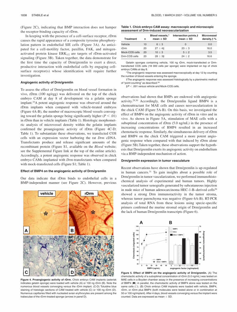
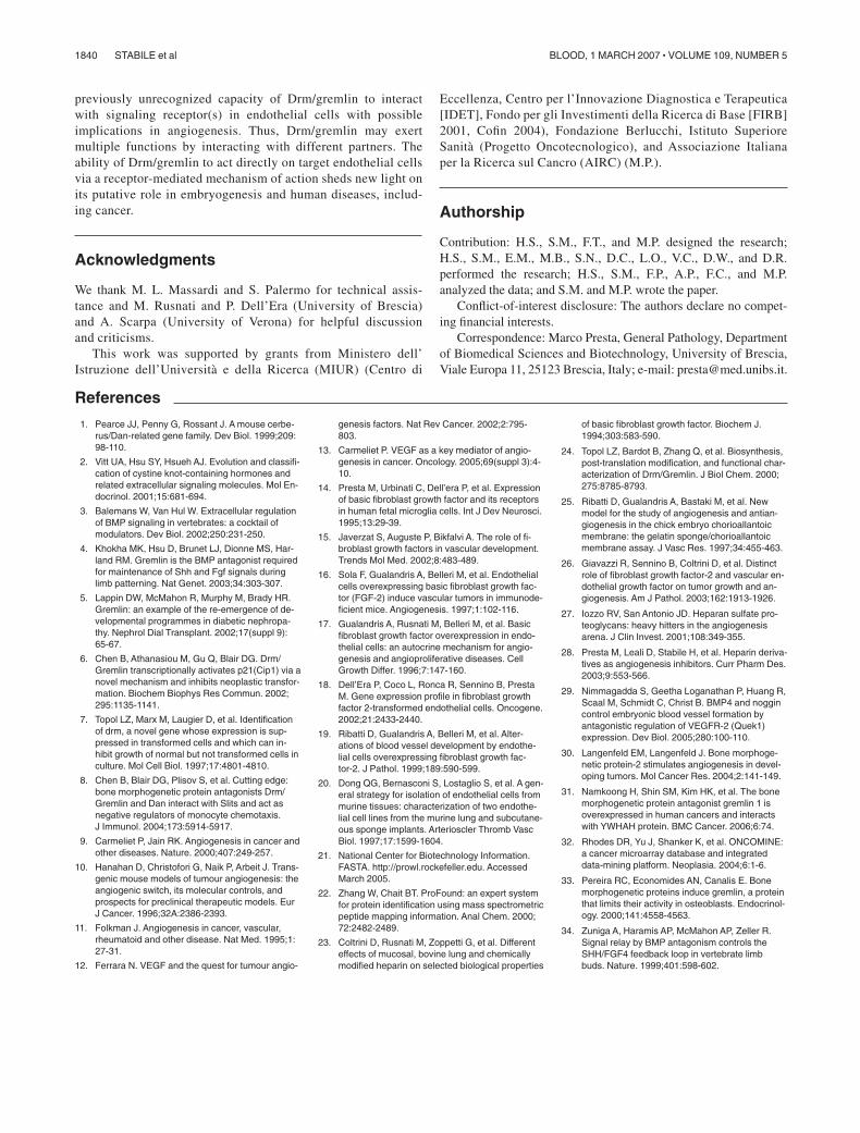
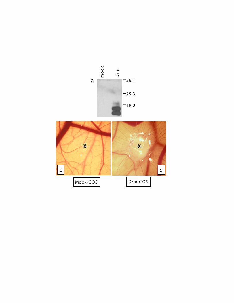
To assess the effect of Drm/gremlin on blood vessel formation invivo, rDrm (100 ng/egg) was delivered on the top of the chickembryo CAM at day 8 of development via a gelatin spongeimplant.25A potent angiogenic response was observed around therDrm implants when compared with vehicle-treated embryos(Figure 4A-B), the number of macroscopic blood vessels converg-ing toward the gelatin sponge being significantly higher (P � .01)in rDrm than in vehicle implants (Table 1). Histologic morphomet-ric analysis of microvessel density within the gelatin implantsconfirmed the proangiogenic activity of rDrm (Figure 4C-D;Table 1). To substantiate these observations, we transfected COScells with an expression vector harboring the rat Drm cDNA.Transfectants produce and release significant amounts of therecombinant protein (Figure S1, available on the Blood website;see the Supplemental Figure link at the top of the online article).Accordingly, a potent angiogenic response was observed in chickembryo CAMs implanted with Drm-transfectants when comparedwith mock-transfected cells (Figure S1; Table 1).
Effect of BMP4 on the angiogenic activity of Drm/gremlin
Our data indicate that rDrm binds to endothelial cells in aBMP-independent manner (see Figure 2C). However, previous
observations had shown that BMPs are endowed with angiogenicactivity.29,30 Accordingly, the Drm/gremlin ligand BMP4 is achemoattractant for MAE cells and causes neovascularization inthe chick CAM (Figure 5A-B). On this basis, we investigated theeffect of BMP4 on the angiogenic activity of rDrm in vitro and invivo. As shown in Figure 5A, stimulation of MAE cells with asuboptimal concentration of rDrm (5.0 ng/mL) in the presence ofincreasing concentrations of BMP4 resulted in an increasedchemotactic response. Similarly, the simultaneous delivery of rDrmand BMP4 on the chick CAM triggered a more potent angio-genic response when compared with that induced by rDrm alone(Figure 5B).Taken together, these observations support the hypoth-esis that Drm/gremlin exerts its angiogenic activity on endotheliumvia a BMP-independent mechanism of action.
Drm/gremlin expression in tumor vasculature
Recent observations have shown that Drm/gremlin is up-regulatedin human cancers.31 To gain insights about a possible role ofDrm/gremlin in tumor vascularization, we performed immunohisto-chemical analysis of experimental and human tumors. Highlyvascularized tumor xenografts generated by subcutaneous injectionin nude mice of human adenocarcinoma HEC-1-B–derived cells26
showed a strong Drm immunoractivity in the tumor stroma,whereas tumor parenchyma was negative (Figure 6A-B). RT-PCRanalysis of total RNA from these lesions using specie-specificprimers confirmed the murine stromal origin of Drm/gremlin andthe lack of human Drm/gremlin transcripts (Figure 6).
Figure 4. Proangiogenic activity of rDrm. Chick embryo CAM implants (asteriskindicates gelatin sponge) were loaded with vehicle (A) or 100 ng rDrm (B). Note thenumerous blood vessels converging versus the rDrm implant. (C-D) Toluidine bluestaining of histologic sections of CAM treated with vehicle (C) or 100 ng rDrm (D).Numerous capillaries filled with nucleated avian erythrocytes are present among thetrabeculae of the rDrm-treated sponge (arrows in panel D).
Figure 5. Effect of BMP4 on the angiogenic activity of Drm/gremlin. (A) Thechemotactic activity of a suboptimal concentration of rDrm (5.0 ng/mL) was tested onMAE cells in a Boyden chamber assay in the presence of increasing concentrationsof BMP4 (F); in parallel, the chemotactic activity of BMP4 alone was tested on thesame cells (E). (B) Chick embryo CAM implants were loaded with vehicle, BMP4,rDrm, or rDrm plus BMP4 (both molecules were tested alone or in combination at50 or 100 ng/implant). After 4 days, blood vessels converging versus the implant werecounted. Data are expressed as mean � SD.
Table 1. Chick embryo CAM assay: macroscopic and microscopicassessment of Drm-induced neovascularization
Treatment nBlood vessels,*
mean � SDIntersection points,†
mean � SDMicrovesseldensity,† %
Vehicle 10 6 � 3 0 0.0
rDrm 20 27 � 4‡ 23 � 3 16.0
Mock-COS cells 20 10 � 3 5 � 2 3.5
Drm-COS cells 20 28 � 3‡ 24 � 2 16.6
Gelatin sponges containing vehicle, 100 ng rDrm, mock-transfected or Drm-transfected COS cells (18 000 cells per sponge) were implanted on top of chickembryo CAMs at day 8.
*The angiogenic response was assessed macroscopically at day 12 by countingthe number of blood vessels entering the sponge.
†The angiogenic response was assessed histologically by a planimetric methodof “point counting” as described.24
‡P � .001 versus vehicle and Mock-COS cells.
1838 STABILE et al BLOOD, 1 MARCH 2007 � VOLUME 109, NUMBER 5
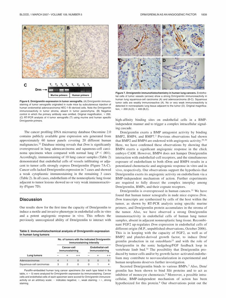
The cancer profiling DNA microarray database Oncomine 2.0contains publicly available gene expression sets generated fromapproximately 60 tumor panels covering 20 different humanmalignancies.32 Database mining reveals that Drm is significantlyoverexpressed in lung adenocarcinoma and squamous-cell carci-noma specimens when compared with normal lung (P � .001).Accordingly, immunostaining of 10 lung cancer samples (Table 2)demonstrated that endothelial cells of vessels infiltrating or adja-cent to tumor cells strongly express Drm/gremlin (Figure 7A-C).Cancer cells lacked Drm/gremlin expression in 7 cases and showeda weak cytoplasmic immunostaining in the remaining 3 cases(Table 2). In all cases, endothelium of the nonneoplastic lung tissueadjacent to tumor lesions showed no or very weak immunoreactiv-ity (Figure 7D).
Discussion
Our results show for the first time the capacity of Drm/gremlin toinduce a motile and invasive phenotype in endothelial cells in vitroand a potent angiogenic response in vivo. This reflects thepreviously unrecognized ability of Drm/gremlin to interact with
high-affinity binding sites on endothelial cells in a BMP-independent manner and to trigger a complex intracellular signal-ing cascade.
Drm/gremlin exerts a BMP antagonist activity by bindingBMP2, BMP4, and BMP7.3 Previous observations had shownthat BMP2 and BMP4 are endowed with angiogenic activity.29,30
Here, we have confirmed these observations by showing thatBMP4 exerts a significant angiogenic response in the chickembryo CAM. However, BMP4 does not hamper Drm/gremlininteraction with endothelial-cell receptors, and the simultaneousexposure of endothelium to both rDrm and BMP4 results in apotentiated chemotactic and angiogenic response in vitro and invivo, respectively. Our observations support the hypothesis thatDrm/gremlin exerts its angiogenic activity on endothelium via aBMP-independent mechanism of action. Further experimentsare required to fully dissect the complex interplay amongDrm/gremlin, BMPs, and their cognate receptors.
Drm/gremlin is overexpressed in human cancers.31 We havefound that human tumor xenografts in nude mice express Drm.Drm transcripts are synthesized by cells of the host within thetumor, as shown by RT-PCR analysis using specific murineprimers, and Drm/gremlin protein accumulates in the stroma ofthe tumor. Also, we have observed a strong Drm/gremlinimmunoreactivity in endothelial cells of human lung tumorsamples, absent in adjacent nonneoplastic lung tissue. Recombi-nant FGF2 up-regulates Drm expression in endothelial cells ofdifferent origin (M.P., unpublished observations, October 2006).This is in keeping with the capacity of FGF2, as well as ofBMP2 and platelet-derived growth factor, to induce Drm/gremlin production in rat osteoblasts33 and with the role ofDrm/gremlin in the sonic hedgehog/FGF feedback loop invertebrate limb bud.34 The possibility that Drm/gremlin pro-duced by tumor cells and/or by growth factor–activated endothe-lium may contribute to neovascularization in experimental andhuman neoplasms deserves further investigation.
Secreted Drm/gremlin binds to various BMPs.3 Also, Drm/gremlin has been shown to bind Slit proteins and to act asinhibitor of monocyte chemotaxis.8 Moreover, a possible intra-cellular, BMP-independent mechanism of action has beenhypothesized for this protein.6 Our observations point out the
Figure 6. Drm/gremlin expression in tumor xenografts. (A) Drm/gremlin immuno-staining of tumor xenografts originated in nude mice by subcutaneous injection ofhuman endometrial adenocarcinoma HEC-1-B–derived cells. Note the Drm/gremlinimmunoreactivity in tumor stroma, absent in tumor parenchyma. (B) Negativecontrol in which the primary antibody was omitted. Original magnification, � 200.(C) RT-PCR analysis of 4 tumor xenografts (T) using murine and human specificDrm/gremlin primers.
Table 2. Immunohistochemical analysis of Drm/gremlin expressionin human lung tumors
Lung tumors
No. of tumors with the indicated Drm/gremlinimmunostaining intensity
Cancer-cellimmunostaining
Endothelial-cellimmunostaining
� � �� � � ��
Adenocarcinomas 4 1 0 0 0 5
Squamous-cell carcinomas 3 2 0 0 1 4
Paraffin-embedded human lung cancer specimens (for each type listed in thetable, n � 5) were analyzed for Drm/gremlin expression by immunostaining. Cancercells and endothelial cells of each specimen were scored for Drm/gremlin immunore-activity on an arbitrary scale: � indicates negative; �, weak staining; ��, strongstaining.
Figure 7. Drm/gremlin immunohistochemistry in human lung cancers. Endothe-lial cells of tumor vessels (arrows) show a strong Drm/gremlin immunoreactivity inhuman lung squamous-cell carcinoma (A) and adenocarcinoma (B-C). Squamoustumor cells are weakly immunoreactive (A). No or very weak immunoreactivity isdetected in nonneoplastic lung tissue adjacent to the tumor (D). Original magnifica-tion, � 200 (A,D); � 400 (B,C).
DRM/GREMLIN AS AN ANGIOGENIC FACTOR 1839BLOOD, 1 MARCH 2007 � VOLUME 109, NUMBER 5
previously unrecognized capacity of Drm/gremlin to interactwith signaling receptor(s) in endothelial cells with possibleimplications in angiogenesis. Thus, Drm/gremlin may exertmultiple functions by interacting with different partners. Theability of Drm/gremlin to act directly on target endothelial cellsvia a receptor-mediated mechanism of action sheds new light onits putative role in embryogenesis and human diseases, includ-ing cancer.
Acknowledgments
We thank M. L. Massardi and S. Palermo for technical assis-tance and M. Rusnati and P. Dell’Era (University of Brescia)and A. Scarpa (University of Verona) for helpful discussionand criticisms.
This work was supported by grants from Ministero dell’Istruzione dell’Universita e della Ricerca (MIUR) (Centro di
Eccellenza, Centro per l’Innovazione Diagnostica e Terapeutica[IDET], Fondo per gli Investimenti della Ricerca di Base [FIRB]2001, Cofin 2004), Fondazione Berlucchi, Istituto SuperioreSanita (Progetto Oncotecnologico), and Associazione Italianaper la Ricerca sul Cancro (AIRC) (M.P.).
Authorship
Contribution: H.S., S.M., F.T., and M.P. designed the research;H.S., S.M., E.M., M.B., S.N., D.C., L.O., V.C., D.W., and D.R.performed the research; H.S., S.M., F.P., A.P., F.C., and M.P.analyzed the data; and S.M. and M.P. wrote the paper.
Conflict-of-interest disclosure: The authors declare no compet-ing financial interests.
Correspondence: Marco Presta, General Pathology, Departmentof Biomedical Sciences and Biotechnology, University of Brescia,Viale Europa 11, 25123 Brescia, Italy; e-mail: [email protected].
References
1. Pearce JJ, Penny G, Rossant J. A mouse cerbe-rus/Dan-related gene family. Dev Biol. 1999;209:98-110.
2. Vitt UA, Hsu SY, Hsueh AJ. Evolution and classifi-cation of cystine knot-containing hormones andrelated extracellular signaling molecules. Mol En-docrinol. 2001;15:681-694.
3. Balemans W, Van Hul W. Extracellular regulationof BMP signaling in vertebrates: a cocktail ofmodulators. Dev Biol. 2002;250:231-250.
4. Khokha MK, Hsu D, Brunet LJ, Dionne MS, Har-land RM. Gremlin is the BMP antagonist requiredfor maintenance of Shh and Fgf signals duringlimb patterning. Nat Genet. 2003;34:303-307.
5. Lappin DW, McMahon R, Murphy M, Brady HR.Gremlin: an example of the re-emergence of de-velopmental programmes in diabetic nephropa-thy. Nephrol Dial Transplant. 2002;17(suppl 9):65-67.
6. Chen B, Athanasiou M, Gu Q, Blair DG. Drm/Gremlin transcriptionally activates p21(Cip1) via anovel mechanism and inhibits neoplastic transfor-mation. Biochem Biophys Res Commun. 2002;295:1135-1141.
7. Topol LZ, Marx M, Laugier D, et al. Identificationof drm, a novel gene whose expression is sup-pressed in transformed cells and which can in-hibit growth of normal but not transformed cells inculture. Mol Cell Biol. 1997;17:4801-4810.
8. Chen B, Blair DG, Plisov S, et al. Cutting edge:bone morphogenetic protein antagonists Drm/Gremlin and Dan interact with Slits and act asnegative regulators of monocyte chemotaxis.J Immunol. 2004;173:5914-5917.
9. Carmeliet P, Jain RK. Angiogenesis in cancer andother diseases. Nature. 2000;407:249-257.
10. Hanahan D, Christofori G, Naik P, Arbeit J. Trans-genic mouse models of tumour angiogenesis: theangiogenic switch, its molecular controls, andprospects for preclinical therapeutic models. EurJ Cancer. 1996;32A:2386-2393.
11. Folkman J. Angiogenesis in cancer, vascular,rheumatoid and other disease. Nat Med. 1995;1:27-31.
12. Ferrara N. VEGF and the quest for tumour angio-
genesis factors. Nat Rev Cancer. 2002;2:795-803.
13. Carmeliet P. VEGF as a key mediator of angio-genesis in cancer. Oncology. 2005;69(suppl 3):4-10.
14. Presta M, Urbinati C, Dell’era P, et al. Expressionof basic fibroblast growth factor and its receptorsin human fetal microglia cells. Int J Dev Neurosci.1995;13:29-39.
15. Javerzat S, Auguste P, Bikfalvi A. The role of fi-broblast growth factors in vascular development.Trends Mol Med. 2002;8:483-489.
16. Sola F, Gualandris A, Belleri M, et al. Endothelialcells overexpressing basic fibroblast growth fac-tor (FGF-2) induce vascular tumors in immunode-ficient mice. Angiogenesis. 1997;1:102-116.
17. Gualandris A, Rusnati M, Belleri M, et al. Basicfibroblast growth factor overexpression in endo-thelial cells: an autocrine mechanism for angio-genesis and angioproliferative diseases. CellGrowth Differ. 1996;7:147-160.
18. Dell’Era P, Coco L, Ronca R, Sennino B, PrestaM. Gene expression profile in fibroblast growthfactor 2-transformed endothelial cells. Oncogene.2002;21:2433-2440.
19. Ribatti D, Gualandris A, Belleri M, et al. Alter-ations of blood vessel development by endothe-lial cells overexpressing fibroblast growth fac-tor-2. J Pathol. 1999;189:590-599.
20. Dong QG, Bernasconi S, Lostaglio S, et al. A gen-eral strategy for isolation of endothelial cells frommurine tissues: characterization of two endothe-lial cell lines from the murine lung and subcutane-ous sponge implants. Arterioscler Thromb VascBiol. 1997;17:1599-1604.
21. National Center for Biotechnology Information.FASTA. http://prowl.rockefeller.edu. AccessedMarch 2005.
22. Zhang W, Chait BT. ProFound: an expert systemfor protein identification using mass spectrometricpeptide mapping information. Anal Chem. 2000;72:2482-2489.
23. Coltrini D, Rusnati M, Zoppetti G, et al. Differenteffects of mucosal, bovine lung and chemicallymodified heparin on selected biological properties
of basic fibroblast growth factor. Biochem J.1994;303:583-590.
24. Topol LZ, Bardot B, Zhang Q, et al. Biosynthesis,post-translation modification, and functional char-acterization of Drm/Gremlin. J Biol Chem. 2000;275:8785-8793.
25. Ribatti D, Gualandris A, Bastaki M, et al. Newmodel for the study of angiogenesis and antian-giogenesis in the chick embryo chorioallantoicmembrane: the gelatin sponge/chorioallantoicmembrane assay. J Vasc Res. 1997;34:455-463.
26. Giavazzi R, Sennino B, Coltrini D, et al. Distinctrole of fibroblast growth factor-2 and vascular en-dothelial growth factor on tumor growth and an-giogenesis. Am J Pathol. 2003;162:1913-1926.
27. Iozzo RV, San Antonio JD. Heparan sulfate pro-teoglycans: heavy hitters in the angiogenesisarena. J Clin Invest. 2001;108:349-355.
28. Presta M, Leali D, Stabile H, et al. Heparin deriva-tives as angiogenesis inhibitors. Curr Pharm Des.2003;9:553-566.
29. Nimmagadda S, Geetha Loganathan P, Huang R,Scaal M, Schmidt C, Christ B. BMP4 and noggincontrol embryonic blood vessel formation byantagonistic regulation of VEGFR-2 (Quek1)expression. Dev Biol. 2005;280:100-110.
30. Langenfeld EM, Langenfeld J. Bone morphoge-netic protein-2 stimulates angiogenesis in devel-oping tumors. Mol Cancer Res. 2004;2:141-149.
31. Namkoong H, Shin SM, Kim HK, et al. The bonemorphogenetic protein antagonist gremlin 1 isoverexpressed in human cancers and interactswith YWHAH protein. BMC Cancer. 2006;6:74.
32. Rhodes DR, Yu J, Shanker K, et al. ONCOMINE:a cancer microarray database and integrateddata-mining platform. Neoplasia. 2004;6:1-6.
33. Pereira RC, Economides AN, Canalis E. Bonemorphogenetic proteins induce gremlin, a proteinthat limits their activity in osteoblasts. Endocrinol-ogy. 2000;141:4558-4563.
34. Zuniga A, Haramis AP, McMahon AP, Zeller R.Signal relay by BMP antagonism controls theSHH/FGF4 feedback loop in vertebrate limbbuds. Nature. 1999;401:598-602.
1840 STABILE et al BLOOD, 1 MARCH 2007 � VOLUME 109, NUMBER 5
19.0
25.3
36.1
Drm
mo
ck
a
**
cb
Drm-C OSMoc k-C OS