Bone Infection (osteomyelitis)
72
Bone Infection (osteomyelitis) รศ.นพ.ยงศักดิ หวังรุ่งทรัพย์ ภาควิชาออร์โธปิดิกส์ คณะแพทยศาสตร์ จุฬาลงกรณ์มหาวิทยาลัย
Transcript of Bone Infection (osteomyelitis)
Bone Infection
(osteomyelitis)
รศ.นพ.ยงศักดิ์ หวังรุ่งทรัพย์ ภาควิชาออร์โธปิดิกส์ คณะแพทยศาสตร์ จุฬาลงกรณ์มหาวิทยาลัย

Types of organism
Pyogenic osteomyelitis or arthritis
Chronic granulomatous reaction
Fungal infection
Parasitic infestation

Route of Infection
Hematogenous system
Direct invasion: Open Fx,
operation, skin puncture
Direct spreading
Acute
Hematogenous
Osteomyelitis

Acute Hematogenous
Osteomyelitis Common in children
Adult – lowered resistance by drug:
immunosuppressive drug, debility disease:
DM, AIDS
- more common in vertebrae than
long bone
Post-trauma: hematoma or fluid collection
in bone
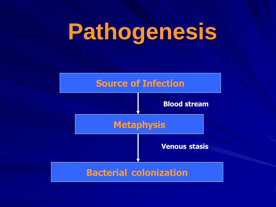
Pathogenesis
Source of Infection
Metaphysis
Bacterial colonization
Blood stream
Venous stasis


Etiology Aerobic organisms -Gram positive : Staphylococcus aureus , Streptococcus pyogens
Streptococcus pneumoniae
-Gram negative : Haemophilus influenza,
E.coli, Pseudomonas aeruginosa,
Proteus mirabilis,
Anaerobic organisms Bacteroides fragilis

Pathology
Inflammation
Suppuration
Necrosis
New bone formation
Resolution

Inflammation
First 24 hours
Vascular congestion
Polymorphonuclear leukocyte infiltration
Exudation
Intraosseus pressure intense pain
intravascular thrombosis ischemia

Suppuration
2-3 days
Pus formation
Subperiosteal abscess
via Volkmann canals
Pus spreading epiphysis
joint
medullary cavity
soft tissue

Necrosis Bone death by the end of a week
Bone destruction ← toxin
← ischemia
Epiphyseal plate injury
Sequestrum formation – small removed by
macrophage,osteoclast.
– large remained

New bone formation
By the end of 2nd week
Involucrum (new bone
formation from deep
layer of periosteum )
surround infected tissue.
If infection persist- pus
discharge through sinus
to skin surface Chronic
osteomyelitis

Resolution
Antibiotics Surgical drainage
Infection is controlled
Bone remodeling
Resolution
Infection is controlled
Intraosseous pressure release
With healing – new bone formation +
periosteal reaction bone thickening and
sclerosis
Remodeling to normal contour or deformity

Infection persist
Chronic drainage
Chronic Osteomyelitis
Signs and Symptoms in infant
Drowsy
Irritable
Fails to thrive
history of birth difficulties
History of umbilical artery
catheterization
Metaphyseal tenderness and
resistance to joint movement
Signs and Symptoms in child
Severe pain
Malaise
Fever
Toxemia
History of recent infection
Local inflammation pus
escape from bone
Lymphadenopathy
Acute osteomyelitis in adult
1.Uncommon
2.History of DM.
3.Immunosuppressive drug
4.Drug addict
5.Elderly patients.

Signs and Symptoms in adult
Fever
Pain
Inflammation
Acute tenderness
Common site is thoraco-
lumbar spine

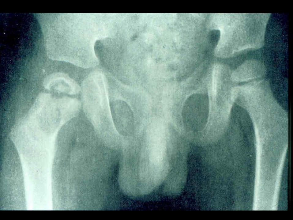
Radiographic studies
มักจะเปล่ียนแปลงหลังจากการติดเช้ือนานกว่า 10 วัน
เริ่มจาก rarefaction, area of lytic and
sclerotic lesion, sequestrum and involucrum.
ควรเริ่มให้การรักษาทันทีก่อนจะเห็นการเปล่ียนแปลงในภาพถ่าย X-ray


Bone Scan
99m TC-HDP - sensitive
- not specific
67 Ga-citrate or 111 In-labeled
leukocyte more specific

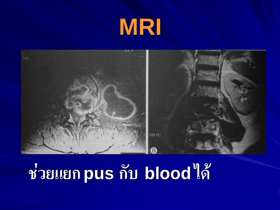
MRI
ช่วยแยก pus กับ blood ได้

Aspiration pus
confirm diagnosis
smear for cell and organism
culture and sensitivity test

Investigations
CBC
ESR
Hemoculture positive ~ 50%
Antistaphylococcal antibody
titer (in doubtful case)
Differential diagnosis
Cellulitis
Acute suppurative arthritis
Acute rheumatism
Gaucher’s disease – Pseudo- osteitis, resembling osteomyelitis, enlargement of spleen and liver. Because of predisposing to infection, antibiotics should be given.
Sickle-cell crisis – mimic osteomyelitis, in endemic area of Salmonella, it is wise to treat with antibiotics until infection is excluded

Treatment for acute
osteomyelitis
Supportive treatment
Splint
Antibiotic therapy
Surgical drainage

Supportive treatment
Analgesics
Correction of dehydration

Splint
- Plaster slab
- traction
- Prevent joint contracture


Surgical drainage
Early treatment no need surgery
Late treatment surgical
drainage about 1/3 of cases. If pus
found and release no need to drill bone.
But drilling one or two holes if no
obvious abscess.

Antibiotics
Initial antibiotics “ BEST GUESS ”
- according to smear findings
- according to incidences , age.
Proper antibiotics
- according to culture and
sensitivities test

Guideline for initial antibiotics
Age Pathogen Drugs
1.Older children and
previously fit adults
-Staphylococcal
infection
- Fluclaxocillin and
fusidic acid IV 3-4 day
oral 3-6 wks
2.Children <4 years -Gram neg. infection
-Haemophilus
infection
-2nd generation
Cephalosporins or
Amoxycillin with
clavulanic acid
3.Sickle-cell patient -Salmonella infection - Co-trimoxazole
- Amoxycillin with
clavulanic acid
4.Heroin addicts and
immuno-compromised
patients
-Unusual infection :
pseudomonas ,
proteus, bacteroides
-3rd or newer generation
Cephalosporins

Acute osteomyelitis
When infection subside, movement is
encourage. Walk with crutches and
full weight bearing is possible after 3-
4 weeks.

Complication
lethal outcome – rare
metastatic infection (multifocal
infection)
suppurative arthritis
very young patient
metaphysis is intracapsular
metastatic infection

Complication
altered bone growth
chronic osteomyelitis
- delay diagnosis and
treatment
- debilitated patients
- compromised host
Chronic
Osteomyelitis

Chronic osteomyelitis
Sequel to acute hematogenous osteomyelitis
Usual organisms are staph. aureus, Escherichia coli, Strep. pyogens, Proteus and Pseudomonas (always mixed infections)
In the presence of foreign implants : Staph. Epidermidis is the commonest pathogen.

Pathology of chronic
osteomyelitis Bone is destroyed in a discrete area or diffuse
Cavities containing pus and sequestrum are surrounded by vascular bone and sclerosis bone resulted from reactive new bone formation
Sequestra, foreign implants act as substrates for bacterial adhesion, ensuring the persistence of infection and sinus drainage
Pathological fracture
Signs and Symptoms of
chronic osteomyelitis
Pain
Pyrexia
Redness
Tenderness
Draining sinus
Excoriation of skin


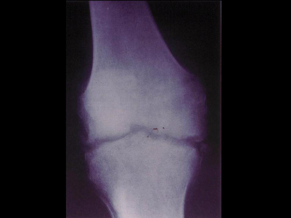
Radiographic study
A patchy loss of bone density with
thickening and sclerosis of the
surrounding bone
Sequestra : dense fragment in contrast
to surrounding vascularized bone
Sinogram may help to localize the site
of infection

Sequestrum

Radioisotope scanning
99m TC-HDP Up take 67 Ga-citrate or 111In-labelled
leukocyte more specific
CT – Scan and MRI
Show extent of bone destruction
and reactive edema, hidden abscess
and sequestrum
Pre-op planning investigation

Other Investigations
CBC
ESR
Antistayphylococcal antibody titers – Dx hidden infection and tracking progress to recovery
C/S from draining discharge R/O resistance bacteria

Treatment for chronic
osteomyelitis
Medical treatment
Local treatment
Surgical treatment

Antibiotics
To stop spreading of infection
To control acute flare
Capable of penetrating sclerotic
bone and non-toxic to body

Surgical treatment
Sequestrectomy :
sulphan blue
stained only vital
tissue
Continuous
irrigation 3-6
weeks.
Gentamicin beads

Space filling techniques
Papineau technique (Papineau et al
1979)
Muscle flap + skin graft (Fitzgerald et al
1985)
Myocutaneous island flap. (Yoshimura
et al 1989)

Prognosis
Local trauma must be avoided
Any recurrent of symptoms should be
taken seriously and investigated

Acute Suppurative Arthritis
Route of infection
1. direct invasion
2. eruption of a bone abscess
3. hematogenous spreading

Causal Organisms
Staphylococcus aureus
Hemophilus influenza
E. coli
Streptococcus
Proteus

Oganism
Synovial membrane
Seropurulent exudate pus
Bacterial enzyme Synovial enzyme
Joint destruction
Acute inflammatory
reaction

Septic Arthritis

TB Arthritis

Signs and symptoms in newborn
Clinical of septicemia : irritable,
refuses to feed, rapid pulse
Joint swelling
Tenderness and resistance to
movement of the joint
Look for umbilical infection

Signs and symptoms
in children
acute pain in single joint : hip.
Pseudoparesis.
Swelling and inflammation of the
joint.
Child looks ill.
Limit movement of the joint.
Look for a source of infection : toe,
boil, otitis media

Signs and symptoms in adult
Often superficial joint : knee, wrist,
ankle
Pain
Swelling and inflammation
Restricted movement
Examined for gonococcal infection or
drug abuse.

Radiographic study
Early : usually normal , joint space
may seem to be widened (because of
fluid in the joint)
Late : osteoporosis ,narrowing and
irregularity of the joint apace.
with E. coli infection there is
sometime gas in the joint


Investigation
CBC
ESR
Gram stain of synovial
fluid
C/S
Differential diagnosis
Acute osteomyelitis: in children indistinguishable from septic joint
Trauma: traumatic synovitis
Irritable joint : the patient does not look ill
Hemophilic bleeding
Rheumatic fever
Gout and pseudogout
Treatment of septic arthritis
Supportive care
: analgesics, fluid supplement ,
splint, traction
Antibiotics
: same as acute osteomyelitis
Drainage
: Aspiration, arthrotomy

Once the conditions improved, if the
articular cartilage is preserved – gentle and gradually increasing active motion
If articular cartilage is destroyed – the joint
is immobilized in optimal position until ankylosis is sound
Treatment of septic arthritis

Outcome After Healing
Complete resolution
Partial loss articular cartilage and
fibrosis of joint.
Loss of articular cartilage and bony
ankylosis
Bone destruction and permanent
deformity of the joint.

Complication
Cartilage destruction
Growth disturbance
Bone destruction