Bone disease and tissue repair - pathkids.com
46
1 Bone disease and tissue repair Dr Tim Bracey MBChB BScHons MRCS PhD FRCPath Consultant Pathologist
Transcript of Bone disease and tissue repair - pathkids.com
1
Bone disease and
tissue repair
Dr Tim Bracey
MBChB BScHons MRCS PhD FRCPath
Consultant Pathologist

Outline of talk
Bone structure and function
Disorders of bone leading to increased
fractures
Bone fracture and repair
Comparison with tissue repair at other
anatomical sites
Factors affecting healing and repair
Disorders of healing and repair

Introduction
Function of bones
Mechanical support for movement
Determines body size and shape
Protect viscera
Haemopoietic and endocrine role
Composition
65% calcium hydroxyapatite
(contains 99% of body's calcium, 85% of phosphorus, 65% of sodium, also magnesium)
35% organic (cells and proteins)

What are the main differences
between compact and
cancellous bone?
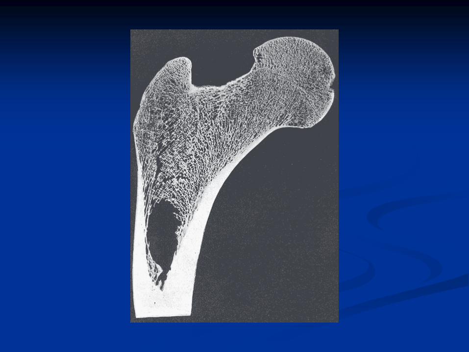

Compact = cortical bone
Cement lines
Haversian canal
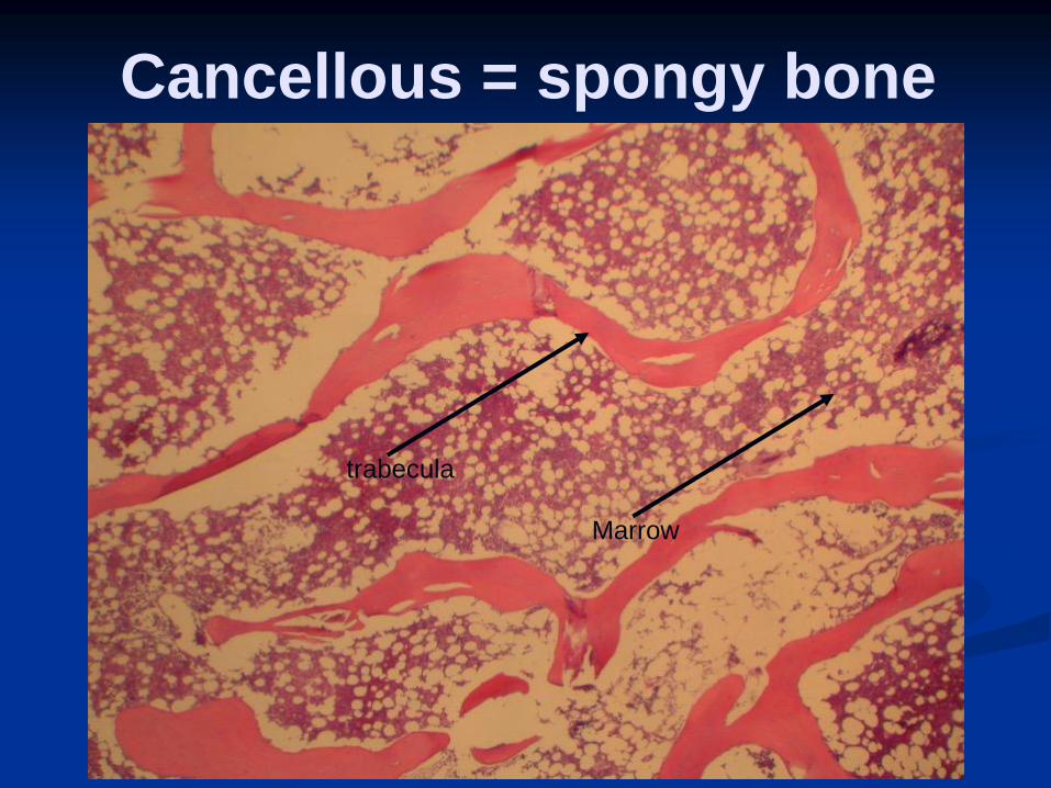
Cancellous = spongy bone
trabecula
Marrow

What are the three main cell
types in bone?
(clue: they all start with
osteo-) and what are their
functions?
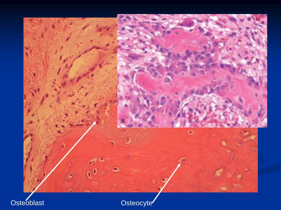
Osteoblast Osteocyte
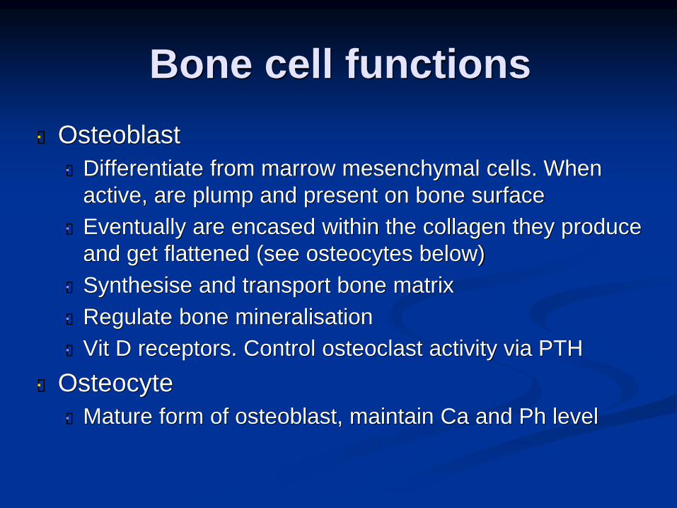
Bone cell functions
Osteoblast
Differentiate from marrow mesenchymal cells. When
active, are plump and present on bone surface
Eventually are encased within the collagen they produce
and get flattened (see osteocytes below)
Synthesise and transport bone matrix
Regulate bone mineralisation
Vit D receptors. Control osteoclast activity via PTH
Osteocyte
Mature form of osteoblast, maintain Ca and Ph level
Osteoclast
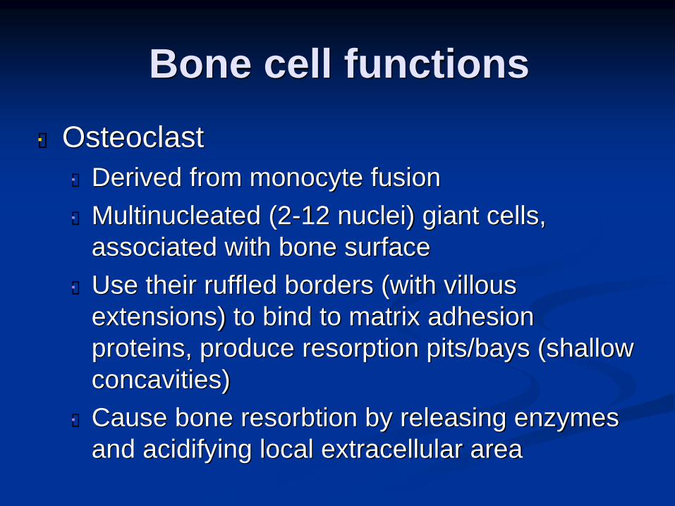
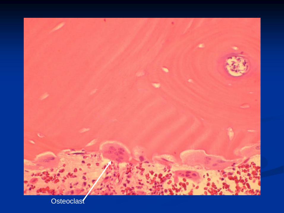
Bone cell functions
Osteoclast
Derived from monocyte fusion
Multinucleated (2-12 nuclei) giant cells,
associated with bone surface
Use their ruffled borders (with villous
extensions) to bind to matrix adhesion
proteins, produce resorption pits/bays (shallow
concavities)
Cause bone resorbtion by releasing enzymes
and acidifying local extracellular area

What is bone remodeling?
What factors influence peak
bone mass?

Remodeling and peak bone
mass Collections of osteocytes, osteoblasts and
osteoclasts work as a functional unit known as the basic multicellular unit (BMU) to control bone formation and resorbtion
In early life bone formation predominates
In fourth decade bone resorbtion predominates and there is a steady decrease in skeletal mass!
PBM is determined by genetics (mainly growth factor and hormone receptors), activity, weight, exogenous drugs and hormones
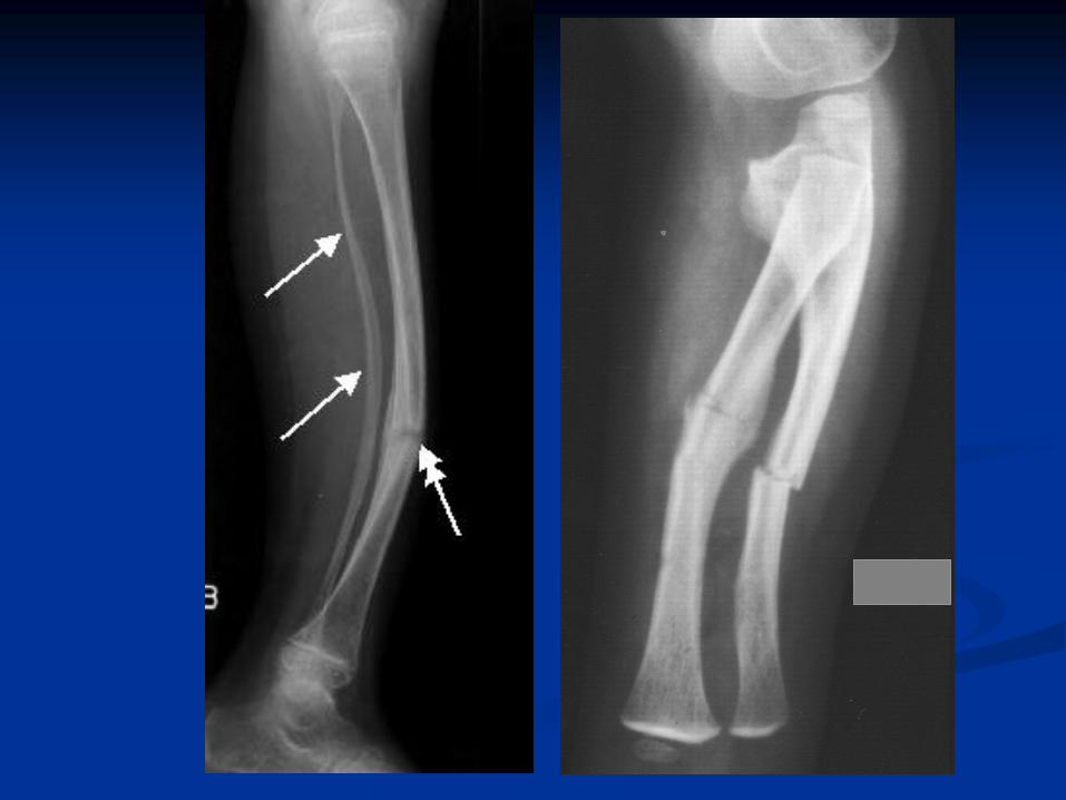
Give definitions of the
following types of fracture
complete
compound
comminuted
displaced
stress

Name an inherited connective
tissue disorder that results in
reduced bone density and
increased susceptibility to
fractures
Other than bone what other tissues
does it affect and what are their
clinical manifestations?
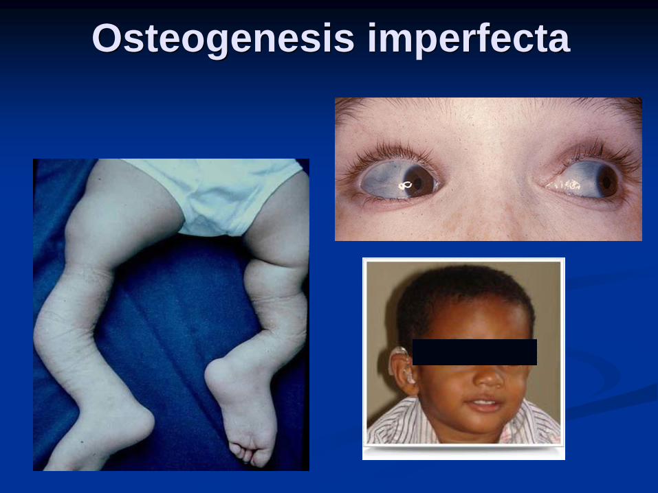
Osteogenesis imperfecta
What are the main risk factors for
osteoporosis?
Which bones do osteoporotic fractures
most commonly affect?
What are the complications of
osteoporotic fractures in the elderly?
How reliable are plain X-rays in
diagnosis of osteoporosis?

Primary osteoporosis
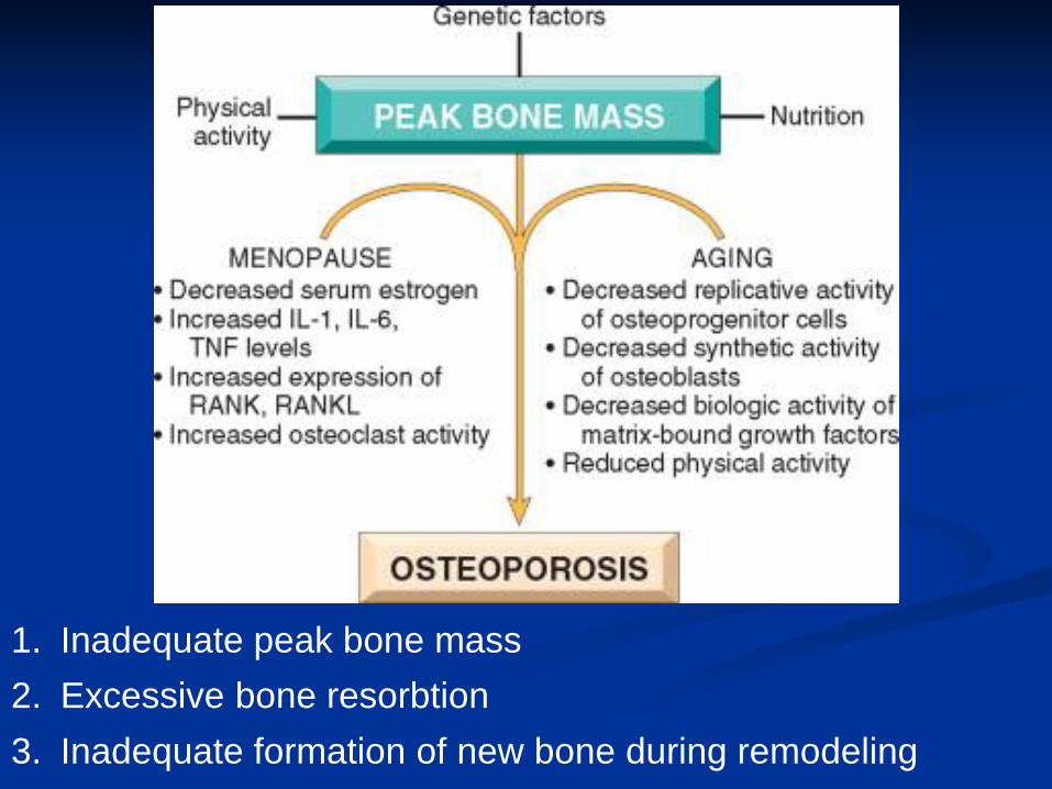
1. Inadequate peak bone mass
2. Excessive bone resorbtion
3. Inadequate formation of new bone during remodeling
Osteoporosis is the most
common disorder associated
with reduced bone mass.
What disorder is associated
with increased bone mass yet
still predisposes to fractures?

Paget’s disease of bone

Paget’s disease of bone
Micrograph showing Paget's disease of the bone with the
characteristic jigsaw puzzle-like/mosaic pattern. H&E stain.
Name the 4 main phases of
fracture healing
What is “woven bone”?

1 – haematoma phase a few days

2 – inflammatory phase ~a week
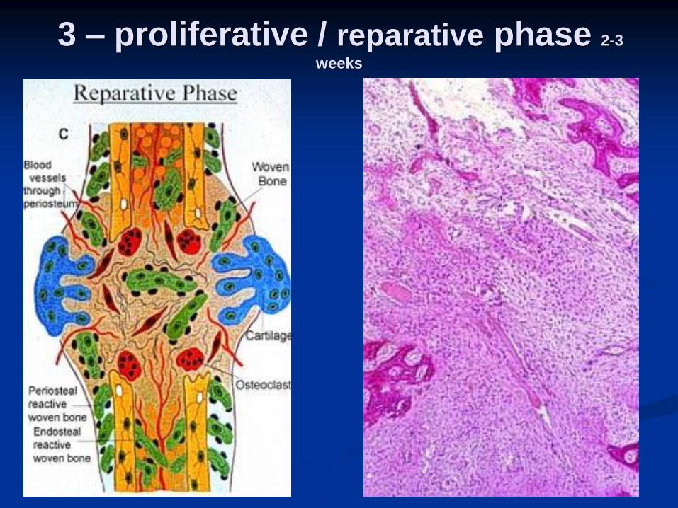
3 – proliferative / reparative phase 2-3
weeks
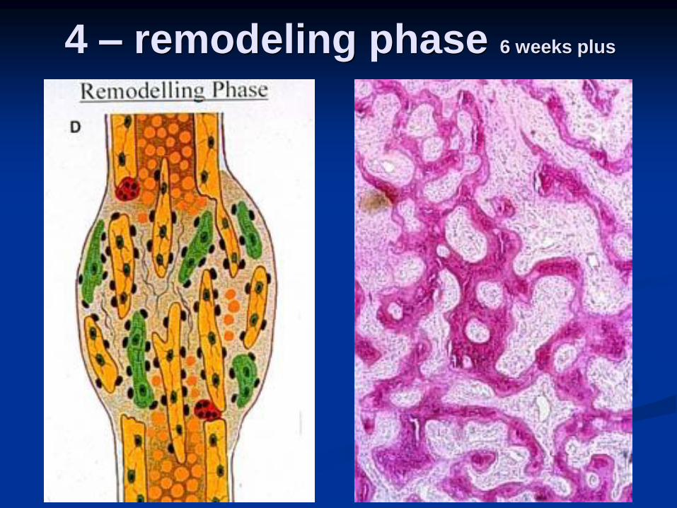
4 – remodeling phase 6 weeks plus

What factors influence duration
of fracture healing and non-
union?

The phases of bone healing
are similar to the mechanisms
of repair at other sites
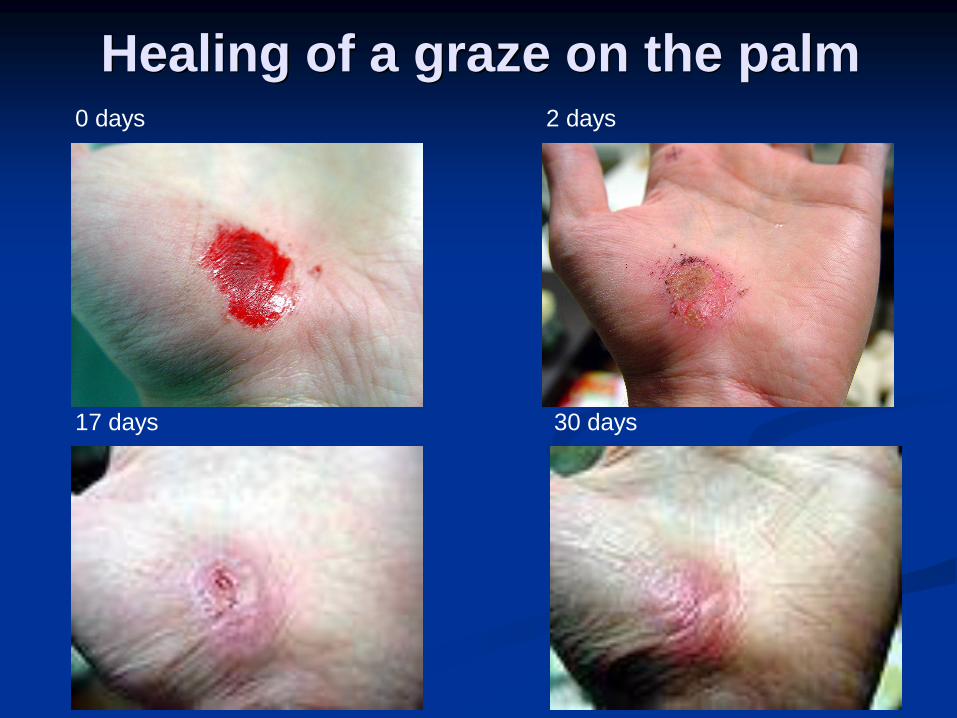
0 days 2 days
17 days 30 days
Healing of a graze on the palm
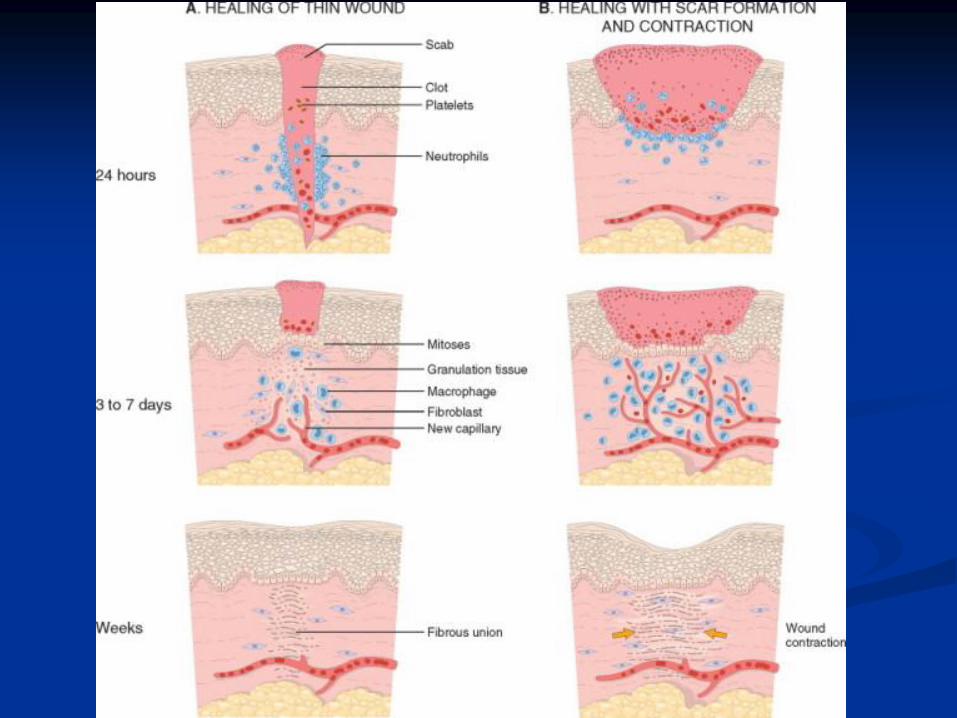

Granulation tissue is the hallmark
of tissue repair
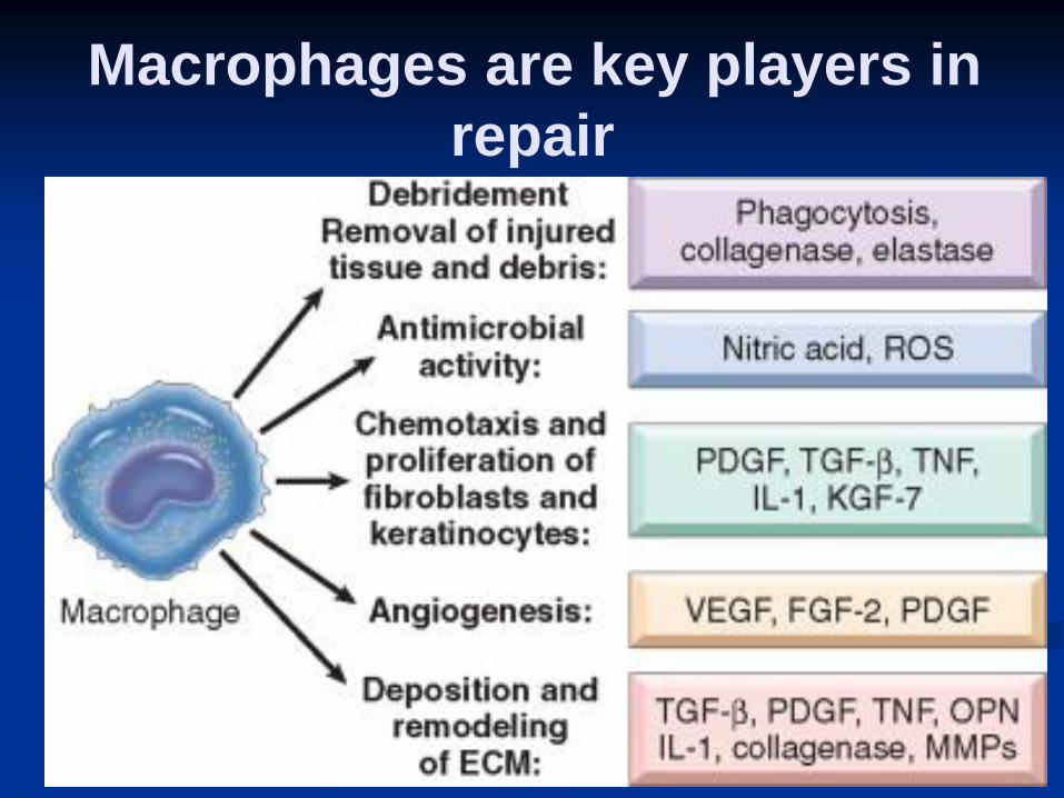
Macrophages are key players in
repair
• Scar formation • Fibroblast migration and proliferation
many GFs trigger above (TGF-beta, PDGF, EGF,
FGF, etc).
VEGF, as well as angiogenic role, causes increased
vascular permeability; exudation and deposition of
proteins in ECM.
• deposition of extracellular matrix
collagen synthesis
• tissue remodelling
above replaces granulation tissue with scar
balance of synthesis and degradation
Matrix metaloproteinases (degrade collagen)

Pathology of Tissue Repair
Complications in skin healing
• deficient scar: dehiscence, ulceration
• excessive scar: hypertrophic, keloid,
proud flesh, desmoid
• contractures
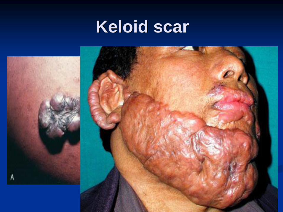
Keloid scar
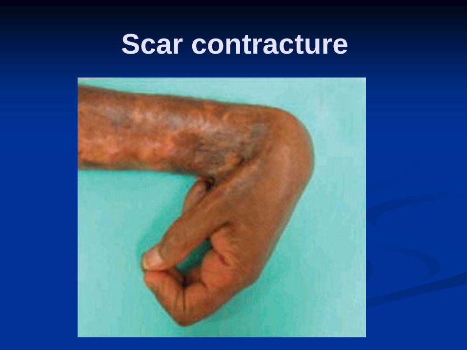
Scar contracture
Tissue Repair
Factors affecting wound healing
Local
systemic

Tissue Repair
Local
blood supply and venous drainage
denervation
contamination and infection
haematoma
mechanical stress
necrotic tissue
Foreign body

Tissue Repair
Tissue Repair
Systemic
age
drugs
hormones
diabetes mellitus (microangiopathy)
malignancy
Nutrition and obesity
uraemia
vitamin C deficiency
Trace elements (zinc and copper deficiency)
Summary and Conclusions
Tissue repair is important in normal
homeostasis and disease
Any abnormality in bone constituents can lead
to increased fracture risk
Bone fracture repair has similarities to healing
at other sites
Future research will aim at manipulating
inflammation and minimising scarring
TGF-beta healing without scarring?
45

Before and after competition!
Please email me with any questions [email protected] or [email protected]
www.pathkids.com