Bone Density Update - Squarespace . SOS calcaneus/radius/phalanges . ... 210 -218 ** After Genant HK...
57
Bone Density Update Southwest Chapter Meeting SNMMI 4/14/2016
Transcript of Bone Density Update - Squarespace . SOS calcaneus/radius/phalanges . ... 210 -218 ** After Genant HK...
Bone Density Update
Southwest Chapter Meeting SNMMI
4/14/2016
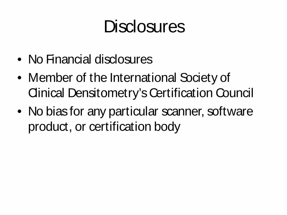
Disclosures
• No Financial disclosures • Member of the International Society of
Clinical Densitometry’s Certification Council • No bias for any particular scanner, software
product, or certification body

Objectives
• Pathophysiology Underlying Bone Loss – in Normal Aging – in Osteoporosis – Molecular Mediators
• Comparison of Current Methods to assess bone density
• Best Practices for DEXA Measurement and Reporting – ISCD Guidance
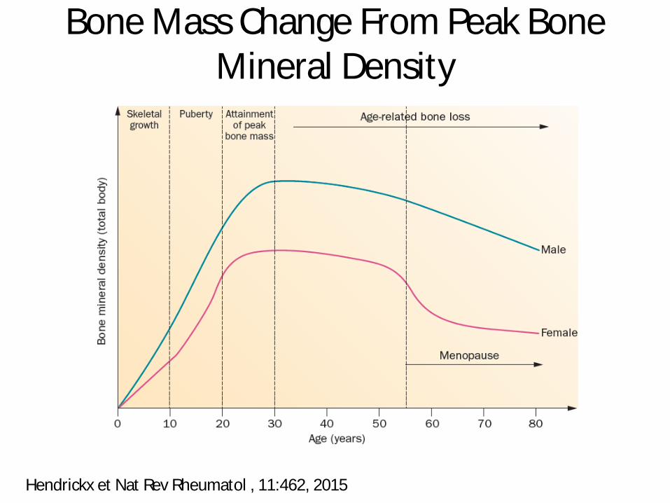
Bone Mass Change From Peak Bone Mineral Density
Hendrickx et Nat Rev Rheumatol , 11:462, 2015

Values for vBMD (mg/cm3) of cortical bone at the distal radius and vertebral body in a population sample of Rochester, Minnesota women and men between the ages of 20 and 97 years.
Sundeep Khosla J Gerontol A Biol Sci Med Sci 2013;68:1226-1235
Bone Density Changes in Aging
Red: Pre-meno F; Blue: Post-meno Fem; Black: Men
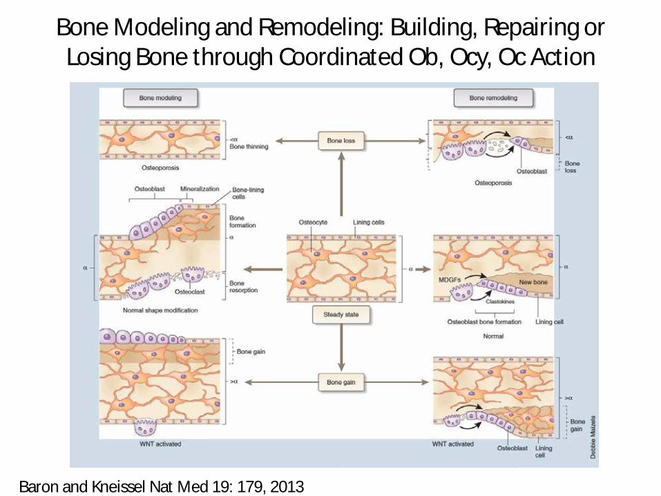
Bone Modeling and Remodeling: Building, Repairing or Losing Bone through Coordinated Ob, Ocy, Oc Action
Baron and Kneissel Nat Med 19: 179, 2013

Multi-factorial Pathogenesis of Primary Osteoporosis
• Age-associated factors – Sex steroid deficiency – Oxidative stress – Apoptosis – Macroautophagy
• Life-style related Factors – Inadequate intake of calcium and vitamin D – Smoking – Physical Inactivity – Excessive Alcohol intake
• Genetic – Monogenic forms: e.g. Type I collagen in Osteogenesis Imperfecta, LRP5 in
Osteoporosis-Pseudoglioma syndrome, Runx2 in cleidocranial dysplasia – Polygenic trait: many contributors each with small effects

Age Associated Factors Contributing to Bone Fragility and Loss: ROS buildup, Cell Death
Hendrickx et Nat Rev Rheumatol , 11:462, 2015

Wnt Signaling Pathways in Bone Regulation
Baron and Kneissel Nat Med 19: 179, 2013
Note Connection to: Hormones, e.g. PTH Therapies e.g.anti-DKK1, anti-SOST

Working Model of Estrogen Regulation of Bone Turnover
Khosla et al Trends Endocrinol Metab 23:576, 2012;

Evidence for Critical Role of Estrogen Deficiency in Male Skeletal Health
• Estrogen (E) receptor alpha deficient males have high E but low BMD – NEJM 331:1056, 1994
• Aromatase Deficient Males have low bone mass – JCEM 80:3689, 1995
• In the Osteoporotic Fracture in Older Men Study (MrOS), bio-available E not bio-T was associated with increased risk of non-spine fracture
• JCEM 94:3337, 2009 – Similar observation for hip however low bio-T was also
associated with increased fracture risk and faster bone loss • JCEM 95:4314, 2010
– Note that estrogens are made from androgens
Reviewed in Cauley, Steroids 99:11, 2015; SAM Question

Osteoporosis
• The most common bone disease in humans • Systemic Disease characterized by low bone
mass, deterioration of bone tissue and disruption of bone architecture, compromised bone strength and an increase in the risk fracture
• Note this definition involves bone quantity and bone quality

Types of Bone Density Tests
• RA: Radiographic Absorptiometry • SPA: Single Photon Absorptiometry • SXA: single x-ray absorptiometry • DPA: Dual Photon Absorptiometry • DXA: dual energy x-ray absorptiometry • pDXA peripheral dual energy x-ray absorptiometry • QUS: quantitative ultrasound • QCT: Quantitative Computed Tomography • pQCT: peripheral quantitative computed tomography
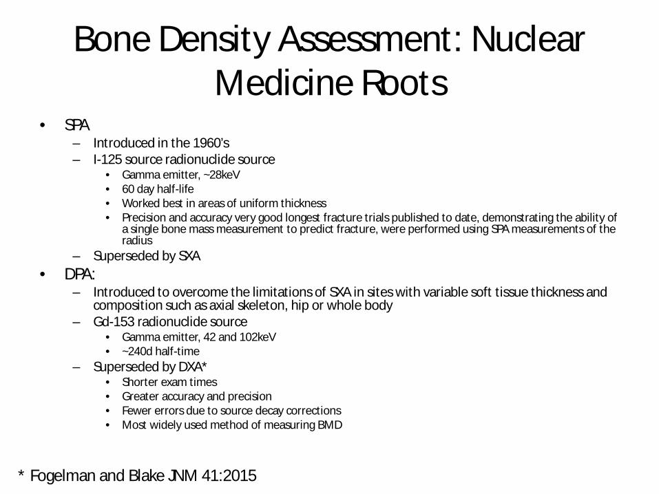
Bone Density Assessment: Nuclear Medicine Roots
• SPA – Introduced in the 1960’s – I-125 source radionuclide source
• Gamma emitter, ~28keV • 60 day half-life • Worked best in areas of uniform thickness • Precision and accuracy very good longest fracture trials published to date, demonstrating the ability of
a single bone mass measurement to predict fracture, were performed using SPA measurements of the radius
– Superseded by SXA • DPA:
– Introduced to overcome the limitations of SXA in sites with variable soft tissue thickness and composition such as axial skeleton, hip or whole body
– Gd-153 radionuclide source • Gamma emitter, 42 and 102keV • ~240d half-time
– Superseded by DXA* • Shorter exam times • Greater accuracy and precision • Fewer errors due to source decay corrections • Most widely used method of measuring BMD
* Fogelman and Blake JNM 41:2015

Comparison of Available Modalities for Bone Status Assessment**
Technique Precision Error (%)
Accuracy Error (%)
Effective Dose (mSv)
Advantages Disadvantages
RA Low cost per test/equipment Limited to the hands Phalanges/metacarpal Equipment mobile 1-2 5 ~5 SXA/pDXA Radius/calcaneus
1-2
4-6
<1
Low cost per test/equipment Equipment mobile Low radiation dose
Limited to hands, wrist or heel Area density Limited correlation to spine/hip
DXA PA spine Lat spine Proximal femur Forearm Whole body
1-1.5 2-3 1.5-3 ~1 ~1
4-10 5-15 6 5 3
~1 ~3 ~1 <1 ~3
Multiple sites capability Low radiation exposure
Limited Mobility Areal density Moderate cost of equipment
QCT
spine trabecular/integral
hip trabecular/integral
2-3
2-3
5-15
4-10
~50
~50
Volumetric density High radiation exposure
“Limited access”
pQCT
radius trabecular
radius total
1-2
1-2
?
2-8
~1
~1
Volumetric density
Equipment mobile
Low radiation dose
Limited to wrist Limited correlation with spine/hip
QUS SOS calcaneus/radius/phalanges BUA calcaneus
0.3-1.2 1.3-3.8
? ?
0 0
Radiation free Low cost per test/equipment Equipment mobile
Limited to peripheral sites Limited correlation to the spine/hip
*Dose for annual background~2,000 mSv, for abdominal radiograph~500mSv and for abdominal CT~4000mSv , Semin Nucl Med 27(3):210-218 ** After Genant HK et al JBMR 11(6): 707, 1996

WHO Operational Definition of Osteoporosis Based on DXA
• Import slides from Oz DXA lecture • Import slide on DXA guidelines from lecture on
PMH NM

DXA Indications • Women aged 65 and older • For post-menopausal women younger than age 65 a bone
density with a risk factor for low bone mass • Low body weight • Prior fracture • High risk medication use • Disease or condition associated with bone loss
• Women during the menopausal transition with clinical risk factors for fracture
• low body weight • prior fracture • high-risk medication use

DXA Indications: Males • Men aged 70 and older • For men < 70 years of age a bone with a risk factor
• Low body weight • Prior fracture • High risk medication use • Disease or condition associated with bone loss.

Other Indications • Adults with a fragility fracture. • Adults with a disease or condition associated with low bone
mass or bone loss. • Adults taking medications associated with low bone mass or
bone loss. • Anyone being considered for pharmacologic therapy. • Anyone being treated, to monitor treatment effect. • Anyone not receiving therapy in whom evidence of bone
loss would lead to treatment. • Women discontinuing estrogen should be considered for
bone density testing

World Health Organization Operational Definition of Interpretation of Bone Density
Patient’s T-Score -0.5 T -1.0 T -1.5 T -2.0 T -2.5 T
Classification
Normal
Osteopenia
Osteoporosis Severe Osteoporosis (with fragility fractures)

Young Adult T-score
• 1T = 1 Standard Deviation (SD) • Compares patient’s result to average Young Adult
reference • Example:
If T-score = -2.3, then subject’s BMD is 2.3 SD below average Young Adult BMD
Age-Matched Z-score • 1Z = 1 Standard Deviation (SD) • Compares patient’s result to Age-Matched reference

Gradient Relationship between T-score and Fracture risk: Approximates Hip-
Fracture Risk and Hip BMD Relationship

WHO Fracture Risk Assessment
Tool (FRAX) • Fracture risk assessment develop by the World Health
Organization • Complex computer algorithm designed to compute 10-year
fracture risk • Based on meta-analysis of multiple studies • Used to help predict those untreated patients at high risk
for fracture who might benefit from therapy • Country specific threshold assigned to the risk depending on
the economic model the country uses to calculate most cost-effective time to intervene
• A guide not a substitute for clinical judgment and acumen • Our clinicians tend to follow NOF guidelines on treatment • May incorporate FN BMD data but not absolutely necessary

WHO Fracture Risk Assessment Tool (FRAX)
https://www.shef.ac.uk/FRAX/tool.jsp
N.B. Does not include fall history; SAM Question
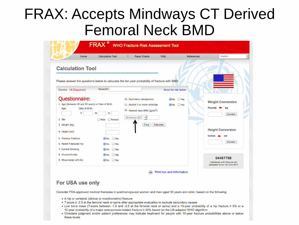
FRAX: Accepts Mindways CT Derived Femoral Neck BMD

Trabecular Bone Score (TBS) • Textural index that evaluates pixel gray-level variations in the lumbar spine
DXA image • Not a direct physical measurement of microarchitecture but correlates but
is related to 3D measures such as Tb.N, Tb.S, and connectivity density • Principle: Dense architecture yields image with small variations in pixel
values, porous architecture yields image with low number of pixel variations of high amplitude
• Elevated TBS appears to represent strong, fracture resistant microarchitecture
• Low TBS reflects weak, fracture prone microarchitecture • Predicts Fracture risk independent of BMD • TBS can differentiate microarchitecture from 3D objects with the same
density but different microarchitecture • Generally derived from re-analysis of lumbar spine BMD, hence papers are
accumulating rapidly and it represents an opportunity for NM DXA labs

TBS Proposed Normal Range for Post-menopausal Women
• Normal: – TBS≥ 1.350
• Partially Degraded Microarchitecture – TBS >1.200 and <1.350
• Degraded Microarchitecture – TBS ≤ 1.200
• Normal range for men not yet proposed

TBS Principles and Example Cases with same BMD but Different TBS
Normal
Partially degraded

TBS in Aging and Medications • Normative reference curve
– J Clin Densitom. 2014 Apr-Jun;17(2):314-9. doi: 10.1016/j.jocd.2013.09.002. Epub 2014 Feb 25.; similar study done for French women
• Healthy Aging – Little change between 30 and 45y/o, thereafter progressive decrease with advancing age – %Decrease with age similar to that of lumbar spine aBMD as is short term reproducibility – Br J Radiol. 2015 Aug;88(1052):20140865. doi: 10.1259/bjr.20140865.
• Improved fracture discrimination – J Clin Densitom. 2014 Jan-Mar;17(1):60-5. doi: 10.1016/j.jocd.2013.05.001. Epub 2013 Jun 13.
• Anti-resorptives: Degrades – Osteoporos Int. 2013 Mar;24(3):1073-8. doi: 10.1007/s00198-012-2155-y. Epub 2012 Oct 3.
• PTH and Ibandronate: Improves or preserves – Osteoporos Int. 2014 Jul;25(7):1945-51. doi: 10.1007/s00198-014-2703-8. Epub 2014 Apr 24.
• Aromatase inhibitors: Degrades – J Endocrinol Invest. 2014 Sep;37(9):871-4. doi: 10.1007/s40618-014-0125-2. Epub 2014 Jul 19.

TBS and Diseases • Diabetes:
– Type I • Osteoporos Int. 2016 Jan;27(1):127-33. doi: 10.1007/s00198-015-3222-y. Epub 2015 Jul
18.; – Type 2
• Bone Res. 2016 Mar 22;4:16001. doi: 10.1038/boneres.2016.1. eCollection 2016. • Hyperparathyroidism:
– J Clin Endocrinol Metab. 2013 May;98(5):1963-70. doi: 10.1210/jc.2012-4255. Epub 2013 Mar 22.
• Review article in Rheumatology: Curr Rheumatol Rep. 2014 Aug;16(8):436. doi: 10.1007/s11926-014-0436-5.
• Treatment of Breast Cancer Patients with Zoledronic Acid: – Osteoporos Int. 2015 Jan;26(1):353-60. doi: 10.1007/s00198-014-2955-3.
Epub 2014 Nov 8. • Others: Crohn‘s Disease, HIV patients

FRAX: Trabecular Bone Score Adjustment
Some clinicians are using the TBS adjusted FRAX score to make treatment decisions

TBS Adjusted Fracture Risk in Osteopenia
Lewiecki et al., Endocr Res, Early Online: 1–14, 2015


Recommendations: DXA Acquisition, Analysis and Interpretation and Reporting
SAM Question


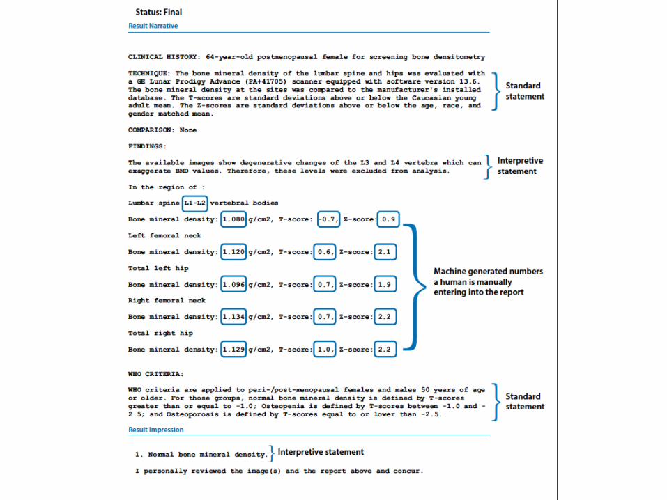
Reporting
• ISCD Recommendations • Automated • Manufacturer’s Reporter • Pre-populating software
– iMorgan – EPIC based workflow tool developed in-house


Drop Smart Menus in EPIC

Decreased Report Turn-around time with Machine to Machine Communication and EPIC Driven WorkFlow

} Our solution § $6175 install, no yearly maintenance cost § $2500 for vendor modality HL7 interface § $1675 for vendor install support § $2000 for internal implementation (45 man hours)
} Commercially available solution § Known from associated institution § $80,000 install and $80,000 for 5 year maintenance § $65,000 for software and license § $15,000 for implementation project § $20,000 for annual service and support

Opportunistic Bone Mass Screening
• Refers to • FDA approved software • Figures from dual modality scan • Peel off Nuc scan • Show CT in axial projection • Show Jim’s analysis and a report • Show DXA equivalent from Mindways software • Show FEA modeling • Conclusion: Dual modality scanning puts us in prime
position to assess in one setting bone mass and bone mechanical competence

Sagittal F-18 PET-CT

Dual Use of CT: QCT from Dual Modality Scans
• QCT was introduced prior to DXA at the end of the 1970s
• Originally bone mineral density of the spine by 2D analysis
• Early CT scanners had limited availability • With the development of DXA, QCT lost ground
• May be performed on any CT system • Calibration used to convert HU to BMD. • Newer systems enable volumetric (3D)
analysis of spine and hip measurement
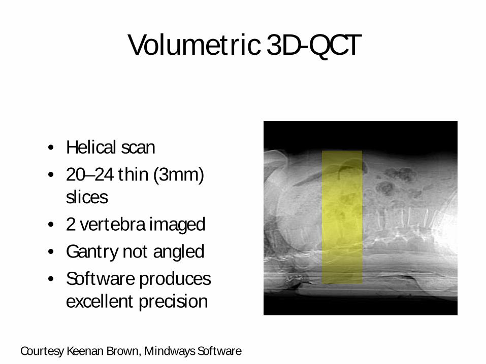
• Helical scan • 20–24 thin (3mm)
slices • 2 vertebra imaged • Gantry not angled • Software produces
excellent precision
Volumetric 3D-QCT
Courtesy Keenan Brown, Mindways Software
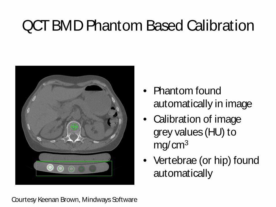
QCT BMD Phantom Based Calibration
• Phantom found
automatically in image • Calibration of image
grey values (HU) to mg/cm3
• Vertebrae (or hip) found automatically
Courtesy Keenan Brown, Mindways Software

QCT Spine Region of Interest
• Elliptical Cylinder ROI • Trabecular Bone
– No cortical bone – No extraosseous mineral – Higher metabolic rate
• Measurement precision: CoV of 0.7%
Courtesy Keenan Brown, Mindways Software

Analysis and Reporting
Analysis Reporting
Courtesy Keenan Brown, Mindways Software

QCT Reporting
· Should not be interpreted using WHO T-Score guidelines for fracture-risk categorization
· The American College of Radiology Guidelines on the use of QCT has the following fracture risk categories for Spine BMD equivalent to DXA T-score categories:
· BMD > 120 mg/cm3 è “Normal” · 80 mg/cm3 ≤ BMD ≤ 120 mg/cm3 è “Osteopenia” · BMD < 80 mg/cm3 è “Osteoporosis”
Courtesy Keenan Brown, Mindways Software
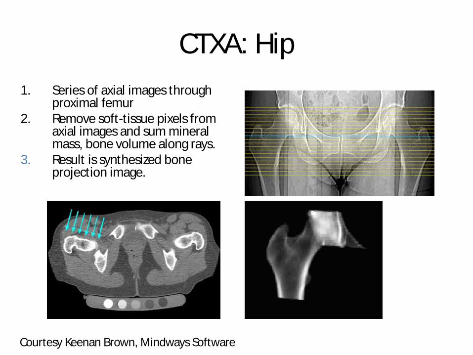
CTXA: Hip 1. Series of axial images through
proximal femur 2. Remove soft-tissue pixels from
axial images and sum mineral mass, bone volume along rays.
3. Result is synthesized bone projection image.
Courtesy Keenan Brown, Mindways Software

QCT & DXA: Hip Areal Density • Both technique give essentially equivalent
results when the same bone is measured with both techniques.
· Same subject · QCT & DXA · Essentially same
T-scores · 0.012 g/cm2 long
term precision
Courtesy Keenan Brown, Mindways Software

Correlation of BMD derived from virtual non-contrast CT Using Asynchronous Calibration
y = 0.6373x - 16.082 R² = 0.9359
(Arterial)
y = 0.607x - 12.672 R² = 0.9207
(Venous)
y = 0.5547x - 8.1797 R² = 0.8697
(Nephrogram + delayed)
0
20
40
60
80
100
120
140
50 100 150 200 250
BMD
from
VN
C of
con
tras
t sca
ns (m
g/m
l)
BMD from non-contrast scans (mg/ml)
Arterial
Venouse
Nephrogram+delayed
Linear (Arterial)
Linear (Venouse)
Linear(Nephrogram+delayed)
Figure. Linear regression of BMD from non-contrast scans and VNC of contrast scans.

HR-pQCT
Link, Can Assoc Radiol Journal, 2016
• 40-80 um resolution
• Wrist and Distal Tibia
• Research Tool

Atypical Subtrochanteric Fractures
• Associated with long term bisphosphonate use (>3y)
• Maybe genetic • Very Rare (<1.0%) • Look for cortical beaked
thickening • Do bone scan or MRI if
focal pain but negative radiograph

Summary • Bone loss is multi-factorial with aging, sex steroids, oxidative stress,
and Wnt signaling being strong contributors • DXA is the number clinical tool for diagnosis of osteoporosis,
treatment response, and monitor age related changes • TBS is a rapidly evolving tool that uses lumbar spine DXA data for
enhanced fracture risk assessment and response to diseases, medications and aging
• Best Practice Guidelines have been published by ISCD • Explore in-house solutions for “automated” reporting of DXA values
to avoid high costs and to decrease reporting errors • BMD from dual use of CT from hybrid dual modality (PET/CT),
SPECT, CT) scanning offers Nuclear Medicine an opportunity to expand its role in bone health assessment
• Nuclear Medicine is uniquely suited for bone health assessment since we have the tools to measure bone mass and turnover

Bone Density Update, Orhan Oz, MD 1. Which of the following steroids is the major regulator of bone metabolism in men and women and low levels of which are associated with low bone density and fracture risk?
A. Vitamin D B. Estradiol (KEY) C. Testosterone D. Dehydroepiandrosterone
Correct Answer: B. Reference: Steroids 99: 11-15, 2015. 2. What is the most commonly used diagnostic imaging method to assess bone density for osteoporosis?
A. Dual Photon Absorptiometry B. Single Photon Absorptiometry C. Dual Energy X-ray Absorptiometry (DXA) (KEY) D. Quantitative Computed Tomography?
Correct answer: C. Reference: J Nucl Med 41:2015-2025, 2000. Or www.webmd.com/osteoporosis/guide/osteoporosis-diagnosis-tests 3. Which of the following is a recommended DXA best practice?
A. Use the manufacturer’s scanner precision as the least significant change for serial BMD measurements B. Only two-thirds of the technologists need to perform an in vivo precision assessment C. At least one practicing DXA technologist and one practicing DXA interpreter should have valid certification in bone densitometry
(KEY) D. Spine phantom BMD measurements should be performed quarterly
Correct answer: C. Source: J Clin Densitometry: Assessment and Management of Musculoskeletal Health http:dx.doi.org/10.1016/j.jocd.2016.03.003. 4. The Fracture Risk Assessment Tool, FRAX, algorithm includes:
A. Glucocorticoid Use B. Smoking History

C. Fracture History D. Fall History E. All of the above F. A-C (KEY) G. B-D
Correct Answer: F. Source: FRAX WHO Fracture Risk Assessment Tool online, https://www.shef.ac.uk/FRAX/tool.aspx?country=8. accessed 3/30/2016. 5. The Trabecular Bone Score is?
A. A parameter obtained from the trabecular compartment of a vertebral body B. A parameter that is derived from DXA scans C. Independent of bone mineral density and FRAX risk variables in fracture prediction D. A and B are true E. B and C are true (KEY)
Correct Answer: E. Sources: J Bone Miner Res 29(3): 518-530, 2014; Bone 78:216-224, 2015.