BODY FLUIDS AND CIRCULATION - SPIRO...
31
BODY FLUIDS AND CIRCULATION www.gneet.com 1 It is the movement of body fluids inside the body of animals so as to transport materials from the region of formation to the region of utilization or disposal. A circulatory system is a complex of structures involved in the flow of body fluids of an organism so as to accomplish transport of materials Circulation of body fluids can be of the following types 1. Intracellular circulation It occurs inside the individual cells through cyclosis or cytoplasmic streming. Examples : Paramecium, Amoeba. 2. Extracellular circulation In multi-cellular animals, the living cells are bathed in an intercellular or extra cellular fluid which circulates in the body for transport of materials Extra cellular circulation can be a) Extra-organismic circulation: Outside water circulates in the body of an organism b) Intra-organismic circulation: It involves circulation of body fluid i) Parenchymal circulation In flatworms, fluid filled spaces present in parenchyma tissue between body wall and internal organs are used in distribution of substances. ii) Coelomic Circulation Coelomic fluid is employed in transport of substances,. Pseudocelom is used for this purpose in roundworms. Haemocoel does so in arthropods iii) Blood vascular system It contains blood and a pumping structure ( heart) for circulation of materials inside the body. Lymphatic system accompanies blood vascular system. FUNCTIONS OF CIRCULATORY SYSTEM 1) Transport of nutrients 2) Transport of waste products. 3) Transport of respiratory gases 4) Transport of metabolic intermediates like lactic acid from muscles to liver 5) Transport of hormones. 6) Regulation of pH by means of buffer. 7) Regulation of temperature. 8) Distribution of water. 9) Support or turgidity of certain organs like penis and nipples 10) Prevention of diseases by means of antibodies and antitoxin present in it 11) Disposal of cell wreckage. 12) Homeostatis or providing a stable internal environment for cells
Transcript of BODY FLUIDS AND CIRCULATION - SPIRO...

BODY FLUIDS AND CIRCULATION www.gneet.com
1
It is the movement of body fluids inside the body of animals so as to transport
materials from the region of formation to the region of utilization or disposal. A
circulatory system is a complex of structures involved in the flow of body fluids of
an organism so as to accomplish transport of materials
Circulation of body fluids can be of the following types
1. Intracellular circulation
It occurs inside the individual cells through cyclosis or cytoplasmic streming.
Examples : Paramecium, Amoeba.
2. Extracellular circulation
In multi-cellular animals, the living cells are bathed in an intercellular or extra
cellular fluid which circulates in the body for transport of materials
Extra cellular circulation can be
a) Extra-organismic circulation: Outside water circulates in the body of an
organism
b) Intra-organismic circulation: It involves circulation of body fluid
i) Parenchymal circulation
In flatworms, fluid filled spaces present in parenchyma tissue between body
wall and internal organs are used in distribution of substances.
ii) Coelomic Circulation
Coelomic fluid is employed in transport of substances,. Pseudocelom is used
for this purpose in roundworms. Haemocoel does so in arthropods
iii) Blood vascular system
It contains blood and a pumping structure ( heart) for circulation of materials
inside the body. Lymphatic system accompanies blood vascular system.
FUNCTIONS OF CIRCULATORY SYSTEM
1) Transport of nutrients
2) Transport of waste products.
3) Transport of respiratory gases
4) Transport of metabolic intermediates like lactic acid from muscles to liver
5) Transport of hormones.
6) Regulation of pH by means of buffer.
7) Regulation of temperature.
8) Distribution of water.
9) Support or turgidity of certain organs like penis and nipples
10) Prevention of diseases by means of antibodies and antitoxin present in it
11) Disposal of cell wreckage.
12) Homeostatis or providing a stable internal environment for cells
spiro
Typewritten text
BODY FLUIDS AND CIRCULATION

BODY FLUIDS AND CIRCULATION www.gneet.com
2
13) Determination of pigmentation in case of blood vascular system.
14) Plugging the area of injury.
15) As connective tissue
OPEN CIRCULATORY SYSTEM
Open circulation occurs in arthropods and mollusks.
The blood is not completely enclosed within vessels, the heart pumps blood
through arteries into large cavities or sinuses, where it mixes with interstitial
fluid and bathes the cells of the body.
Blood is a combination of blood and interstitial fluid called haemolymph, while
the spaces and lacumal are together called haemocoel.
The blood is slowly returned to the heart through small pores called ostia e.g.
arthropods ( cockroach)
Circulation is slower in an open system, because with some of the blood
pooled in sinuses, the heart cannot build up enough pressure to make blood
flow rapidly.
Open system cannot achieve the high rates of oxygen transport that active
animals require.
Animals with open systems are either small and sluggish or use open system
only for transport of food and wastes and use a different system for the
transport of gases.
Respiratory pigment, if present, is dissolved in the plasma, no red corpuscles
are present.
CLOSED CIRCULATORY SYSTEM
Closed circulatory system is a type of blood vascular system in which blood
remains confined and flows inside blood vessels only, never coming in direct
contact with body cells. It occurs in most annelids, cephalopods and
vertebrates. Annelids are the simplest animals to have closed circulatory
system.
Flow of blood is
Heart artery arteriole capillary venule vein heart
Circulatory system as discovered by William Harvey ( 1628), blood capillaries
by malpighi ( 1661) blood pressure by Halls (1732) and sphygmanometer by
Riva Rocci( 1896 )
BODY FLUIDS AND CIRCULATION www.gneet.com
3
HEART
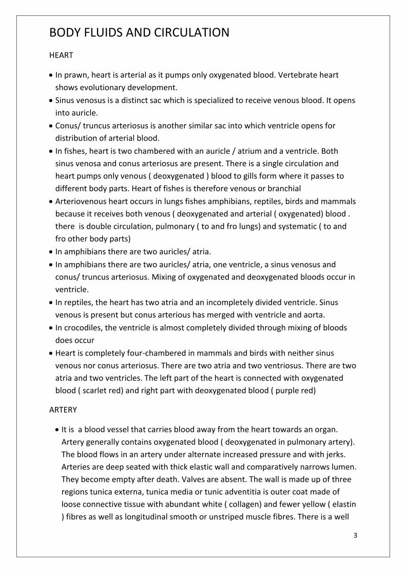
In prawn, heart is arterial as it pumps only oxygenated blood. Vertebrate heart
shows evolutionary development.
Sinus venosus is a distinct sac which is specialized to receive venous blood. It opens
into auricle.
Conus/ truncus arteriosus is another similar sac into which ventricle opens for
distribution of arterial blood.
In fishes, heart is two chambered with an auricle / atrium and a ventricle. Both
sinus venosa and conus arteriosus are present. There is a single circulation and
heart pumps only venous ( deoxygenated ) blood to gills form where it passes to
different body parts. Heart of fishes is therefore venous or branchial
Arteriovenous heart occurs in lungs fishes amphibians, reptiles, birds and mammals
because it receives both venous ( deoxygenated and arterial ( oxygenated) blood .
there is double circulation, pulmonary ( to and fro lungs) and systematic ( to and
fro other body parts)
In amphibians there are two auricles/ atria.
In amphibians there are two auricles/ atria, one ventricle, a sinus venosus and
conus/ truncus arteriosus. Mixing of oxygenated and deoxygenated bloods occur in
ventricle.
In reptiles, the heart has two atria and an incompletely divided ventricle. Sinus
venous is present but conus arterious has merged with ventricle and aorta.
In crocodiles, the ventricle is almost completely divided through mixing of bloods
does occur
Heart is completely four-chambered in mammals and birds with neither sinus
venous nor conus arteriosus. There are two atria and two ventriosus. There are two
atria and two ventricles. The left part of the heart is connected with oxygenated
blood ( scarlet red) and right part with deoxygenated blood ( purple red)
ARTERY
It is a blood vessel that carries blood away from the heart towards an organ.
Artery generally contains oxygenated blood ( deoxygenated in pulmonary artery).
The blood flows in an artery under alternate increased pressure and with jerks.
Arteries are deep seated with thick elastic wall and comparatively narrows lumen.
They become empty after death. Valves are absent. The wall is made up of three
regions tunica externa, tunica media or tunic adventitia is outer coat made of
loose connective tissue with abundant white ( collagen) and fewer yellow ( elastin
) fibres as well as longitudinal smooth or unstriped muscle fibres. There is a well
BODY FLUIDS AND CIRCULATION www.gneet.com
4
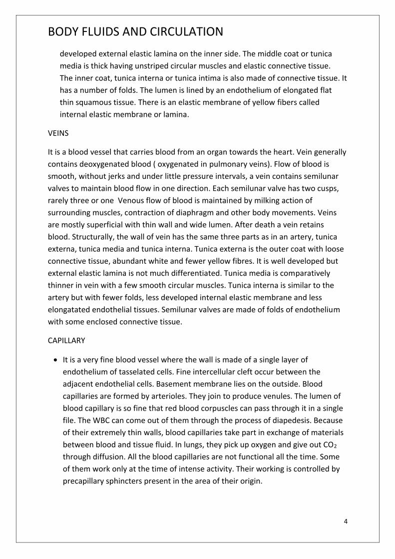
developed external elastic lamina on the inner side. The middle coat or tunica
media is thick having unstriped circular muscles and elastic connective tissue.
The inner coat, tunica interna or tunica intima is also made of connective tissue. It
has a number of folds. The lumen is lined by an endothelium of elongated flat
thin squamous tissue. There is an elastic membrane of yellow fibers called
internal elastic membrane or lamina.
VEINS
It is a blood vessel that carries blood from an organ towards the heart. Vein generally
contains deoxygenated blood ( oxygenated in pulmonary veins). Flow of blood is
smooth, without jerks and under little pressure intervals, a vein contains semilunar
valves to maintain blood flow in one direction. Each semilunar valve has two cusps,
rarely three or one Venous flow of blood is maintained by milking action of
surrounding muscles, contraction of diaphragm and other body movements. Veins
are mostly superficial with thin wall and wide lumen. After death a vein retains
blood. Structurally, the wall of vein has the same three parts as in an artery, tunica
externa, tunica media and tunica interna. Tunica externa is the outer coat with loose
connective tissue, abundant white and fewer yellow fibres. It is well developed but
external elastic lamina is not much differentiated. Tunica media is comparatively
thinner in vein with a few smooth circular muscles. Tunica interna is similar to the
artery but with fewer folds, less developed internal elastic membrane and less
elongatated endothelial tissues. Semilunar valves are made of folds of endothelium
with some enclosed connective tissue.
CAPILLARY
It is a very fine blood vessel where the wall is made of a single layer of
endothelium of tasselated cells. Fine intercellular cleft occur between the
adjacent endothelial cells. Basement membrane lies on the outside. Blood
capillaries are formed by arterioles. They join to produce venules. The lumen of
blood capillary is so fine that red blood corpuscles can pass through it in a single
file. The WBC can come out of them through the process of diapedesis. Because
of their extremely thin walls, blood capillaries take part in exchange of materials
between blood and tissue fluid. In lungs, they pick up oxygen and give out CO2
through diffusion. All the blood capillaries are not functional all the time. Some
of them work only at the time of intense activity. Their working is controlled by
precapillary sphincters present in the area of their origin.

BODY FLUIDS AND CIRCULATION www.gneet.com
5
ARTERIO-VENOUS ANASTOMOSIS
It is a direct vascular connection between an arteriole and venule bypassing capillary
supply. The connection occurs in certain exposed parts like finger tips, nose, pinnae,
eye lids, lips, tongue etc. It is meant for controlling blood supply and temperature of
the exposed parts.
VASCULAR PLEXUS
Anastomosis of blood vessels is like arteries in certain regions to provide extra blood
e.g. cutaneous plexus, papillary plexus, nasal plexus.
EFFIENCY OF CLOSED RESPIRATORY SYSTEM
i) It can regulate blood flow into an organ.
ii) Blood flows rapidly in blood vessels than in open spaces
iii) There is quick supply and removal of materials.
iv) Blood flows to all parts of the body with equal efficiency and speed.
BLOOD
It is complex mobile fluid connective tissue of reddish colour in which the fluid matrix
is not synthesized by the contained cells. An adult human has 5-5.5 litres of blood. pH
is 7.4. Blood consists of two parts, plasma and blood corpuscles ( formed elements)
PLASMA
It is slightly alkaline non-living intercellular substance which constitutes about 60%
part of the blood. It is a pale yellow but transparent and clear fluid.
It is composed of 91-92% of water, 7% proteins, 0.9% inorganic substances, 0.1%
glucose and traces of other constituents ( amino acids, fatty acid, fat drops,
cholesterol, anticoagulants, hormones, excretory products, vitamins etc.)
The main categories of protein are albumins, globulins and fibrinogen. Albumins
produce colloidal osmotic pressure. It also carry ca and some fatty acid α-globulin,
β-globulin carry fat soluble vitamins, cholesterol and ions other globulin are
prothrombin, thromoplastin and anti haemophiliac factors. Fibrinogen takes part in
blood coagulation by forming fibrin
Mineral salts like chlorides, bicarbonates, sulphates and phosphate of sodium,
potassium calcium, ironanad magnesium constitute about 0.9% of plasma. Buffer of
the blood is sodium bicarbonates

BODY FLUIDS AND CIRCULATION www.gneet.com
6
FUNCTION OF BLOOD PLASMA
i) Transport
ii) Retention of fluid in blood.
iii) Maintenance of blood pH
iv) Body immunity
v) Prevention of blood loss
vi) Conducting heat to skin for dissipation
vii) Uniform distribution of heat all over body
BLOOD GLUCOSE
Usually blood glucose level is about 80-100 mg per 100 ml of blood 12 hours
after a normal meal
It blood glucose level exceeds 180 mg per 100 ml, it starts appearing in urine.
This condition is called glucosuria. If it is less it causes hypoglycemia and if it is
higher it causes hyperglycemia.
BLOOD CHOLESTEROL
Its normal amount is 80-180 mg in 100 ml of blood plasma. Increased blood
cholesterol may lead to its deposition in the internal wall of the blood vessels like
arteries and veins which causes high blood pressure and heart problem
FORMED ELEMENTS
Erythrocytes ( Rd blood corpuscles or RBCs)
A normal adult man and woman havae 5 to 4.5 million RBCs per cubic
millimeter of blood respectively.
Less amount of haemo globin leads to anemia which may be caused by loss of
blood or destruction of RBCs.
An abnormal rise in RBC count is called polycythemia. Decrease in the number
of RBC count is called erythrocytopenia which causes shortage in the blood
and tissues
They are biconcave, disc-shaped enucleate reddish coloured cells of 7-8μm in
diameter and 1-2μm thick. Red colour is due to the presence of haemoglobin
Haemoglobin is a conjugate protein which is made up of a protein called
globin and a non protein group heme (=haeme) hence the haemoglobin.
BODY FLUIDS AND CIRCULATION www.gneet.com
7
Haemoglobin is oxygen carrying pigment. 100ml of blood of a normal man
contains 15g of haemoglobin and of normal woman an average of 13g of
haemoglobin
Erythropoiesis is the process by which red blood cells are produced. In human
adults, this usually occurs within the bone marrow.
The life of an RBC is about 120 days. The worn out RBCs are destroyed in the
spleen and liver
Their iron is returned to the red bone marrow for reuse in the synthesis of
fresh haemoglobin
Their pigment is degraded to yellowish pigment bilirubin which is excreted in
bile.
ERYTHROCYTE SEDIMENTATION RATE ( ESR)
If blood containing an anticoagulant (oxalate) is allowed to stand in a narrow vertical
tube the erythrocyte settle to the bottom half of the tube. The rate at which this
occurs is called the erythrocyte sedimentation rate. ESR is very useful in diagnosing
various diseases including tuberculosis. ESR in men is 0-5 mm/hr and in women it is
0-7mm/ hour.
Leucocytes ( White bold corpuscles or WBCs)
They are colourless, active and mobile nucleated blood corpuscles with a
number 7000±3500 /mm3. Leucytes are of two types granulocytes ( with
granules and polymorphic nucleus) and agranulocytes ( without granules and
monomorphic nucleus).
The life of granuloctes is normally 40 to 8 hours circulating in the blood and
another 4-5 days in the tissue
Monoctes have a short life span of 10-20 hours. The lymphocytes have life
span of few days or months or years
Granulocytes are of three types ( neutrophiles, basophiles, losinophiles) while
agranulocytes are of two types ( monocytes and lymphocytes)
Neutrophile
They have granules that stain with neutral dyes nucleus 2-7 lobed, nearly
circular, 62% of all leucoctes, phagocytic.
Esoinophile
Coarse granules that get stained with acidic dyes ( bright red with cosine), nucleus
bilobed, size 10-14μm, 2-3% of total leucocytes, number increases in asthama.
Basinophile
BODY FLUIDS AND CIRCULATION www.gneet.com
8
Fewer coaese granyles stained with basic dye ( methylene blue ), nucleus S-
shaped and 3 lobed, 0.5% - 1%, allergic reactions by releasing histamine, also
heparin and serotonin.
Lymohocytes: Large nucleus with granule free pale blue cytoplasm, 30% of total
leucocytes, manufacture globins some of which function as antibodies in
immunological reactions. Lymphocytes have size of 7-10μm, nucleus stains more
deeply with basic dyes than surrounding cytoplasm. Large lymphocytes have 10-
14μm, nucleus stains more deeply with basic dyes than surrounding cytoplasm.
Large lymphocytes have 10-14 μm and more cytoplasm. On basis of site of
maturation, two kinds β-lymphocytes and T-lymphocytes
Monocytes
Largest leucocytes, 10-18 μm kidney shaped nucleus, 5-6% of total leucocytes
motile, phagocytic, scavengers, production of interleukin and pyrogen.
THROMBOCYTES ( BLOOD PLATELETS)
There are about 250,000 platelets in a cubic millimeters of blood. Increase and
decrease in the number of platilets is known as thrombocytosis and
thrombocytopenin respectively.
They are rounded or oval disc like bodies platelets are 2-3 μm in diameter.
They are colourless
Platelets are formed from the megakaryocytes( very large cells of the bone
marrow). Formation of thrombocytes is called thrombopoiesis.
Normal life span of blood platelets is about a week
When an injury is caused, the blood platelets release certain chemicals which
are called the platelet factors ( thrombo plastin). The platelet factors help in
the clotting of blood
BLOOD COAGULATION ( BLOOD CLOTTING)
When an injury is caused to a blood vessel, bleeding starts which is topped by a
process called blood clotting or blood coagulation
First step: At the site of an injury, the blood patelets disintegrate and release a
phospholipid called platelet factor-3 ( Thromboplastin). Injured tissues also
release a lipoprotein factor called hromoplastin. These two factors combine
with calcium ions Ca+2 and certain protein of the blood to form an enzyme
called pro-thrombinase.
BODY FLUIDS AND CIRCULATION www.gneet.com
9
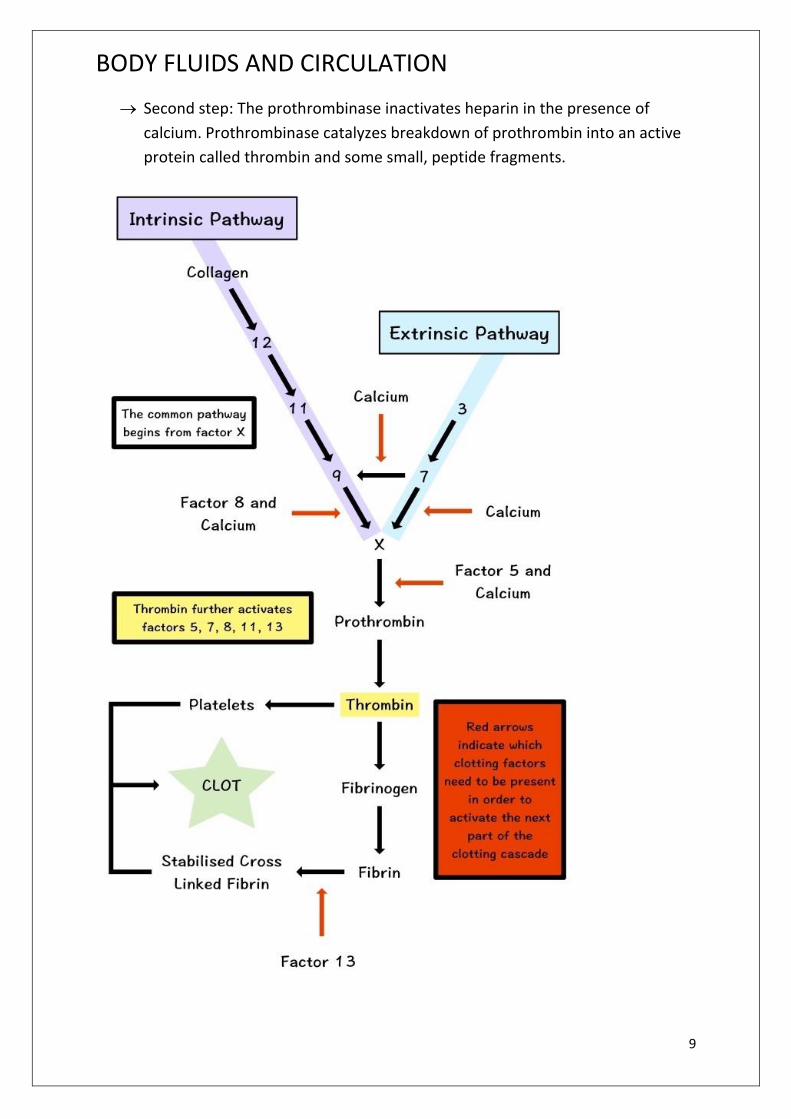
Second step: The prothrombinase inactivates heparin in the presence of
calcium. Prothrombinase catalyzes breakdown of prothrombin into an active
protein called thrombin and some small, peptide fragments.
BODY FLUIDS AND CIRCULATION www.gneet.com
10
Third step: Thrombin acts as enzyme and first brings about depolymerization
of these monomers. Later thrombin stimulates repolymerization of these
monomers into long insoluble fibres – like polymers called fibrin. The thin,
long and solid fibres of fibrin from a dense network upon the wound and trap
blood corpuscles to form a clot. The clot seals the wound and stops bleeding.
Soon after the clot seals the wound and stops bleeding. Soon after the clot
starts contracting and a pale yellow fluid, the serum, starts oozing out. This
serum is blood plasma minus fibrinogen and blood corpuscles.
Vitamin K is essential for blood clotting as it is necessary for the synthesis of
prothrombin in the liver
List of Clotting Factors
Factor I
Name :Fibrinogen
Source :Liver
Pathway : Both extrinsic and intrinsic
Activator :Thrombin
Actions : When fibrinogen is converted into fibrin by thrombin, it forms long strands
that compose the mesh network for clot formation.
FactorII Name :Prothrombin Source :Liver Pathway : Both extrinsic and intrinsic Activator : Prothrombin activator Actions : Prothrombin is converted into thrombin which then activated fibrinogen into fibrin.
Factor III
Name : Thromboplastin / Tissue factor Source : Platelets (intrinsic) and damaged endothelium (cells) lining the blood vessel (extrinsic). Pathway : Both extrinsic and intrinsic Activator : Injury to blood vessel Action : Activates factor VII (VIIa).

BODY FLUIDS AND CIRCULATION www.gneet.com
11
Factor IV
Name : Calcium Source : Bone and absorption from food in gastrointestinal tract Pathway : Both extrinsic and intrinsic Action : Works with many clotting factors for activation of the other clotting factors. These are called calcium-dependent steps.
Factor V
Name : Proaccerin / Labile factor / Ac-globulin (Ac-G) Source : Liver and platelets Pathway : Both extrinsic and intrinsic Activator : Thrombin Action : Works with Factor X to activate prothrombin (prothrombin activator).
Factor VII
Name : Proconvertin / Serum prothrombin conversion accelerator (SPCA) / stable factor Source : Liver Pathway : Extrinsic Activator : Factor III (tissue factor) Actions : Activates Factor X which works with other factors to convert prothrombin into thrombin.
Factor VIII
Name : Anti-hemoplytic factor / Antihemophilic factor (AHF) or globulin (AHG) / antihemophilic factor A Source : Endothelium lining blood vessel and platelets (plug) Pathway : Intrinsic Activator : Thrombin Actions : Works with Factor IX and calcium to activate Factor X. Deficiency : Hemophilia A
Factor IX
Name : Christmas factor / Plasma thromboplastin component (PTC) / Antihemophilic factor B Source : Liver Pathway : Intrinsic Activator : Factor XI and calcium
BODY FLUIDS AND CIRCULATION www.gneet.com
12
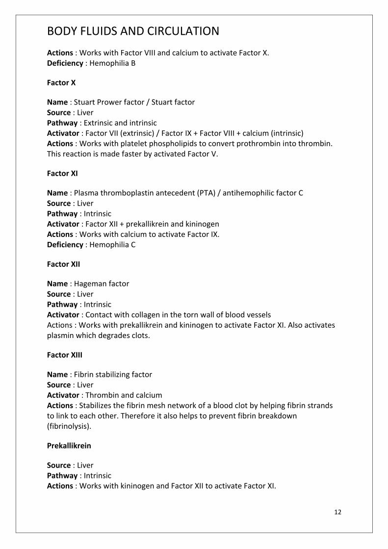
Actions : Works with Factor VIII and calcium to activate Factor X. Deficiency : Hemophilia B
Factor X
Name : Stuart Prower factor / Stuart factor Source : Liver Pathway : Extrinsic and intrinsic Activator : Factor VII (extrinsic) / Factor IX + Factor VIII + calcium (intrinsic) Actions : Works with platelet phospholipids to convert prothrombin into thrombin. This reaction is made faster by activated Factor V.
Factor XI
Name : Plasma thromboplastin antecedent (PTA) / antihemophilic factor C Source : Liver Pathway : Intrinsic Activator : Factor XII + prekallikrein and kininogen Actions : Works with calcium to activate Factor IX. Deficiency : Hemophilia C
Factor XII
Name : Hageman factor Source : Liver Pathway : Intrinsic Activator : Contact with collagen in the torn wall of blood vessels Actions : Works with prekallikrein and kininogen to activate Factor XI. Also activates plasmin which degrades clots.
Factor XIII
Name : Fibrin stabilizing factor Source : Liver Activator : Thrombin and calcium Actions : Stabilizes the fibrin mesh network of a blood clot by helping fibrin strands to link to each other. Therefore it also helps to prevent fibrin breakdown (fibrinolysis).
Prekallikrein
Source : Liver Pathway : Intrinsic Actions : Works with kininogen and Factor XII to activate Factor XI.

BODY FLUIDS AND CIRCULATION www.gneet.com
13
Kininogen
Source : Liver Pathway : Intrinsic Actions : Works with prekallikrein and Factor XII to activate Factor XI.
FUNCTIONS OF BLOOD
i) Transport o f food materials : Blood transports the digested food from the
alimentary canal to the different body cells
ii) Transport of respiratory gases : Oxygen is carried from the respiratory organs to
the tissues and carbon dioxide from the tissue to the respiratory organ by blood.
iii) Transport of hormones: Hormones are carried by blood from the endocrine
glands to the places of use
iv) Transport of excretory matter: Blood transport the excretory matter to the kidney
or other excretory organs.
v) Transport of heat: Blood allows the transfer of heat from the deeper tissue to
surface of the body where it can be lost.
vi) Defense against infection: Some white blood corpuscles are phagocytic in action,
however, certain blood corpuscles produce antitoxins to neutralize the toxins
released by the foreign germs.
vii) Temperature regulation : Blood maintains the body temperature to a constant
level after distributing heat within the body.
viii) Water balance: Blood maintains water to a constant level by bringing about
constant exchange of water between circulating blood and the tissue fluid
ix) Maintenance of pH: Blood helps to regulate the pH of the body.
x) Prevention of eccessive loss of blood: When any part of the body is injured, loss
of blood is prevented by the formation of a clot.
xi) Helps in healing: Blood maintains necessary supplies for the repair of damaged
tissue. Eosinophils and basophils help in the healing of wound.
xii) Maintenance of physiological co-operation: Blood maintains a physiological co-
oeration between parts of the body by circulating from one to other parts.
BLOOD GROUP
Karl Landsteiner reported first time ABO blood groups in human being ( 1900). AB
blood group was found out by de castellan and Steini ( 1902)
BODY FLUIDS AND CIRCULATION www.gneet.com
14
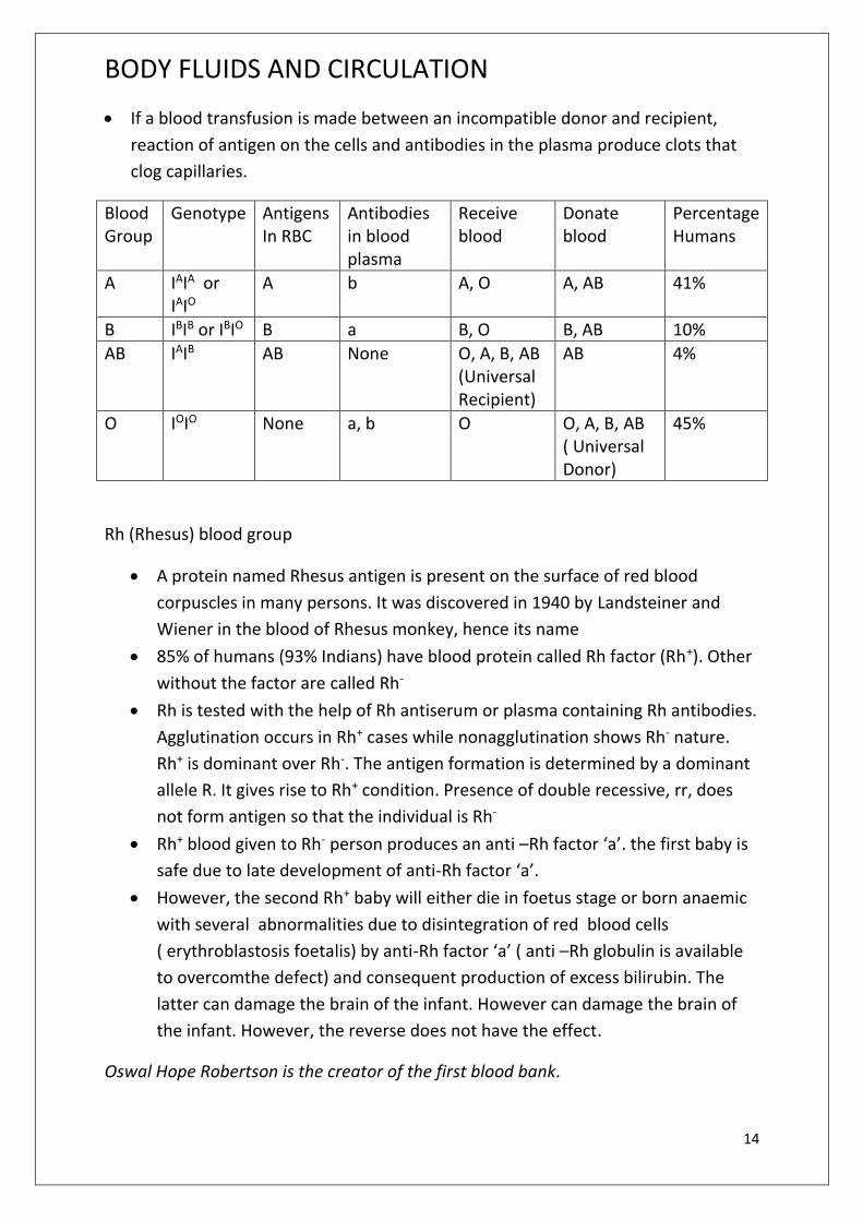
If a blood transfusion is made between an incompatible donor and recipient,
reaction of antigen on the cells and antibodies in the plasma produce clots that
clog capillaries.
Blood Group
Genotype Antigens In RBC
Antibodies in blood plasma
Receive blood
Donate blood
Percentage Humans
A IAIA or IAIO
A b A, O A, AB 41%
B IBIB or IBIO B a B, O B, AB 10%
AB IAIB AB None O, A, B, AB (Universal Recipient)
AB 4%
O IOIO None a, b O O, A, B, AB ( Universal Donor)
45%
Rh (Rhesus) blood group
A protein named Rhesus antigen is present on the surface of red blood
corpuscles in many persons. It was discovered in 1940 by Landsteiner and
Wiener in the blood of Rhesus monkey, hence its name
85% of humans (93% Indians) have blood protein called Rh factor (Rh+). Other
without the factor are called Rh-
Rh is tested with the help of Rh antiserum or plasma containing Rh antibodies.
Agglutination occurs in Rh+ cases while nonagglutination shows Rh- nature.
Rh+ is dominant over Rh-. The antigen formation is determined by a dominant
allele R. It gives rise to Rh+ condition. Presence of double recessive, rr, does
not form antigen so that the individual is Rh-
Rh+ blood given to Rh- person produces an anti –Rh factor ‘a’. the first baby is
safe due to late development of anti-Rh factor ‘a’.
However, the second Rh+ baby will either die in foetus stage or born anaemic
with several abnormalities due to disintegration of red blood cells
( erythroblastosis foetalis) by anti-Rh factor ‘a’ ( anti –Rh globulin is available
to overcomthe defect) and consequent production of excess bilirubin. The
latter can damage the brain of the infant. However can damage the brain of
the infant. However, the reverse does not have the effect.
Oswal Hope Robertson is the creator of the first blood bank.

BODY FLUIDS AND CIRCULATION www.gneet.com
15
IMPORTANCE OF BLOOD GROUPS
i) Knowledge of blood group is essential for blood transfusion
ii) Rh compatibility is required for both marriage and transfusion in order to
prevent erythroblastosis
iii) Preliminary information about disputed parentage and progeny is provided
by blood grouping.
iv) Blood grouping is used in forensic identification of blood stains.
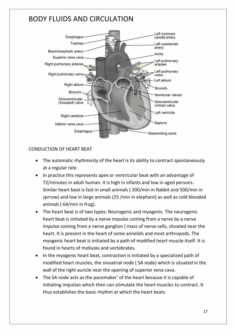
Human Heart
It is a reddish conical muscular mesodermal hollow organ of a about 12cm
length 9 cm breadth, weighs about 300gm and lies behind the sternum in the
mediastinum space of thoader cavity in between the two lungs. Broader base
is upwards.
The mammalian heart comprises of four complete chambers two ventricles
and two auricles ( atria)
Heart wall consists of connective tissue, blood vessels and cardiac muscle
fibres. The latter form a cross – connected network for smooth passage of
constriction wave. The cardiac muscle or myocardium does not tire due to
a) Alternate rest and activity
b) Non- formation of lactic acid
Heart is covered by a double fibrosenous sac or pericardium. It has two
components outer non-distensible tough fibrous pericardium ( prevents
excessive expansion of heart) and inner thin serous pericardium.
Serous pericardium has two thin secretory membranes, outer parietal and
inner visceral. It encloses a narrow pericardial cavity having pericardial fluid
for frictionless movement, protection from shock and mechanical injury
There is a depression or coronary sulcus between atria and ventricles, inter-
atrial sulcus ( two parts, anterior and posterior) between two ventricles.
Coronary arteries are housed in these sulci. They supply blood to walls of
heart.
Atrial appendages is protruded part of atria which overhangs the ventricles.
Low ridges occur internally in the region of atrial appendage. They are called
musculi pectinati. Blood vessels connected to heart are known as great blood
vessels
Deoxygenated blood flows through right half of heart and oxygenated blood
flows through left half of heart. The right and left atria are separated by inter
atrial septum. It bears a depression called fossa ovalis ( in the area of foetal

BODY FLUIDS AND CIRCULATION www.gneet.com
16
opening called foramen ovale). Right atrium / auricle receives deoxygenated
blood from superior vena cava ( upper part of body), inferior vena cava (
middle and lower part of body) and coronary sinus ( heart walls). The basius
valve occurs at the opening of cornonary sinus and Eustachian valve at the
opening of inferior vena cava
Back flow in superior vena cava is prevented by obliquity of opening. The left
atrium / auricle receives oxygenataed blood from two lungs through four
pulmonary veins. Right and left ventricles are separated by an interventricular
septum
Left ventricle is larger, includes the apex part and has extra thick wall as
compared to right ventricle due to its mechanical requirement of pumping
oxygenated blood to all parts of body, walls of ventricle posses a network of
low ridges or columnal carnea and a few large muscular projection or papillary
muscles/musculi papillares.
Right ventricle contains a moderator band that extends between upper
papillary muscle and inter-ventricular septum. Atria opens into ventricles
through atrio-ventricular apertures is guarded by valves. Right atrio-
ventricular aperture is guarded by tricuspid valve possessing three flaps and
left atrio-ventricular aperture is guarded by bicuspid and mitral valve
possessing two flaps.
The flaps of the valves are held in their position by fine inelastic cords or
chordae tendineae connected to papillary muscles. Left ventricle opens into
aorta. The opening is guarded by an aortic semilunar valve between two.
BODY FLUIDS AND CIRCULATION www.gneet.com
17
CONDUCTION OF HEART BEAT
The automatic rhythmicity of the heart is its ability to contract spontaneously
at a regular rate
In practice this represents apex or ventricular beat with an advantage of
72/minutes in adult human. It is high in infants and low in aged persons.
Similar heart beat is fast in small animals ( 200/min in Rabbit and 500/min in
sprrow) and low in large animals (25 /min in elephant) as well as cold blooded
animals ( 64/min in frog).
The heart beat is of two types: Neurogenic and myogenic. The neurogenic
heart beat is initiated by a nerve impulse coming from a nerve by a nerve
impulse coming from a nerve ganglion ( mass of nerve cells, situated near the
heart. It is present in the heart of some annelids and most arthropods. The
myogenic heart beat is initiated by a path of modified heart muscle itself. It is
found in hearts of mollusks and vertebrates.
In the myogenic heart beat, contraction is initiated by a specialized path of
modified heart muscles, the sinoatrial node ( SA node) which is situated in the
wall of the right auricle near the opening of superior vena cava.
The SA node acts as the pacemaker’ of the heart because it is capable of
initiating impulses which then can stimulate the heart muscles to contract. It
thus establishes the basic rhythm at which the heart beats

BODY FLUIDS AND CIRCULATION www.gneet.com
18
The impulse of contraction emitted by the sinoatrial node spreads as a wave
of contraction over the right and left atrial wall pushing the blood through the
strio ventricular valves into the ventricles.
This wave of contraction next reaches the atrio-ventricular ( AV – node) or
pacesetler. Which is stinulated to emit an impulse of contraction spreading to
the ventricular muscle in the atrioventricular bundle and the purkinje fibres.
The atrial muscle fibres are separated from those of the ventricles by a fibrous
tissue ring. These is no functional continuity between the atria and ventricles.
They only conducting tissue between the atria and the ventricles is the atrio
ventricular bundle or the Bundle of His).
The atrioventricular bundle ( Bundle of His) was discovered by His (1983) and
consists of a set of specialized muscle strands originating from AV node and
pass downwards into the inter-ventricular septum. This bundle then divided
into the left and right bundle branches, one going to each ventricle.
Within the myocardium of the ventricles the branches break up into a
network of fine branching, anastomising filaments of fibres known as Purkinje
fibres.
The bundle of His and the Purkinje fibres convey the impulse of contraction
from the AV node to the myocardium of Ventricles.
BODY FLUIDS AND CIRCULATION www.gneet.com
19
PACE-MAKER
SA node is called natural pace maker of heart as impulse generated by it
spreads to booth atria and through AV node to ventricles for their rhythmic
contraction.
Disruption or insufficiency of any component of this impulse conducting
system results in slowing down or irregularity of heart rhythm or independent
contraction of atria and ventricles. Failure of atrial impulse to pass into
ventricles for a few seconds to few hours is called ventricular escape or
stokes- Adam syndrome. In all such cases an artificial pace – maker is
implanted.
It is an electric device first developed by Greatbatch and Chardack ( 1960)
which is connected to heart for covering up any deficiency of myogenic
functioning so as to make it beat normally ( 72-80/ min) A pacemaker has a
pulse generator having long lasting lithium halide cells ( with over 10 years of
life) and a biocompatible plastic covered fine metallic string for functioning as
muscles stimulating electrode. There are various types of pacemaker.
i) External pacemaker
ii) Epicardial pacemaker
iii) Endocardial pacemaker
iv) Demand pacemaker
v) Atrial synchronized
vi) Temporary pacemaker
vii) Permanent pacemaker
In common type, the pulse generator is placed below skin under right clavicle
while the string / cable is passed via superior vena cava right atrium and
allowed to rest against the tip of right ventricle.
Pacemaker are liable to be influenced by microwave ovens, metal detectors,
electric shavers cellphone etc.
CARDIAC CYCLE
The cardiac cycle consists of one heart or one cycle of contraction and
relaxation of the carbiac muscle. The contraction phase is called the systole
while the relaxation phase is called diastole.
When both the atria and ventricles are in diastolic or relaxed phase, this is
referred to as a joint diastole. During this phase, the blood flows from the
superior vena cava and inferior vena cava into the atria and from ateria to the
respective ventricles through auriculo ventricular valves. But there is no flow
BODY FLUIDS AND CIRCULATION www.gneet.com
20
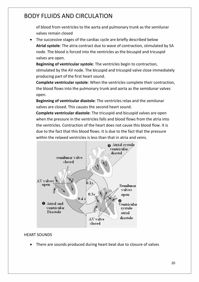
of blood from ventricles to the aorta and pulmonary trunk as the semilunar
valves remain closed
The successive stages of the cardiac cycle are briefly described below
Atrial systole: The atria contract due to wave of contraction, stimulated by SA
node. The blood is forced into the ventricles as the bicuspid and tricuspid
valves are open.
Beginning of ventricular systole: The ventricles begin to contraction,
stimulated by the AV node. The bicuspid and tricuspid valve close immediately
producing part of the first heart sound.
Complete ventricular systole: When the ventricles complete their contraction,
the blood flows into the pulmonary trunk and aorta as the semidunar valves
open.
Beginning of ventricular diastole: The ventricles relax and the semilunar
valves are closed. This causes the second heart sound.
Complete ventricular diastole: The tricuspid and bicuspid valves are open
when the pressure in the ventricles falls and blood flows from the atria into
the ventricles. Contraction of the heart does not cause this blood flow. It is
due to the fact that this blood flows. It is due to the fact that the pressure
within the relaxed ventricles is less than that in atria and veins.
HEART SOUNDS
There are sounds produced during heart beat due to closure of valves

BODY FLUIDS AND CIRCULATION www.gneet.com
21
Lubb (S1) first sound, systolic sound) is the first heart sound which is dull, loud
or low pitched, of long duration ( 0.16 and 0.19 seconds) and is produced due
to closure of atrio ventricular valves ( tricuspid and bicuspid valves)
Dup ( S2, second sound, diastolic sound) is the second heart sound which is
sharp high pitched, of shorted duration ( 0.1 sec) and is produced due to
closer of semilunar valves at the base of great arteries. A pause or gap occurs
between the second sound and the first sound of next cycle. It coincides with
ventricular diastole.
Incomplete closure of valves due to disease or other defect produces
abnormal heart sound called murmur. Heat sounds are listened by means of
instrument called stethoscope.
CARDIAC OUTPUT.
The volume of blood pumped by each ventricle per minute is called the
cardiac output
It is determined by multiplying the heart rate with the volume of blood
ejected by each ventricle during each beat, which is called the stroke volume.
Cardiac output = Heart rate × stroke volume
= 72 beats /min × 0.08 litre/ beat = 5.5 litres/min
Cardiac index is the minute volume per sq.m. of body surface area. Its normal
value is 3.3 litre/min/sq.m
REGULATION OF HEART BEAT.
Neural Regulation:
The cardiac centre lies in the medulla oblongata of the brain. The cardiac
centre is formed of cardio-inhibitor and cardio-accelerated parts. The former
decreases the rate of heart beat and the latter accelerates it. The cardio-
inhibitor is connected with the heart through vagus nerve ( it carries –
parasympathetic nerve fibres) and cardio accelerator through sympathetic
nerve fibres. Sensory fibres extended from the receptors present in the
superior vena cava aorta and carotid sinuses to the cardiovascular centre in
medulla oblongata. The impulses received from the aorta and carotid sinuses
decreases the heart rate, whereas the impulses from Vena cava increases the
heart rate.
Hormonal regulation:
Adrenaline and noradrenaline hormones are secreted by the medulla of the
adrenal glands. Noradrenaline accelerates the heart beat under normal

BODY FLUIDS AND CIRCULATION www.gneet.com
22
conditions while adrenaline does this function at the time of emergency.
These hormones directly influence the SA node. Thyroxine hormone secreted
by thyroid glands increases oxidative metabolism of the body cells. This
requires more oxygen and thus indirectly increases heart beat.
Body temperature also affect the pacemaker. Just 1OC rise in temperature
increases with exercise to provide additional oxygen and food to muscles.
ELECTROCARDIOGRAM ( ECG )
ECG is graph record of the elastic current produced by the excitation of the
cardiac muscles. The instrument used to record the changes in an
electrocardiograph. Waller ( 1887) first recorded the electrocardiogram but
Einthoven ( 1906) studied ECG in detail and got Nobel Prize. He is also
considered “father of the electrocardiography”
A normal ECG is composed of P waves, a QRS wave ( complex) and a T wave.
The letters are arbitrarily selected and do not stand for any particular words
The P wave is a small upward wave that indicates the depolarization of the
atria ( atrial contraction). It is caused by the activation of SA node.
The QRS wave ( complex) begins after a fraction of second of the P wave. It
begins as a small downward deflection (Q) and continues as large upright ®
and triangular wave, ending as downward wave (S) at its base. It represents
ventricular depolarization ( ventricular contraction )
The T wave is a dome-shaped which indicates ventricular repolarisation (
ventricular relaxation)

BODY FLUIDS AND CIRCULATION www.gneet.com
23
Each large square represents 0.2 second. Normal P-R interval is 0.12 to 0.2
second. Normal QRS complex duration is 0.12 second. Normal Q-T interval is
0.4 second.
Enlargement of the P wave indicates enlargement of atria. During
atherosclerotic heart diseases and rheumatic fever, the P-R interval is
lengthened. This is due to the inflammation of atria and AV nose
The enlarged Q and R waves indicate a mycocardial infection ( heart attack).
The S-T segment is elevated in acute myocardial infection and depressed
when the heart muscle receives insufficient oxygen.
T wave is flat when the heart muscles receives insufficient oxygen as in
atherosclerotic heart disease. It may be elevated when the body’s potassium
level is increased.
When ECG of person to be recorded, four leads ( metal electrodes) are
attached in the arms and legs. It is done after leaning and putting special jelly,
which improves electrical conduction. With the help of rubber suction cup, an
additional electrode is placed on the chest. Now the electrocardiograph is
switched on which detects and amplifies the electrical current of the heart
and transmits to the recording pen. The latter draws a wavy line that is called
deflection wave.
The importance of ECG is that it gives accurate information about the heart.
Therefore, ECG is of great diagnostic value in cardiac diseases.
BLOOD PRESSURE
It is the pressure exerted by the flow of blood on the walls of arteries and
measured as millimeters of mercury by the instrument is called
sphygmanometer ( Riva-Rocci). It has a high systolic value ( normal 120
mmHg) and low diastolic value ( normal 80 mmHg). The difference between
two is called pulse pressure.
Hypertension ( hyperpiesis)
It is sustained rise in arterial blood pressure or high blood pressure with
systolic more than 140 mmHg and diastolic more than 90mmHg. The reason is
stiffening of arterial walls due to cholesterol walls, varicose veins, obesity,
toxins, hormones, defective kidney etc.
Hypertension caused by hormones is called hypertension. Other forms of
hypertension are known as primary hypertension. It accounts to 90% of the
cases.
BODY FLUIDS AND CIRCULATION www.gneet.com
24
High blood pressure harms three vital organs-heart, brian and kidney. It makes
heart to overwork due to which congestive heart disease develops quite early.
A blood pressure of 220/120 mmHg may cause internal haemorrhage due to
rupturing of some blood vessel. Cerebral haemorrhage causes stroke or CVA.
Damage to optic arteries leads to blindness while a similar damage to renal
vessels causes nephritis. It leads to renal failure.
Hypotension ( Hypopiesis)
It is low blood pressure with systolic below 110 mmHg and diastolic below 70
mmHg
Hyptension is caused by low metabolic rate, starvation, anaemia, chronic
vasodilation of arterioles, lower pumping activity, valvular defects, nervous
disorders, Addison’s disease.
There is an increase relationship between rate of heart beat and blood
pressure. The phenomenon is called marey’s law of heart
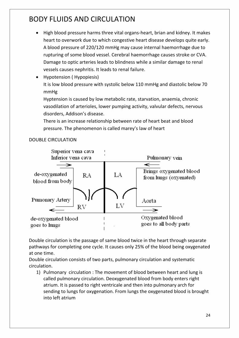
DOUBLE CIRCULATION
Double circulation is the passage of same blood twice in the heart through separate pathways for completing one cycle. It causes only 25% of the blood being oxygenated at one time. Double circulation consists of two parts, pulmonary circulation and systematic circulation.
1) Pulmonary circulation : The movement of blood between heart and lung is called pulmonary circulation. Deoxygenated blood from body enters right atrium. It is passed to right ventricale and then into pulmonary arch for sending to lungs for oxygenation. From lungs the oxygenated blood is brought into left atrium

BODY FLUIDS AND CIRCULATION www.gneet.com
25
2) Systematic circulation: This is movement of blood between heart and different parts of body except lungs. Oxygenataed blood is received by left atrium. It is passed to left ventricle which pumps it into aorata for supply to body parts including walls of heart. On deoxygenation the blood passes back into right atrium of heart through coronary sinus, inferior vena cava and superior vena cava. Purpose of systematic circulation is to transport O2 and nutrients to tissue and remove CO2 and nitrogenous waste from them.
ARTERIAL SYSTEM
It comprises all the arteries coming out of heart and supplying blood to different parts of body. The heart gives out two main arterial vessels, pulmonary arch ( from right ventricle) and aorta.
Pulmonary arch carries deoxygenated blood. It divides into two pulmonary arteries one for each lung. Aorta carries oxygenated blood. It is swollen into aortic oxygenated blood. It is swollen into aortic sinus at its origin. Aortic sinus gives out right and left coronary arteries to the heart. Aorta then produces a short and wide innominate on right side, a left common carotid and a left subclaxian before bending down as dorsal aorata. Innominate or branchiocephalic forms a right common carotid and a right subclavian. Subclavin provide oxygenated blood to fore limbs, chest and spinal cord. Carotids supply oxygenated blood to neck, face, mouth, eyes, scalp and brain
Dorsal aorta has two parts, thoracic and abdominal. Thoracic aorta gives out oesophageal ( to oesohagus), phrenic ( to diaphragm), branches to back and intercoastals ( to intercoastal muscles) in thoracic cavity. Abdominal aorta supplies blood to visceral organs and lower extremities. It first gives out thick celiac artery with branches like hepatic ( liver), gastric ( stomach), splenic (spleen), duodenal ( duodenum) and pancreatic ( pancreas). Below coelic, abdominal aorta gives out a superior mesenteric artery ( small intestine), two super renal ( adrenal or supra-renal glands), two renals ( kidneys), two genitalis and inferior ( posterior) mesenteric artery ( large intestine) and then divides into two iliacs ( pelvic region and lower limbs)
4% of arterial blood passes into heart, 10% to liver, 8% to brain, 15% to digestive tract and the remaining to rest of the body.
VENOUS SYSTEM
It comprises all the veins that bring blood to the heart. Venous system consists
of pulmonary veins, coronary sinus, portal system and venae cavae.
Pulmonary veins are four in number, two from each lung. They bring
oxygenated blood to left atrium. Coronary sinus collects deoxygenated blood
from all the walls of heart. It opens into right atrium. Superior vena cava is
formed by two branchiocephalic veins each of which receives deoxygenated
blood from a jugular vein ( from head and neck), subclavian vein ( upper limb)
and internal thoracic vein ( part of chest). Before opening into right atrium,

BODY FLUIDS AND CIRCULATION www.gneet.com
26
superior vena cava receives a small ozygos vein from oesophagus and
intercoastal area.
Inferior vena cava is formed by the union of two common iliac veins ( pelvis
and lower limbs). While on its way to heart, it receives genital veins ( gonads),
lumbar veins ( muscles of back), renal ( liver) and phrenic veins ( diaphragm).
It then opens into right atrium.
PORTAL SYSTEM
It is a system made of a portal vein and the capillary complex formed by it in
an organ than the one of its origin. A portal vein is the vein which collects
blood from one organ by a set of capillaries and distributes that blood into a
second organ through another set of capillaries instead of sending blood into
heart. There are three types of portal systems – hepatic, hypophysial and
renal.
Hepatic portal system : It occurs in all vertebrates and is meant for taking
blood from digestive tract, pancreas and spleen into liver. The system has a
large heapatic portal vein that is formed by four veins-splenic ( spleen),
inferior mesenteric ( rectum and distial part of colon), superior mesenteric (
small intestine, calcum and proximal part of colon) and gastroepiploic ( from
stomach and pancreas). Hepatic portal vein enters liver and breaks into
capillaries. The system function as a short circuit for
(i) Removal of glucose, amino acids and other nutrients.
(ii) Deamination of extra amino acids and conversion of harmful ammonia into
urea
(iii) Separation of toxic chemicals and their detoxification
(iv) Direct pouring of liver products into venous blood
Hypophysical portal system : It is a minor portal system that occurs in higher
vertebrates. The system consists of a single hypophysial portal vein. The portal
vein is formed by capillaries in the hypothalamus. It passes into anterior lobe
of pituitary glands and breaks up into capillaries there. Hypophyseal portal
sytam is meant for pouring hormones secreated by hypothalamus directly into
anterior part of pituitary.
Renal portal system: It occurs in lower vertebrates ( fish and amphibians),
reduced in reptiles and aves and is absent in mammals. It consists of renal
portal veins that bring blood from posterior part of the body directly into
kidneys for removal of waste products.
BODY FLUIDS AND CIRCULATION www.gneet.com
27
LYMPHATIC SYSTEM
It comprise lymph, lymphatic capillaries, lymphatic vessels, lymphatic nodes and
lymphatic ducts.
LYMPH
Lymph, a colourless fluid is a part of tissue fluid, which in turn, is a part of blood
plasma. So the composition of tissue fluid and lymph is same as that of blood plasma
but it lacks RBCs and large plasma proteins. As compared to the tissue fluid, the
lymph contains very small amount of nutrients and oxygen, but contains abundant
carbon dioxide and other metabolic wastes. Amoeboid shaped white blood
corpuscles may be present in the lymph.
LYMPHATIC CAPILLARIES
Lymphatic capillaries lie close to the blood capillaries but differ from them to extent
that they end blindly. Moreover, they have extremely thin walls. They are composed
of a single layer of endothelial cells. The lymphatic capillaries of intestine absorb the
digested fats. They are milky in appearance and are, therefore, called the lacteals.
LYMPHATIC VESSEL
The lymphatic capillaries unite to form large lymphatic vessels. They are composed of
an outer coat of fibrous tissue, middle coat of muscular tissue and an inner lining of
endothelial cells. The lymphatic vessels have numerous valves.
LYMPH NODER
These are small oval or bean shaped structures located along the length of
lymphatic vessels. Lymph nodes are most numerous in the thoracic
mediastinum on the posterior abdominal wall in the abdominal mesenteries
and in the pelvis neck and proximal ends of the limbs.
Lymphatic nodes perform the following main functions.
Both B-lymphocytes and T-lymphocytes are produced here.
Macrophages of lymph nodes remove bacteria, foreign material and cell
debris from the lymph.
B-lymphocytes change to plasma cells that produce antibodies against
invading antigens, while T-lymphocytes attack cells that are ‘foreign’ to the
host body.
BODY FLUIDS AND CIRCULATION www.gneet.com
28
THORACIC DUCT
The lymphatic vessels of left side unite to form a thoracic duct. This duct begins at
the cisternal chili, which is a sac-dilation situated in the front of the first and second
number vertebrate. The thoracic duct contains several valves. It discharges its lymph
into the left subclavian vein.
RIGHT LYMPHATIC DUCT
The lymphatic vessel of the right side of the thorax, head and neck unite to form
right lymphatic duct. It is about 1 cm in length. It discharges its lymph into the right
subclavian vein.
LYMPH MOVEMENT
The lymph flows in lymphatic vessels very slowly. Forcing out of fluid from the blood
capillaries sets up some pressure in the tissue fluid. This establishes a pressure
gradient in the lymphatics, causing flow of lymph in the latter. Movements of viscera
and contractions of the body muscles help considerably in squeezing the lymph
along. The valves present in lymphatic vessels prevent its back flow. Movement of
villi assist flow of lymph in the lacteals. Gravity helps in moving the lymph down the
lymphatic vessels of head and neck.
FUNCTIONS OF LYMPH
The lymph or lymphatic system serves functions as:
It drains excess tissue fluid from the extracellular spaces bin to the fluid.
Some of fluid from the digestive tract is absorbed into the lymph. The
lymphatic vessels store this fluid temporarily and release it gradually so that
the kidney do not face a sudden pressure of urine execration.
It carries carbon dioxide and nitrogenous waste materials that diffuse into the
tissue fluid to the blood.
It takes lymphocytes and antibodies from lymphatic nodes to the blood.
It transported fat that is digested and absorbed in the intestine to the blood in
the form of chylomicron droplets.
It destroys the invading microorganisms and foreign particles in the lymphatic
nodes.
It maintains quality and quantity of the blood by restoring the fluid and solute
that leaves it.
BODY FLUIDS AND CIRCULATION www.gneet.com
29
It brings plasma protein macromolecules synthesized in the liver cells and
hormones produced in the endocrine glands to the blood.
SPLEEN
Spleen is the largest component of the lymphatic system. It is large ( 7-10 cm in
diameter), bean-shaped, vascular, dark red organ located in the abdomen just below
the diaphragm at the tail of the pancreas behind the stomach.
The spleen is composed of red pulp ( reticular tissue rich in RBCs) having small
patches of white pulp ( lymphatic nodes) scattered in it. The red pulp is enclosed by a
capsule of white fibrous tissue. The capsule sends trabeculae into the pulp and is
surrounded by viscernal peritoneum.
FUNCTIONS
i) Destruction of worn-out red corpuscles
ii) Reservoir of rd corpuscles
iii) Formation of agranulocytes
iv) Production of antibodies
v) Storage of iron
vi) Erythropoiesis
vii) Disposal of foreign elements
THYMUS
Thymus is also a lymphatic organ. It lies in the upper chest near the neck. It is
prominent in children but begins to degenerate in early childhood. It educates the
lymphocytes in the foetus to distinguish cells from foreign cells.
TONSILS
Tonsils too are lymphatic tissues. They are located in the throat. They do not filter
lymph. They are thought to protect against infection.
SOME COMMON CARDIOVASCULAR DEFFECTS
1. Arteriousclerosis:
Sclerosis and hardening of walls of generally smaller arteries and arterioles is
called arteriosclerosis. The common cause is deposition of calcium in tunica
media cholesterol may get calcified. The walls of arteries become stiff and
rigid. There is a loss of elasticity. The phenomenon is called hardening of
arteries. Limb arteries are usually the first to undergo arteriosclerosis. Lesions

BODY FLUIDS AND CIRCULATION www.gneet.com
30
develop at branch points. It ultimately leads to distal obstruction causing pain,
numbness of extrimities, peripheral oedema, cyanosis etc. rupturing of some
vessels also occur. It forms blood clot and blocks the flow of blood.
2. Atherosclerosis:
It is wall thicknening and narrowing of lumen of medium and large arteries. In
atherosclerosis, yellowish plaques ( atheromas) of cholesterol and other lipids
are deposited within tunica intima and inner part of tunica media where
smooth muscles abound. They are mostly caused by low density lipoprotenins
or LDL which can pass through endothelium. Plaques grow. The smooth
muscles also proliferate probably caused by release of platelet derived growth
factor ( PDGF). This occurs due to roughness of inner arterial lining.
Thickneining of arterial wall reduces the lumen size. In extreme cases growth
of plaques may completely block an artery. Atherosclerosis leads to
hypertension, reduced blood supply to limbs and other organs resulting in
their dysfunctioning. Atherosclerosis in coronary arteries results in reduced O2
supply to heart walls causing angina, myocardial infaction or heart attack or
stroke.
3. Coronary artery disease ( CAD)
Coronary arteries undergo atherosclerosis. There is deposition of calcium, fat
and fibrous tissue which results in narrowing of the arterial lumen. Flow of
blood in the affected arteries is reduced. The cardiac muscles supplied by the
affected arteries will begin to deteriorate. There is thoracic pain, nausea,
perspiration and E.C. G changes. The defect can be treated through
angioplasty ( breaking of arterial blockage by balloon catheter) and bypass
surgery.
4. Angina or Angina pectoris
It is recurrent, spasmodic suffocating thoracic ( or heart ) pain which often
radiates to left arm. Angina is generally caused by deficient blood supply to
heart muscles. It is precipitated by excitement or strenuous physical activity.
Angina pectoris can occur in all types of individuals. Both men and women of
any age. However, it is more common in middle aged and elderly persons.
Reduced blood supply to myocardial muscles occurs either due to constriction
or obstruction of blood vessels.
5. Heart Failure
It is the inability of heart to supply blood in adequate quantities to all parts of
the body. Heart failure is a syndrome of ventricular dysfunction. The person
suffering from heart failure has reduced exercise capacity. Health of different
muscles of the body would also be affected. Heart failure should not be
BODY FLUIDS AND CIRCULATION www.gneet.com
31
confused with heart attack ( heart muscle damaged due to inadequate blood
supply) or cardiac arrest in which case there is stoppage of heart beat.
6. Caardiomegaly : hypertrophy of heart. Inflammation of heart is carditis.
7. Cardiomyopathy : Noninflammatory disease of heart muscle.
8. Ischaemic heart: Heart with degenerate or defective components due to
rheumatic disorder or fever in childhood.
9. Rheumatic heart : Heart with degenerate or defective components due to
rheumatic disorder of fever in childhood.
10. Embolus: Mass of clotted blood, other formed elements, fragments, air,
calcium etc. coming from a larger blood vessel is forced into a smaller or
narrow blood vessel resulting in its blockage and hence obstruction of bllod
circulation.
11. Myocardial Infarction: Complication due to reduced blood supply to heart
wall-pain, pallor, perspiration, nausea, ECG changes.
12. Heart Burn ( Pyrosis). Sensation of burning occurring in waves in oesophagus
tending to rise upward towards neck often with reflex into mouth. It has
nothing to do with heart.
13. Varicose Veins: Unnatural permanently distended veins.
14. Haematoma: Localised collection of usually clotted blood in a tissue or organ
due to injury and rupturing of blood vessel.