Blunt Cerebrovascular Injury and Angiography · Blunt Cerebrovascular Injury and Angiography Janeen...
35
Blunt Cerebrovascular Injury and Angiography Janeen Jordan, PGY3 Grand Rounds April 3, 2006
Transcript of Blunt Cerebrovascular Injury and Angiography · Blunt Cerebrovascular Injury and Angiography Janeen...
Blunt Cerebrovascular Injury and Angiography
Janeen Jordan, PGY3Grand RoundsApril 3, 2006

Epidemiology
♦ 1 – 2% of trauma admissions♦Rapid deceleration assoc with hyperflexion
and rotation♦ Intimal tears, dissection, psa, transection
– Avf, ccsf, thrombus, emboli♦ 20-40% of patients have bilateral injuries♦ 13-15% of patients have both CAI and VAI
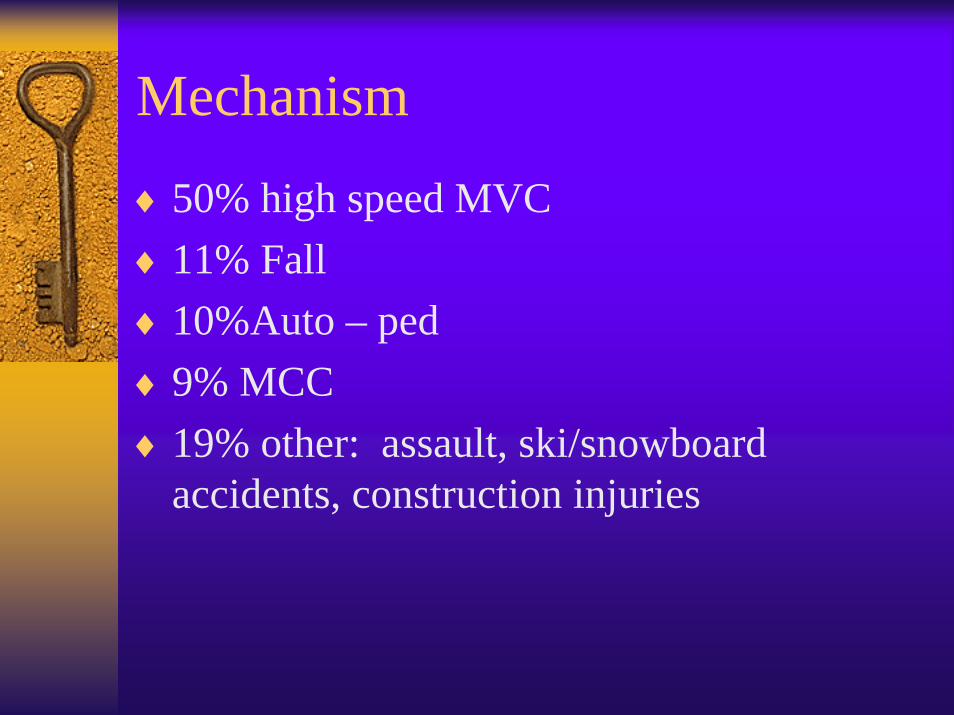
Mechanism
♦ 50% high speed MVC♦ 11% Fall♦ 10%Auto – ped♦ 9% MCC♦ 19% other: assault, ski/snowboard
accidents, construction injuries
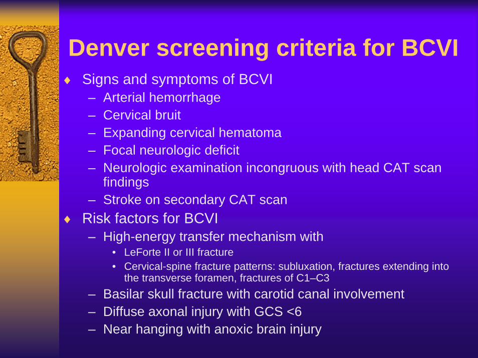
Denver screening criteria for BCVI♦ Signs and symptoms of BCVI
– Arterial hemorrhage– Cervical bruit– Expanding cervical hematoma– Focal neurologic deficit– Neurologic examination incongruous with head CAT scan
findings– Stroke on secondary CAT scan
♦ Risk factors for BCVI– High-energy transfer mechanism with
• LeForte II or III fracture• Cervical-spine fracture patterns: subluxation, fractures extending into
the transverse foramen, fractures of C1–C3– Basilar skull fracture with carotid canal involvement– Diffuse axonal injury with GCS <6– Near hanging with anoxic brain injury
Pathophysiology
♦ Intimal injury incites plaque formation ♦Thrombogenesis♦Resolution of the plaque, pseudoanuerysm
formation, continued dissection and plaque thrombus
♦ stroke

Table 1. Denver grading scale for blunt carotid artery injury
Grade AngiographicFindings
Prognosis Treatment
I Vessel wall irregularity or dissection with <25% of luminal diameter
Good (7% progress) Systemic anticoagulation controversial
II Raised Intimal flap, thrombus, dissection, or hematomas >25% of luminal diameter
Fair with treatment (70% progress)
Systemic anticoagulation
III Pseudoaneurysms Require intervention Surgery or stenting
IV Total vessel occlusion Outcome usually assured at time of diagnosis
Systemic anticoagulation
V Transection Very poor, high mortality
Surgery

♦Average Age– 35 – 38yrs (67% male)
♦Average time to sx onset– 12 – 72hours (30% symptomatic)
♦Average time to diagnosis– 18 – 53hrs
♦ 9% grade I injuries progress to higher grade♦ 43% grade II injuries progress
Morbidity & Mortality
♦Overall mortality– 21 – 31%
♦Morbidity– 37 – 58% have permanent neurologic disability

Benefit of early diagnosis and treatment♦Fabian et al
– Significant reduction in morbidity and mortality with heparin therapy
♦Cothren et al– Untreated pt – 21% stroke rate– CAI increase by grade– VAI ~20% stroke
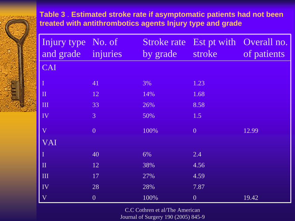
C.C Cothren et al/The American Journal of Surgery 190 (2005) 845-9
Table 3 . Estimated stroke rate if asymptomatic patients had not been treated with antithrombotics agents Injury type and grade
Injury type and grade
No. of injuries
Stroke rate by grade
Est pt with stroke
Overall no. of patients
CAI
I 41 3% 1.23
II 12 14% 1.68
III 33 26% 8.58
IV 3 50% 1.5
V 0 100% 0 12.99
VAII 40 6% 2.4
II 12 38% 4.56
III 17 27% 4.59
IV 28 28% 7.87
V 0 100% 0 19.42

Diagnosis
♦Arteriography– Invasive– Resource intensive ($6800)– Complications
• Catheter insertion site injuries (1-2% hematomas, PSA formation)
• Contrast administration (1-2% renal dysfunction, allergic reaction)
• Stroke (less than 1%)

Angiogram vs CT angiographyand MRA♦Miller et al♦Univ Tenn, Memphis♦Prospective analysis, 2002♦Screened 216pts over 2yrs
– (Jan 2000 – Mar 2002)– Angiogram– CTA– MRI

Screening Triggers for suspected blunt Cerebrovascular injuryCervical Spine fracture
Neurologic exam not explained by brain imaging
Horner’s Syndrome
LeFort II or III facial fractures
Skull base fractures involving the foramen lacerum
Neck soft tissue injury (e.g., seatbelt injury or hanging)
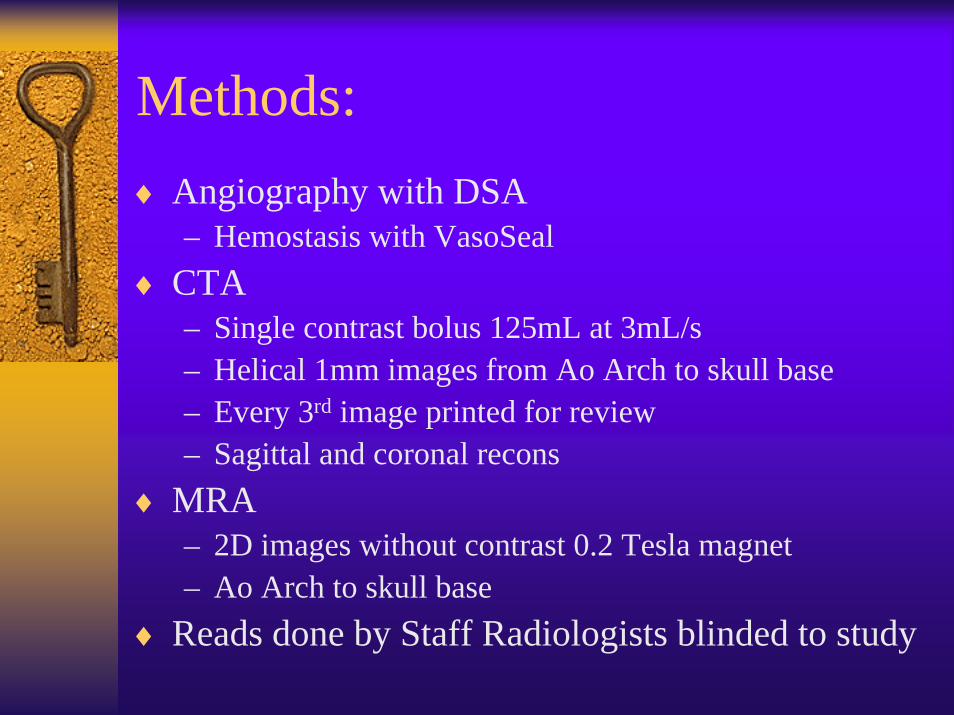
Methods:♦ Angiography with DSA
– Hemostasis with VasoSeal♦ CTA
– Single contrast bolus 125mL at 3mL/s– Helical 1mm images from Ao Arch to skull base– Every 3rd image printed for review– Sagittal and coronal recons
♦ MRA– 2D images without contrast 0.2 Tesla magnet– Ao Arch to skull base
♦ Reads done by Staff Radiologists blinded to study

Results:♦ 216pt screened (212 with 1 screening criteria)♦ 63pt with BCVI (29%); 1.03% incidence♦ Avg time to diagnosis 29.8hrs♦ CVA
– 50% CCF (1/2)– 100% occlusions (3/3)– 23% dissections (5/22)
♦ 79% diagnosed prior to sxs
CAI (n=27) VAI (n=49)
Dissection 22(82%) 27(55%)
Occlusion 3(11%) 22(45%)
Carotid/cavernous fistula
2(7%) --

Results:
♦ 143pt Angio and CTA– 17:8 CAI (47%)– 30:16 VAI (53%)– 1 false positive
♦ 21pt MRA– 4:2 CAI (50%)– 17:8 VAI (47%)– 1 false positive

Miller. Annals of Surgery. Vol 236(3).386-95
Results: Sensitivity and SpecificityCTA CAI VAI
Sensitivity 47% 53%
Specificity 99% 99%
MRA
Sensitivity 50% 47%
Specificity 100% 97%
Conclusion: CTA/MRA currently inadequate screening tools

Angiogram vs CT Angiographyand MRA♦Biffl, Moore, Johnson, Burch, Mestek, Ray♦Denver Health♦Prospective analysis, 2002♦ 46pts underwent CTA and angio♦ 16pts underwent MRI and angio

Biffl: J trauma, Vol 53(5). Nov 2002. 850-6.

CTA♦ Figure 5a. Pseudoaneurysm in a patient with a
gunshot wound to zone II of the neck. (a) Axial contrast-enhanced CT image shows focal irregularity of the left vertebral artery and a change in its caliber (arrow). Note the well-defined contour and normal appearance of the right vertebral artery. (b) Selective left vertebral arteriogram shows segmental narrowing of the vessel with a pseudoaneurysm.

Angiogram♦ Figure 5b. Pseudoaneurysm in a patient with a
gunshot wound to zone II of the neck. (a) Axial contrast-enhanced CT image shows focal irregularity of the left vertebral artery and a change in its caliber (arrow). Note the well-defined contour and normal appearance of the right vertebral artery. (b) Selective left vertebral arteriogram shows segmental narrowing of the vessel with a pseudoaneurysm.


Biffl: J Trauma, Vol 53(5). Nov 2002. 850-6
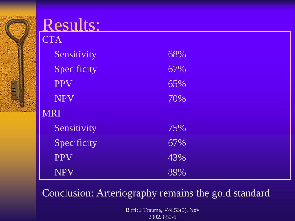
Results:CTA
Sensitivity 68%Specificity 67%PPV 65%NPV 70%
MRISensitivity 75%Specificity 67%PPV 43%NPV 89%
Conclusion: Arteriography remains the gold standard

Conclusions
♦ CAI and VAI incidence increasing with improved screening protocols
♦ Morbidity and mortality can be decrease with early diagnosis and treatment
♦The risk of angiography is less that the risk of an undiagnosed grade I BCVI.
♦ CTA and MRA are insufficient diagnostic modalities and risk increasing morbidity and mortality by delaying diagnosis and treatment


Works Cited♦ Miller PR, Fabian TC, Croce MA, et al. Prospective screening for blunt cerebrovascular injuries: analysis of diagnostic
modalities and outcomes. Ann Surg 2002; 236(3): 386-95.♦ Biffl WL, Ray CE Jr, Moore EE, et al . Noninvasive diagnosis of blunt cerebrovascular injuries: a preliminary report. J Trauma
2002; 53(5): 850-6.♦ Biffl WL, Ray CE Jr, Moore EE, et al. Treatment-related outcomes from blunt cerebrovascular injuries: importance of routine
follow-up arteriography. Ann Surg 2002; 235(5): 699-707.♦ Biffl WL. Diagnosis of blunt cerebrovascular injuries. Current opinion in Critical Care. 2003;9(6): 530-4.♦ Fabian TC, Patton JH Jr, Croce MA, et al. Blunt carotid injury: Importance of early diagnosis and anticoagulant therapy. Ann
Surg 1996; 223(5):513-25.♦ Cothren CC, Moore EE, Ray CE Jr, et al. Screening for blunt cerebrovascular injuries is cost effective. Am J of Surg 2005;
190(6): 845-49.♦ Berne JD, Norwood SH, McAuley CE, Villareal DH. Helical Computed Tomographic Angiography: an excellent screening test
for blunt cerebrovascular injury. J Trauma 2004; 57(1): 11-19.♦ Munera F, Soto JA, Palacio D, et al. Diagnosis of Arterial Injuries caused by penetrating trauma to the neck: comparison of
helical CT angiography and conventional angiography. Neuroradiology 2000; 216: 356-62.♦ Rogers FB, Baker EF, Osler TM, et al. Computer tomographic angiography as a screenign modality for blunt cervical arterial
injuries: preliminary results. J Trauma 1999; 46(3): 380-85.♦ Nunez DB, Torres-Leon M, Munera F. Vascular injuries of the neck and thoracic inlet: helical CT-angiography correlation.
Radiographics 2004; 24:1087-98.♦ Baker WE, Wassermann J. Unsuspected vascular trauma: blunt arterial injuries. Emerg Med Clin N Am 2004; 22: 1081-98.♦ Cogbill TH, Moore EE, Meissner M, et al. The spectrum of blunt injury to the carotid artery: a multicenter perspective. J
Trauma 1994; 37(3): 473-9.♦ Biffl WL, Moore EE, Offner PJ, Burch JM. Blunt carotid and vertebral arterial injuries. World J Surg 2001; 25(8): 1036-43♦ Chomel A, Vernet M, Lile A, et al. Traumatic bilateral dissections of the internal carotid artery: an infrequent diagnosis not to
be missed. J Neurosurgical Anesthesia 2002; 14(4): 309-12.♦ Mayberry JC, Brown CV, Mullins RJ, et al. Blunt carotid artery injury: the futility of aggressive screening and diagnosis. Arch
Surg 2004; 139(6): 609-13.♦ Cothren CC, Moore EE, Biffl WL, et al. Cervical spine fracture patterns predictive of blunt vertebral artery injury. J Trauma
2005; 55(5): 811-13.♦ Biffl WL, Moore EE, Patrick J, et al. Optimizing screening for blunt cerebrovascular injuries. Am J Surg 1999; 178: 517-522.

CTA♦ Figure 1b. Right internal carotid
arterial occlusion. (a-c) Transverse source images obtained caudad to cephalad at helical CT angiographic examination. In a and b, there is progressive narrowing of the right internal carotid arterial lumen (arrow). In the more cephalic image, c, the artery (arrow) is no longer opacified with contrast material. (d) Sagittalhelical CT angiographic image reformatted by means of maximum intensity pixel projection depicts the site of the internal carotid arterial occlusion (arrow).
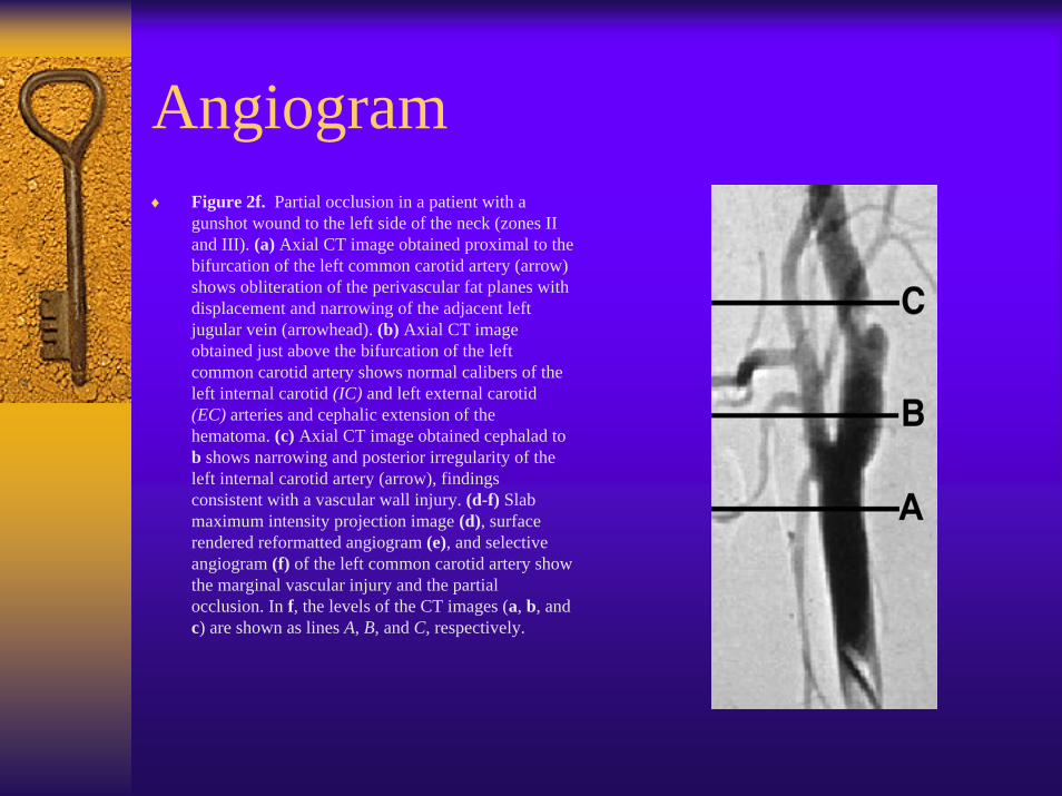
Angiogram♦ Figure 2f. Partial occlusion in a patient with a
gunshot wound to the left side of the neck (zones II and III). (a) Axial CT image obtained proximal to the bifurcation of the left common carotid artery (arrow) shows obliteration of the perivascular fat planes with displacement and narrowing of the adjacent left jugular vein (arrowhead). (b) Axial CT image obtained just above the bifurcation of the left common carotid artery shows normal calibers of the left internal carotid (IC) and left external carotid (EC) arteries and cephalic extension of the hematoma. (c) Axial CT image obtained cephalad to b shows narrowing and posterior irregularity of the left internal carotid artery (arrow), findings consistent with a vascular wall injury. (d-f) Slab maximum intensity projection image (d), surface rendered reformatted angiogram (e), and selective angiogram (f) of the left common carotid artery show the marginal vascular injury and the partial occlusion. In f, the levels of the CT images (a, b, and c) are shown as lines A, B, and C, respectively.
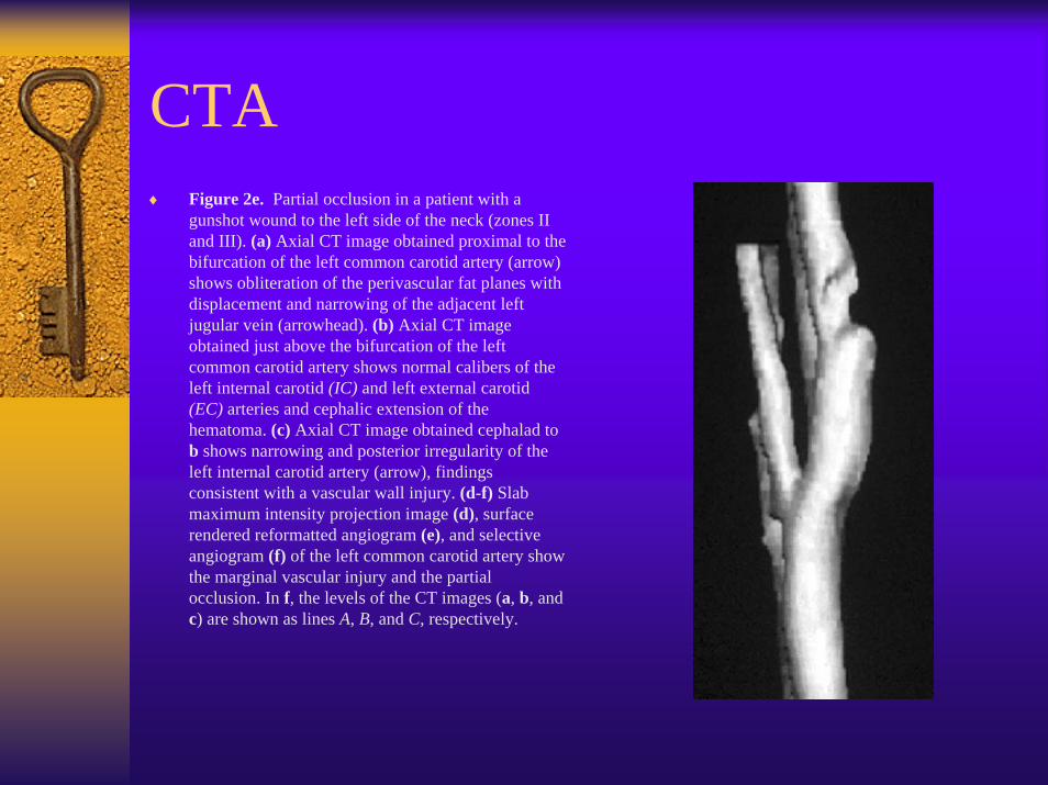
CTA♦ Figure 2e. Partial occlusion in a patient with a
gunshot wound to the left side of the neck (zones II and III). (a) Axial CT image obtained proximal to the bifurcation of the left common carotid artery (arrow) shows obliteration of the perivascular fat planes with displacement and narrowing of the adjacent left jugular vein (arrowhead). (b) Axial CT image obtained just above the bifurcation of the left common carotid artery shows normal calibers of the left internal carotid (IC) and left external carotid (EC) arteries and cephalic extension of the hematoma. (c) Axial CT image obtained cephalad to b shows narrowing and posterior irregularity of the left internal carotid artery (arrow), findings consistent with a vascular wall injury. (d-f) Slab maximum intensity projection image (d), surface rendered reformatted angiogram (e), and selective angiogram (f) of the left common carotid artery show the marginal vascular injury and the partial occlusion. In f, the levels of the CT images (a, b, and c) are shown as lines A, B, and C, respectively.
CTA
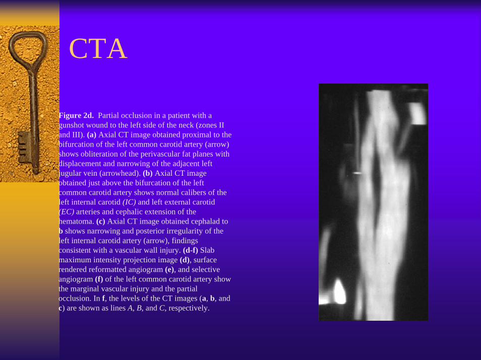
♦ Figure 2d. Partial occlusion in a patient with a gunshot wound to the left side of the neck (zones II and III). (a) Axial CT image obtained proximal to the bifurcation of the left common carotid artery (arrow) shows obliteration of the perivascular fat planes with displacement and narrowing of the adjacent left jugular vein (arrowhead). (b) Axial CT image obtained just above the bifurcation of the left common carotid artery shows normal calibers of the left internal carotid (IC) and left external carotid (EC) arteries and cephalic extension of the hematoma. (c) Axial CT image obtained cephalad to b shows narrowing and posterior irregularity of the left internal carotid artery (arrow), findings consistent with a vascular wall injury. (d-f) Slab maximum intensity projection image (d), surface rendered reformatted angiogram (e), and selective angiogram (f) of the left common carotid artery show the marginal vascular injury and the partial occlusion. In f, the levels of the CT images (a, b, and c) are shown as lines A, B, and C, respectively.
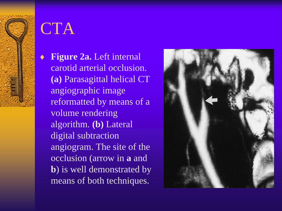
CTA♦ Figure 2a. Left internal
carotid arterial occlusion. (a) Parasagittal helical CT angiographic image reformatted by means of a volume rendering algorithm. (b) Lateral digital subtraction angiogram. The site of the occlusion (arrow in a and b) is well demonstrated by means of both techniques.

CTA♦ Figure 2c. Partial occlusion in a patient with a
gunshot wound to the left side of the neck (zones II and III). (a) Axial CT image obtained proximal to the bifurcation of the left common carotid artery (arrow) shows obliteration of the perivascular fat planes with displacement and narrowing of the adjacent left jugular vein (arrowhead). (b) Axial CT image obtained just above the bifurcation of the left common carotid artery shows normal calibers of the left internal carotid (IC) and left external carotid (EC) arteries and cephalic extension of the hematoma. (c) Axial CT image obtained cephalad to b shows narrowing and posterior irregularity of the left internal carotid artery (arrow), findings consistent with a vascular wall injury. (d-f) Slab maximum intensity projection image (d), surface rendered reformatted angiogram (e), and selective angiogram (f) of the left common carotid artery show the marginal vascular injury and the partial occlusion. In f, the levels of the CT images (a, b, and c) are shown as lines A, B, and C, respectively.
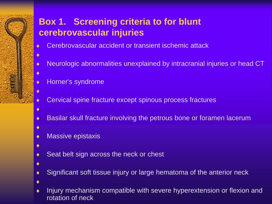
Box 1. Screening criteria to for blunt cerebrovascular injuries♦ Cerebrovascular accident or transient ischemic attack♦♦ Neurologic abnormalities unexplained by intracranial injuries or head CT♦♦ Horner's syndrome♦♦ Cervical spine fracture except spinous process fractures♦♦ Basilar skull fracture involving the petrous bone or foramen lacerum♦♦ Massive epistaxis♦♦ Seat belt sign across the neck or chest♦♦ Significant soft tissue injury or large hematoma of the anterior neck♦♦ Injury mechanism compatible with severe hyperextension or flexion and
rotation of neck