Blood Supply Of Eye and Optic Nerve
37
BLOOD AND NERVE SUPPLY OF EYE AND OPTIC NERVE Presenter – Dr. Om Patel Moderator – Dr. Rashi Shar
-
Upload
om-patel -
Category
Health & Medicine
-
view
115 -
download
1
Transcript of Blood Supply Of Eye and Optic Nerve

BLOOD AND NERVE SUPPLY OF EYE AND OPTIC NERVE
Presenter – Dr. Om PatelModerator – Dr. Rashi Sharma

Arterial supply• Internal carotid artery• External carotid artery

Arterial supply
• All structures are supplied by branches of Internal Carotid Artery
• Except eyelids and conjunctiva which receives blood supply from the branches of both internal and external carotid artery

INTE
RNAL
CAR
OTI
D CERVICAL
PETROUS
CAVERNOUS
CEREBRAL
OPHTHALMIC


Post.Ciliary(supply the choroid and parts of the optic nerve)
Internal carotid a.
Supratrochlear a. &Dorsal Nasal a.
Ant. ethmoidal a.
Post. ethmoidal a.
Ophthalmic a.
Lacrimal a.Muscular Branches.(Ant.ciliary arteries are derived from it)
Central Retinal Art.
Branches of ophthalmic artery:

Central retinal artery
• First branch from the ophthalmic artery
• End arteries
• Divides into equal superior & inferior branches, then another division (nasal & temporal)
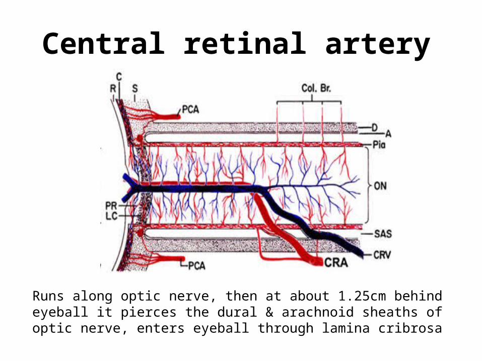
Central retinal artery
Runs along optic nerve, then at about 1.25cm behind eyeball it pierces the dural & arachnoid sheaths of optic nerve, enters eyeball through lamina cribrosa

9
Central retinal artery
Meningeal Branches Central Collateral Branches
Pial Sheath Macular Nerve Fibres

Two Ciliary arteries (on each side of the optic nerve)
• Divide into 2 long posterior ciliary arteries and ~20 short posterior ciliary arteries
• The short posterior ciliary arteries directly supply the choroid and take part in formation of Circle of Zinn
• The long posterior ciliary arteries travel in the suprachoroidal space anteriorly then supply the choroid anteriorly via recurrent branches
Ciliary arteries

Circle Of Zinn
• Circular anastomosis between short posterior ciliary arteries when they are piercing the sclera
• Gives branches to choriod, optic nerve and pial network
• Cilioretinal artery – helps to maintain vision in case of CRA occlusion

Anterior Ciliary arteries– 7 arteries 2 for each rectus
muscle except LR muscle
– Takes part in formation of Major arterial circle along with long posterior ciliary artery
– Supplies ciliary body and iris

Eyelids• Arterial blood supply:– Lateral Palpebral artery
(from lacrimal artery) – Medial Palpebral artery
( from dorsal nasal artery)
• Marginal and peripheral palpebral arcades

Venous drainage:
• Medially: to angular vein
• Laterally: to superficial temporal vein

Nerve supply:Sensory Supply• Lower lid:– infra-orbital (from V2)
– Medial Aspect infra-trochlear nerve (V1)
• Upper lid: – Supra-orbital nerve– Supra-trochlear nerve– Lacrimal nerve (v1)

Nerve Supply - Eyelids
• Arrangement of sensory nerves– Submuscular plane – Site of injection for anesthesia
• Motor Supply– Facial Nerve – Orbicularis muscle– Oculomotor nerve – LPS

Conjunctiva
• Palpebral conjunctiva marginal and peripheral arcades (from medial and lateral palpebral artery)
• Bulbar conjunctiva anterior and posterior conjuctival arteries

Conjunctiva
• Venous drainage: – superior and inferior ophthalmic vein
• Nerve supply: – Long ciliary nerve (nasociliary branch of ophthalmic nerve)

Blood supply of the ACLong posterior ciliary artery anastomose with anterior ciliary artery– Major arterial arcade (ciliary stroma)– Minor arterial arcade (At the collarete of the iris)
• They are the major blood supply to the iris and ciliary body
• Venous drainage: minor venous circle directly into the vortex veins (not into the corresponding major circle)


Macula RegionSupplied by Superior & Inferior temporal branches of central retinal artery**In 20% population Cilioretinal Artery supplies macula.(in case of CRA occlusion it helps to retain vision)
• Outer-plexiform layer • CRA + partially by choriocapillaries
• Inner nuclear layer, Inner Plexiform Layer, Ganglion cell layer, Nerve Fibre Layer, Internal Limiting Membrane
• Supplied by Central Retinal Artery
• RPE, Rodes and Cones ,External Limiting Membrane, Outer Nuclear Layer
• Supplied by choriocapillaries
Retina

Venous Drainage Of Eye
• No valves
• Tortuous & freely anastomose with one another

Venous Channels
• Superior ophthalmic vein• Inferior ophthalmic vein• Middle ophthalmic vein• Medial ophthalmic vein• Angular Vein• Cavernous Sinus

Venous drainage of eye

Superior ophthalmic vein
• Formed by union of supraorbital & angular veins
• Communicates with Central Retinal Vein, receives Inferior Ophthalmic Vein & 2 vorticose veins from the upper part of the eyeball
• Leaves the orbit through superior orbital fissure to join the cavernous sinus

Inferior Ophthalmic Vein
• Arises from venous plexus in orbital floor
• Communicates with pterygoid venous plexus
• Receives muscular branches & 2 inferior vorticose vein
• Joins superior ophthalmic vein or drains directly into the cavernous sinus

Angular Vein
• Formed by supratrochlear and supraorbital veins
• Runs down the side of nose about 8 mm from the medial canthus
• Important landmark for lacrimal sac surgery
• Continues as facial vein

Blood supply of Optic Nerve:A. Intraocular part/ optic nerve head
• Cilioretinal artery• Peripapillary choroidal vessels• Vessels from zinn and heller
Surface nerve fiber layer and
Prelaminar part
• Short Posterior Ciliary ArteriesLamina cribrosa region
• Central retinal artery• Pial vesselsRetrolaminar

Periaxial System Axial System
Derived From branches of ICA Derived from branches of Central Retinal Artery
Ophthalmic Artery Cental Retinal Artery
Long posterior ciliary artery Central collateral branches
Short posterior ciliary artery Intraneural branch
Lacrimal Artery
Central Retinal Artery
B. Intraorbital part :

• The anterior portion of the nerve derives its blood supply from the posterior ciliary arteries (PCA) and the choroid (C)
• The posterior optic nerve derives its blood supply from penetrating pial arteries (Col br) and branches of the central retinal artery (CRA)

C .Intracanalicular part : Periaxial system of vessels
D . Intracranial part : Pial system of vessels

Venous drainage :
Optic nerve head
• Central retinal vein
Orbital part
• Peripheral pial plexus
• Central retinal vein
Intracranial part
• Pial plexus which ends in anterior cerebral & basal vein

NERVE SUPPLY• Sensory N/S – *Trigeminal nerve (Mixed nerve) V *Ophthalmic (V1) & Maxillary(V2) divison play main role of eye sensation
• Motor N/S -- *Cranial nerve 3rd ,4th,6th for ocular movements *Cranial nerve 7th for eyelid closure
• Sympathetic N/s --* Nasociliary branch of ophthalmic nerve
• Parasympathetic N/S:-- * Occulomotor (3rd)

TRIGEMINAL NERVE
OPHTHALMIC MAXILLARY MANDIBULAR

•SUPRA-ORBITAL
•SUPRA-TROCHLEAR
•Ant.Ethmoidal –----- Supplies Ethmoidal Sinus •Post.Ethmoidal ------ Supplies Sphenoidal Sinus
•Infratrochlear -------- Medial Eyelid
•Long Ciliary Nerves---Pierces Sclera & Supply Cornea, Iris, Ciliary Body
•Sympathetic Fibres---Dilate Pupil
Supply Forehead + Scalp + Conjunctiva + upper & central eyelid
Supplies to lateral eyelid + lacrimal gland + conjunctiva
OPHTHALMIC
Frontal Nerve
Lacrimal Nerve
Nasociliary Nerve

The nerves of the orbit that enter through the superior orbital fissure and supply the ocular muscles :
• Oculomotor (CN III)
• Trochlear (CN IV)
• Abducent (CN VI)

Thank You