Blood SBI 3U Ms. Raper. Function of Blood Transport oxygen - oxyhemoglobin Transport nutrients: -...
52
Blood SBI 3U Ms. Raper
-
Upload
lora-ami-george -
Category
Documents
-
view
230 -
download
0
Transcript of Blood SBI 3U Ms. Raper. Function of Blood Transport oxygen - oxyhemoglobin Transport nutrients: -...

Blood
SBI 3U
Ms. Raper

Function of Blood
• Transport oxygen - oxyhemoglobin• Transport nutrients: - glucose, amino acids,
• Transport wastes – CO2 , urea, water
• Transport hormones – adrenalin, sex hormones etc.
• Transport heat – • Clotting during injury• Provide immune response: - white blood cells


Plasma
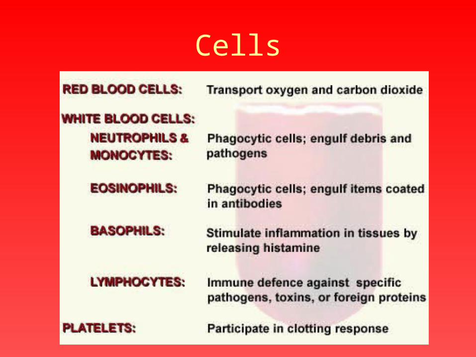
Cells
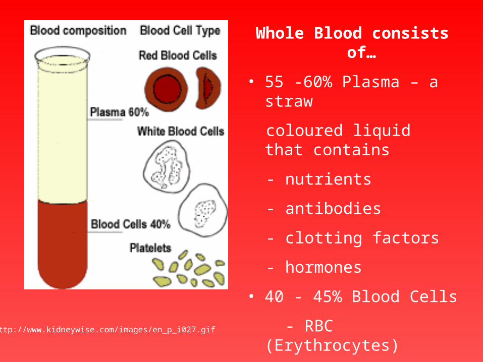
Whole Blood consists of…
• 55 -60% Plasma – a straw
coloured liquid that contains
- nutrients
- antibodies
- clotting factors
- hormones
• 40 - 45% Blood Cells
- RBC (Erythrocytes)
- WBC (Leucocytes)
- Platelets (Thrombocytes)http://www.kidneywise.com/images/en_p_i027.gif

http://www.cardioliving.com/consumer/Circulatory/Images/Cells.JPG


http://users.rcn.com/jkimball.ma.ultranet/BiologyPages/H/Hematopoiesis2.gif

http://www.24dr.com/reference/pictures/5.jpg
http://www.webshots.com/g/55/294-sh/23689.html

http://www.redcross.org/news/bm/blooddonation/images/kidlearn1.jpg
Red Blood cells …
• Made in the Bone Marrow and
• Destroyed in the Spleen.
• Live for about 120 days
• Are flexible to squeeze through blood capillaries.
• Contain Hemoglobin
• Are Bi-concave discs
• Have no nucleus
http://www.psbc.org/education/hematology/blood/_frm/frm_made.htm

Red Blood cells are BICONCAVE discs.

How do Red blood Cells carry Oxygen?
• Hemoglobin + Oxygen = Oxyhemoglobin
dark purple/red bright red
Oxyhemoglobin is unstable and readily dissociates back into hemoglobin and free oxygen.

Red blood cell diseases• Anaemia – too few red blood cells maybe
because of low Iron.
Normal Low Iron

• Sickle Cell disease – RBC are not round but sickle shaped, (genetic mutation for assisting in Malaria prevention) results in blood cells being destroyed prematurely.
Malaria Parasite – Plasmodium inside a RBC http://www.sicklecellfoundationofalberta.org/sic3.jpg
http://www-cxro.lbl.gov/microscopy/ALS_Abstracts_97/IMG00012.GIF

Liver or Kidney disease causes RBC to be damaged or destroyed
Your normal RED BLOOD CELL COUNT or Hb is between 12 and 14,(some hospitals measure this as 120 to 140, both are correct, just different units used).

White Blood Cells….
• are made in the bone marrow
• are responsible for “fighting” disease.
• are various types
• contain a nucleus
Together they make up the total white blood count - normally 4 to 10.(Which is actually 4,000 to 10,000 white blood cells per cubic millimetre of blood!!!)

Neutrophil Monocyte Lymphocyte Eosinophil Basophil
http://www.psbc.org/education/hematology/blood/_frm/frm_made.htm
http://hsc.unm.edu/som/biomedcom/Photography/PIX/blood%20cells-b-7x10jpg.JPG

Normal blood smear (right), compared to Infection (left)
http://www.psbc.org/education/hematology/blood/_frm/frm_blood.htm

http://www.iranblood.org/slide/slid1.16.jpg
Infectious Mononucleosis (“Mono”) shows Atypical Lymphocytes.
http://www.wadsworth.org/chemheme/heme/microscope/pix/atyplymph_nw.jpg

http://www.pathguy.com/lectures/mono.gif

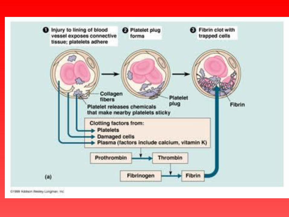
Platelets… are made in the bone marrow
• Concerned with blood clotting.
•Contain an enzyme that is released when the platelets are damaged. This enzyme triggers clotting
• Circulate in the blood for about 10 days then die.http://www.sirinet.net/~jgjohnso/circulation.html
Your normal PLATELET COUNT is between 150 and 400(Which is actually 150,000 to 400,000 per cubic millimetre of blood!!!)

Platelets form part of the network of a clot.
http://chapters.redcross.org/tn/nashvilleblood/Platelets.jpg


Blood Groups
“In 1901 Karl Landsteiner demonstrated the existence of blood group antigens on human red blood cells as well as antibodies directed against those antigens in human sera.” (http://ntri.tamuk.edu/immunology/blood.html)

Vienna, Nov 20th, 1890“In the last few days several patients with more or less serious wounds were taken to hospital. One patients had suffered an open fracture during an accident, another one had internal injuries and a third one had suffered a knife wound during a quarrel. Even though these patients had completely different injuries, all of them had suffered great losses of blood so that they needed a blood transfusion. Some patients recovered from their injuries quickly whereas the condition of other patients turned from bad to worse and some of them even died within a short time. When they were examined to find out the reason for their sudden death it was found that their red blood cells had clumped together i. e. formed sort of blood clots in the blood vessels.”

Landsteiner's experimentBloodRed blood cells
Serum StörkPletschnig
Sturli Erdheim ZaritschLandsteiner
Störk
Pletschnig
Sturli
Erdheim
Zaritsch
Landsteiner
clumping no clumping
http://www.ginkgo-web.de/bilingual/blgroups.html

GenotypeBlood group phenotype
Antigens on erythrocytes
Serum antibodies
AA or AO A A Anti-B
BB or BO B B Anti-A
AB AB A and B None
OO O None Anti-A and Anti-B
http://ntri.tamuk.edu/immunology/blood.html

http://www.sirinet.net/~jgjohnso/bloodtype.jpg

Fig 2. ABO Tile Grouping
anti-A anti-B anti-A+B
group A
group B
group AB
group O
http://www.umds.ac.uk/tissue/bludgrp2.html#Abbs
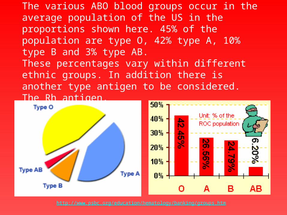
The various ABO blood groups occur in the average population of the US in the proportions shown here. 45% of the population are type O, 42% type A, 10% type B and 3% type AB. These percentages vary within different ethnic groups. In addition there is another type antigen to be considered. The Rh antigen.
http://www.psbc.org/education/hematology/banking/groups.htm

http://www.transweb.org/journey/recip_journey/kidney/kidney_blue/kb_14.htm

Remember! Every cell has
surface proteins that helps the
body identify it
www.nlm.nih.gov/.../ency/images/ ency/fullsize/9125.jpg
Rh Factor
• Another key substance in the blood is the Rh or Rhesus factor (named after the monkeys in which it was located)
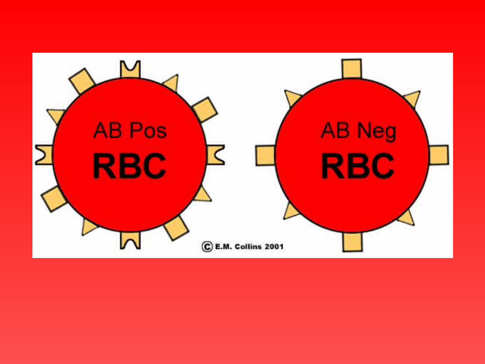
• People either have the factor and are then Rh + (positive)
• or you don’t have the factor - Rh - (negative)
• Rh is a dominant trait.


Rh + Phenotype Rhesus +
Rh + Genotype either RR or Rr
Rh - Phenotype & Genotype - rr

According to the blood grouping systems, you can belong to either of
following 8 blood groups:
A Rh+ B Rh+ AB Rh+ 0 Rh+
A Rh- B Rh- AB Rh- 0 Rh-
This means that there are 8 possible ABO Blood groups.

Rh. Problems in Pregnancy. Rh + Man and Rh - Woman
Because Rh is a dominant allele there is a 50:50 chance the man
will have the Genotype Rr or RR If he is RR then his children will
inherit the dominant allele and be Rh + this will cause the woman to
develop antibodies to her unborn babies blood and try to destroy it.

A red blood cell (RBC) with three different antigens
on the surface of its membrane.
The antigens are glycoproteins with unique
molecular shapes.


Agglutination


http://gslc.genetics.utah.edu/units/basics/blood/images/ABObloodsystem.gif

Distribution of the B type blood allele in native populations of the world

Distribution of the A type blood allele in native populations of the world

Distribution of the O type blood in native populations of the world
http://anthro.palomar.edu/vary/vary_3.htm

http://waynesword.palomar.edu/bio100.htm
Also….


![Blood Circulation and Transport Module[1]](https://static.fdocuments.net/doc/165x107/547fe879b4af9fc3158b5bd5/blood-circulation-and-transport-module1.jpg)