Blood - brazosport.edubrazosport.edu/Assets/pdfs/Mickey-Dufilho/Anatomy-and-physiology-2...17.2...
45
© Annie Leibovitz/Contact Press Images Chapter 17 Blood MDufilho 12/28/2015 1
-
Upload
truongkhanh -
Category
Documents
-
view
218 -
download
4
Transcript of Blood - brazosport.edubrazosport.edu/Assets/pdfs/Mickey-Dufilho/Anatomy-and-physiology-2...17.2...

© Annie Leibovitz/Contact Press Images
Chapter 17
Blood
MDufilho 12/28/2015 1

17.2 Composition of Blood
• Blood is the only fluid tissue in body
• Type of connective tissue
– Matrix is nonliving fluid called plasma
– Cells are living blood cells called formed
elements
• Cells are suspended in plasma
• Formed elements
– Erythrocytes (red blood cells, or RBCs)
– Leukocytes (white blood cells, or WBCs)
– Platelets
MDufilho 12/28/2015 2

Blood
12/28/2015 MDufilho
3

Figure 17.1 The major components of whole
blood.
MDufilho
Withdraw blood
and place in tube.
1 2 Centrifuge the
blood sample.
Plasma
• 55% of whole blood
• Least dense component
Buffy coat
• Leukocytes and platelets
• <1% of whole blood
Erythrocytes
• 45% of whole blood (hematocrit)
• Most dense component
Formed
elements
12/28/2015 4

Physical Characteristics and Volume
• Blood is a sticky, opaque fluid with metallic
taste
• Color varies with O2 content -
• pH -
• Makes up ~8% of body weight
• Average volume:
– Males:
– Females:
MDufilho 12/28/2015 5

17.1 Functions of Blood
• Functions include
– Transport
• What?
– Regulation
• Which substances?
– Protection from what?
MDufilho 12/28/2015 6

Figure 17.2b Blood cells.
MDufilho
Platelets Erythrocytes
Neutrophil
Eosinophil
Monocyte Lymphocyte
Photomicrograph of a human blood smear,
Wright’s stain (610) 12/28/2015 7

17.3 Erythrocytes
Structural Characteristics
• Erythrocytes are small-diameter (7.5 m)
cells that contribute to gas transport
• Cell has biconcave disc shape, is anucleate,
and essentially has no organelles
• Filled with hemoglobin (Hb) for gas transport
• RBC diameters are larger than some
capillaries
• Contain plasma membrane protein spectrin
and other proteins
– Spectrin provides flexibility to change shape MDufilho 12/28/2015 8

Figure 17.3 Structure of erythrocytes (red blood cells).
MDufilho
Side view (cut)
Top view
7.5 m
2.5 m
12/28/2015 9

Figure 17.4 Structure of hemoglobin.
MDufilho
Hemoglobin consists of
globin (two alpha and two
beta polypeptide chains)
and four heme groups.
Iron-containing
heme pigment.
Heme
group Globin
chains
Globin
chains
12/28/2015 10
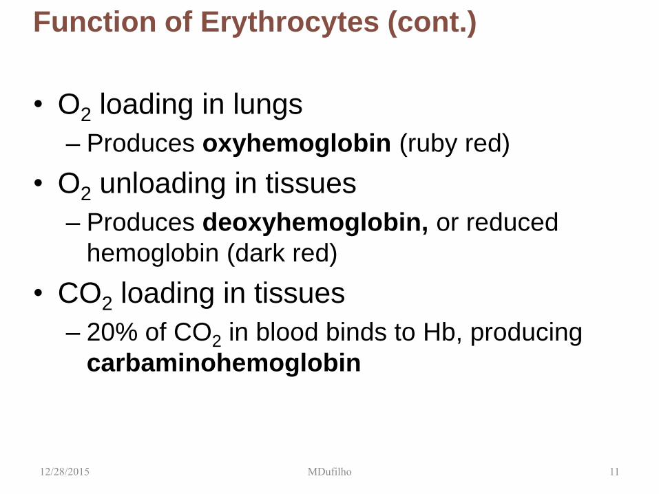
Function of Erythrocytes (cont.)
• O2 loading in lungs
– Produces oxyhemoglobin (ruby red)
• O2 unloading in tissues
– Produces deoxyhemoglobin, or reduced
hemoglobin (dark red)
• CO2 loading in tissues
– 20% of CO2 in blood binds to Hb, producing
carbaminohemoglobin
MDufilho 12/28/2015 11

Sickle-Cell Anemia
• Results from a
defective gene coding
for an abnormal
hemoglobin called
hemoglobin S (HbS)
– HbS has a single
amino acid substitution
in the beta chain
– This defect causes
RBCs to become
sickle-shaped in low
oxygen situations
MDufilho 12/28/2015 12

Homeostatic Imbalance - Sickle-cell Anemia
• Black people of African
malarial belt and
descendants
• Protects against Malaria
• Sickle-cell gene
– Two copies Sickle-
cell anemia
– One copy Sickle-
cell trait; milder
disease; better
chance to survive
malaria
12/28/2015 MDufilho 13
Sickle-cell Anemia: Treatments
• Acute crisis treated with transfusions; inhaled nitric oxide
• Preventing sickling
–Hydroxyurea induces fetal hemoglobin (which does not sickle) formation
–Stem cell transplants
–Gene therapy
–Nitric oxide for vasodilation
12/28/2015 MDufilho 14

Hematopoiesis
• Blood cell formation in
red bone marrow
• In adult, found in axial
skeleton, girdles, and
proximal epiphyses of
humerus and femur
• Hematopoietic stem
cells (Hemocytoblasts)
– Give rise to all formed
elements
12/28/2015 MDufilho 15

Figure 17.5 Erythropoiesis: formation of red blood cells.
12/28/2015 MDufilho 16
Stem cell Committed cell Developmental pathway
Phase 1
Ribosome synthesis
Phase 2
Hemoglobin accumulation
Phase 3
Ejection of nucleus
Hematopoietic stem
cell (hemocytoblast) Proerythroblast Basophilic
erythroblast
Polychromatic
erythroblast
Orthochromatic
erythroblast Reticulocyte Erythrocyte

Regulation and Requirements of
Erythropoiesis
• Too few RBCs lead to tissue hypoxia
• Too many RBCs increase blood viscosity
• > 2 million RBCs are made per second
• Balance between RBC production and
destruction depends on:
– Hormonal controls
– Adequate supplies of iron, amino acids,
and B vitamins
MDufilho 12/28/2015 17

Figure 17.6 Erythropoietin mechanism for regulating erythropoiesis.
MDufilho
5
1
4
2
3
Homeostasis: Normal blood oxygen levels
Enhanced
erythropoiesis
increases RBC count.
Erythropoietin
stimulates red
bone marrow.
Kidney (and liver to
a smaller extent)
releases
erythropoietin.
O2-carrying
ability of blood
rises.
Hypoxia (inadequate
O2 delivery) due to
• Decreased
RBC count
• Decreased amount
of hemoglobin
• Decreased
availability of O2
Stimulus:
12/28/2015 18

Clinical Applications – Possible causes of
kidney hypoxia
• Insufficient number of RBC
• Reduced oxygenation of blood
• Increased aerobic demands
• Treatment
12/28/2015 MDufilho 19

Figure 17.7 Life
cycle of red
blood cells.
MDufilho
Hemoglobin
Heme Globin
Bilirubin is
picked up
by the liver.
Iron is stored
as ferritin or
hemosiderin.
Iron is bound to transferrin
and released to blood
from liver as needed
for erythropoiesis.
Bilirubin is secreted into
intestine in bile where it is
metabolized to stercobilin
by bacteria.
Circulation
Amino
acids
Stercobilin
is excreted
in feces.
Food nutrients
(amino acids, Fe,
B12, and folic acid)
are absorbed from
intestine and enter
blood.
5
4
3
2
1
6
Low O2 levels in blood stimulate
kidneys to produce erythropoietin.
Erythropoietin levels rise in blood.
Erythropoietin and necessary raw
materials in blood promote
erythropoiesis in red bone marrow.
New erythrocytes
enter bloodstream;
function about 120
days.
Aged and damaged
red blood cells are engulfed
by macrophages of spleen,
liver, and bone marrow; the
hemoglobin is broken down.
Raw materials are
made available in blood
for erythrocyte synthesis.
12/28/2015 20

Disorders that produce jaundice
• Liver malfunction – cirrhosis and hepatitis
• Gallstones
• Erythroblastosis fetalis
• Newborn jaundice 12/28/2015 MDufilho 21

Erythrocyte Disorders
• Most erythrocyte disorders are classified
as either anemia or polycythemia
• Anemia
– Blood has abnormally low O2-carrying
capacity that is too low to support normal
metabolism
– Sign of problem rather than disease itself
– Symptoms: fatigue, pallor, dyspnea, and chills
– Three groups based on cause
• Blood loss
• Not enough RBCs produced
• Too many RBCs being destroyed
MDufilho 12/28/2015 22

Anemias - OYO
• Hemorrhagic anemia
• Iron-deficiency anemia
• Pernicious anemia
• Renal anemia
• Aplastic anemia
• Hemolytic anemia
• Sickle Cell anemia
• Thalasemia
12/28/2015 MDufilho 23

Erythrocyte Disorders (cont.)
• Polycythemia
– Abnormal excess of RBCs; increases blood
viscosity, causing sluggish blood flow
– Polycythemia vera: Bone marrow cancer
leading to excess RBCs
• Hematocrit may go as high as 80%
• Treatment: therapeutic phlebotomy
– Secondary polycythemia: caused by low O2
levels (example: high altitude) or increased
EPO production
MDufilho 12/28/2015 24

17.6 Hemostasis
• Hemostasis: fast series of reactions for
stoppage of bleeding
• Requires clotting factors and substances
released by platelets and injured tissues
• Three steps involved
Step 1: Vascular spasm
Step 2: Platelet plug formation
Step 3: Coagulation (blood clotting)
MDufilho 12/28/2015 25

Figure 17.13 Events of hemostasis.
MDufilho
1
2
3
Collagen
fibers
Platelets
Fibrin
• Platelets release chemicals
that make nearby platelets
sticky; platelet plug forms.
• Injury to lining of vessel
exposes collagen fibers;
platelets adhere.
• Fibrin forms a mesh that
traps red blood cells and
platelets, forming the clot.
Coagulation
Vascular spasm
Platelet plug
formation
• Smooth muscle contracts,
causing vasoconstriction.
Slide 1
12/28/2015 26

Step 3: Coagulation (cont.)
• Phase 1: Two pathways to prothrombin
activator
– Initiated by either intrinsic or extrinsic
pathway (usually both)
• Triggered by tissue-damaging events
• Involves a series of procoagulants
• Each pathway cascades toward and ends with the
activation of factor X
– Factor X then complexes with Ca2+, PF3
(platelet factor 3), and factor V to form
prothrombin activator
MDufilho 12/28/2015 27

Figure 17.14-1 The intrinsic and extrinsic pathways of blood clotting (coagulation).
MDufilho
Xa X
Vessel endothelium
ruptures, exposing
underlying tissues
(e.g., collagen)
Tissue cell trauma
exposes blood to
Extrinsic pathway Intrinsic pathway
Platelets cling and their
surfaces provide sites for
mobilization of factors
Tissue factor (TF)
Phospholipid
surfaces of
aggregated
platelets
complex complex
Phospholipid
surface Prothrombin
activator
Phase 1
XII
XI
IX
XIIa
XIa
IXa
VIII
VIIIa
IXa/VIIIa
VIIa
VII
Ca2+
Ca2+
Ca2+
Va V
TF/VIIa
Prothrombin activator
consists of factors Xa,
Va, Ca2+, and
phospholipid surface. 12/28/2015 28

Step 3: Coagulation (cont.)
• Phase 2: Pathway to thrombin
– Prothrombin activator catalyzes
transformation
of prothrombin to active enzyme thrombin
MDufilho 12/28/2015 29

Figure 17.14-2 The intrinsic and extrinsic pathways of blood clotting (coagulation).
MDufilho
Phase 2
Phase 3
Prothrombin (II)
Thrombin (IIa)
Fibrinogen (I)
(soluble) Fibrin
(insoluble polymer)
Cross-linked fibrin mesh
XIIIa
XIII
Ca2+
12/28/2015 30

Step 3: Coagulation (cont.)
• Phase 3: Common pathway to the fibrin
mesh
– Thrombin converts soluble fibrinogen to fibrin
– Fibrin strands form structural basis of clot
– Fibrin causes plasma to become a gel-like trap
catching formed elements
– Thrombin (along with Ca2+) activates factor XIII
(fibrin stabilizing factor), which:
• Cross-links fibrin
• Strengthens and stabilizes clot
– Anticoagulants: factors that normally dominate
in blood to inhibit coagulation MDufilho 12/28/2015 31

Figure 17.14-2 The intrinsic and extrinsic pathways of blood clotting
(coagulation).
MDufilho
Phase 2
Phase 3
Prothrombin (II)
Thrombin (IIa)
Fibrinogen (I)
(soluble) Fibrin
(insoluble polymer)
Cross-linked fibrin mesh
XIIIa
XIII
Ca2+
12/28/2015 32

Figure 17.14 The intrinsic and extrinsic pathways of blood clotting (coagulation).
MDufilho
Phase 1
Phase 2
Phase 3
Platelets cling and their
surfaces provide sites for
mobilization of factors
Phospholipid
surfaces of
aggregated
platelets
Phospholipid
surface Prothrombin
activator
Prothrombin (II)
Thrombin (IIa)
Fibrinogen (I)
(soluble) Fibrin
(insoluble
polymer)
Cross-linked
fibrin mesh
XIIIa
XIII
Ca2+
Prothrombin activator
consists of factors Xa,
Va, Ca2+, and
phospholipid surface.
Ca2+
Ca2+
complex
Vessel endothelium
ruptures, exposing
underlying tissues
(e.g., collagen)
Tissue cell trauma
exposes blood to
Extrinsic pathway Intrinsic pathway
Ca2+
VII
VIIa
XII
XIIa
XI
IX
XIa
IXa
VIII
VIIIa
IXa/VIIIa complex
Tissue factor (TF)
TF/VIIa
Va V
X Xa
12/28/2015 33

Figure 17.15 Scanning electron micrograph of erythrocytes trapped in a fibrin mesh.
MDufilho 12/28/2015 34

Clot Retraction and Fibrinolysis
• Clot must be stabilized and removed when
damage has been repaired
• Clot retraction
– Actin and myosin in platelets contract within
30–60 minutes
– Contraction pulls on fibrin strands, squeezing
serum from clot
• Serum is plasma minus the clotting proteins
– Draws ruptured blood vessel edges together
MDufilho 12/28/2015 35

Clot Retraction and Fibrinolysis (cont.)
• Vessel is healing even as clot retraction
occurs
• Platelet-derived growth factor (PDGF) is
released by platelets
– Stimulates division of smooth muscle cells
and fibroblasts to rebuild blood vessel wall
• Vascular endothelial growth factor (VEGF)
stimulates endothelial cells to multiply and
restore endothelial lining
MDufilho 12/28/2015 36
Clot Retraction and Fibrinolysis (cont.)
• Fibrinolysis
– Process whereby clots are removed after
repair is completed
– Begins within 2 days and continues for
several days until clot is dissolved
– Plasminogen, plasma protein that is trapped
in clot, is converted to plasmin, a fibrin-
digesting enzyme
• Tissue plasminogen activator (tPA), factor XII,
and thrombin all play a role in conversion process
MDufilho 12/28/2015 37

Clinical Application – Thrombolytic Agents
• Thrombolytic Agents – Clot busting drugs
– tPA – alteplase, streptokinase, urokinase
• Given for ischemic strokes and myocardial
infarctions up to 4.5 hours after onset of event.
12/28/2015 MDufilho 38

Factors Limiting Clot Growth or Formation
• Two mechanisms limit clot size
– Swift removal and dilution of clotting factors
– Inhibition of activated clotting factors
• Thrombin bound onto fibrin threads
• Antithrombin III inactivates unbound
thrombin
• Heparin in basophil and mast cells inhibits
thrombin by enhancing antithrombin III
12/28/2015 MDufilho 39

Factors Preventing Undesirable Clotting
• Platelet adhesion is prevented by
– Smooth endothelium of blood vessels
prevents platelets from clinging
– Antithrombic substances nitric oxide and
prostacyclin secreted by endothelial cells
– Vitamin E quinone acts as potent
anticoagulant
12/28/2015 MDufilho 40

Prevention of Undesirable Clots
• Substances used to prevent undesirable clots include:
– TPA – given in stoke and heart attack
– Heparin – an anticoagulant used clinically for pre- and postoperative cardiac care
– Warfarin (Coumadin)– used for those prone to atrial fibrillation
– Aspirin – an antiprostaglandin that inhibits thromboxane A2 – How does this reduce odds of heart attack and stroke?????
– Dabigatran
12/28/2015 MDufilho 41

Disorders of Hemostasis
• Two major types of disorders
– Thromboembolic disorders: result in
undesirable clot formation
– Bleeding disorders: abnormalities that
prevent normal clot formation
• Disseminated intravascular coagulation
(DIC)
– Involves both types of disorders
MDufilho 12/28/2015 42

Disorders of Hemostasis (cont.)
• Thromboembolic conditions
– Thrombi and emboli
• Thrombus:
• Embolus:
• Embolism:
• Risk factors: atherosclerosis, inflammation, slowly
flowing blood or blood stasis from immobility
• How can these be prevented?
MDufilho 12/28/2015 43

Thrombus vs. Embolus
12/28/2015 MDufilho 44
Bleeding Disorders - OYO
• Thrombocytopenia
• Vitamin K Deficiency
• Cirrhosis
• Hepatitis
• Hemophilia
12/28/2015 MDufilho 45