Blood Bank 4
20
The Os ler I nstitute P}Chaffin (2/11/2013) Blood Bank IV page 1 Blood Bank IV D. Joe Chaffin, MD Cedars-Sinai Medical Center, Los Angeles, CA Transfusion Reactions A. Scope of the problem 1. Transfusions still harm, despite great reductions in transfusion-transmitt ed diseases Figure 1: FDA Transfusion Fatalities FY 2007-2011 (Source: http://www.fda.gov) B. Suspected reaction workup 1. When? a. Indicated when possible react ion is suspected by a combination of signs/symptoms b. Just a few: 1) Inflammatory: a) Fever/chills, s kin changes, infusion sit e pain 2) Circulatory: a) BP changes, s hock, hemoglobinemia/ur ia 3) Pulmonary: a) Dyspnea, orthopnea, wheezing, full failure 4) Coagulation: a) Unexplained increa se in bleeding, DI C 5) Psychological: a) Sense of unease or impending “doom”! 2. General philosophy (my opinions) : a. Opinion 1: Assume all suspected react ions are hemolytic, and work to disprove your assumption b. Opinion 2: Anyone involved in a transfusion should be allowed to initiate a transfusion reaction workup 3. STEP ONE: STOP THE TRANSFUSION! a. Don’t disconnect the unit (though that will eventually happen); at least stop the incoming flow of blood. b. Main indicator of survival of an acute HTR: amount of incompatible blood infused
-
Upload
moonfire2009 -
Category
Documents
-
view
238 -
download
0
Transcript of Blood Bank 4

8/11/2019 Blood Bank 4
http://slidepdf.com/reader/full/blood-bank-4 1/20
The Osler I nstitu te
P}Chaffin (2/11/2013) Blood Bank IV page 1
Blood Bank IV D. Joe Chaffin, MD
Cedars-Sinai Medical Center, Los Angeles, CA
Transfusion Reactions
A. Scope of the problem1. Transfusions still harm, despite great reductions in
transfusion-transmitted diseases
Figure 1: FDA Transfusion Fatalities FY 2007-2011
(Source: http://www.fda.gov)
B. Suspected reaction workup1. When?
a. Indicated when possible reaction is suspected by a
combination of signs/symptoms
b. Just a few:
1) Inflammatory:a) Fever/chills, skin changes, infusion site pain
2) Circulatory:
a) BP changes, shock, hemoglobinemia/uria3) Pulmonary:
a) Dyspnea, orthopnea, wheezing, full failure
4) Coagulation:a) Unexplained increase in bleeding, DIC
5) Psychological:
a) Sense of unease or impending “doom”!
2. General philosophy (my opinions):
a. Opinion 1: Assume all suspected reactions arehemolytic, and work to disprove your assumption
b. Opinion 2: Anyone involved in a transfusion should beallowed to initiate a transfusion reaction workup
3. STEP ONE: STOP THE TRANSFUSION!
a. Don’t disconnect the unit (though that will eventually
happen); at least stop the incoming flow of blood. b. Main indicator of survival of an acute HTR:
amount of incompatible blood infused

8/11/2019 Blood Bank 4
http://slidepdf.com/reader/full/blood-bank-4 2/20
Pathology Review Course
page 2 Blood Bank IV P}Chaffin (2/11/2013)
c. Leave the line open with saline.
Figure 2: Transfusion Reaction Workup
4. Necessary parts of workup (things everyone should do):
a. Clerical check 1) Bedside paperwork/bag check
2) Blood bank paperwork/computer check3) Includes basic inspection of the unit for
discoloration or obvious issues
a) Darkened color: Susp. of bacterial contamination b) Clots, aggregates, or anything out of the ordinary
b. Visible hemoglobinemia check
1) Spin a post-transfusion EDTA sample and examine
visually for a pink-red color change2) Compare to pretransfusion sample if abnormal.
3) Detects > 2.5 to 5 ml of hemolysis occurringanywhere in the body
4) Most sensitive way to detect intravascular
hemolysis; not specific, though
c. Direct antiglobulin (Coombs) test (DAT)
1) Demonstrates coating of RBCs with antibody and/orcomplement in-vivo (see figure 3 below)
2) Most commonly polyspecific method (IgG + C3d)
3) If positive, compare to pretransfusion DAT4) Positive DAT does not prove an acute HTR
a) Nonspecific positives in hospitalized patients
(20%), autoantibodies, drugs, passiveadministration of other things like RhIG or IVIG
5) Also note that a negative DAT does not disprove an
acute hemolytic reaction
a) Donor RBCs all destroyed gives neg. DAT

8/11/2019 Blood Bank 4
http://slidepdf.com/reader/full/blood-bank-4 3/20
The Osler I nstitu te
P}Chaffin (2/11/2013) Blood Bank IV page 3
Figure 3: Direct Antiglobulin Test (DAT)
Image credit: A Rad 2006
d. Repeat ABO/Rh testing
1) Another check for right patient, right blood
2) Check both pre- and post-reaction specimens5. Other things that may be done (but not required)
a. Repeat antibody screen (on both pre- and post-
transfusion samples b. Repeat crossmatch with pre- and post samples
1) Best done with tube technique including immediate
spin and IAT phase readings +/- 37 C reading
c. Elution if DAT is + to determine specificityd. Haptoglobin
1) Haptoglobin binds to free HGB molecules, cleared
by monocytes and macrophages in the RE system2) Levels decrease sharply in acute intravascular
hemolysis (as well as extravascular)
3) Long turnaround time and acute phase reaction
make for limited usefulness in acute setting.a) If you must use, compare pre- and post levels.
e. Direct and indirect bilirubin
1) Really more useful to confirm, not make diagnosis
2) Both will rise quickly, peak in less than 10 hours,may be normal within 24 hours (if liver is OK)
f. Lactate dehydrogenase (LDH)
1) Abundant in RBCs (especially LD2 and LD1)2) Not specific for intravascular hemolysis
g. Urine hemoglobin
1) Not as sensitive or as fast as hemoglobinemia
2) Hematuria does not equal hemoglobinuria!c. Additional testing for suspected septic reactions:
1) Should be done if suggested by clinical data
a) Temperature greater than 102 F or >2 or 3oF
b) Severe rigors or other clinical findings
2) Both patient and product must be evaluated
a) Patient:
• Blood cultures• Consider culture of all intravenous fluids

8/11/2019 Blood Bank 4
http://slidepdf.com/reader/full/blood-bank-4 4/20
Pathology Review Course
page 4 Blood Bank IV P}Chaffin (2/11/2013)
b) Product:• Gram stain and culture of actual residual
product in the bagd. Additional testing for suspected respiratory reactions
(see details in TRALI/TACO sections):
1) Chest X-ray2) BNP levels
3) ABG4) Donor testing for anti-HLA/HNA antibodies
e. Additional testing for suspected severe allergicreactions:
1) Serum IgA levels (pretransfusion sample!)
2) Consider anti-IgA if serum IgA is non-detectable
C. Classification of reactions
Presenting With Fever
Acute
Acute Hemolytic
Febrile Non-hemolytic
Transfusion-related Sepsis
TRALI
Delayed
Delayed Hemolytic
TA-GVHD
Presenting Without Fever
Acute
Allergic
Hypotensive
Tx-associated Dyspnea
TACO
Delayed
Delayed Serologic
Post-transfusion Purpura
Iron Overload
D. Acute reactions presenting with fever1. Acute hemolytic transfusion reactions (AHTRs)
a. Incidence: 1:76,000 transfusions, (1:1.8 million
transfusions fatal HTR)
b. Clerical errors are most common causec. RBC destruction may be intravascular or extravascular
1) ABO-related, intravascular usually more severe
d. Signs/symptoms
1) Timinga) Severe reactions may occur early in transfusion
(first 15 minutes; see figure 4)
b) Milder reactions may present later, but usually before end of transfusion
2) Specific signs/symptoms:
a) Fever and chills
• Most common presenting symptom (> 80%)

8/11/2019 Blood Bank 4
http://slidepdf.com/reader/full/blood-bank-4 5/20
The Osler I nstitu te
P}Chaffin (2/11/2013) Blood Bank IV page 5
Figure 4
b) Back or infusion site painc) Hypotension/shock
d) Hemoglobinuria (1st indication anesthetized pts)
e) DIC/increased bleeding
f) Sense of “impending doom” e. Lab findings
1) Hemoglobinemia (pink or red serum/plasma); lasts
several hours in those with adequate renal function2) Hemoglobinuria (us clears by the end of one day)
3) Positive DAT (unless all donor cells destroyed);may be “mixed field”
4) Elevated indirect and direct bilirubin5) D-dimers, decreased fibrinogen, etc. (DIC)
6) RBC abnormalities
a) Schistocytes: Intravascular hemolysis
b) Spherocytes: Extravascular hemolysis
f. Pathophysiology1) Intravascular hemolysis due to ABO incompatibility
typifies these reactions
a) ABO antibodies fix complement well and this
leads to rapid RBC lysis
b) Other antibodies (e.g., Kidd) may also fixcomplement and lyse RBCs
c) Less commonly due to incompat. donor plasma
Figure 5: Classical Complement Pathway Image credit: http://www.twiv.tv/classical-complement.jpg
2) Hemolysis leads to a complex array of events:
a) Release of free HGB and HGB-free stroma

8/11/2019 Blood Bank 4
http://slidepdf.com/reader/full/blood-bank-4 6/20
Pathology Review Course
page 6 Blood Bank IV P}Chaffin (2/11/2013)
b) Stimulation of intrinsic coag pathway and bradykinin via Ag-Ab complexes
c) C3a and C5a generation (“anaphylatoxins”)
d) Production of several very important cytokines:
• TNF-, IL-1, IL-6, IL-8
Substance Effect
C3a/C5a Increases: Nitric Oxide (NO), cytokines,histamine, leukotrienes
TNF- Increases: NO, tissue factor expressionDecreases: Thrombomodulin (anticoagulant;assists protein C)
Interleukin-1
(IL-1
)Increases: NODecreases: Thrombomodulin
Free HGB Scavenges NO (local, possible global decrease)
Bradykinin Transient hypotension
RBC Stroma Direct renal tubular damage
Table 1: Substances generated duri ng acute HTRs and their ef fects
3) Net effects on various systems:
a) Inflammatory consequences:
• TNF- , IL-1 , IL-6 strongly promote fever• WBCs activated and stimulated by all
b) Coagulation consequences:
• Direct intrinsic path activation by Ag-Abcomplex interaction with factor XII
• Indirect activation of extrinsic path by TNF- stimulation of tissue factor
c) Circulatory consequences:
• Increased C3a/5a, IL-1 , TNF- stimulateincreased nitric oxide levels (vasodilation)• Bradykinin from Ag-Ab complexes likewise
promotes transient systemic hypotensiond) Renal consequences:
• Sympathetic response leads to renalvasoconstriction
• Free hemoglobin scavenges renal NO, promoting vasoconstriction
• Renal microthrombi decrease renal flow• HBG-free RBC stroma damages renal tubules• Resultant oliguric renal failure in about 1/3 of
confirmed acute HTRse) Respiratory consequences:
• Anaphylatoxins promote histamine release,with resultant wheezing/dyspnea
• Aggressive hydration during resuscitationgives pulmonary edema risk
4) Extravascular hemolysis (e.g., Rh/Kell/Duffy, etc.)is usually but not always less severe due to lack of
systemic complement and cytokine activation

8/11/2019 Blood Bank 4
http://slidepdf.com/reader/full/blood-bank-4 7/20
The Osler I nstitu te
P}Chaffin (2/11/2013) Blood Bank IV page 7
g. Treatment1) Hydration/diuresis critical early components for
hypotension treatment and renal fx preservation
a) Urine output > 1 mL/Kg/hr with saline +/-
furosemide b) Low-dose dopamine use is controversial
2) Consider DIC; (+/-) heparin
3) Consider early exchange transfusion, esp. for high-volume incompatible transfusion
h. Prevention possibilities
1) Training and careful attention to phlebotomy,
labeling, issue, and administration2) Two separate ABO/Rh types before transfusion
3) Advanced methods (RFID, bar codes, etc.)
2. Febrile nonhemolytic transfusion reactions (FNHTRs) a. Historically most frequently reported reaction
1) Now 0.1 to <1% due to leukoreduction
2) Still more common than any reaction in heme-onc
and transplant patients, especially with PLTs b. Unexplained increase in temperature of 1C or 2
oF
1) Don’t require to diagnose or work up rxn
c. Cause: Increased pyrogenic substances (e.g., TNF-,
IL-1 ), mostly from WBCs1) Cytokines produced before transfusion (e.g., PLTs)
• Donor WBCs secrete while in the storage bag.
• Transfusion infuses pre-formed substances2) OR, cytokines made after transfusion (e.g., RBCs)
• Recipient anti-HLA/HNA antibodies attack
donor WBCs, or donor antibodies attack
recipient WBCs• Leads to substance release after transfusion
d. Signs/symptoms
1) Transient fever/chills (+/- rigors?) during or up to 2
hours after transfusion
Figure 6
2) Symptoms usually later in transfusion (esp. PLTs)
3) Chills may be first; fever may be delayed up to one
hour or more after transfusion in up to 10% of cases4) Premedicated or head injury patients may never
have fever

8/11/2019 Blood Bank 4
http://slidepdf.com/reader/full/blood-bank-4 8/20
Pathology Review Course
page 8 Blood Bank IV P}Chaffin (2/11/2013)
e. Differential diagnosis:1) Acute HTR
a) Most AHTRs present with fever/chills alone
b) Impossible to distinguish early AHTR from
FNHTR clinically2) Transfusion-related sepsis
a) Especially sepsis from PLT transfusions (RBC-
related sepsis usually presents earlier) b) Timing may be identical
f. Lab findings
1) None (diagnosis of exclusion)
g Treatment1) Antipyretics (acetaminophen)
2) Meperidine (Demerol) for more severe chills
h. Prevention1) Acetaminophen premedication (doesn’t work well
as a routine prophylactic; use with repeat reactions)
2) Preventing FNH during RBC transfusions
a) Leukocyte reduction works extremely well to prevent vast majority of these reactions
3) Preventing FNH during platelet transfusions
a) Bedside leukoreduction is ineffective;substances are already in the bag!
b) Pre-storage leukoreduction best, but reactions in
~0.1 to 1% of PLT transfusions
3. Transfusion-related sepsis (septic transfusion reaction) a. General statements
1) #1 infectious risk from transfusion
2) As many as 1 in 3000 platelet units are
contaminated (many fewer reactions, however) b. How does it happen?
1) Platelets tend to be contaminated by skin
contaminants from collection process2) RBCs more often contaminated by an organism
growing in the donor’s blood (often asymptomatic)
c. Organisms identified depend on product.1) Red cells
a) Gram-negative rods (endotoxin-makers that like
growing in cold temperatures):
• Y. enterocolit ica (most common hi stori cally)
• E. coli • Enterobacter/Pantoea sp.
• Serratia marcescens and S. liquifaciens • Pseudomonas species
b) Gram-positive organisms (much less commonly)
• Staph Epidermidis, Staph aureus
• Propionibacteria
2) Platelets
a) Vast majority are gram-positive cocci (skin)

8/11/2019 Blood Bank 4
http://slidepdf.com/reader/full/blood-bank-4 9/20
The Osler I nstitu te
P}Chaffin (2/11/2013) Blood Bank IV page 9
b) Gram neg rods less common but more likely tocause fatalities (Serratia, E. coli, and Klebsiella)
3) Plasma products
a) Reports water bath contam. with Pseudomonas
d. Signs/symptoms1) Earlier symptoms seen in more severe reactions and
more often with RBC transfusions (see fig 7)
2) Rapid onset high fever (often greater than 4oF/2C)3) Rigors (true shaking chills with rigidity)
4) Abdominal cramping, nausea/vomiting
5) Hypotension/shock
6) DICe. Differential diagnosis:
1) Acute HTR (always!); Septic reactions usually more
“acute” and dramatic than the typical AHTR 2) Anaphylactic transfusion reaction: Similar
presentation but usually NOT febrile.
3) FNHTR: Milder septic reactions and many PLT
contaminations overlap with FNHTR (many cultureunits as part of FNHTR workup protocol)
4) Sepsis from non-transfusion source (infected lines
and/or fluids, coincidental presentation)
Figure 7
f. Lab findings
1) Discolored RBC product (+/-); contaminated RBCs
may turn DARK or purple 2) May have hemoglobinemia/uria (non-immune)
3) DAT negative (unless coincidental)
4) Gram stain + in only half to 2/3 of proven cases!a) Source of gram stain/culture is very important
b) Avoid culturing or staining a segment
c) Also culture associated IV fluids and consider
that an indwelling IV catheter as a source5) Culture is proof positive (same organism cultured
from unit and recipient; better if from donor too!)
g. Treatment1) Immediate IV antibiotics; treat presumptively with
broad spectrum coverage, then adjust as necessary
2) Pressure/respiratory/general support as needed3) Notify blood collection agencies promptly!

8/11/2019 Blood Bank 4
http://slidepdf.com/reader/full/blood-bank-4 10/20
Pathology Review Course
page 10 Blood Bank IV P}Chaffin (2/11/2013)
h. Prevention1) Careful donor history
2) Proper phlebotomy technique; diversion pouches
(divert contaminated skin plugs), strict attention to
possible site contaminants3) Leukocyte reduction filters may decrease risk
(decrease in Yersinia concentration)
4) Routine detection of platelet contamination required by AABB Standard
a) Culture-based methods (24 hrs post-collection):
• BacT/ALERT system: Microbiology standard blood culture equipment
• Pall enhanced bacterial detection system(eBDS): Detects bacterial use of oxygen
b) Pre-issue methods (shortly before issue)
• VeraxPGD system (LR apheresis PLTs and pooled WBD PLTs); detects bacterial cellwall antigens
• BacTx system (approved only for pooledWBD PLTs)
5) Despite detection methods, false negatives occur,and pathogen reduction may be the ultimate answer
4. Transfusion-related acute lung injury (TRALI)
a. #1 cause of transfusion-related fatality in the US! 1) Incidence varies: 1:1200 to 1:190,000 transfusions
b. Two almost identical standard definitions:
1) NHLBI Working Group and Canadian Consensus
Conference Panela) New acute lung injury < 6 hours post
transfusion; ALI defined:• Hypoxemia with PaO2/FiO2 < 300 mm Hg
(or O2 sat <90%) and bilat CXR infiltrates
b) Lack of other risk factors for pulmonary edema
c) No pre-existing acute lung injury
2) Usually fever, chills, transient hypertension thenhypotension
3) PLTs/plasma transfusions, but also RBCs/WB
Figure 8
c. Clinical differential diagnosis:
1) ARDS: TRALI may look exactly like ARDS, butTRALI usually resolves in 24-48 hours.

8/11/2019 Blood Bank 4
http://slidepdf.com/reader/full/blood-bank-4 11/20
The Osler I nstitu te
P}Chaffin (2/11/2013) Blood Bank IV page 11
2) Transfusion-associated circulatory overload
(TACO): TRALI usually febrile and does not
respond to diuretics
3) Anaphylactic reactions (generally afebrile)
4) Acute pulmonary and myocardial disordersd. Pathophysiology: Two currently accepted pathways
1) The neutrophil is the villain in the pathophysiology!
a) Secretes toxic oxygen free radicals and othersubstances damage endothelial cells, leads to
vascular leakage and TRALI
b) Almost 30% of PMNs are in lungs at all times!
2) Donor antibody pathway for TRALI (Figure 9)
Figure 9: Donor Antibody-mediated TRALI
(Image courtesy of Dr. Chris Silliman)
a) Anti-HLA or anti-neutrophil antibodies from
donor bind to antigens on recipient PMNs b) Antibody-PMN complexes deposit in pulmonary
vasculature, activate PMN bactericidal responsec) Damage to pulmonary endothelial cells lining
capillaries, with resultant leakage of fluids intothe alveolar spaces (pulmonary edema)
d) This mechanism may also occur less commonly
with recipient antibodies against donor WBCse) Problem: Does not explain all TRALI reactions,
nor does it explain why TRALI does NOT occur
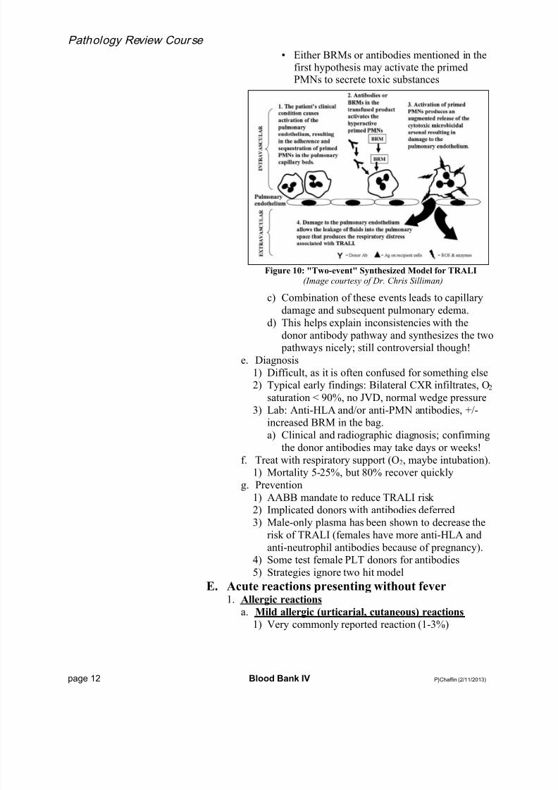
in settings where it SHOULD3) “Two-event” pathway for TRALI (Figure 10)
a) First event: Pre-existing condition, activates lung
endothelial cells and primes PMNs
• Sepsis, major surgery, massive transfusion b) Second event: Transfusion of stored blood
product (+/- antibodies)
• Stored blood products accumulate substancescalled “biologic response modifiers” (BRMs)that can prime/activate destructive PMNs
8/11/2019 Blood Bank 4
http://slidepdf.com/reader/full/blood-bank-4 12/20
Pathology Review Course
page 12 Blood Bank IV P}Chaffin (2/11/2013)
• Either BRMs or antibodies mentioned in thefirst hypothesis may activate the primedPMNs to secrete toxic substances
Figure 10: "Two-event" Synthesized Model for TRALI
(Image courtesy of Dr. Chris Silliman)
c) Combination of these events leads to capillary
damage and subsequent pulmonary edema.
d) This helps explain inconsistencies with the
donor antibody pathway and synthesizes the two pathways nicely; still controversial though!
e. Diagnosis
1) Difficult, as it is often confused for something else2) Typical early findings: Bilateral CXR infiltrates, O2
saturation < 90%, no JVD, normal wedge pressure
3) Lab: Anti-HLA and/or anti-PMN antibodies, +/-increased BRM in the bag.
a) Clinical and radiographic diagnosis; confirming
the donor antibodies may take days or weeks!
f. Treat with respiratory support (O2, maybe intubation).1) Mortality 5-25%, but 80% recover quickly
g. Prevention
1) AABB mandate to reduce TRALI risk2) Implicated donors with antibodies deferred
3) Male-only plasma has been shown to decrease the
risk of TRALI (females have more anti-HLA and
anti-neutrophil antibodies because of pregnancy).4) Some test female PLT donors for antibodies
5) Strategies ignore two hit model
E. Acute reactions presenting without fever1. Allergic reactions
a. Mild allergic (urticarial, cutaneous) reactions
1) Very commonly reported reaction (1-3%)

8/11/2019 Blood Bank 4
http://slidepdf.com/reader/full/blood-bank-4 13/20
The Osler I nstitu te
P}Chaffin (2/11/2013) Blood Bank IV page 13
2) Localized hives, +/- more severe swelling aroundeyes and lips (angioedema), mild respiratory
symptoms, and mild laryngeal edema
3) Mechanism
a) Type I (IgE-mediated) hypersensitivity totransfused plasma proteins
b) Mast cell secretion of histamine and other
mediators of allergic reactions4) Prevention and treatment options
a) Diphenhydramine IV 25-50 mg as treatment,
oral form as prophylaxis (not cost-effective)
b) Washed products work too (not usually done)c) May restart transfusion after hives clear.
b. Moderate allergic reactions
1) Some reactions fall between the two extremes2) May present with upper/lower airway obstruction
+/ – cutaneous manifestations
a) Upper airway:
• Stridor, hoarseness, “lump” in throat b) Lower airway:
• Wheezing, chest tightness, dyspnea3) Some may respond to IV diphenhydramine, while
others will need epinephrine
c. Severe allergic (anaphylactic) reactions 1) Opposite end of hypersensitivity reaction spectrum
2) Uncommon (1:20,000 to 50,000 transfusions)
3) Presentationa) Anaphylaxis very early in the transfusion
b) Acute hypotension, lower airway obstruction,
abdominal distress, systemic crashc) Virtually all of these patients have skin
findings (urticaria, generalized pruritis)
4) What’s the allergen?
a) Classic: IgA deficient recipient with IgE anti-
IgA (induces severe type I reaction)• Seen only in those with undetectable IgA
levels (i.e. IgA < 0.05 mg/dL)• Problem: Labs don’t detect IgE anti-IgA!• Vast minority of patients with anaphylactic-
type reactions have demonstrable IgA
deficiency with detectable anti-IgA b) Haptoglobin deficiency in Asian patients; can
give same type of severe reaction as anti-IgA
c) Latex, drugs, foods in donors can lead to severereactions in susceptible recipients
d) Scattered reports of donors with IgE antibodies
transmitting temporary hypersensitivitye) While all above are possible, very uncommon to
find specific reason for a severe allergic reaction
8/11/2019 Blood Bank 4
http://slidepdf.com/reader/full/blood-bank-4 14/20
Pathology Review Course
page 14 Blood Bank IV P}Chaffin (2/11/2013)
5) Differential diagnosis:a) Acute HTR
• Typically febrile• Most acute HTRs don’t present this early
b) Septic transfusion reaction
• High fevers, no skin findings in sepsis• If unclear, give epinephrine anyway
c) Acute hypotensive reactions• These reactions (see below) have hypotension
only, without respiratory or skin findings6) Confirmation
a) ALL with a severe allergic reaction should have,
at minimum, check of pretransfusion IgA level
b) Those with very low/undetectable levels (<0.05mg/dL) are tested for anti-IgA (detects IgG
antibody but could predict the possibility of IgE)
7) Prevention
a) IgA-deficient (IgAD) products given for those
with antibodies, but sometimes for low IgA only• IgAD: Washed cellular products (RBCs,
PLTs), or products from IgA-deficient donors• If possible, may also bank autologous units
b) Washed cellular products for those with severe
reactions and no demonstrable IgA deficiency
• MOST patients with history of severe allergicreactions can get future untreated transfusionswithout harm (monitor carefully however!)
c) Benadryl insufficient by itself; may use
corticosteroids +/- additional histamine blockers
8) Treatmenta) Epinephrine (0.2-0.5 ml of 1:1000 IM/SQ)
b) SQ or IM preferred, but IV ok if already crashed
2. Acute hypotensive reactions
a. Reactions similar to severe allergic reactions but noskin symptoms, no GI or respiratory issues
1) CDC definition:
a) > 30 mm Hg drop in systolic BP; diastolic < 80
b) Occurs < 15 minutes after start of transfusionc) Resolves < 10 minutes after transfusion stopped
b. Classically associated with two situations
1) Patients taking angiotensin-converting enzymeinhibitors (ACEi) a) Rapid onset of flushing and hypotension in
transfused patients who are on ACEi (e.g.,
Vasotec, Lotensin, Zestril, Capoten). b) Probably accumulation of increased bradykinin
or metabolites during storage
• ACEi prevent bradykinin metabolism
8/11/2019 Blood Bank 4
http://slidepdf.com/reader/full/blood-bank-4 15/20
The Osler I nstitu te
P}Chaffin (2/11/2013) Blood Bank IV page 15
• Marked but transient hypotension (bradykininhalf-life is on the order of seconds
2) Receiving blood via negatively charged filters
a) Seen historically with certain negatively charged
bedside leukoreduction filters
b) Also in LDL apheresis and plasma exchangewith albumin replacement (esp. if on ACEi)
c) Reported with reinfusion of intraoperative blood(cell saver) that is filtered before infusion
c. Diagnosis1) Clearly a diagnosis of exclusion
2) Rule out:
a) Acute HTR by workup and lack of fever
b) Severe allergic reaction by lack of skin andrespiratory findings (as well as transience)
c) Septic reaction by lack of high fever, GI
complaints, and transienced. Management
1) STOP the transfusion! (rapid resolution)2) Give fluids, consider epinephrine if not resolved
e. Prevention
1) No routine prophylactic measures necessary
2) Avoid bedside leukoreduction filters (not really a
problem in most places)3) Stop ACEi before therapeutic apheresis procedures
3. Transfusion-associated dyspnea (TAD)
a. A total garbage can diagnosis, in my view! b. Definition:
1) Acute resp. distress < 24 hours after transfusion
2) TRALI, TACO, and allergy ruled out4. Transfusion-associated circulatory overload (TACO)
a. Acute onset of congestive heart failure as a direct
result of blood transfusion
1) Dyspnea, orthopnea, bilateral rales, with hypoxia
2) Systolic hypertension (widened pulse pressure),tachycardia, JVD, pedal edema, headache
3) Usually afebrile
4) X-rays with bilateral basilar infiltrates, widenedcardiac silhouette
5) Proposed diagnostic criteria include some of the
above signs/symptoms PLUS:a) Hypoxemia (<90% saturation on room air)
b) Bilateral CXR infiltrates
c) Reaction occurring within 6 hours of transfusion
b. Patients most at risk (though any patient may getTACO if transfused rapidly):
1) Patients with pre-existing CHF
2) Very old (>85% occur in patients over age 60) andvery young (to a lesser extent)
8/11/2019 Blood Bank 4
http://slidepdf.com/reader/full/blood-bank-4 16/20
Pathology Review Course
page 16 Blood Bank IV P}Chaffin (2/11/2013)
3) Renal failure4) Chronic anemias due to compensation for anemia
with increased plasma volume
c. Differential diagnosis:
1) TRALI (see below)2) Allergic/anaphylactic reactions
a) May present VERY early in transfusion (first
few drops) b) Not responsive to diuretics or positional changes
3) Coincidental cardiac or pulmonary issues unrelated
to transfusion
a) Acute MI or pulmonary embolism b) Valvular heart disease with decompensation
d. Distinguishing from TRALI
1) Clinical (response to diuretics/positional changes inTACO, fever in TRALI)
2) CXR: Less cardiac silhouette widening in TRALI
3) Lab: Elevated brain natriuretic peptide (BNP)
suggests TACO (some use ratio of pre- to post-transfusion >1.5 AND elevated post-transfusion)
4) Finding antibodies as above establishes TRALI
e. Treatment1) Stop the transfusion, evaluate, sit patient up
2) Give supplemental oxygen
3) Diuretics to decrease blood volume
4) In severe cases, consider therapeutic phlebotomyf. Prevention in at-risk patients
1) Control infusion rates (1 mL/Kg/hour).
2) Split units into aliquots when possible.
3) Consider lower volume units or volume reduction
F. Delayed reactions presenting with fever1. Delayed hemolytic transfusion reactions (DHTRs)
a. Hemolysis occurring at least 24 hours but less than 28
days after transfusion (rare reports up to 6 weeks).
b. Pathophysiologic possibilities1) Anamnestic response:
a) Patient exposed to non-self RBC antigen(s)
b) Antibody formed, but fades over time
c) Patient re-exposed to antigen in futuretransfusion (antibody screen is negative)
d) Anamnestic rapid production of IgG antibodye) Typical for Kidd, Duffy, Kell antibodies
2) Primary responsea) Patient exposed to non-self RBC antigen(s)
b) Antibody is formed quickly, and attacks still-
circulating transfused red cellsc) MUCH less common than anamnestic
c. Classically leads to extravascular hemolysis
1) IgG coats RBCs, removal in liver/spleen

8/11/2019 Blood Bank 4
http://slidepdf.com/reader/full/blood-bank-4 17/20
The Osler I nstitu te
P}Chaffin (2/11/2013) Blood Bank IV page 17
2) Peripheral smear will commonly show spherocytes3) NOTE: DHTRs due to Kidd (Jk) antibodies may be
intravascular and severe (complement fixation)
d. Signs/symptoms
1) Often completely asymptomatic2) Fever and anemia of unknown origin
3) Mild jaundice/scleral icterus may be seen
e. Lab findings1) Icteric serum
2) DAT positive (classically “mixed field”)
3) Anemia
4) Newly identified RBC antibody5) Spherocytes on peripheral smear
6) Elevated LDH and indirect bilirubin, decreased
haptoglobin (even if extravascular)f. Treatment
1) As for AHTR if severe and intravascular
2) Often no treatment necessary
g. DHTR vs. “delayed serologic reaction” (DSTR) 1) Official definition of DSTR:
a) New, clinically significant RBC antibody in a
patient transfused > 24 hrs and < 28 days, AND: b) Complete lack of evidence of hemolysis
2) Consider: Repeat antibody screen on pretransfusion
sample; evaluate bilirubin, haptoglobin, LDH,
peripheral smear, etc.a) Any evidence of hemolysis in study above
changes the diagnosis from DSTR to DHTR
2. Transfusion-associated graft-vs-host disease (TA-
GVHD) a. Results from an attack on recipient cells by viable T-
lymphocytes in a transfused blood product
b. TA-GVHD sequence/requirements:1) Viable, active T-lymphocytes are transfused
2) Donor and recipient are not HLA-identical
3) Recipient is unable to respond to neutralize theeffect of the transfused WBCs
c. The normal response:
1) Transfused T-lymphocytes (CD4, CD8, and NKcells) mount immune response vs foreign HLA host
2) Normally, host T-lymphs (CD8 and NK cells)counterattack and neutralize the response (fig 11)
d. Lack of host neutralization (figure 12) may lead toTA-GVHD, with continued T-lymph attack on host
1) Almost uniformly fatal, so thankfully rare
2) Patients present with:a) Fever 7-10 days post-transfusion
b) Face/trunk rash that spreads to extremities
c) Mucositis, nausea/vomiting, watery diarrhea

8/11/2019 Blood Bank 4
http://slidepdf.com/reader/full/blood-bank-4 18/20
Pathology Review Course
page 18 Blood Bank IV P}Chaffin (2/11/2013)
d) Hepatitise) Pancytopenia and subsequent marrow aplasia
• Most patients die from infections
Figure 11: Normal Sequence
Figure 12: TA-GVHD
e. Radiation deactivates T-lymphs in transfused products1) 2500 cGy (“rad”) dose required targeted to center of
bag, with at least 1500 cGy in all parts of the bag
2) Doesn’t significantly damage other cells
3) Why not leukocyte reduction?a) Minimum threshold is not known
b) Reports of TA-GVHD from leukoreduced units
f. Patients potentially at-risk for TA-GVHD:1) Immunosuppressed patients
a) Congenital T-cell deficiencies (DiGeorge’s,
SCID, Wiskott-Aldrich) b) Stem cell or marrow transplant recipientsc) Patients taking chemo agents that attack T-cells
(Fludarabine, purine analogs)
d) Aplastic anemia patientse) Patients with solid tumors getting intensive
chemotherapy/radiation
2) Intrauterine transfusions, premature neonatal
transfusions, and neonatal exchange transfusions

8/11/2019 Blood Bank 4
http://slidepdf.com/reader/full/blood-bank-4 19/20
The Osler I nstitu te
P}Chaffin (2/11/2013) Blood Bank IV page 19
3) Hematologic malignancies (esp. Hodgkin’s)
a) Inherent cellular defect in HD
b) Other heme malignancies at risk due to treatment
4) Patients with solid tumors and intense treatment
5) Granulocyte transfusion recipients
a) Fresh T-lymphs in short-shelf life product
6) Receiving blood from a first-degree relative
donor or receiving HLA-matched units a) Specific: HLA-heterozygous recipient from an
HLA-homozygous donor (“One-way HLA
match”); see Figure 13 below
• Child 2 gets blood from child (child 1 HLAhomozygous, child 2 shares one haplotype)
• Child 1 sees child 2 as “non-self,” but child 2does NOT see child 1 as “non-self” (nocounterattack)
Figure 13: One-way HLA Match
b) Occurs most frequently in families, but also in
less HLA-diverse populations (Japan)
d) Can lead to TA-GVHD in a completelyimmunocompetent recipient
g. Patients probably NOT at risk:
1) Solid organ transplant recipients2) Term neonates
3) AIDS patients (CD8 cells that counterattack
preserve function until late in disease).
4) Patients receiving previously frozen plasma products (FFP, cryoprecipitate)
a) Disagreement over previously frozen RBCs need
for irradiation; no case reports of TA-GVHDh. Don’t use irradiation for:
1) Preventing CMV transmission (leukocyte reduction)
2) Peripheral progenitor cell infusions (think about it)
i. Gamma irradiation and x-ray irradiation are usedinterchangeably and are equally effective
j. Maximum storage: 28 days after irradiation or regular
expiration date, whichever comes first1) K+ and free hemoglobin increase in plasma
G. Delayed reactions presenting without fever1. Delayed Serologic Transfusion Reaction (DSTR)
a. Described above in the DHTR section
8/11/2019 Blood Bank 4
http://slidepdf.com/reader/full/blood-bank-4 20/20
Pathology Review Course
2. Post-transfusion Purpura (PTP) a. Rare, with marked thrombocytopenia and increased
risk of bleeding about ten days following transfusion
(may be below 10,000/L)1) Bleeding mucocutaneous (mouth and nose, GI
tract); intracranial hemorrhage < 10% of cases
2) Triggering transfusion platelets or RBCs
3) RBC products contain substantial amounts of platelets and soluble platelet antigens
b. Multiparous females at risk (5:1 female-male ratio)
c. Caused by antibody vs common PLT antigen
1) Anti-HPA-1A (PLA1
; 98% frequency) 70-80%2) HPA-1A neg pts exposed via pregnancy/transfusion
3) HPA-1A-positive transfused platelets and HPA-
1a-negative patient platelets are both destroyed! a) Antibody probably has autoantibody activity b) Passive adsorption of Ag/Ab complexes or
soluble PLT Ags also suggested
d. Differential diagnosis is challenging and difficult1) TTP, ITP, DIC, HIT all can share features
2) Even more difficult if already thrombocytopenic
d. IVIG normalizes platelet count in about 3-5 days
1) Use plasma exchange if IVIG fails only2) Mortality 10% without treatment; now near 0%
with treatment.
e. Avoid platelet transfusion if possiblef. Future PLT transfusions negative for target antigen
3. Iron overload
a. Each unit of RBCs: 200-250 mg iron (generally, 1 mg
iron per 1 mL RBCs) b. Lifetime load of ~50-100 transfusions in 70 Kg person
= risk for overload (big risk in chronically transfused)
1) Hepatic, cardiac, endocrine organ, RE systemdeposition is especially damaging
2) May present with hepatic or cardiac failure,
diabetes, thyroid abnormalities
c. Exchange transfusions reduce riskd. Iron chelators (deferoxamine, deferiprone, deferasirox)
remove iron from hepatic stores and from RE system
H. Consequences of significant reactions1. FDA requirements
a. If there is suspicion that a death is transfusion-related,
FDA requires notification “as soon as possible” by phone, fax, or e-mail (formerly 24 hours)
b. Full investigation and written report within 7 days
2. Joint Commissiona. AHTRs are “sentinel events” and requir e Root Cause
Analysis and reporting