Blood
107
Blood
description
Blood. Introduction. Blood, a type of connective tissue, is a complex mixture of cells, chemicals, and fluid. Blood transports substances throughout the body, and helps to maintain a stable internal environment. The blood includes red blood cells, white blood cells, platelets, and plasma. - PowerPoint PPT Presentation
Transcript of Blood

Blood

Introduction Blood, a type of connective tissue,
is a complex mixture of cells, chemicals, and fluid.
Blood transports substances throughout the body, and helps to maintain a stable internal environment.
The blood includes red blood cells, white blood cells, platelets, and plasma.

Red Blood Cells Red blood cells (erythrocytes) are
biconcave disks that contain one-third oxygen-carrying hemoglobin by volume.
When oxygen combines with hemoglobin bright red oxyhemoglobin results.
Deoxygenated blood (deoxyhemoglobin) is darker.
RBCs discard their nuclei during development and so cannot reproduce or produce proteins.

Figure

RBC Production, Control & Counts Red blood cell production occurs in
the red bone marrow after birth. The average life span of a red blood
cell is 120 days. The number of RBCs is a measure of
the blood’s oxygen-carrying capacity.

Dietary Factors Affecting RBC Production Vitamins B12 and folic acid are
needed for DNA synthesis, so they are necessary for the reproduction of all body cells
Iron is needed for hemoglobin synthesis.
A deficiency in red blood cells or quantity of hemoglobin results in anemia.

White Blood Cells White blood cells (leukocytes) help
defend the body against disease. Five types of WBCs are in circulating
blood and are distinguished by size, granular appearance of the cytoplasm, shape of the nucleus, and staining characteristics.
The types of WBCs are the granular neutrophils, eosinophils, and basophils, and the agranular monocytes and lymphocytes.

Functions of White Blood Cells Neutrophils and monocytes are
phagocytic, with monocytes engulfing the larger particles.
Eosinophils moderate allergic reactions as well as defend against parasitic infections.
Basophils migrate to damaged tissues and release histamine to promote inflammation and heparin to inhibit blood clotting.
Lymphocytes are the major players in specific immune reactions and some produce antibodies.

White Blood Cell Counts A differential WBC count can help
pinpoint the nature of an illness, indicating whether it is caused by bacteria or viruses.
Leukocytosis occurs after an infection when excess numbers of leukocytes are present; leukopenia occurs from a variety of conditions, including AIDS.

Blood Plateletes Platelets help repair damaged
blood vessels by adhering to their broken edges.

Blood Plasma Plasma is the clear, straw-colored
fluid portion of the blood.Plasma is mostly water but
contains a variety of substances.Plasma functions to transport
nutrients and gases, regulate fluid and electrolyte balance, and maintain a favorable pH.

Blood Plasma – Gases & Nutrients The most important blood gases
are oxygen and carbon dioxide. The plasma nutrients include amino
acids, monosaccharides, nucleotides, and lipids.

Plasma Electrolytes Plasma electrolytes are absorbed
by the intestine or are by-products of cellular metabolism.
They include sodium, potassium, calcium, magnesium, chloride, bicarbonate, phosphate, and sulfate ions.
Some of these ions are important in maintaining osmotic pressure and pH of the plasma.

Hemostasis
Hemostasis refers to the stoppage of bleeding.Following injury to a vessel,
three steps occur in hemostasis: blood vessel spasm, platelet plug formation, and blood coagulation.

Figure

Blood Coagulation Once a blood clot forms, it
promotes still more clotting through a positive feedback system.
After a clot forms, fibroblasts invade the area and produce fibers throughout the clots.
A clot that forms abnormally in a vessel is a thrombus; if it dislodges, it is an embolus.

Blood Groups and Transfusions
After mixed success with transfusions, scientists determined that blood was of different types and only certain combinations were compatible.

Antigens and Antibodies Clumping of red blood cells following
transfusion is called agglutination. Agglutination is due to the
interaction of proteins on the surfaces of red blood cells (antigens) with certain antibodies carried in the plasma.
Only a few of the antigens on red blood cells produce transfusion reactions.These include the ABO group and
Rh group.

ABO Blood Group Type A blood has A antigens on red
blood cells and anti-B antibodies in the plasma.
Type B blood has B antigens on red blood cells and anti-A antibodies in the plasma.
Type AB blood has both A and B antigens, but no antibodies in the plasma.
Type O blood has neither antigen, but both types of antibodies in the plasma.

ABO Blood Group Cont.
Adverse transfusion reactions are avoided by preventing the mixing of blood that contains matching antigens and antibodies.Adverse reactions are due to the
agglutination of red blood cells.

Figure

Rh Blood Group The Rh factor was named after the
rhesus monkey. If the Rh factor surface protein is
present on red blood cells, the blood is Rh positive; otherwise it is Rh negative.
There are no corresponding antibodies in the plasma unless a person with Rh- negative blood is transfused with Rh- positive blood; the person will then develop antibodies for the Rh factor.
Erythroblastosis fetalis develops in Rh- positive fetuses of Rh-negative mothers but can now be prevented.

Figure

Cardiovascular System

Introduction
The cardiovascular system consists of the heart, and vessels, arteries, capillaries and veins.
A functional cardiovascular system is vital for supplying oxygen and nutrients to tissues and removing wastes from them.

Structure of the Heart The heart is a hollow, cone-shaped,
muscular pump within the thoracic cavity.
Size and Location of the Heart The average adult heart is 14 cm
long and 9 cm wide.The heart lies in the mediastinum
under the sternum; its apex extends to the fifth intercostal space.
The pericardium encloses the heart.

Figure

Wall of the Heart The wall of the heart is composed of
three distinct layers. The outermost layer, the epicardium, is
made up of connective tissue and epithelium; it houses capillaries along with coronary arteries. It is the same as the visceral pericardium.
The middle layer called myocardium consists of cardiac muscle and is the thickest layer of the heart wall.
The inner endocardium is smooth and is made up of connective tissue and epithelium, and is continuous with the endothelium of major vessels joining the heart.

Figure

Heart Chambers and Valves The heart has four internal
chambers: two atria on top and two ventricles below.Atria receive blood returning to
the heart and have thin walls and ear-like auricles projecting from their exterior.
The thick-muscled ventricles pump blood to the body.

Heart Chambers and Valves cont. A septum divides the atrium and
ventricle on each side. Each also has an atrioventricular (A-V) valve to ensure one way flow of blood.The right A-V valve (tricuspid) and left
A-V valve (bicuspid or mitral valve) have cusps to which chordae tendinae attach.
Chordae tendinae are, in turn, attached to papillary muscles in the inner heart wall that contract during ventricular contraction to prevent the backflow of blood through the A-V valves.

Heart Chambers and Valves cont.
The superior and inferior vena cavae bring de-oxygenated blood from the body to the right atrium.
The right ventricle has a thinner wall than does the left ventricle because it must pump blood only as far as the lungs, compared to the left ventricle pumping to the entire body.

Heart Chambers and Valves cont.
At the base of the pulmonary trunk leading to the lungs is the pulmonary valve, which prevents a return flow of blood to the ventricle.
The left atrium receives blood from four pulmonary veins.
The left ventricle pumps blood into the entire body through the aorta, guarded by the aortic valve that prevents backflow of blood into the ventricle.

Figure

Figure

Path of Blood through the Heart
Blood low in oxygen returns to the right atrium via the venae cavae and coronary sinus.
The right atrium contracts, forcing blood through the tricuspid valve into the right ventricle.

Path of Blood through the Heart
The right ventricle contracts, closing the tricuspid valve, and forcing blood through the pulmonary valve into the pulmonary trunk and arteries.
The pulmonary arteries carry blood to the lungs where it can rid itself of excess carbon dioxide and pick up a new supply of oxygen.

Path of Blood through the Heart
Freshly oxygenated blood is returned to the left atrium of the heart through the pulmonary veins.
The left atrium contracts, forcing blood through the left bicuspid valve into the left ventricle.
The left ventricle contracts, closing the bicuspid valve and forcing open the aortic valve as blood enters the aorta for distribution to the body.

Figure

Blood Supply to the Heart
The first branches off of the aorta, which carry freshly oxygenated blood, are the right and left coronary arteries that feed the heart muscle itself.
Branches of the coronary arteries feed many capillaries of the myocardium.

Blood Supply to the Heart cont.
The heart muscle requires a continuous supply of freshly oxygenated blood, so smaller branches of arteries often have anastomoses as alternate pathways for blood, should one pathway become blocked.
Cardiac veins drain blood from the heart muscle and carry it to the coronary sinus, which empties into the right atrium.

Figure

Figure

Heart Actions The cardiac cycle consists of the
atria beating in unison (atrial systole) followed by the contraction of both ventricles, (ventricular systole) then the entire heart relaxes for a brief moment (diastole).

Cardiac Cycle During the cardiac cycle, pressure
within the heart chambers rises and falls with the contraction and relaxation of atria and ventricles.
When the atria fill, pressure in the atria is greater than that of the ventricles, which forces the A-V valves open.
Pressure inside atria rises further as they contract, forcing the remaining blood into the ventricles.

Cardiac Cycle cont.
When ventricles contract, pressure inside them increases sharply, causing A-V valves to close and the aortic and pulmonary valves to open.As the ventricles contract,
papillary muscles contract, pulling on chordae tendinae and preventing the backflow of blood through the A-V valves.

Heart Sounds Heart sounds are due to vibrations
in heart tissues as blood rapidly changes velocity within the heart.
Heart sounds can be described as a "lubb-dupp" sound.
The first sound (lubb) occurs as ventricles contract and A-V valves are closing.
The second sound (dupp) occurs as ventricles relax and aortic and pulmonary valves are closing.

Blood Vessels
The blood vessels (arteries, arterioles, capillaries, venules, and veins) form a closed tube that carries blood away from the heart, to the cells, and back again.

Arteries and Arterioles Arteries are strong, elastic vessels
adapted for carrying high-pressure blood.
Arteries become smaller as they divide and give rise to arterioles.
The wall of an artery consists of smooth muscle and connective tissue.
Arteries are capable of vasoconstriction as directed by the sympathetic impulses; when impulses are inhibited, vasodilation results.

Figure

Capillaries Capillaries are the smallest
vessels, consisting only of a layer of endothelium through which substances are exchanged with tissue cells.
Capillary permeability varies from one tissue to the next, generally with more permeability in the liver, intestines, and certain glands, and less in muscle and considerably less in the brain (blood-brain barrier).

Capillaries cont. The pattern of capillary density also
varies from one body part to the next.Areas with a great deal of metabolic
activity (leg muscles, for example) have higher densities of capillaries.
Precapillary sphincters can regulate the amount of blood entering a capillary bed and are controlled by oxygen concentration in the area. If blood is needed elsewhere in the
body, the capillary beds in less important areas are shut down.

Figure

Venules and Veins Venules leading from capillaries
merge to form veins that return blood to the heart.
Veins have the same three layers as arteries have and have a flap-like valve inside to prevent backflow of blood.Veins are thinner and less
muscular than arteries; they do not carry high-pressure blood.
Veins also function as blood reservoirs.

Blood Pressure
Blood pressure is the force of blood against the inner walls of blood vessels anywhere in the cardiovascular system, although the term "blood pressure“ usually refers to arterial pressure.

Arterial Blood Pressure Arterial blood pressure rises and falls
following a pattern established by the cardiac cycle.During ventricular contraction,
arterial pressure is at its highest (systolic pressure).
When ventricles are relaxing, arterial pressure is at its lowest (diastolic pressure).
The surge of blood that occurs with ventricular contraction can be felt at certain points in the body as a pulse.

Factors that Influence Arterial BP Arterial pressure depends on heart
action, blood volume, resistance to flow, and blood viscosity.
Heart ActionHeart action is dependent upon
stroke volume and heart rate (together called cardiac output); if cardiac output increases, so does blood pressure.

Factors that Influence Arterial BP cont.
Blood Volume Blood pressure is normally directly
proportional to the volume of blood within the cardiovascular system.
Blood volume varies with age, body size, and gender.
Peripheral Resistance Friction between blood and the walls of
blood vessels is a force called peripheral resistance.
As peripheral resistance increases, such as during sympathetic constriction of blood vessels, blood pressure increases.

Factors that Influence Arterial BP cont. Blood Viscosity
The greater the viscosity (ease of flow) of blood, the greater its resistance to flowing, and the greater the blood pressure.

Venous Blood Flow Blood flow through the venous
system is only partially the result of heart action and instead also depends on skeletal muscle contraction, breathing movements, and vasoconstriction of veins.Contractions of skeletal muscle
squeeze blood back up veins one valve at a time.
Differences in thoracic and abdominal pressures draw blood back up the veins.

Figure

Paths of Circulation
The body's blood vessels can be divided into a pulmonary circuit, including vessels carrying blood to the lungs and back, and a systemic circuit made up of vessels carrying blood from the heart to the rest of the body and back.

Pulmonary & Systemic Circuit The pulmonary circuit is made up of
vessels that convey blood from the right ventricle to the pulmonary arteries to the lungs, alveolar capillaries, and pulmonary veins leading from the lungs to the left atrium.
The systemic circuit includes the aorta and its branches leading to all body tissues as well as the system of veins returning blood to the right atrium.

Arterial System
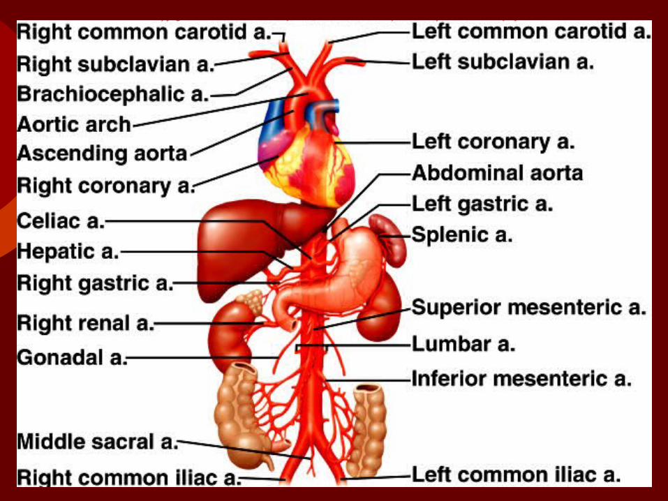
The aorta is the body's largest artery.

Principal Branches of the Aorta The branches of the ascending aorta are
the right and left coronary arteries that lead to heart muscle.
Principal branches of the aortic arch include the brachiocephalic, left common carotid, and left subclavian arteries.
The descending aorta (thoracic aorta) gives rise to many small arteries to the thoracic wall and thoracic viscera.
The abdominal aorta gives off the following branches: celiac, superior mesenteric, suprarenal, renal, gonadal, inferior mesenteric, and common iliac arteries.
Figure

Figure

Figure

Figure

Venous System Veins return blood to the heart
after the exchange of substances has occurred in the tissues.
Characteristics of Venous PathwaysLarger veins parallel the courses
of arteries and are named accordingly; smaller veins take irregular pathways and are unnamed

Characteristics of Venous Pathways
Veins from the head and upper torso drain into the superior vena cava.
Veins from the lower body drain into the inferior vena cava.
The vena cavae merge to join the right atrium.

Figure

Figure

Figure

Respiratory System

IntroductionA. The respiratory system consists of tubes
that filter incoming air and transport it into the microscopic alveoli where gases are exchanged.
B. The entire process of exchanging gases between the atmosphere and body cells is called respiration and consists of the following: ventilation, gas exchange between blood and lungs, gas transport in the bloodstream, gas exchange between the blood and body cells, and cellular respiration.

I. Organs of the Respiratory System
A. The organs of the respiratory tract can be divided into two groups: the upper respiratory tract (nose, nasal cavity, sinuses, and pharynx), and the lower respiratory tract (larynx, trachea, bronchial tree, and lungs).
B. The nose is supported by bone and cartilage, provides an entrance for air in which air is filtered by coarse hairs inside the nostrils.

Figure

Nasal CavityC. Nasal Cavity
1. The nasal cavity is a space posterior to the nose that is divided medially by the nasal septum.
2. Nasal conchae divide the cavity into passageways that are lined with mucous membrane, and help increase the surface area available to warm and filter incoming air.
3. Particles trapped in the mucus are carried to the pharynx by ciliary action, swallowed, and carried to the stomach where gastric juice destroys any microorganisms in the mucus.

Paranasal SinusesD. Paranasal
1. Sinuses are air-filled spaces within the maxillary, frontal, ethmoid, and sphenoid bones of the skull.
2. These spaces open to the nasal cavity and are lined with mucus membrane that is continuous with that lining the nasal cavity.
3. The sinuses reduce the weight of the skull and serve as a resonant chamber to affect the quality of the voice.

Pharynx
E. Pharynx1. The pharynx is a common
passageway for air and food.2. The pharynx aids in producing
sounds for speech.

Figure

LarynxF. Larynx
1.The larynx is an enlargement in the airway superior to the trachea and inferior to the pharynx.
2.It helps keep particles from entering the trachea and also houses the vocal cords.
3. The larynx is composed of a framework of muscles and cartilage bound by elastic tissue.

Larynx Cont.4. Inside the larynx, two pairs of folds
of muscle and connective tissue covered with mucous membrane make up the vocal cords.a. The upper pair- false vocal cordsb. The lower pair- true vocal cordsc. Changing tension on the vocal
cords controls pitch, while increasing the loudness depends upon increasing the force of air vibrating the vocal cords.

Larynx Cont.5. During normal breathing, the
vocal cords are relaxed and the glottis is a triangular slit.
6. During swallowing, the false vocal cords and epiglottis close off the glottis.

Figure
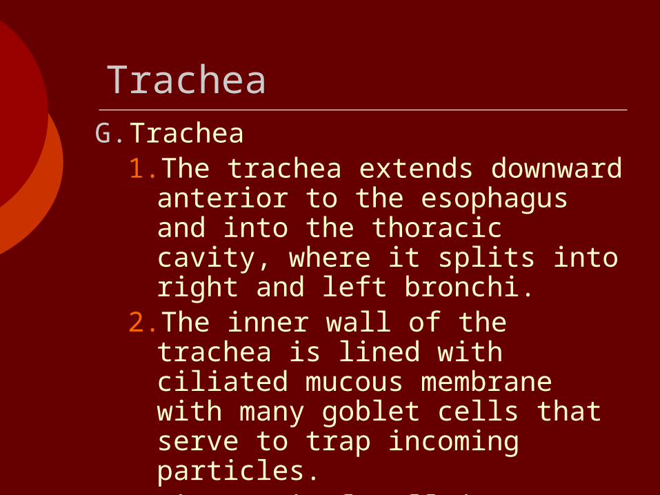
TracheaG. Trachea
1.The trachea extends downward anterior to the esophagus and into the thoracic cavity, where it splits into right and left bronchi.
2.The inner wall of the trachea is lined with ciliated mucous membrane with many goblet cells that serve to trap incoming particles.
3.The tracheal wall is supported by 20 incomplete cartilaginous rings.

Bronchial TreeH. Bronchial Tree
1. The bronchial tree consists of branched tubes leading from the trachea to the alveoli.
2. The bronchial tree begins with the two primary bronchi, each leading to a lung.
3. The branches of the bronchial tree from the trachea are right and left primary bronchi; these further subdivide until bronchioles give rise to alveolar ducts which terminate in alveoli.
4. It is through the thin epithelial cells of the alveoli that gas exchange between the blood and air occurs.

Figure

Figure

LungsI. Lungs
1. The right and left soft, spongy, cone-shaped lungs are separated medially by the mediastinum and are enclosed by the diaphragm and thoracic cage.
2. The bronchus and large blood vessels enter each lung.
3. A layer of serous membrane, the visceral pleura, folds back to form the parietal pleura.

Lungs cont.4. The visceral pleura is attached to
the lung, and the parietal pleura lines the thoracic cavity; serous fluid lubricates the “pleura cavity” between these two membranes.
5. The right lung has three lobes, the left has two.
6. Each lobe is composed of lobules that contain air passages, alveoli, nerves, blood vessels, lymphatic vessels, and connective tissues.

II. Breathing MechanismA. Ventilation (breathing), the
movement of air in and out of the lungs, is composed of inspiration and expiration.

Figure

III. Control of BreathingA. Normal breathing is a rhythmic,
involuntary act even though the muscles are under voluntary control.
B. Respiratory Center 1. Groups of neurons in the brain stem
comprise the respiratory center, which controls breathing by causing inspiration and expiration and by adjusting the rate and depth of breathing.
2. The components of the respiratory center include the rhythmicity center of the medulla and the pneumotaxic area of the pons.

Respiratory Center Cont.3. The medullary rhythmicity center
includes two groups of neurons: the dorsal respiratory group and the ventral respiratory group.a. The dorsal respiratory group is
responsible for the basic rhythm of breathing.
b. The ventral respiratory group is active when more forceful breathing is required.
4. Neurons in the pneumotaxic area control the rate of breathing.

Factors Affecting BreathingC. Factors Affecting Breathing
1. Chemicals, lung tissue stretching, and emotional state affect breathing.
2. Chemosensitive areas (central chemoreceptors) are associated with the respiratory center and are sensitive to changes in the blood concentration of carbon dioxide and hydrogen ions.a. If either carbon dioxide or hydrogen
ion concentrations rise, the central chemoreceptors signal the respiratory center, and breathing rate increases.

Factors Affecting Breathing Cont.3. Peripheral chemoreceptors in the
carotid sinuses and aortic arch sense changes in blood oxygen concentration, transmit impulses to the respiratory center, and breathing rate and tidal volume increase.
4. An inflation reflex, triggered by stretch receptors in the visceral pleura, bronchioles, and alveoli, helps to prevent overinflation of the lungs during forceful breathing.
5. Hyperventilation lowers the amount of carbon dioxide in the blood.

IV. Alveolar Gas Exchanges
A. The alveoli are the only sites of gas exchange between the atmosphere and the blood.
B. Alveoli 1. The alveoli are tiny sacs
clustered at the distal ends of the alveolar ducts.

Respiratory Membrane
C. Respiratory Membrane1. The respiratory membrane
consists of the epithelial cells of the alveolus, the endothelial cells of the capillary, and the two fused basement membranes of these layers.
2. Gas exchange occurs across this respiratory membrane.

Figure

Diffusion across the Respiratory Membrane
D. Diffusion across the Respiratory Membrane1.Gases diffuse from areas of higher
pressure to areas of lower pressure.
2.In a mixture of gases, each gas accounts for a portion of the total pressure; the amount of pressure each gas exerts is equal to its partial pressure.

Diffusion across the Respiratory Membrane Cont.
3. When the partial pressure of oxygen is higher in the alveolar air than it is in the capillary blood, oxygen will diffuse into the blood.
4. When the partial pressure of carbon dioxide is greater in the blood than in the alveolar air, carbon dioxide will diffuse out of the blood and into the alveolus.
5. A number of factors favor increased diffusion; more surface area, shorter distance, greater solubility of gases, and a steeper partial pressure gradient.

V. Gas Transport
A. Gases are transported in association with molecules in the blood or dissolved in the plasma.
Oxygen Transport
B. Oxygen Transport1.Over 98% of oxygen is carried in
the blood bound to hemoglobin of red blood cells, producing oxyhemoglobin.
2.Oxyhemoglobin is unstable in areas where the concentration of oxygen is low, and gives up its oxygen molecules in those areas.

Oxygen Transport cont.
3. More oxygen is released as the blood concentration of carbon dioxide increases, as the blood becomes more acidic, and as blood temperature increases.
4. A deficiency of oxygen reaching the tissues is called hypoxia and has a variety of causes.

Figure