Blocking Neurogenic Inflammation for the Treatment of Acute
17
Hindawi Publishing Corporation International Journal of Inflammation Volume 2013, Article ID 578480, 16 pages http://dx.doi.org/10.1155/2013/578480 Review Article Blocking Neurogenic Inflammation for the Treatment of Acute Disorders of the Central Nervous System Kate Marie Lewis, Renée Jade Turner, and Robert Vink Adelaide Centre for Neuroscience Research, School of Medical Sciences, e University of Adelaide, North Terrace, SA 5005, Australia Correspondence should be addressed to Robert Vink; [email protected] Received 6 February 2013; Accepted 8 May 2013 Academic Editor: Christopher D. Buckley Copyright © 2013 Kate Marie Lewis et al. is is an open access article distributed under the Creative Commons Attribution License, which permits unrestricted use, distribution, and reproduction in any medium, provided the original work is properly cited. Classical inflammation is a well-characterized secondary response to many acute disorders of the central nervous system. However, in recent years, the role of neurogenic inflammation in the pathogenesis of neurological diseases has gained increasing attention, with a particular focus on its effects on modulation of the blood-brain barrier BBB. e neuropeptide substance P has been shown to increase blood-brain barrier permeability following acute injury to the brain and is associated with marked cerebral edema. Its release has also been shown to modulate classical inflammation. Accordingly, blocking substance P NK1 receptors may provide a novel alternative treatment to ameliorate the deleterious effects of neurogenic inflammation in the central nervous system. e purpose of this paper is to provide an overview of the role of substance P and neurogenic inflammation in acute injury to the central nervous system following traumatic brain injury, spinal cord injury, stroke, and meningitis. 1. Introduction Acute disorders of the central nervous system (CNS), includ- ing traumatic brain injury (TBI), spinal cord injury (SCI), stroke, and meningitis, account for a significant disease burden worldwide, with CNS injury being the leading cause of death aſter trauma [1]. ese acute neurological conditions affect individuals of all ages and both sexes alike resulting in significant morbidity and mortality. Despite the prevalence of these conditions, current treatments remain limited and largely inadequate. New therapies are urgently required in order to reduce the death and disability associated with these conditions. One feature which is central to each of these con- ditions is disruption to the blood-brain barrier (BBB)/blood- spinal cord barrier (BSCB) and subsequent development of vasogenic edema. As such, targeting this aspect of the injury cascade is likely to produce beneficial outcomes in each of these conditions. Recent reports on the role of the neuropeptide substance P (SP) and neurogenic inflammation in BBB dysfunction and genesis of cerebral edema following acute brain injury suggest that this pathway provides a novel target for therapeutic intervention. e current paper will provide an overview of the BBB and vasogenic edema, followed by a discussion of the role of SP and neurogenic inflammation in CNS injury. 2. Blood-Brain Barrier/Blood-Spinal Cord Barrier e BBB is a highly selective barrier that serves to protect the fragile brain microenvironment. It is the interface between the blood and the brain, separating the brain parenchyma from the blood within cerebral capillaries, and involves the interactions between endothelial cells, astrocytes, pericytes, and the capillary basement membrane. Within the spinal cord, the blood-spinal cord barrier (BSCB) is similar in function to the BBB [2] and serves to protect the spinal cord by modulating the entry of blood-borne substances. e fundamental structures of the BBB and BSCB are the same although there are some specific differences in the BSCB including glycogen deposits, decreased P-glycoprotein transporters, and decreased expression of tight junctional protein expression [3]. e main function of these barriers is to facilitate a constant supply of nutrients, preserve ion homeostasis within the brain/spinal cord microenvironment, and protect against
Transcript of Blocking Neurogenic Inflammation for the Treatment of Acute

Hindawi Publishing CorporationInternational Journal of InflammationVolume 2013, Article ID 578480, 16 pageshttp://dx.doi.org/10.1155/2013/578480
Review ArticleBlocking Neurogenic Inflammation for the Treatment ofAcute Disorders of the Central Nervous System
Kate Marie Lewis, Renée Jade Turner, and Robert Vink
Adelaide Centre for Neuroscience Research, School of Medical Sciences, The University of Adelaide, North Terrace, SA 5005, Australia
Correspondence should be addressed to Robert Vink; [email protected]
Received 6 February 2013; Accepted 8 May 2013
Academic Editor: Christopher D. Buckley
Copyright © 2013 Kate Marie Lewis et al. This is an open access article distributed under the Creative Commons AttributionLicense, which permits unrestricted use, distribution, and reproduction in any medium, provided the original work is properlycited.
Classical inflammation is a well-characterized secondary response tomany acute disorders of the central nervous system. However,in recent years, the role of neurogenic inflammation in the pathogenesis of neurological diseases has gained increasing attention,with a particular focus on its effects on modulation of the blood-brain barrier BBB.The neuropeptide substance P has been shownto increase blood-brain barrier permeability following acute injury to the brain and is associated with marked cerebral edema. Itsrelease has also been shown to modulate classical inflammation. Accordingly, blocking substance P NK1 receptors may providea novel alternative treatment to ameliorate the deleterious effects of neurogenic inflammation in the central nervous system. Thepurpose of this paper is to provide an overview of the role of substance P and neurogenic inflammation in acute injury to the centralnervous system following traumatic brain injury, spinal cord injury, stroke, and meningitis.
1. Introduction
Acute disorders of the central nervous system (CNS), includ-ing traumatic brain injury (TBI), spinal cord injury (SCI),stroke, and meningitis, account for a significant diseaseburden worldwide, with CNS injury being the leading causeof death after trauma [1].These acute neurological conditionsaffect individuals of all ages and both sexes alike resulting insignificant morbidity and mortality. Despite the prevalenceof these conditions, current treatments remain limited andlargely inadequate. New therapies are urgently required inorder to reduce the death and disability associated with theseconditions. One feature which is central to each of these con-ditions is disruption to the blood-brain barrier (BBB)/blood-spinal cord barrier (BSCB) and subsequent developmentof vasogenic edema. As such, targeting this aspect of theinjury cascade is likely to produce beneficial outcomes ineach of these conditions. Recent reports on the role of theneuropeptide substance P (SP) and neurogenic inflammationin BBB dysfunction and genesis of cerebral edema followingacute brain injury suggest that this pathway provides anovel target for therapeutic intervention. The current paperwill provide an overview of the BBB and vasogenic edema,
followed by a discussion of the role of SP and neurogenicinflammation in CNS injury.
2. Blood-Brain Barrier/Blood-SpinalCord Barrier
The BBB is a highly selective barrier that serves to protect thefragile brain microenvironment. It is the interface betweenthe blood and the brain, separating the brain parenchymafrom the blood within cerebral capillaries, and involves theinteractions between endothelial cells, astrocytes, pericytes,and the capillary basement membrane. Within the spinalcord, the blood-spinal cord barrier (BSCB) is similar infunction to the BBB [2] and serves to protect the spinalcord by modulating the entry of blood-borne substances.The fundamental structures of the BBB and BSCB are thesame although there are some specific differences in theBSCB including glycogen deposits, decreased P-glycoproteintransporters, and decreased expression of tight junctionalprotein expression [3].
The main function of these barriers is to facilitate aconstant supply of nutrients, preserve ion homeostasis withinthe brain/spinal cord microenvironment, and protect against

2 International Journal of Inflammation
noxious chemicals, variations in blood composition, and thebreakdown of concentration gradients. The gate functionof the BBB and BSCB is afforded by tight and adherensjunctions, comprised of a complex network of transmem-brane and cytosolic proteins [4, 5]. Specifically, claudins,occludins, junctional adhesion molecules (JAMs), and zonaoccludens (ZOs) are the proteins that make up this network.Tight junctions are located on the most apical region of thecleft between cerebral capillary endothelial cells and forma seal to prevent substances from passing between them[6]. Claudins, predominately caludin-5, are involved in theprimary makeup or backbone of tight junctions, formingdimers which interact with opposing claudin molecules toform the primary seal of the tight junction [6, 7]. JAM hasa single transmembrane segment, which initiates cell-to-cellattachment and is able to mediate permeability through thisavenue [7]. Occludin has four transmembrane segments andis present in higher concentrations in endothelial cells ofthe BBB than in those in systemic capillary endothelial cells.It induces high membrane resistance, which is indicativeof low ion permeability [7, 8]. Occludin interacts with thecytoskeleton of BBB/BSCB endothelial cells through ZO1,ZO2, and ZO3 molecules [6, 7]. A further obstacle to preventthe entry of unwanted substances into the brain is providedby the basement membrane of the BBB, which is made up ofproteins found within the extracellular matrix including col-lagens, vitronectin, fibronectin, tenascin, and proteoglycans[9]. These components provide stability to the structure ofthe blood vessels and a surface upon which cerebral capillaryendothelial cells can rest.
Astrocytes are central to the structure and function of theBBB/BSCB. Their end feet surround 99% of BBB endothelialcells and act to support and enhance the tight junctionsbetween them [7, 10]. Furthermore, astrocytes mediate theconnection between neurones and endothelial cells [11], andthe gap junctions between astrocytes allow for quick transferof substances and information [12].They become activated inresponse to pathological stimuli, which results in the hyper-trophy of the astrocytic processes and overexpression of inter-mediate filaments, namely, glial fibrillary acidic protein [12].
Pericytes have a stellate appearance and cytoplasmicprocesses and act as support cells that play an important rolein the BBB/BSCB.They cover 22–32% of the capillary cell sur-faces [13], and the gap junctions between pericytes and cere-bral capillary endothelial cells allow communication to occur[7]. The main function of pericytes is thought to be bloodflow regulation, particularly in the precapillary arterioles thatsupply the brain with blood [14]. The structure of pericytesmakes them ideal for this function, as they are contractile andexpress the smooth muscle actin isoform [13]. Collagen typeIV glycosaminoglycans and laminin are also synthesised inpericytes to be used in formation of the basement membrane[13]. They have the ability to regulate endothelial cell prolif-eration, survival, migration, and differentiation [7].
3. Edema
Of the secondary injury factors that occur in the settingof CNS injury, edema within the brain or spinal cord is
of particular concern given its association with increasedmortality and morbidity [15, 16]. Edema is defined as theabnormal accumulation of fluid within the CNS tissue.Klatzo [17] was the first to classify edema into two broadcategories based upon the integrity of the BBB: cytotoxic andvasogenic edema. Cytotoxic edema is an intracellular edemathat occurs as a result of cellular injury. It is characterizedby a shift of water from the extracellular compartment to theintracellular compartment, accompanied by shrinkage of theextracellular space. Cytotoxic edema occurs independentlyof alterations in the BBB/BSCB and appears to be moreprominent in the grey matter [18]. Failure of the Na+/K+ATPase in regions of energy failure and subsequent loss ofion homeostasis, leading to influx of water into cells, is centralto the development of cytotoxic edema [19, 20]. Conversely,vasogenic edema has been shown to be more prevalent inthe white matter [18] and involves the escape of proteinsfrom the vasculature in the setting of BBB/BSCB disruptionand injury to cerebral blood vessels. Protein accumulation inthe brain/spinal cord extracellular space causes an osmoticincrease at the site of injury and the subsequent movement ofwater down its osmotic gradient.There is a strong correlationbetween extravasation of proteins into the extracellular spaceand the development of vasogenic edema [21, 22].
The temporal profile of edema pathogenesis after injuryvaries greatly with injury type and severity [23] and hasbeen extensively studied in order to characterize the periodin which anti-inflammatory pharmacological interventionsmay be effective. In a mouse model of cerebral contusion,permeability of the BBB to large proteins was resolved byapproximately 5 hours following injury, whereas smallermolecules of 10 kDa were still able to pass through the BBBfor up to 4 days [24]. Similarly, the BSCBmay be disrupted forseveral days following traumatic SCI [25, 26]. Furthermore,in ischemic stroke, it has been shown that edema continuesto develop for up to 7 days, with the initial cytotoxic edemabeing followed by vasogenic edema [27]. Thus, there issubstantial opportunity for amelioration of barrier dysfunc-tion and subsequent cerebral edema through manipulationof mediators of BBB/BSCB permeability. Further studiesare required to elucidate the exact mechanisms of barrierdisruption and subsequent edema pathogenesis to developtargeted therapeutic agents.
The development of edema is associated with significantmortality and morbidity in the setting of CNS injury. Suchoutcomes are related to the ability of vasogenic edema tolead to an increase in pressure within the cranium or spinalcanal. Given that the skull is rigid structure, any increase inthe intracranial contents (blood, brain, and cerebral spinalfluid)must be compensated by a decrease in the volume of theother components.The same is truewithin the spinal column.Within both the brain and the spinal cord, there is limitedcapacity for compensation through reductions in blood orcerebrospinal fluid volume to accommodate for an increase inthe intracranial volume.This compensation is responsible forthe initial plateau in the intracranial pressure/volume curve,which becomes exponential once compensatorymechanismsare exhausted [28]. When such compensatory mechanismsfail, profound increases in intracranial pressure (ICP) or

International Journal of Inflammation 3
intrathecal pressure (ITP) may result. The sequelae of ele-vated ICP/ITP include reduced blood flow to CNS tissue,ischemia and infarct extension, deformation and herniationof the brain and spinal cord tissue, and in severe cases, death[18, 29–31].
With the mortality of malignant cerebral edema approa-ching 80% [18], the reduction of cerebral edema and itsassociated rise in ICP is now widely recognised as an impor-tant clinical management target. Current treatments seekto reduce brain swelling and ICP though administration ofhyperosmotic agents and barbiturates, induction of hyper-ventilation or hypothermia, and surgical interventions suchas cerebrospinal fluid (CSF) drainage, or in severe cases,decompressive craniectomy [23, 30, 32, 33]. In the case ofhemorrhage, evacuation of space occupying lesions likehematomas may be warranted [34]. Clinical signs of edemahave been linked with poor functional outcome followingSCI [16]. The use of steroids in an attempt to minimize SCI-induced edema and inflammation is common, despite thecontroversy surrounding their effectiveness and safety [35].Decompressive surgery is also a current standard treatmentfollowing SCI [36].
With respect to patient morbidity and mortality, currentclinical treatment regimens for acute disorders of the CNShave proven somewhat ineffective, mainly because they donot address the specific mechanisms that are associated withthe genesis of edema in cerebral ischemia. Recent studies haveidentified substance P (SP) release as a feature of acute CNSinjury and have delineated a critical role for SP in increasedBBB permeability and the development of vasogenic edema.
4. Neurogenic Inflammation
Neurogenic inflammation is a neurally elicited, local inflam-matory response characterized by vasodilation, increasedvascular permeability, mast cell degranulation, and therelease of neuropeptides including SP and calcitonin gene-related peptide (CGRP) [37]. In addition, there are alsotissue-specific responses including smooth muscle contrac-tion/relaxation in the bladder and bronchoconstriction in theairways, amongst others [38]. Neurogenic inflammation hasbeen demonstrated in tissue receiving trigeminal innervationandmay be stimulated bymany agents including prostanoids,leukotrienes, histamine, and serotonin, as well as by changesin the extracellular environment such as decreased pH,increased osmolarity, heat, inflammatory conditions, andtissue (mechanical) injury [39, 40]. The changes in bloodvessel size and permeability that occur with neurogenicinflammation lead to edema formation within the tissue[21, 22]. Perhaps the most important factor in this responseis SP, having been identified as the most potent initiator ofneurogenic inflammation [41, 42].
Neurogenic inflammatory mediators such as SP andCGRP and their respective receptors are found in abundancein both the rodent and human CNSs, and whilst neu-rogenic inflammation and classical inflammation are bothinflammatory processes, neurogenic inflammation in thebrain differs from classical inflammation in that neurogenicinflammation is neurally elicited and results in an increased
Insult tothe brain/
spinalcord
Release ofsubstance P
Neurogenicinflammation
BBB/BSCBpermeability
Movement ofplasma proteins +
H2O into CNStissue
Vasogenicedema
↑ volumecranial cavity/spinal cavity
Failure ofcompensatorymechanisms
↑ ICP/ITP
Classicalinflammation
Release ofinflammatory
mediators
Activation ofinflammatory
cells
Cellularinjury
Cell death
∙ Ischemia∙ Infarct extension
+ ↓ cerebral blood flow∙ Blood vessel compression
herniation∙ Tissue compression+
∙ Death
Figure 1: Acute CNS injury leads to the initiation of both neuro-genic inflammation and classical inflammation.
permeability of the BBB through the release of neuropeptides.In contrast, classical inflammation involves the accumulationand proliferation of microglia, perivascular macrophages,and other inflammatory cells (Figure 1) [43, 44]. These cellssubsequently release classical inflammatory mediators likebradykinin, which drive vascular changes [45]. Nevertheless,there is an interaction between the two processes as manyof the factors within each cascade may initiate or potentiatethe other. For example, the classical inflammatory mediatorbradykinin causes release of the neurogenic inflammatorymediator SP, which in turn is well known to cause mastcell degranulation along with bradykinin and nitric oxiderelease by endothelial cells and thus potentiation of classicalinflammation (Figure 1). Inflammation in the brain may playmany roles, including the maintenance of tissue homeostasis,although when these processes are unable to be controlled,tissue damage occurs. Thus, this paper focuses on the phar-macological blockade of neurogenic inflammation for thetreatment of acute disorders of the CNS.
There aremultiple pathways by which neurogenic inflam-mation may be initiated. It is well documented, using both

4 International Journal of Inflammation
Injury CapsaicinHeat
ProtonsBradykinin
Tryptaseetc.
Plasma extravasation
Vasogenic edema
PLCSensoryneuroneending
Vasodilation
CRLR +RAMP
CGRP
SP
Increasedpermeability
Bloodvessel EC
NK1
IP3
↑ (Ca2+)i
Figure 2: Neurogenic inflammatory initiation of vasogenic edema.PLC—phospholipase C, IP
3—inositol triphosphate, (Ca2+)
𝑖—
intracellular calcium ions, CGRP—calcitonin gene-related peptide,SP—substance P, CRLR—calcitonin receptor-like receptor, RAMP—receptor activity modifying protein, NK1—NK1 receptor, EC—endothelial cell.
animal models and isolated neurons in vitro, that capsaicin,heat, protons, bradykinin, and tryptase are upstream reg-ulators of the intracellular calcium influx, which results ininflammatory neuropeptide release [46–48]. In contrast, it isthought that prostaglandins E
2and I2, cytokines, interleukin-
1, interleukin-6, and tumor necrosis factor do not cause neu-rotransmitter release themselves, but rather excite sensoryneurons and thus lower the threshold for firing and causeaugmented release of neuropeptides [48, 49].
While neurogenic inflammation has been extensivelystudied and well documented in peripheral tissues [50,51], until recently the concept of neurogenic inflammationwithin the CNS has remained largely unexplored. Given thecapacity for neurogenic inflammation to influence vascularpermeability and lead to the genesis of edema (Figure 2), ithas now been widely investigated for its potential to influenceBBB permeability and vasogenic edema within the brain andspinal cord under varying pathological conditions.
4.1. Capsaicin. Capsaicin activates transient receptor poten-tial vanilloid-1 (TRPV1) channels on polymodal nociceptivefibers, thus, resulting in the release of neurogenic inflamma-tory mediators and subsequent vasodilation and increasedvascular permeability [52, 53]. Under experimental condi-tions, capsaicin is commonly used to cause release and/ordepletion of neuropeptides [54]. Thus, capsaicin initiallyelicits a neurogenic inflammatory response, followed by arefractory period in which there is no response to factorsthat would ordinarily induce neurogenic inflammation. Athigh doses in young animals, capsaicinmay cause permanentdamage to the sensory neurons so that neurogenic inflamma-tory mediators are no longer synthesized, meaning that theneurogenic response is permanently abolished.
4.2. Substance P. SP is an 11 amino acid peptide that is amem-ber of the tachykinin family, so named for their fast-actingproperties [55], which also includes neurokinin A (NKA),neurokinin B (NKB), neuropeptide K (NPK), and neuropep-tide 𝛾 (NP𝛾), amongst others. SP is released fromboth centraland peripheral endings of primary afferent neurons where itfunctions as a neurotransmitter [41, 55]. SP, along with othertachykinins, is produced from the preprotachykinin (PPT) Aand B genes. Alternate splicing of the PPTA gene yields the𝛼- and 𝛿-transcripts giving rise to SP, NKA, NPK, and NK𝛾,whereas the 𝛽- and 𝛾-transcripts only produce SP. The PPTBgene only encodes for NKB. SP synthesis occurs at the cellbody ribosomes, where it is then packaged into vesicles andaxonally transported to the terminal endings for final enzy-matic processing [56]. Precursor proteins are stored in secre-tory granules along with processing enzymes for posttransla-tional modifications and release of the active peptide [39, 57].The biologically active peptide is then stored in large, densevesicles ready for release. Under normal conditions, substan-tial amounts of SP are synthesised and stored within neurons[56]. However, activation or damage of these neurons resultsin the rapid release of SP and other neuropeptides [39].
SP is widely distributed throughout the central and peri-pheral nervous systems, with 𝛼-PPTA transcriptsmore abun-dant within the brain and𝛽-PPTA transcriptsmore abundantin peripheral tissues. Specifically, in the brain, SP immunore-activity has been demonstrated in the rhinencephalon, telen-cephalon, basal ganglia, hippocampus, amygdala, septalareas, diencephalon, hypothalamus, mesencephalon, meten-cephalon, pons, myelencephalon, and spinal cord. SP hasalso been found localized within brain endothelial cells andmicroglia [58–60]. In peripheral tissues, SP and other sensoryneuropeptides are distributed throughout the gut, respiratorysystem, urinary system, immune system, blood, and bloodvessels [37]. SP is localized in capsaicin sensitive neurons andis released from central and peripheral endings of primaryafferent neurons in response to various noxious stimuli [39].Of interest is the fact that SP is colocalizedwith other classicaltransmitters such as serotonin and glutamate, and otherneuropeptides such as CGRP and NKA [56, 59].
Once released, SPmay be cleared and inactivated bymanydifferent proteolytic enzymes including neutral endopep-tidase (NEP) [61, 62] and angiotensin-converting enzyme(ACE) [61, 63, 64], amongst others. BothNEP andACE catal-yse the degradation of the hydrolytic bonds of SP, rendering itinactive without the carboxyl terminus required to bind to itsreceptor [56]. Specifically, NEP has been shown to degrade SPwithin the brain [65], spinal cord [66], and peripheral tissues[39], whereas ACE has been shown to degrade SP in plasma,CSF, and brain, in particular the substantia nigra [67].
The biological actions of SP are mediated through itsbinding at tachykinin NK receptors which are rhodopsin-like membrane structures comprised of 7 transmembranedomains connected by intra- and extracellular loops andcoupled to G proteins [68]. To date, 3 mammalian tachykininreceptors have been identified, theNK1,NK2, andNK3 recep-tors [69].The tachykinins share a common carboxyl terminalsequence that reflects their common biological action, and, asa result, some cross-reactivity amongst tachykinin receptors

International Journal of Inflammation 5
exists [70]. Each of the tachykinins may act on all receptortypes with varying affinities depending upon receptor avail-ability and neuropeptide concentration. Under normal con-ditions, SP has a high affinity for the NK1 receptor, NKA forthe NK2 receptor, andNKB for the NK3 receptor [38, 71].TheNK1 receptor is a 403 amino acid protein that is highly con-served with only discrete variations amongst species. AnNK1autoreceptor has also been characterized to be involved inthe regulation of SP release [72–75]. NK1 receptors are foundin their highest levels in the caudate putamen and superiorcolliculus; however, they are also found in low to moderatelevels in the inferior colliculus, olfactory bulb, hypothalamus,cerebral cortex, septum, striatum,mesencephalon, and dorsalhorn of the spinal cord [75]. NK1 receptors are expressedby a wide variety of cell types including neurons, astrocytes,oligodendrocytes, endothelial cells, and microglia [76].
SP release is initiated in response to Ca2+-dependent dep-olarisation of neurons, induced by a variety of stimuli includ-ing electrical stimulation, pH changes, and ligand binding totheir receptors, including capsaicin [37, 57]. Once released,SP has several effects including direct postsynaptic actionsas a neurotransmitter, modulatory function at postsynapticsites, and/or paracrine functions on nonneuronal targets [57].Transduction of the SP signal then occurs through the actionof G proteins associated with the intracellular domain of theNK1 receptor, leading to an elevation in cAMP as a secondarymessenger, which through a cascade of events, leads to theregulation of ion channels, enzyme activity, and changes ingene expression [48, 77]. Although normally confined to thecell membrane, the NK1-SP complex is rapidly internalisedfollowing SP binding. SP is then removed by endosomalacidification and targeted by the lysosomes for degradation,whilst the NK1 receptor is recycled to the cell membrane [57].
In addition to its role in neurogenic inflammation, SPmayinduce classical inflammatory reactions through the releaseof cytokines and recruitment of immune cells. In the skin, SPacts in a dose-dependant fashion to induce mast cell degran-ulation and histamine and tumour necrosis factor-𝛼 alongwith variable release of leukotriene B4 [78, 79]. SP also actsto induce widespread microvascular permeability. Virtuallyall blood vessels are surrounded by primary sensory nervefibers that secrete SP, and the cerebral blood vessels are partic-ularly well innervated. Intravenous injection of SP has beenshown to increase the permeability of dural blood vessels asevidenced by leakage of horseradish peroxidase in associationwith widening of junctions between endothelial cells and anincrease in the number of cytoplasmic vesicles [80].
In brain endothelial cells, the normal resting level of freeCa2+ is 100 nM [81]. SP causes calcium responses in the endo-thelial cells of the BBB of approximately 1000 nM and henceincrease Ca2+ levels leading to increased BBB permeabilitythrough cell contraction [81, 82]. In conjunction with this,treatmentwith SP of cerebral capillary endothelial cells cocul-tured with astrocytes has been shown to decrease the con-centration of ZO-1 and claudin-5 tight junctional proteins,resulting in increased permeability of the simulated BBB [83].
SP is present in cerebral capillary endothelial cells, and itssecretion by these cells can be increased through treatment
with high doses of cytokines, including interleukin-1𝛽 andtumour necrosis factor 𝛼 [60, 84].This increase in SP releasedfrom brain endothelial cells was found to be associatedwith an increase in the expression of 𝛽-preprotachykininmRNA, a precursor for SP, inside the cells [60]. Spantide, aNK1 antagonist, reversed this increase in SP release fromendothelial cells and the subsequent increased permeabilityof the BBB in a dose-dependent fashion [84].Through the useof electron microscopy, it was shown that the morphologicalchanges associated with SP interactions with endothelial cellswere also neutralized [84].
SP has been implicated in the pathogenesis of manyneurological diseases, due to its effects on BBB permeability.Thus, NK1 antagonists have been investigated for the treat-ment of chronic diseases such as Parkinson’s [85], depression[86], brain tumours [43, 87, 88], and migraine [89] withvariable success. However, this paper focuses primarily onacute disorders of the CNS.
The only NK1 receptor antagonist that is currently avail-able and approved for use clinically is aprepitant. This drugis used as an antiemetic to combat chemotherapy-inducednausea in cancer patients and is generally well tolerated [90].Thus, NK1 receptor antagonist treatment is an appealingalternative to classical anti-inflammatory drugs, the use ofwhich are often limited by detrimental side effects for thetreatment of acute and chronic CNS diseases.
4.3. Calcitonin Gene-Related Peptide. CGRP is a neuropep-tide that is commonly colocalized and released with SP,particularly within sensory C fibers that innervate cerebralvasculature [91–94]. CGRP is the most potent endogenousvasodilator [95] and has been shown to increase the diameterof large cerebral arteries and arterioles. This vasodilation hasbeen shown in many species, including the carotid arterialbed of rabbits, piglet arterioles, pial artery of cats, and guineapigs [96–99]. Furthermore, CGRP infusion in healthy humansubjects causes middle meningeal artery dilation [100]. Therelaxation of blood vessels by CGRP is mediated by proteinkinase C [101]. There are two isoforms of CGRP, CGRP𝛼and CGRP𝛽, which are encoded by alternate RNA processingof the gene for calcitonin located on chromosome 11 andCGRP𝛽 [102, 103]. These isoforms differ in only a singleamino acid and are functionally similar, although CGRP𝛼 isthe predominate form found in the CNS [104]. CGRP exertsits function through binding at theCGRP receptor, which likethe NK1 receptor, has seven transmembrane domains and iscoupled to a G protein. The receptor interacts with a singletransmembrane receptor activity modifying protein to allowfor activation to occur [105, 106]. These receptor complexesare commonly located on neurons, astrocytes, smoothmusclecells, and endothelial cells, particularly those lining duralblood vessels [107–109]. CGRP potentiates the actions of SP[110], which is thought to be through interference with SPbreakdown processes [111, 112].
5. Traumatic Brain Injury
TBI results from physical trauma to the head that conse-quently causes injury to the brain. It is currently the leading
6 International Journal of Inflammation
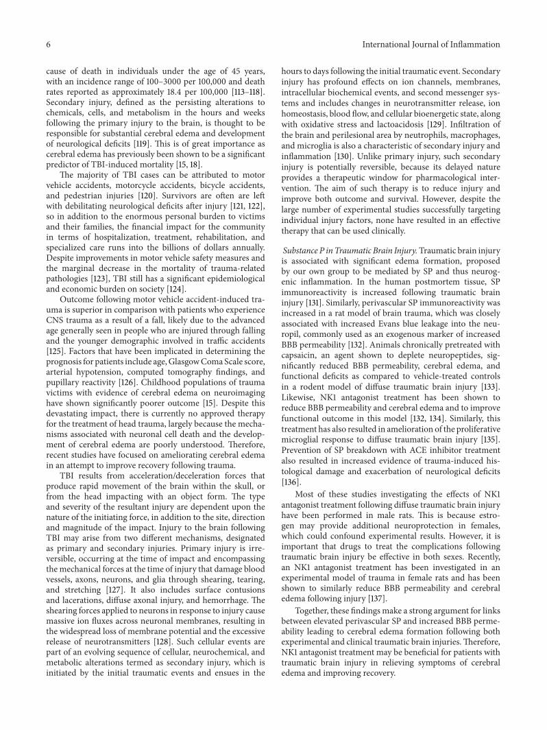
cause of death in individuals under the age of 45 years,with an incidence range of 100–3000 per 100,000 and deathrates reported as approximately 18.4 per 100,000 [113–118].Secondary injury, defined as the persisting alterations tochemicals, cells, and metabolism in the hours and weeksfollowing the primary injury to the brain, is thought to beresponsible for substantial cerebral edema and developmentof neurological deficits [119]. This is of great importance ascerebral edema has previously been shown to be a significantpredictor of TBI-induced mortality [15, 18].
The majority of TBI cases can be attributed to motorvehicle accidents, motorcycle accidents, bicycle accidents,and pedestrian injuries [120]. Survivors are often are leftwith debilitating neurological deficits after injury [121, 122],so in addition to the enormous personal burden to victimsand their families, the financial impact for the communityin terms of hospitalization, treatment, rehabilitation, andspecialized care runs into the billions of dollars annually.Despite improvements in motor vehicle safety measures andthe marginal decrease in the mortality of trauma-relatedpathologies [123], TBI still has a significant epidemiologicaland economic burden on society [124].
Outcome following motor vehicle accident-induced tra-uma is superior in comparison with patients who experienceCNS trauma as a result of a fall, likely due to the advancedage generally seen in people who are injured through fallingand the younger demographic involved in traffic accidents[125]. Factors that have been implicated in determining theprognosis for patients include age,GlasgowComaScale score,arterial hypotension, computed tomography findings, andpupillary reactivity [126]. Childhood populations of traumavictims with evidence of cerebral edema on neuroimaginghave shown significantly poorer outcome [15]. Despite thisdevastating impact, there is currently no approved therapyfor the treatment of head trauma, largely because the mecha-nisms associated with neuronal cell death and the develop-ment of cerebral edema are poorly understood. Therefore,recent studies have focused on ameliorating cerebral edemain an attempt to improve recovery following trauma.
TBI results from acceleration/deceleration forces thatproduce rapid movement of the brain within the skull, orfrom the head impacting with an object form. The typeand severity of the resultant injury are dependent upon thenature of the initiating force, in addition to the site, directionand magnitude of the impact. Injury to the brain followingTBI may arise from two different mechanisms, designatedas primary and secondary injuries. Primary injury is irre-versible, occurring at the time of impact and encompassingthemechanical forces at the time of injury that damage bloodvessels, axons, neurons, and glia through shearing, tearing,and stretching [127]. It also includes surface contusionsand lacerations, diffuse axonal injury, and hemorrhage. Theshearing forces applied to neurons in response to injury causemassive ion fluxes across neuronal membranes, resulting inthe widespread loss of membrane potential and the excessiverelease of neurotransmitters [128]. Such cellular events arepart of an evolving sequence of cellular, neurochemical, andmetabolic alterations termed as secondary injury, which isinitiated by the initial traumatic events and ensues in the
hours to days following the initial traumatic event. Secondaryinjury has profound effects on ion channels, membranes,intracellular biochemical events, and second messenger sys-tems and includes changes in neurotransmitter release, ionhomeostasis, blood flow, and cellular bioenergetic state, alongwith oxidative stress and lactoacidosis [129]. Infiltration ofthe brain and perilesional area by neutrophils, macrophages,and microglia is also a characteristic of secondary injury andinflammation [130]. Unlike primary injury, such secondaryinjury is potentially reversible, because its delayed natureprovides a therapeutic window for pharmacological inter-vention. The aim of such therapy is to reduce injury andimprove both outcome and survival. However, despite thelarge number of experimental studies successfully targetingindividual injury factors, none have resulted in an effectivetherapy that can be used clinically.
Substance P in Traumatic Brain Injury.Traumatic brain injuryis associated with significant edema formation, proposedby our own group to be mediated by SP and thus neurog-enic inflammation. In the human postmortem tissue, SPimmunoreactivity is increased following traumatic braininjury [131]. Similarly, perivascular SP immunoreactivity wasincreased in a rat model of brain trauma, which was closelyassociated with increased Evans blue leakage into the neu-ropil, commonly used as an exogenous marker of increasedBBB permeability [132]. Animals chronically pretreated withcapsaicin, an agent shown to deplete neuropeptides, sig-nificantly reduced BBB permeability, cerebral edema, andfunctional deficits as compared to vehicle-treated controlsin a rodent model of diffuse traumatic brain injury [133].Likewise, NK1 antagonist treatment has been shown toreduce BBB permeability and cerebral edema and to improvefunctional outcome in this model [132, 134]. Similarly, thistreatment has also resulted in amelioration of the proliferativemicroglial response to diffuse traumatic brain injury [135].Prevention of SP breakdown with ACE inhibitor treatmentalso resulted in increased evidence of trauma-induced his-tological damage and exacerbation of neurological deficits[136].
Most of these studies investigating the effects of NK1antagonist treatment following diffuse traumatic brain injuryhave been performed in male rats. This is because estro-gen may provide additional neuroprotection in females,which could confound experimental results. However, it isimportant that drugs to treat the complications followingtraumatic brain injury be effective in both sexes. Recently,an NK1 antagonist treatment has been investigated in anexperimental model of trauma in female rats and has beenshown to similarly reduce BBB permeability and cerebraledema following injury [137].
Together, these findingsmake a strong argument for linksbetween elevated perivascular SP and increased BBB perme-ability leading to cerebral edema formation following bothexperimental and clinical traumatic brain injuries.Therefore,NK1 antagonist treatment may be beneficial for patients withtraumatic brain injury in relieving symptoms of cerebraledema and improving recovery.

International Journal of Inflammation 7
6. Traumatic Spinal Cord Injury
Spinal cord injury (SCI) is an insult to the spinal tissue thatresults in altered motor, sensory, and autonomic functioning.The incidence and mortality estimates for SCI range from 1.3to 8.3 per 100,000 and 0.3–1.8 per 100,000, respectively, whichis approximately 10% of the rates reported for TBI [138–140].Common mechanisms of SCI are vertebral dislocation andburst fracture injury [141]. Similar to TBI, initial primaryinjuries including laceration of blood vessels, bone fracture,and axonal injury, are followed by persistent secondaryinflammatory processes. Specifically, this commonly includesimmune cell accumulation and inflammatory mediatoryrelease, which have been linked to BSCBdisruption [142, 143].The BSCB controls the passage of substances between capil-laries and spinal tissue and is disrupted to cause vasogenicedema [144]. This increased permeability of the BSCB maybe evident over several segments rostral and caudal fromthe injury epicenter, particularly following severe spinal cordinjury [25, 26]. The importance of this process is illustratedby the established link between edema formation and SCI-induced mortality [16].
Nearly 80% of spinal trauma occurs in males, with twopeak age groups affected, young adults in their 20s andthe elderly over 60 years of age. This bimodal demographicis thought to be associated with traffic accidents and falls,respectively [145]. Brain injury is a common comorbidityfor spinal trauma, which is unsurprising as it has manycommon epidemiological features. The most common siteof traumatic spinal cord injury is the cervical level, withdecreasing incidence in the lumbar and thoracic regions ofthe cord [146]. The clinical deficits increase in severity as theSCI occurs at a higher, or more superior, level.
Spinal cord injury is a highly inflammatory process,resulting in immune cell chemotaxis. In a rodent modelof thoracic contusion, inflammatory cytokine release wasevident in the spinal cord following injury, which replicatesthe human condition where a similar pattern of cytokineexpression was evident in the CSF, although at a later timepoint [147]. Additionally, following T9 spinal contusion,neutrophil, macrophages/microglia, and T cells infiltrate theinjured spinal cord and remain evident up to 180 daysfollowing trauma [148].
The promising research on the role of SP in edemadevelopment following brain trauma has led researchers toconsider that the pathogenesis of secondary injury followingspinal cord injury may have similar mechanisms. Moreover,it is thought that this injury type may too respond to manip-ulation of inflammatory neuropeptides, as it has previouslybeen shown that resolution of BSCB permeability and edemaresults in improved functional outcome in animal models ofSCI [149, 150].
Neurogenic Inflammation in Spinal Cord Injury. Previousstudies have shown that SP expression is altered followingtraumatic SCI in both the human condition and in experi-mental animal models. In a combined human cohort of bothperipheral nerve and SCI patients, increased SP levels in thecerebrospinal fluidwere observed in comparisonwith control
patients [151]. Similarly, at both 1 and 2 hours after focalthoracic injury, there was a significant increase in SP foundup to 5mm from the site of injury [152]. In addition, therewas a significant increase in brain SP 5 hours after injury[152]. Therefore, the modulation of SP following trauma tothe spinal cord may occur throughout the entire CNS. Therewas also an increase in SP evident following T12 transectionof the spinal cord in female cats [153]. In contrast, a weightdrop model of trauma in rodents resulted in decreased SP atthe site of injury [154]. Furthermore, NK1 receptors have beenshown to be significantly increased 1 week after injury using aratmodel of thoracic cordotomy [155].The alterations in bothSP and NK1 receptor expression in the spinal cord followingtrauma suggest that SP may play a part in the pathogenesisof spinal cord injury and its complications. However, furtherstudies are required to determine its exact role.
There has been limited research on the role of CGRP intraumatic spinal cord injury. It has been shown, followingeither C4 or T13 hemisection, that primary afferents axonsimmunostaining for CGRP grow into the area of injury [156,157]. However, the functional or mechanistic significance ofthis is yet to be elucidated. Therefore, the evidence for arole of CGRP and possible therapeutic benefit following itsmanipulation is far less compelling for spinal injury whencompared to the results seen for other pathologies.
7. Stroke
Stroke is the third most common cause of disability-adjustedlife years and as such is a major health problem worldwide[158]. Specifically, a staggering 15 million people worldwidesuffer a stroke each year, of which 10 million either die orare left permanently disabled [159]. The social and economiccosts of stroke are consequently enormous. Despite this,there is currently only one approved treatment for use instroke, that being tissue plasminogen activator within 4.5 hof symptom onset [160]. However, as little as 5–15% of strokepatients are eligible for and receive such treatment. In the caseof hemorrhagic stroke, little can be done beyond evacuationof the hemorrhage if surgically accessible. As such, noveltherapies that can limit or reverse ischemic injury followingstroke are urgently required.
Stroke is defined as an interruption in the cerebral bloodflow of vascular origin that restricts the supply of vital oxygenand substrates for neurons. Stroke can be broadly classifiedinto two types, ischemic and hemorrhagic. Ischemic strokemost frequently involves a thrombus (local origin) or embo-lus (distant origin) obstructing blood flow, although whenblood flow is reestablished, reperfusion injury may occur.This involves the interaction of blood with oxygen-deprivedtissue resulting in substantial inflammation and oxidativestress. Hemorrhagic stroke refers to a bleed within the brain.In both instances, cerebral ischemia results, and if bloodflow is not rapidly restored, death of cells may result withassociated long-term functional deficits [161]. Restoration ofblood flow is seen as an urgent priority in reducing theextent of tissue injury and preserving function. However, it isnow well accepted that secondary injury processes continueto evolve many hours to days following stroke and also

8 International Journal of Inflammation
contribute to the size of the infarct [162]. With respect tooutcome, hemorrhagic stroke generally has a poorer outcomethan ischemic stroke with mortality rates in the order of37% and 11%, respectively [163]. Hemorrhagic stroke maybe classified as either intracerebral hemorrhage (ICH) orsubarachnoid hemorrhage (SAH). The rupture of charcot-bouchard microaneurysms on small arterioles commonlyleads to ICH, whereas ruptured berry aneurisms within theCircle of Willis are often the cause of SAH [164, 165].
Following stroke, the resultant tissue injury and infarc-tion can be considered as being made up of two components,the infarct core and the surrounding penumbral tissue [166].The infarct core is widely considered to be irreversiblydamaged during ischemic stroke, with cell death occurringrapidly within this region. In the penumbral tissue, however,blood flow is less restricted and so there exists an opportunityfor neuronal tissue to survive the insult. Nevertheless, celldeath may continue to occur here as a result of secondarybiochemical and physiological mechanisms that manifestover the hours to days following stroke [162, 166]. Similar toTBI, there are diverse arrays of secondary injury processesthat contribute to injury and cell loss following stroke, includ-ing excitotoxicity, oxidative stress, inflammation, apopto-sis, increased vascular permeability, and cerebral edema,amongst others [167]. Given the delayed nature of secondaryinjury following stroke, there is an opportunity for pharma-cological intervention to limit tissue damage and cell death.
Both SAH and ICH can often result in rapid death,meaning that there is only a small window for therapeuticadministration or surgical intervention. Furthermore, giventhat themass effect of such hemorrhagic lesions is substantial,the contribution of secondary injury processes to functionalimpairments is smaller compared with ischemic lesions.In contrast, ischemic stroke has a pattern of injury morecomparable to TBI, with increased permeability of the BBBand cerebral edema as common features. Mortality ratesincrease with time following stroke, demonstrating that evenif patients survive the initial insult, the condition may stillbe fatal due to persistent secondary injury mechanismssuch as the development of cerebral edema [168]. The typeand severity of edema may be influenced by the durationand severity of ischemia and reperfusion status, amongstother factors, and may also differ between the core and thepenumbra of the stroke lesion.
Cerebral edema is a major cause of clinical deteriorationwithin the first 24 h, is the leading cause of death within thefirst week, and is a predictor of poor outcome following stroke[30]. Clinical studies report that it is maximal between 1 and3 d following stroke [18], whilst experimental studies reportits presence as early as 15mins after the onset of vascularocclusion [169]. The presence of vasogenic edema is of par-ticular concern, not only because it increases brain volume,but also because in the setting of vascular recanalization, itincreases risk of hemorrhagic transformation from damagedblood vessels and excess fluid accumulation [170].
7.1. Substance P in Stroke. To date, few groups have investi-gated SP in cerebral ischemia [171], and only our researchgroup has explored the role of neurogenic inflammation
following stroke [172–175]. Our own studies have recentlyshown that SP is increased following experimental ischemicstroke, indicative of neurogenic inflammation. Specifically,increased SP immunoreactivity was observed within penum-bral tissue at 24 h following stroke, being particularly markedin perivascular tissue. Such an increase in SP was confirmedthrough SP ELISA of the ischemic hemisphere [174]. Theincrease in SP was associated with marked disruption to theBBB, as evidenced by increased Evan’s blue extravasation intothe brain parenchyma at 24 h after stroke, thus, supportingprevious observations of a delayed opening of the BBB [176].The increased BBB permeability was observed in the settingof profound cerebral edema, suggesting that the edema hada vasogenic component [174]. Furthermore, profound andpersistent functional deficits with respect to motor, sensory,and neurological function were observed [174].
A role for SP in clinical stroke has also been docu-mented by Bruno and colleagues [177], suggesting that theremay be a role for neurogenic inflammation in this diseasepathogenesis. They observed that patients with transientischemic attack and complete stroke showed elevated serumSP when compared to the control group [177]. Interestingly,individuals with transient ischemic attack showed a greaterelevation than complete stroke [177].
Early studies reported that hypoxia of the rabbit carotidbody increased SP release as a function of the severity of thehypoxic insult [178]. This finding suggested that SP releasemay be a tissue response to hypoxia/ischemia. Consistentwith this, capsaicin pre- or posttreatment was shown toconfer protection from neonatal hypoxia-ischemia injurywith a reduction in infarct volume and apoptosis, in additionto improved vascular dynamics [179].
Given the clear increase in SP that has been docu-mented in both experimental and clinical stroke studies,NK1 tachykinin receptor antagonists have been investigatedfor their potential utility in reducing BBB dysfunction andvasogenic edema in the setting of ischemic stroke. Yu andcolleagues [171] reported a reduction in infarct volume andan improvement in neurological outcome asmeasured at 24 hpoststroke following administration of the NK1 tachykininreceptor antagonist SR-14033. Recently, our group hasextended these initial observations and extensively character-ized the effect of NK1 tachykinin receptor antagonist treat-ment in experimental ischemic stroke. Specifically, we haveshown that intravenous NK1 antagonist treatment adminis-tered 4 hours following stroke resulted in decreased evidenceof cerebral edema [174]. Furthermore, when combined withthe current standard clot dissolution treatment, tissue plas-minogen activator (tPA), NK1 antagonist treatment resultedin equal or better performance in functional outcome testswhen compared to NK1 antagonist or tPA alone [175].
7.2. Substance P in Subarachnoid Hemorrhage. Similar toischemic stroke, altered SP expression has been reported fol-lowing SAH. Perivascular SP expression was increased in twomodels of SAH, injection of autologous blood into the prechi-asmatic cistern and following puncture of themiddle cerebralartery to cause an endogenous bleed [180]. However, NK1

International Journal of Inflammation 9
tachykinin receptor antagonist treatment was unable to ame-liorate the raised ICP, cerebral edema, or impaired functionaloutcome that resulted in either of these models of SAH [180].A possible reason for this is that the pathogenesis of SAHdiffers greatly from ischemic stroke, in whichNK1 tachykininreceptor antagonists have shown promise. SAH presents lessopportunity for therapeutic intervention, due to the masseffect of the bleed, such that therapeutic interventions thatact to modulate the permeability of BBB have limited effects.Thus, the functional deficits that result from SAH may bemore related to the space occupying blood and damage fromits breakdown products rather than to cerebral edema.
7.3. Calcitonin Gene-Related Peptide in Ischemic Stroke. Thewell-established vasodilatory actions of CGRP have ledresearchers to postulate that it may play a protective role topromote cerebral blood flow following ischemic stroke. Thiseffect was demonstrated in a rat model of middle cerebralartery reperfusion stroke. Following injury, treatment withCGRP administered at the beginning of reperfusion resultedin a reduction of arterial blood pressure, decreased the infarctvolume, and ameliorated the increased BBB permeabilitysubsequently inhibiting cerebral edema formation [181, 182].
Along with the vasodilatory actions of CGRP, the mech-anism of neuroprotection following ischemic reperfusionstroke may be through modulation of water channels andother elements of the BBB. As such, in two studies usingthe middle cerebral artery reperfusion model of rodentstroke, CGRP treatment resulted in decreased aquaporin 4mRNA and protein expression [181, 182]. In conjunction,the reduction in tight junction proteins normally associatedwith stroke was ameliorated, along with reduced evidence ofultrastructural damage of endothelial cells [181, 182]. Simi-larly, increased expression of basic fibroblast growth factorhas been found following experimental ischemic reperfusionstroke treated with CGRP, which likely acts to improve thestructural integrity of the BBB basement membrane [182].Furthermore, the neuroprotective effects of leptin in a mousemodel of middle cerebral artery occlusion and reperfusioninjury have been shown to bemediated by CGRP, resulting inincreased blood flow and once again reduced infarct volume[183]. Thus, CGRP may be a promising treatment to improvefunctional outcome following cerebral ischemia throughmultiple actions on the BBB to reduce the severity of injury.
7.4. Calcitonin Gene-Related Peptide in Subarachnoid Hem-orrhage. Akin to ischemic stroke, CGRP is thought to bebeneficial following SAH. CGRP has been measured in thecranial venous outflow of 34 patients following SAH and wasfound to be elevated when compared to the control group,although there was no change in SP levels [184]. In contrast,following subarachnoid hemorrhage, autopsy brain concen-trations of CGRP were reduced in comparison with controlsin the location of the proximal middle cerebral artery [185].Therefore, SAH results in modulation of CGRP levels in boththe blood and the brain. A possible reason for the differentialeffectsmay be that the study in which CGRPwas elevated wasconducted on patients who had survived their SAH, whilst
decreased CGRP was evident following the fatal condition.Thus, the severity of SAH may determine the extent anddirection of changes in CGRP. It is postulated that exhaustionof CGRP may be involved in vasospasm, which is most com-mon following severe SAH, and is often a fatal complication.
CGRP has been tested in both clinical SAH patients andin experimental models of SAH showing protection fromabnormal blood vessel contraction. Intravenous administra-tion of human 𝛼CGRP significantly inhibited vasoconstric-tion in comparison to that evident prior to infusion in 5patients [186]. Similarly, when rabbit basilar artery strips wereisolated following experimental subarachnoid hemorrhage,responsiveness to in vitro application of CGRP to induceblood vessel relaxation was impaired when compared tothose from the control group [101]. This result suggests thatincreased CGRP levels are required in stroke patients. Itis likely that CGRP treatment may hold promise for theprevention of complications associated with subarachnoidhemorrhage.
Taken together, both animal and clinical studies showthat neurogenic inflammation plays an integral role in thepathogenesis of both ischemic stroke and SAH. However,there is a differential effect of inflammatory neuropeptidesin these conditions. The role of neurogenic inflammation inICH has not been widely investigated, although it is likelythat, similar to SAH, the edema component of this conditionmay not contribute as significantly as blood volume to thedevelopment of neurological deficits. There is evidence of SPmediation of many deleterious secondary injurymechanismsfollowing ischemic reperfusion injury, including cerebraledema formation.Thus theNK1 receptor is a promising targetfor pharmacological manipulation to improve patient out-comes. In contrast, SP does not seem to play a significant rolein the immediate injury following subarachnoid SAH.CGRP-induced vasodilation may improve blood flow to hypoxicbrain tissue during cerebral ischemia and prevent vasospasmfollowing subarachnoid hemorrhage. This indicates that spe-cific neurogenic inflammatory mediators need to be targetedin different ways to optimize treatment following stroke.
8. Bacterial Meningitis
Meningitis is characterized by infection and subsequent acuteinflammation of the meninges that cover the outside ofthe brain. The most common causative infectious agent isbacteria, specifically Neisseria meningitidis and Streptococcuspneumoniae. There is a marked adult incidence of bacte-rial meningitis but generally children are most susceptible[187]. Meningitis is associated with CNS symptoms such asneck stiffness, headache, photophobia, phonophobia, alteredconsciousness, and neurological state, as well as systemicsigns of inflammation such as fever, nausea, and vomiting.Additionally, individual bacteria typesmay be associatedwithspecific features, for example,Neisseriameningitidis producesa characteristic rash.
The introduction of vaccinations against specific strainsof bacteria has substantially reduced the incidence of thismeningitis [188]. Despite this, the availability of antibiotics to
10 International Journal of Inflammation
combat bacterial infection of the meningitis remains a med-ical emergency due to the close proximity of inflammationto neurological tissue. This poses a critical threat to braintissue, not only due to the presence of bacteria, but alsothe contribution of secondary injury processes. Specifically,inflammatory processes are associated with increased per-meability of the BBB and cerebral edema, which worsenthe prognosis associated with this disease [6]. Furthermore,cytokine production and leukocyte accumulation are keyfeatures in the pathogenesis of bacterial meningitis.
Currently, anti-inflammatory agents are used in anattempt to combat the symptoms of meningitis, although thedose and duration of treatment are limited by deleteriousside effects of the commonly used drugs like the syntheticcorticosteroid, dexamethasone. Therefore, alternative ther-apeutic agents that combat secondary injury and inflam-matory processes are attractive targets for investigation.Neurogenic inflammation may be a worthy target given itsdocumented role in BBB permeability and cerebral edema inthe setting of acute brain injury and stroke. Specifically, NK1tachykinin receptor antagonists are able to block neurogenicinflammation by modulating neuropeptide action. In thesetting of meningitis, this may prevent deleterious changes indiameter and permeability of cerebral blood vessels and thusleukocyte infiltration and edema formation.
8.1. Substance P in Meningitis. In vitro, SP has been shownto increase the production of inflammatory cytokines byastrocytes and microglia when exposed to Neisseria menin-gitidis and Borrelia burgdorferi gram-negative bacteria [189].Similarly, SP treatment of microglia in vitro, which wereexposed to the gram-negative Borrelia burgdorferi bacteria,results in augmented secretion of prostaglandin E2 [190].Thiseffect was ameliorated by NK1 tachykinin receptor antagonisttreatment and in NK1 knockout cell lines [190]. Furthermore,microglial cells respond to the presence of the gram-positivebacteria Streptococcus pneumoniae with upregulation of NK1receptors by this cell type [191].These results suggest that NK1antagonist treatment may act to inhibit many inflammatoryprocesses associated with bacterial meningitis that causesubstantial tissue damage and worsen outcome.
Positive results from in vitro studies led to in vivoexperiments to determine the effectiveness of NK1 receptorantagonists in experimental mouse models of both gram-positive and gram-negative bacterial meningitis. Intrac-erebral inoculation of Neisseria meningitidis and Borreliaburgdorferi into C57BL/6 mice resulted in increased inflam-matory cytokine and decreased immunosuppressive cytokinesecretion, resulting in a substantially proinflammatory envi-ronment [189]. Correspondingly, intracerebral inoculation offemale C57BL/6 mice with Streptococcus pneumoniae causeda similar pattern of cytokine expression along with gliosis,demyelination, and increased BBB permeability [191]. Thesefeatures were abolished with both NK1 antagonist treatmentand in NK1 knockout mice [189, 191]. Therefore, NK1 antag-onist treatment may be able to limit infection-associatedinflammation and subsequent edema formation through itsability to inhibit inflammatory cytokine secretion and mod-ulate the permeability of the BBB. The results suggest that in
the future, this class of agents could be used as an alternativeto classical anti-inflammatory drugs like dexamethasone.However, the effect of NK1 receptor antagonist treatment hasonly been demonstrated in experimental animal models ofmeningitis; thus, further investigation into the role of SP inthe human condition is required.
8.2. Calcitonin Gene-Related Peptide in Meningitis. The proi-nflammatory nature of meningitis makes CGRP a likelycandidate in the pathogenesis of associated vascular changes,although there has been limited investigation into this area.Nevertheless, patients with acute bacterial meningitis andsepsis have shown evidence of increased CGRP in arterialblood samples [192]. Therefore, the possible role of CGRP inthe inflammatory response of bacterial meningitis warrantsadditional examination.
9. Conclusion
Acute injury to the brain and spinal cord is associatedwith a number of deleterious secondary injury processesof which altered vascular permeability and tissue swellingare of particular concern. This is further compounded bythe lack of effective therapies. However, the inhibition ofneurogenic inflammation may provide a novel alternativetherapy for the treatment of barrier dysfunction and tissueswelling in the setting of acute CNS injury. Experimentalstudies of TBI and stroke have shown that blocking theaction of SP with an NK1 tachykinin receptor antagonistproduces profound reductions in BBB permeability, cerebraledema, and functional deficits. Studies of NK1 tachykininreceptor antagonists in SCI, meningitis, and hemorrhagicstroke are ongoing, but early results suggest that neurogenicinflammation does play a role in these pathologies, albeit aless pronounced role than in TBI and stroke. CGRP may beanotherworthy target alongside SPwith experimentalmodelsof both hemorrhagic and ischemic stroke models showingbenefits of CGRP treatment. Further investigations on therole of neurogenic inflammation and the neuropeptides SPand CGRP in the barrier dysfunction and tissue swelling thatare associated with acute brain and spinal cord injury areongoing, and given the encouraging results to date, they arecertainly warranted.
Conflict of Interests
The authors declare that they have no conflict of interests.
Acknowledgment
The authors would like to thank Tavik Morgenstern for thecreation of artwork for this paper.
References
[1] R. Pfeifer, I. S. Tarkin, B. Rocos, and H. C. Pape, “Patterns ofmortality and causes of death in polytrauma patients-Has any-thing changed?” Injury, vol. 40, no. 9, pp. 907–911, 2009.

International Journal of Inflammation 11
[2] A. E.Mautes,M. R.Weinzierl, F. Donovan, and L. J. Noble, “Vas-cular events after spinal cord injury: contribution to secondarypathogenesis,”PhysicalTherapy, vol. 80, no. 7, pp. 673–687, 2000.
[3] V. Bartanusz, D. Jezova, B. Alajajian et al., “The blood-spinalcord barrier: morphology and clinical implications,” Annals ofNeurology, vol. 70, no. 2, pp. 194–206, 2011.
[4] G. L. Suidan, J. R. Mcdole, Y. Chen, I. Pirko, and A. J. Johnson,“Induction of blood brain barrier tight junction protein alter-ations by CD8 T cells,” PLoS ONE, vol. 3, no. 8, Article ID e3037,2008.
[5] C. Silwedel and C. Forster, “Differential susceptibility of cere-bral and cerebellar murine brain microvascular endothelialcells to loss of barrier properties in response to inflammatorystimuli,” Journal of Neuroimmunology, vol. 179, no. 1-2, pp. 37–45, 2006.
[6] M. A. Petty and E. H. Lo, “Junctional complexes of the blood-brain barrier: permeability changes in neuroinflammation,”Progress in Neurobiology, vol. 68, no. 5, pp. 311–323, 2002.
[7] Y. Persidsky, S. H. Ramirez, J. Haorah, and G. D. Kanmogne,“Blood-brain barrier: structural components and functionunder physiologic and pathologic conditions,” Journal of Neu-roimmune Pharmacology, vol. 1, no. 3, pp. 223–236, 2006.
[8] F. Joo, “Endothelial cells of the brain and other organ systems:some similarities and differences,” Progress in Neurobiology, vol.48, no. 3, pp. 255–273, 1996.
[9] E. Baumann, E. Preston, J. Slinn, and D. Stanimirovic, “Post-ischemic hypothermia attenuates loss of the vascular basementmembrane proteins, agrin and SPARC, and the blood-brain bar-rier disruption after global cerebral ischemia,” Brain Research,vol. 1269, pp. 185–197, 2009.
[10] M. Bundgaard andN. J. Abbott, “All vertebrates started out witha glial blood-brain barrier 4-500 million years ago,” GLIA, vol.56, no. 7, pp. 699–708, 2008.
[11] J. H. Kim, J. A. Park, S. W. Lee et al., “Blood-neural barrier:intercellular communication at glio-vascular interface,” Journalof Biochemistry and Molecular Biology, vol. 39, no. 4, pp. 339–345, 2006.
[12] C. Escartin and G. Bonvento, “Targeted activation of astrocytes:a potential neuroprotective strategy,” Molecular Neurobiology,vol. 38, no. 3, pp. 231–241, 2008.
[13] M. Fisher, “Pericyte signaling in the neurovascular unit,” Stroke,vol. 40, no. 3, supplement, pp. S13–S15, 2009.
[14] F. Joo, “The blood-brain barrier in vitro: the second decade,”Neurochemistry International, vol. 23, no. 6, pp. 499–521, 1993.
[15] T. Rhine, S. L.Wade, K. L.Makoroff et al., “Clinical predictors ofoutcome following inflicted traumatic brain injury in children,”Journal of Trauma and Acute Care Surgery, vol. 73, no. 9,supplement 3, pp. S248–S253, 2012.
[16] J. R. Wilson, R. G. Grossman, R. F. Frankowski et al., “A clinicalprediction model for long-term functional outcome after trau-matic spinal cord injury based on acute clinical and imagingfactors,” Journal of Neurotrauma, vol. 29, no. 13, pp. 2263–2271,2012.
[17] I. Klatzo, “Pathophysiological aspects of brain edema,” ActaNeuropathologica, vol. 72, no. 3, pp. 236–239, 1987.
[18] C. Ayata and A. H. Ropper, “Ischaemic brain oedema,” Journalof Clinical Neuroscience, vol. 9, no. 2, pp. 113–124, 2002.
[19] J. Lazovic, A. Basu, H. W. Lin et al., “Neuroinflammation andboth cytotoxic and vasogenic edema are reduced in interleukin-1 type 1 receptor-deficient mice conferring neuroprotection,”Stroke, vol. 36, no. 10, pp. 2226–2231, 2005.
[20] T. Kuroiwa, M. Ueki, Q. Chen, H. Suemasu, I. Taniguchi, andR. Okeda, “Biomechanical characteristics of brain edema: thedifference between vasogenic-type and cytotoxic-type edema,”Acta Neurochirurgica, Supplement, vol. 60, pp. 158–161, 1994.
[21] T. Kuroiwa, R. Cahn, and M. Juhler, “Role of extracellularproteins in the dynamics of vasogenic brain edema,” Acta Neu-ropathologica, vol. 66, no. 1, pp. 3–11, 1985.
[22] T. Kuroiwa, P. Ting, H. Martinez, and I. Klatzo, “The biphasicopening of the blood-brain barrier to proteins following tempo-rary middle cerebral artery occlusion,” Acta Neuropathologica,vol. 68, no. 2, pp. 122–129, 1985.
[23] A. W. Unterberg, J. Stover, B. Kress, and K. L. Kiening, “Edemaand brain trauma,” Neuroscience, vol. 129, no. 4, pp. 1021–1029,2004.
[24] M. D. Habgood, N. Bye, K. M. Dziegielewska et al., “Changes inblood-brain barrier permeability to large and small moleculesfollowing traumatic brain injury in mice,” European Journal ofNeuroscience, vol. 25, no. 1, pp. 231–238, 2007.
[25] L. J. Noble and J. R. Wrathall, “Distribution and time courseof protein extravasation in the rat spinal cord after contusiveinjury,” Brain Research, vol. 482, no. 1, pp. 57–66, 1989.
[26] P. G. Popovich, P. J. Horner, B. B. Mullin, and B. T. Stokes,“A quantitative spatial analysis of the blood-spinal cord barrierI. Permeability changes after experimental spinal contusioninjury,” Experimental Neurology, vol. 142, no. 2, pp. 258–275,1996.
[27] C. Y. Hsu, T. H. Liu, J. Xu et al., “Arachidonic acid and itsmetabolites in cerebral ischemia,” Annals of the New YorkAcademy of Sciences, vol. 559, pp. 282–295, 1989.
[28] M. Smith, “Monitoring intracranial pressure in traumatic braininjury,” Anesthesia and Analgesia, vol. 106, no. 1, pp. 240–248,2008.
[29] D. F. Hanley, “Review of critical care and emergency approachesto stroke,” Stroke, vol. 34, no. 2, pp. 362–364, 2003.
[30] W.Hacke, S. Schwab,M.Horn,M. Spranger,M.DeGeorgia, andR. Von Kummer, “‘Malignant’ middle cerebral artery territoryinfarction: clinical course and prognostic signs,” Archives ofNeurology, vol. 53, no. 4, pp. 309–315, 1996.
[31] G. Gartshore, J. Patterson, and I.M.Macrae, “Influence of ische-mia and reperfusion on the course of brain tissue swelling andblood-brain barrier permeability in a rodent model of transientfocal cerebral ischemia,” Experimental Neurology, vol. 147, no. 2,pp. 353–360, 1997.
[32] A. Torre-Healy, N. F. Marko, and R. J. Weil, “Hyperosmolartherapy for intracranial hypertension,” Neurocritical Care, vol.17, no. 1, pp. 117–130, 2012.
[33] F. Sadaka and C. Veremakis, “Therapeutic hypothermia for themanagement of intracranial hypertension in severe traumaticbrain injury: a systematic review,” Brain Injury, vol. 26, no. 7-8,pp. 899–908, 2012.
[34] J. V. Rosenfeld, A. I. Maas, P. Bragge et al., “Early managementof severe traumatic brain injury,”The Lancet, vol. 380, no. 9847,pp. 1088–1098, 2012.
[35] R. J. Hurlbert, “Strategies of medical intervention in the man-agement of acute spinal cord injury,” Spine, vol. 31, no. 11,supplement, pp. S16–S21, 2006.
[36] J. C. Furlan, V. Noonan, D. W. Cadotte, and M. G. Fehlings,“Timing of decompressive surgery of spinal cord after traumaticspinal cord injury: an evidence-based examination of pre-clinical and clinical studies,” Journal of Neurotrauma, vol. 28,no. 8, pp. 1371–1399, 2011.

12 International Journal of Inflammation
[37] C. Severini, G. Improta, G. Falconieri-Erspamer, S. Salvadori,and V. Erspamer, “The tachykinin peptide family,” Pharmaco-logical Reviews, vol. 54, no. 2, pp. 285–322, 2002.
[38] P. H. Black, “Stress and the inflammatory response: a review ofneurogenic inflammation,” Brain, Behavior, and Immunity, vol.16, no. 6, pp. 622–653, 2002.
[39] S. Harrison and P. Geppetti, “Substance P,” International Journalof Biochemistry andCell Biology, vol. 33, no. 6, pp. 555–576, 2001.
[40] A. Saria and J. M. Lundberg, “Capsaicin pretreatment inhibitsheat-induced oedema in the rat skin,” Naunyn-Schmiedeberg’sArchives of Pharmacology, vol. 323, no. 4, pp. 341–342, 1983.
[41] M. Otsuka and K. Yoshioka, “Neurotransmitter functions ofmammalian tachykinins,” Physiological Reviews, vol. 73, no. 2,pp. 229–308, 1993.
[42] P. Holzer, “Neurogenic vasodilatation and plasma leakage in theskin,” General Pharmacology, vol. 30, no. 1, pp. 5–11, 1998.
[43] K. M. Lewis, E. Harford-Wright, R. Vink et al., “Targetingclassical but not Neurogenic inflammation reduces peritumoraledema in secondary brain tumors,” Journal of Neuroimmunol-ogy, vol. 250, no. 1-2, pp. 59–65, 2012.
[44] M. B. Graeber, W. Li, and M. L. Rodriguez, “Role of microgliain CNS inflammation,” The FEBS Letters, vol. 585, no. 23, pp.3798–3805, 2011.
[45] J. J. Donkin and R. Vink, “Mechanisms of cerebral edema intraumatic brain injury: therapeutic developments,” CurrentOpinion in Neurology, vol. 23, no. 3, pp. 293–299, 2010.
[46] M. J. Caterina, M. A. Schumacher, M. Tominaga, T. A. Rosen,J. D. Levine, and D. Julius, “The capsaicin receptor: a heat-activated ion channel in the pain pathway,”Nature, vol. 389, no.6653, pp. 816–824, 1997.
[47] M. Tominaga, M. J. Caterina, A. B.Malmberg et al., “The clonedcapsaicin receptor integrates multiple pain-producing stimuli,”Neuron, vol. 21, no. 3, pp. 531–543, 1998.
[48] J. D. Richardson and M. R. Vasko, “Cellular mechanismsof neurogenic inflammation,” Journal of Pharmacology andExperimental Therapeutics, vol. 302, no. 3, pp. 839–845, 2002.
[49] G. D. Nicol and M. Cui, “Enhancement by prostaglandin E2of bradykinin activation of embryonic rat sensory neurones,”Journal of Physiology, vol. 480, no. 3, pp. 485–492, 1994.
[50] R. V. Alves, M. M. Campos, A. R. S. Santos, and J. B. Calixto,“Receptor subtypes involved in tachykinin-mediated edemaformation,” Peptides, vol. 20, no. 8, pp. 921–927, 1999.
[51] M. M. Campos and J. B. Calixto, “Neurokinin mediation ofedema and inflammation,”Neuropeptides, vol. 34, no. 5, pp. 314–322, 2000.
[52] A. Starr, R. Graepel, J. Keeble et al., “A reactive oxygen species-mediated component in neurogenic vasodilatation,” Cardiovas-cular Research, vol. 78, no. 1, pp. 139–147, 2008.
[53] D. E. Hu, A. S. Easton, and P. A. Fraser, “TRPV1 activationresults in disruption of the blood-brain barrier in the rat,”British Journal of Pharmacology, vol. 146, no. 4, pp. 576–584,2005.
[54] N. Erin and O. Ulusoy, “Differentiation of neuronal from non-neuronal Substance P,” Regulatory Peptides, vol. 152, no. 1–3, pp.108–113, 2009.
[55] C. A. Maggi, “The mammalian tachykinin receptors,” GeneralPharmacology, vol. 26, no. 5, pp. 911–944, 1995.
[56] T. Hokfelt, C. Broberger, Z. Q. D. Xu, V. Sergeyev, R. Ubink, andM. Diez, “Neuropeptides—an overview,” Neuropharmacology,vol. 39, no. 8, pp. 1337–1356, 2000.
[57] F. T. Lundy and G. J. Linden, “Neuropeptides and neurogenicmechanisms in oral and periodontal inflammation,” CriticalReviews in Oral Biology & Medicine, vol. 15, no. 2, pp. 82–98,2004.
[58] M. Łazarczyk, E.Matyja, andA. Lipkowski, “Substance P and itsreceptors—as potential target for novel medicines in malignantbrain tumour therapies (mini-review),” Folia Neuropathologica,vol. 45, no. 3, pp. 99–107, 2007.
[59] A. Ribeiro-da-Silva and T. Hokfelt, “Neuroanatomical local-isation of substance P in the CNS and sensory neurons,”Neuropeptides, vol. 34, no. 5, pp. 256–271, 2000.
[60] C. Cioni, D. Renzi, A. Calabro, and P. Annunziata, “Enhancedsecretion of substance P by cytokine-stimulated rat brain endo-thelium cultures,” Journal of Neuroimmunology, vol. 84, no. 1,pp. 76–85, 1998.
[61] A. L. Freed, K. L. Audus, and S. M. Lunte, “Investigationof the metabolism of substance P at the blood-brain barrierusing capillary electrophoresis with laser-induced fluorescencedetection,” Electrophoresis, vol. 22, no. 17, pp. 3778–3784, 2001.
[62] R. Matsas, A. J. Kenny, and A. J. Turner, “The metabolism ofneuropeptides. The hydrolysis of peptides, including enkepha-lins, tachykinins and their analogues, by endopeptidase-24.11,”Biochemical Journal, vol. 223, no. 2, pp. 433–440, 1984.
[63] R. A. Skidgel and E. G. Erdos, “The broad substrate specificityof human angiotensin I converting enzyme,” Clinical andExperimental Hypertension A, vol. 9, no. 2-3, pp. 243–259, 1987.
[64] R. A. Skidgel and E. G. Erdos, “Cleavage of peptide bonds byangiotensin I converting enzyme,” Agents and Actions Supple-ments, vol. 22, pp. 289–296, 1987.
[65] N. M. Hooper and A. J. Turner, “Isolation of two differentiallyglycosylated forms of peptidyl-dipeptidase A (angiotensin con-verting enzyme) from pig brain: a re-evaluation of their role inneuropeptide metabolism,” Biochemical Journal, vol. 241, no. 3,pp. 625–633, 1987.
[66] T. Sakurada, A. Hara, H. Matsumura, H. Yamada, S. Sakurada,and K. Kisara, “A substance P analogue reduces amino acidcontents in the rat spinal cord,” Pharmacology and Toxicology,vol. 66, no. 1, pp. 75–76, 1990.
[67] Y.Wang, V. A. Lance, P. F. Nielsen, and J. M. Conlon, “Neuroen-docrine peptides (insulin, pancreatic polypeptide, neuropep-tide Y, galanin, somatostatin, substance P, and neuropeptide 𝛾)from the desert tortoise, Gopherus agassizii,” Peptides, vol. 20,no. 6, pp. 713–722, 1999.
[68] A. Kavelaars, D. Broeke, F. Jeurissen et al., “Activation of humanmonocytes via a non-neurokinin substance P receptor that iscoupled to Gi protein, calcium, phospholipase D, MAP kinase,and IL- 6 production,” Journal of Immunology, vol. 153, no. 8, pp.3691–3699, 1994.
[69] D. Regoli, A. Boudon, and J. L. Fauchere, “Receptors and antag-onists for substance P and related peptides,” PharmacologicalReviews, vol. 46, no. 4, pp. 551–599, 1994.
[70] J. C. Hardwick, G. M. Mawe, and R. L. Parsons, “Tachykinin-induced activation of non-specific cation conductance via NK3neurokinin receptors in guinea-pig intracardiac neurones,”Journal of Physiology, vol. 504, no. 1, pp. 65–74, 1997.
[71] G. A. Carrasco and L. D. Van De Kar, “Neuroendocrine phar-macology of stress,”European Journal of Pharmacology, vol. 463,no. 1–3, pp. 235–272, 2003.
[72] S. Kalsner, “The question of feedback at the somadendriticregion and antidepressant drug action,” Brain Research Bulletin,vol. 52, no. 6, pp. 467–473, 2000.

International Journal of Inflammation 13
[73] M. Levesque, M. J. Wallman, R. Parent, A. Sık, and A. Parent,“Neurokinin-1 and neurokinin-3 receptors in primate substan-tia nigra,”Neuroscience Research, vol. 57, no. 3, pp. 362–371, 2007.
[74] R. Patacchini, C. A. Maggi, and P. Holzer, “Tachykinin autore-ceptors in the gut,” Trends in Pharmacological Sciences, vol. 21,no. 5, p. 166, 2000.
[75] T. V. Dam and R. Quirion, “Pharmacological characterizationand autoradiographic localization of substance P receptors inguinea pig brain,” Peptides, vol. 7, no. 5, pp. 855–864, 1986.
[76] I. Marriott, “The role of tachykinins in central nervous systeminflammatory responses,” Frontiers in Bioscience, vol. 9, pp.2153–2165, 2004.
[77] A. Saria, “The tachykininNK1 receptor in the brain: pharmacol-ogy and putative functions,” European Journal of Pharmacology,vol. 375, no. 1–3, pp. 51–60, 1999.
[78] T. Okabe, M. Hide, O. Koro, N. Nimi, and S. Yamamoto, “Therelease of leukotriene B4 from human skin in response tosubstance P: evidence for the functional heterogeneity of humanskin mast cells among individuals,” Clinical and ExperimentalImmunology, vol. 124, no. 1, pp. 150–156, 2001.
[79] T. Okabe, M. Hide, O. Koro, and S. Yamamoto, “SubstanceP induces tumor necrosis factor-𝛼 release from human skinvia mitogen-activated protein kinase,” European Journal ofPharmacology, vol. 398, no. 2, pp. 309–315, 2000.
[80] M. N. Ghabriel, M. X. Lu, C. Leigh, W. C. Cheung, and G. Allt,“Substance P-induced enhanced permeability of dura matermicrovessels is accompanied by pronounced ultrastructuralchanges, but is not dependent on the density of endothelial cellanionic sites,”Acta Neuropathologica, vol. 97, no. 3, pp. 297–305,1999.
[81] P. A. Revest, N. J. Abbott, and J. I. Gillespie, “Receptor-mediatedchanges in intracellular [Ca2+] in cultured rat brain capillaryendothelial cells,” Brain Research, vol. 549, no. 1, pp. 159–161,1991.
[82] K. Paemeleire, A. De Hemptinne, and L. Leybaert, “Chemically,mechanically, and hyperosmolarity-induced calcium responsesof rat cortical capillary endothelial cells in culture,” Experimen-tal Brain Research, vol. 126, no. 4, pp. 473–481, 1999.
[83] T. S. Lu, H. K. Avraham, S. Seng et al., “Cannabinoids inhibitHIV-1 Gp120-mediated insults in brainmicrovascular endothe-lial cells,” Journal of Immunology, vol. 181, no. 9, pp. 6406–6416,2008.
[84] P. Annunziata, C. Cioni, R. Santonini, and E. Paccagnini,“Substance P antagonist blocks leakage and reduces activationof cytokine-stimulated rat brain endothelium,” Journal of Neu-roimmunology, vol. 131, no. 1-2, pp. 41–49, 2002.
[85] E. Thornton and R. Vink, “Treatment with a substance preceptor antagonist is neuroprotective in the intrastriatal 6-hydroxydopamine model of early Parkinson’s disease,” PLoSONE, vol. 7, no. 4, Article ID e34138, 2012.
[86] M. S. Kramer, A. Winokur, J. Kelsey et al., “Demonstration ofthe efficacy and safety of a novel substance P (NK1) receptorantagonist in major depression,” Neuropsychopharmacology,vol. 29, no. 2, pp. 385–392, 2004.
[87] K. M. Lewis, E. Harford-Wright, R. Vink et al., “Walker 256tumour cells increase substance P immunoreactivity locallyand modify the properties of the blood-brain barrier duringextravasation and brain invasion,” Clinical & ExperimentalMetastasis, vol. 30, no. 1, pp. 1–12, 2012.
[88] C. Palma, M. Bigioni, C. Irrissuto, F. Nardelli, C. A. Maggi, andS. Manzini, “Anti-tumour activity of tachykinin NK1 receptor
antagonists on human glioma U373 MG xenograft,” British Jou-rnal of Cancer, vol. 82, no. 2, pp. 480–487, 2000.
[89] D. J. Goldstein, W. W. Offen, E. G. Klein et al., “Lanepitant, anNK-1 antagonist, in migraine prevention,” Cephalalgia, vol. 21,no. 2, pp. 102–106, 2001.
[90] J. Herrstedt, H. B. Muss, D. G. Warr et al., “Efficacy and tol-erability of aprepitant for the prevention of chemotherapy-induced nausea and emesis over multiple cycles of moderatelyemetogenic chemotherapy,” Cancer, vol. 104, no. 7, pp. 1548–1555, 2005.
[91] G. Skofitsch and D. M. Jacobowitz, “Calcitonin gene-relatedpeptide: detailed immunohistochemical distribution in thecentral nervous system,” Peptides, vol. 6, no. 4, pp. 721–745, 1985.
[92] P. Geppetti, S. Frilli, D. Renzi et al., “Distribution of calcitoningene-related peptide-like immunoreactivity in various rat tis-sues: correlation with substance P and other tachykinins andsensitivity to capsaicin,” Regulatory Peptides, vol. 23, no. 3, pp.289–298, 1988.
[93] G. Skofitsch and D. M. Jacobowitz, “Calcitonin gene-relatedpeptide coexists with substance P in capsaicin sensitive neuronsand sensory ganglia of the rat,” Peptides, vol. 6, no. 4, pp. 747–754, 1985.
[94] J. J. Iliff, L. N. Close, N. R. Selden, and N. J. Alkayed, “A novelrole for P450 eicosanoids in the neurogenic control of cerebralblood flow in the rat,” Experimental Physiology, vol. 92, no. 4,pp. 653–658, 2007.
[95] I. Marquez-Rodas, F. Longo, R. P. Rothlin, and G. Balfagon,“Pathophysiology and therapeutic possibilities of calcitoningene-related peptide in hypertension,” Journal of Physiology andBiochemistry, vol. 62, no. 1, pp. 45–56, 2006.
[96] M. J. Perren, H. E. Connor, and D. T. Beattie, “NK1 and CGRPreceptor-mediated dilatation of the carotid arterial bed of theanaesthetized rabbit,” Neuropeptides, vol. 30, no. 2, pp. 141–148,1996.
[97] K. J. Escott, H. E. Connor, S. D. Brain et al., “The involvementof calcitonin gene-related peptide (CGRP) and substance Pin feline pial artery diameter responses evoked by capsaicin,”Neuropeptides, vol. 29, no. 3, pp. 129–135, 1995.
[98] D. W. Busija and J. Chen, “Effects of trigeminal neurotrans-mitters on piglet pial arterioles,” Journal of DevelopmentalPhysiology, vol. 18, no. 2, pp. 67–72, 1993.
[99] D. T. Beattie, C. M. Stubbs, H. E. Connor, andW. Feniuk, “Neu-rokinin-induced changes in pial artery diameter in the anaes-thetized guinea-pig,” British Journal of Pharmacology, vol. 108,no. 1, pp. 146–149, 1993.
[100] M. S. Asghar, A. E. Hansen, T. Kapijimpanga et al., “Dilation byCGRP of middle meningeal artery and reversal by sumatriptanin normal volunteers,” Neurology, vol. 75, no. 17, pp. 1520–1526,2010.
[101] B. Sutter, S. Suzuki, N. F. Kassell, and K. S. Lee, “Characteristicsof relaxation induced by calcitonin gene-related peptide incontracted rabbit basilar artery,” Journal of Neurosurgery, vol.82, no. 1, pp. 91–96, 1995.
[102] M. Alevizaki, A. Shiraishi, and F. V. Rassool, “The calcitonin-like sequence of the 𝛽 CGRP gene,” The FEBS Letters, vol. 206,no. 1, pp. 47–52, 1986.
[103] S. G. Amara, J. L. Arriza, and S. E. Leff, “Expression in brain of amessenger RNA encoding a novel neuropeptide homologous tocalcitonin gene-related peptide,” Science, vol. 229, no. 4718, pp.1094–1097, 1985.

14 International Journal of Inflammation
[104] R. Quirion, D. Van Rossum, Y. Dumont, S. St-Pierre, and A.Fournier, “Characterization of CGRP1 and CGRP2 receptorsubtypes,” Annals of the New York Academy of Sciences, vol. 657,pp. 88–105, 1992.
[105] L.M.McLatchie, N. J. Fraser,M. J.Main et al., “RAMPS regulatethe transport and ligand specificity of the calcitonin-receptor-like receptor,” Nature, vol. 393, no. 6683, pp. 333–339, 1998.
[106] M.Heroux,M.Hogue, S. Lemieux, andM. Bouvier, “Functionalcalcitonin gene-related peptide receptors are formed by theasymmetric assembly of a calcitonin receptor-like receptorhomo-oligomer and a monomer of receptor activity-modifyingprotein-1,” Journal of Biological Chemistry, vol. 282, no. 43, pp.31610–31620, 2007.
[107] J. K. Lennerz, V. Ruhle, E. P. Ceppa et al., “Calcitonin receptor-like receptor (CLR), receptor activity-modifying protein 1(RAMP1), and calcitonin gene-related peptide (CGRP) immu-noreactivity in the rat trigeminovascular system: differencesbetween peripheral and central CGRP receptor distribution,”Journal of Comparative Neurology, vol. 507, no. 3, pp. 1277–1299,2008.
[108] M. J. Moreno, J. A. Terron, D. B. Stanimirovic, H. Doods, andE. Hamel, “Characterization of calcitonin gene-related peptide(CGRP) receptors and their receptor-activity-modifying pro-teins (RAMPs) in human brain microvascular and astroglialcells in culture,”Neuropharmacology, vol. 42, no. 2, pp. 270–280,2002.
[109] M. J. Moreno, Z. Cohen, D. B. Stanimirovic, and E. Hamel,“Functional calcitonin gene-related peptide type 1 andadrenomedullin receptors in human trigeminal ganglia, brainvessels, and cerebromicrovascular or astroglial cells in culture,”Journal of Cerebral Blood Flow and Metabolism, vol. 19, no. 11,pp. 1270–1278, 1999.
[110] J. J. Brokaw and G. W. White, “Calcitonin gene-related peptidepotentiates substance P-induced plasma extravasation in the rattrachea,” Lung, vol. 170, no. 2, pp. 85–93, 1992.
[111] P. Le Greves, F. Nyberg, T. Hokfelt, and L. Terenius, “Calci-tonin gene-related peptide is metabolized by an endopeptidasehydrolyzing substance P,” Regulatory Peptides, vol. 25, no. 3, pp.277–286, 1989.
[112] F. Nyberg, P. Le Greves, and L. Terenius, “Modulation ofendopeptidase activity by calcitonin gene related peptide: amechanism affecting substance P action?”Biochimie, vol. 70, no.1, pp. 65–68, 1988.
[113] S. R. Finfer and J. Cohen, “Severe traumatic brain injury,”Resuscitation, vol. 48, no. 1, pp. 77–90, 2001.
[114] T. K. McIntosh, D. H. Smith, D. F. Meaney, M. J. Kotapka, T.A. Gennarelli, and D. I. Graham, “Neuropathological sequelaeof traumatic brain injury: relationship to neurochemical andbiomechanical mechanisms,” Laboratory Investigation, vol. 74,no. 2, pp. 315–342, 1996.
[115] B. Jennett, “Epidemiology of head injury,” Archives of Disease inChildhood, vol. 78, no. 5, pp. 403–406, 1998.
[116] R. F. Frankowski, “Descriptive epidemiologic studies of headinjury in the United States: 1974–1984,” Advances in Psychoso-matic Medicine, vol. 16, pp. 153–172, 1986.
[117] W. T. Chiu, S. J. Huang, S. H. Tsai et al., “The impact of time,legislation, and geography on the epidemiology of traumaticbrain injury,” Journal of Clinical Neuroscience, vol. 14, no. 10, pp.930–935, 2007.
[118] V. G. Coronado, L. Xu, S. V. Basavaraju et al., “Surveillancefor traumatic brain injury-related deaths–United States, 1997–2007,” Morbidity and Mortality Weekly Report, vol. 60, no. -5,pp. 1–36, 2011.
[119] R. Vink and A. J. Nimmo, “Multifunctional drugs for headinjury,” Neurotherapeutics, vol. 6, no. 1, pp. 28–42, 2009.
[120] A. O. Asemota, B. P. George, S. M. Bowman et al., “Causes andtrends in traumatic brain injury for United States adolescents,”Journal of Neurotrauma, vol. 30, no. 2, pp. 67–75, 2013.
[121] F. P. Rivara, T. D. Koepsell, J.Wang et al., “Incidence of disabilityamong children 12 months after traumatic brain injury,” Ameri-can Journal of Public Health, vol. 102, no. 11, pp. 2074–2079, 2012.
[122] F. P. Rivara, M. S. Vavilala, D. Durbin et al., “Persistence ofdisability 24 to 36months after pediatric traumatic brain injury:a cohort study,” Journal of Neurotrauma, vol. 29, no. 15, pp.2499–2504, 2012.
[123] M. Siman-Tov, I. Radomislensky, and K. Pelega, “Reduction intrauma mortality in Israel during the last decade (2000–2010):the impact of changes in the trauma system,” Injury, vol. 2012.
[124] A. Garcia-Altes, K. Perez, A. Novoa et al., “Spinal cord injuryand traumatic brain injury: a cost-of-illness study,” Neuroepi-demiology, vol. 39, no. 2, pp. 103–108, 2012.
[125] M. Majdan, W. Mauritz, A. Brazinova et al., “Severity andoutcome of traumatic brain injuries (TBI) with different causesof injury,” Brain Injury, vol. 25, no. 9, pp. 797–805, 2011.
[126] G. Petroni, M. Quaglino, S. Lujan et al., “Early Prognosis ofsevere traumatic brain injury in an urban argentinian traumacenter,” Journal of Trauma, vol. 68, no. 3, pp. 564–570, 2010.
[127] R. Vink and C. van den Heuvel, “Substance P antagonists asa therapeutic approach to improving outcome following trau-matic brain injury,” Neurotherapeutics, vol. 7, no. 1, pp. 74–80,2010.
[128] M. Reinert, A. Khaldi, A. Zauner, E. Doppenberg, S. Choi, andR. Bullock, “High extracellular potassium and its correlates aftersevere head injury: relationship to high intracranial pressure,”Neurosurgical Focus, vol. 8, no. 1, p. e10, 2000.
[129] N. Marklund and L. Hillered, “Animal modelling of traumaticbrain injury in preclinical drug development: where do we gofrom here?” British Journal of Pharmacology, vol. 164, no. 4, pp.1207–1229, 2011.
[130] T. Woodcock and M. C. Morganti-Kossmann, “The role ofmarkers of inflammation in traumatic brain injury,” Frontiersin Neurology, vol. 4, p. 18, 2013.
[131] A. C. Zacest, R. Vink, J. Manavis, G. T. Sarvestani, and P. C.Blumbergs, “Substance P immunoreactivity increases followinghuman traumatic brain injury,” Acta Neurochirurgica. Supple-ment, vol. 106, pp. 211–216, 2010.
[132] J. J. Donkin, A. J. Nimmo, I. Cernak, P. C. Blumbergs, and R.Vink, “Substance P is associated with the development of brainedema and functional deficits after traumatic brain injury,”Journal of Cerebral Blood Flow and Metabolism, vol. 29, no. 8,pp. 1388–1398, 2009.
[133] A. J. Nimmo, I. Cernak, D. L. Heath, X. Hu, C. J. Bennett, andR. Vink, “Neurogenic inflammation is associated with develop-ment of edema and functional deficits following traumatic braininjury in rats,” Neuropeptides, vol. 38, no. 1, pp. 40–47, 2004.
[134] R. Vink, J. J. Donkin, M. I. Cruz, A. J. Nimmo, and I. Cernak,“A substance P antagonist increases brain intracellular freemagnesium concentration after diffuse traumatic brain injuryin rats,” Journal of the American College of Nutrition, vol. 23, no.5, pp. 538S–540S, 2004.

International Journal of Inflammation 15
[135] H. L. Carthew, J. M. Ziebell, and R. Vink, “Substance P-inducedchanges in cell genesis following diffuse traumatic brain injury,”Neuroscience, vol. 214, pp. 78–83, 2012.
[136] E. Harford-Wright, E. Thornton, and R. Vink, “Angiotensin-converting enzyme (ACE) inhibitors exacerbate histologicaldamage and motor deficits after experimental traumatic braininjury,” Neuroscience Letters, vol. 481, no. 1, pp. 26–29, 2010.
[137] F. Corrigan, A. Leonard, M. Ghabriel et al., “A substance pantagonist improves outcome in female sprague dawley ratsfollowing diffuse traumatic brain injury,” CNS Neuroscience &Therapeutics, vol. 18, no. 6, pp. 513–515, 2012.
[138] W. T. Chiu, H. C. Lin, C. Lam, S. F. Chu, Y. H. Chiang, andS. H. Tsai, “Epidemiology of traumatic spinal cord injury:comparisons between developed and developing countries,”Asia-Pacific Journal of PublicHealth, vol. 22, no. 1, pp. 9–18, 2010.
[139] M. Wyndaele and J. J. Wyndaele, “Incidence, prevalence andepidemiology of spinal cord injury: what learns a worldwideliterature survey?” Spinal Cord, vol. 44, no. 9, pp. 523–529, 2006.
[140] A. Divanoglou and R. Levi, “Incidence of traumatic spinalcord injury in Thessaloniki, Greece and Stockholm, Sweden: aprospective population-based study,” Spinal Cord, vol. 47, no. 11,pp. 796–801, 2009.
[141] A. M. Choo, J. Liu, C. K. Lam, M. Dvorak, W. Tetzlaff, and T. R.Oxland, “Contusion, dislocation, and distraction: primary hem-orrhage and membrane permeability in distinct mechanisms ofspinal cord injury,” Journal of Neurosurgery, vol. 6, no. 3, pp.255–266, 2007.
[142] P. Gal, P. Kravcukova, M. Mokry, and D. Kluchova, “Chemo-kines as possible targets inmodulation of the secondary damageafter acute spinal cord injury: a review,” Cellular and MolecularNeurobiology, vol. 29, no. 6-7, pp. 1025–1035, 2009.
[143] E. Thornton, J. M. Ziebell, A. V. Leonard, and R. Vink, “Kininreceptor antagonists as potential neuroprotective agents incentral nervous system injury,” Molecules, vol. 15, no. 9, pp.6598–6618, 2010.
[144] H. S. Sharma, “Pathophysiology of blood-spinal cord barrierin traumatic injury and repair,” Current Pharmaceutical Design,vol. 11, no. 11, pp. 1353–1389, 2005.
[145] M. V. D. Berg, J. M. Castellote, I. Mahillo-Fernandez, and J.De Pedro-Cuesta, “Incidence of traumatic spinal cord injury inaragon, Spain (1972–2008),” Journal of Neurotrauma, vol. 28, no.3, pp. 469–477, 2011.
[146] J. C. Wu, Y. C. Chen, L. Liu et al., “Effects of age, gender, andsocio-economic status on the incidence of spinal cord injury:an assessment using the eleven-year comprehensive nationwidedatabase of Taiwan,” Journal of Neurotrauma, vol. 29, no. 5, pp.889–897, 2012.
[147] A. T. Stammers, J. Liu, and B. K. Kwon, “Expression of inflam-matory cytokines following acute spinal cord injury in a rodentmodel,” Journal of Neuroscience Research, vol. 90, no. 4, pp. 782–790, 2012.
[148] K. D. Beck, H. X. Nguyen, M. D. Galvan, D. L. Salazar, T. M.Woodruff, and A. J. Anderson, “Quantitative analysis of cellularinflammation after traumatic spinal cord injury: evidence fora multiphasic inflammatory response in the acute to chronicenvironment,” Brain, vol. 133, no. 2, pp. 433–447, 2010.
[149] D. S. Tian, J. L. Liu, M. J. Xie et al., “Tamoxifen attenuatesinflammatory-mediated damage and improves functional out-come after spinal cord injury in rats,” Journal of Neurochemistry,vol. 109, no. 6, pp. 1658–1667, 2009.
[150] M. Bilgen, B. Dogan, and P. A. Narayana, “In vivo assessment ofblood-spinal cord barrier permeability: serial dynamic contrast
enhanced MRI of spinal cord injury,” Magnetic ResonanceImaging, vol. 20, no. 4, pp. 337–341, 2002.
[151] H. Cramer, N. Rosler, K. Rissler, M. C. Gagnieu, and B. Renaud,“Cerebrospinal fluid immunoreactive substance P and somato-statin in neurological patients with peripheral and spinal corddisease,” Neuropeptides, vol. 12, no. 3, pp. 119–124, 1988.
[152] H. S. Sharma, F. Nyberg, Y. Olsson, and P. K. Dey, “Alterationof substance P after trauma to the spinal cord: an experimentalstudy in the rat,” Neuroscience, vol. 38, no. 1, pp. 205–212, 1990.
[153] N. E. Naftchi, S. J. Abrahams, and H. M. S. Paul, “Localizationand changes of substance P in spinal cord of paraplegic cats,”Brain Research, vol. 153, no. 3, pp. 507–513, 1978.
[154] A. I. Faden, T. P. Jacobs, andC. J. Helke, “Changes in substance Pand somatostatin in the spinal cord after traumatic spinal injuryin the rat,” Neuropeptides, vol. 6, no. 3, pp. 215–225, 1985.
[155] G. Vita, C. K. Haun, E. F. Hawkins, and W. K. Engel, “Effects ofexperimental spinal cord transection on substance P receptors:a quantitative autoradiography study,”Neuropeptides, vol. 17, no.3, pp. 147–153, 1990.
[156] L. N. Novikova, M. Brohlin, P. J. Kingham et al., “Neuroprotec-tive and growth-promoting effects of bonemarrow stromal cellsafter cervical spinal cord injury in adult rats,” Cytotherapy, vol.13, no. 7, pp. 873–887, 2011.
[157] M. D. Christensen and C. E. Hulsebosch, “Spinal cord injuryand anti-NGF treatment results in changes in CGRP densityand distribution in the dorsal horn in the rat,” ExperimentalNeurology, vol. 147, no. 2, pp. 463–475, 1997.
[158] C. J. Murray, T. Vos, R. Lozano et al., “Disability-adjusted lifeyears (DALYs) for 291 diseases and injuries in 21 regions, 1990–2010: a systematic analysis for the Global Burden of DiseaseStudy 2010,”The Lancet, vol. 380, no. 9859, pp. 2197–2223, 2013.
[159] J. Mackay andG.Mensah,TheAtlas of Heart Disease and Stroke,World Health Organization, Geneva, Switzerland, 2004.
[160] J. B. Zhang, Z. Y. Ding, Y. Yang et al., “Thrombolysis withalteplase for acute ischemic stroke patients with atrial fibrilla-tion,” Neurological Research, vol. 32, no. 4, pp. 353–358, 2010.
[161] P. Lipton, “Ischemic cell death in brain neurons,” PhysiologicalReviews, vol. 79, no. 4, pp. 1431–1568, 1999.
[162] R. R. Leker and E. Shohami, “Cerebral ischemia and trauma—different etiologies yet similar mechanisms: neuroprotectiveopportunities,” Brain Research Reviews, vol. 39, no. 1, pp. 55–73,2002.
[163] Y. Xian, R. G. Holloway, W. Pan et al., “Challenges in assessinghospital-level strokemortality as a qualitymeasure: comparisonof ischemic, intracerebral hemorrhage, and total stroke mortal-ity rates,” Stroke, vol. 43, no. 6, pp. 1687–1690, 2012.
[164] A. E. Doyle, “A review of the short-term benefits of antihyper-tensive treatment with emphasis on stroke,” American Journalof Hypertension, vol. 6, no. 3, pp. 6S–8S, 1993.
[165] M. Venti, “Subarachnoid and intraventricular hemorrhage,”Frontiers in Human Neuroscience, vol. 30, pp. 149–153, 2012.
[166] H. Memezawa, M. L. Smith, and B. K. Siesjo, “Penumbraltissues salvaged by reperfusion followingmiddle cerebral arteryocclusion in rats,” Stroke, vol. 23, no. 4, pp. 552–559, 1992.
[167] E. H. Lo, A. B. Singhal, V. P. Torchilin, and N. J. Abbott, “Drugdelivery to damaged brain,” Brain Research Reviews, vol. 38, no.1-2, pp. 140–148, 2001.
[168] G. Corso, E. Bottacchi, G. Giardini et al., “Epidemiology ofstroke in Northern Italy: the Cerebrovascular Aosta Registry,2004–2008,” Neurological Sciences, 2012.

16 International Journal of Inflammation
[169] T. Kuroiwa, N. Miyasaka, Z. Fengyo et al., “Experimental ische-mic brain edema: morphological and magnetic resonanceimaging findings,” Neurosurgical Focus, vol. 22, no. 5, p. E11,2007.
[170] G. A. Rosenberg, “Ischemic brain edema,” Progress in Cardio-vascular Diseases, vol. 42, no. 3, pp. 209–216, 1999.
[171] Z. Yu, G. Cheng, X. Huang, K. Li, and X. Cao, “Neurokinin-1receptor antagonist SR140333: a novel type of drug to treat cere-bral ischemia,”NeuroReport, vol. 8, no. 9-10, pp. 2117–2119, 1997.
[172] R. J. Turner, P. C. Blumbergs, N. R. Sims, S. C. Helps, K. M.Rodgers, and R. Vink, “Increased substance P immunoreactiv-ity and edema formation following reversible ischemic stroke,”Acta Neurochirurgica, Supplementum, no. 96, pp. 263–266,2006.
[173] R. Turner and R. Vink, “Inhibition of neurogenic inflammationas a novel treatment for ischemic stroke,” Timely Topics inMedi-cine. Cardiovascular Diseases, vol. 11, p. E24, 2007.
[174] R. J. Turner, S. C. Helps, E.Thornton, and R. Vink, “A substanceP antagonist improves outcome when administered 4 h afteronset of ischaemic stroke,” Brain Research, vol. 1393, pp. 84–90,2011.
[175] R. J. Turner and R. Vink, “Combined tissue plasminogenactivator and an NK1 tachykinin receptor antagonist: an effec-tive treatment for reperfusion injury following acute ischemicstroke in rats,” Neuroscience, vol. 220, pp. 1–10, 2012.
[176] E. Preston, G. Sutherland, and A. Finsten, “Three openings ofthe blood-brain barrier produced by forebrain ischemia in therat,” Neuroscience Letters, vol. 149, no. 1, pp. 75–78, 1993.
[177] G. Bruno, F. Tega, A. Bruno et al., “The role of substance P in thecerebral ischemia,” International Journal of Immunopathologyand Pharmacology, vol. 16, no. 1, pp. 67–72, 2003.
[178] D. K. Kim, E. K. Oh, B. A. Summers, N. R. Prabhakar, and G.K. Kumar, “Release of substance P by low oxygen in therabbit carotid body: evidence for the involvement of calciumchannels,” Brain Research, vol. 892, no. 2, pp. 359–369, 2001.
[179] N. H. Khatibi, V. Jadhav, S. Charles et al., “Capsaicin pre-treatment provides neurovascular protection against neonatalhypoxic-ischemic brain injury in rats,” Acta Neurochirurgica,Supplementum, no. 111, pp. 225–230, 2011.
[180] C. M. Barry, S. C. Helps, C. V. Den Heuvel, and R. Vink, “Char-acterizing the role of the neuropeptide substance P in exper-imental subarachnoid hemorrhage,” Brain Research, vol. 1389,pp. 143–151, 2011.
[181] Z. Liu, Q. Liu, H. Cai et al., “Calcitonin gene-related peptideprevents blood-brain barrier injury and brain edema inducedby focal cerebral ischemia reperfusion,”Regulatory Peptides, vol.171, no. 1–3, pp. 19–25, 2011.
[182] G. Liu,H. Cai, X. Xu et al., “The effects of calcitonin gene-relatedpeptide on bFGF and AQP4 expression after focal cerebralischemia reperfusion in rats,” Pharmazie, vol. 65, no. 4, pp. 274–278, 2010.
[183] J. Y. Zhang, G. T. Yan, J. Liao et al., “Leptin attenuates cerebralischemia/reperfusion injury partially by CGRP expression,”European Journal of Pharmacology, vol. 671, no. 1—3, pp. 61–69,2011.
[184] R. Juul, H. Hara, S. E. Gisvold et al., “Alterations in perivasculardilatory neuropeptides (CGRP, SP, VIP) in the external jugularvein and in the cerebrospinal fluid following subarachnoidhaemorrhage in man,” Acta Neurochirurgica, vol. 132, no. 1–3,pp. 32–41, 1995.
[185] L. Edvinsson, R. Juul, and I. Jansen, “Perivascular neuropeptides(NPY, VIP, CGRP and SP) in human brain vessels after sub-arachnoid haemorrhage,” Acta Neurologica Scandinavica, vol.90, no. 5, pp. 324–330, 1994.
[186] R. Juul, S. Aakhus, K. Bjornstad, S. E. Gisvold, A. O. Brubakk,and L. Edvinsson, “Calcitonin gene-related peptide (human 𝛼-CGRP) counteracts vasoconstriction in human subarachnoidhaemorrhage,” Neuroscience Letters, vol. 170, no. 1, pp. 67–70,1994.
[187] D. Rosselli and J. D. Rueda, “Burden of pneumococcal infectionin adults in Colombia,” Journal of Infection and Public Health,vol. 5, no. 5, pp. 354–359, 2012.
[188] S. H. Shin and K. S. Kim, “Treatment of bacterial meningitis: anupdate,” Expert Opinion on Pharmacotherapy, vol. 13, no. 15, pp.2189–2206, 2012.
[189] V. S. Chauhan, D. G. Sterka, D. L. Gray, K. L. Bost, and I.Marriott, “Neurogenic exacerbation of microglial and astrocyteresponses to Neisseria meningitidis and Borrelia burgdorferi,”Journal of Immunology, vol. 180, no. 12, pp. 8241–8249, 2008.
[190] A. Rasley, I. Marriott, C. R. Halberstadt, K. L. Bost, and J.Anguita, “Substance P augments Borrelia burgdorferi-inducedprostaglandin E2 production by murine microglia,” Journal ofImmunology, vol. 172, no. 9, pp. 5707–5713, 2004.
[191] V. S. Chauhan, J. M. Kluttz, K. L. Bost, and I. Marriott, “Pro-phylactic and therapeutic targeting of the neurokinin-1 receptorlimits neuroinflammation in a murine model of pneumococcalmeningitis,” Journal of Immunology, vol. 186, no. 12, pp. 7255–7263, 2011.
[192] R.M.G. Berg,G. I. Strauss, F. Tofteng et al., “Circulating levels ofvasoactive peptides in patients with acute bacterial meningitis,”Intensive Care Medicine, vol. 35, no. 9, pp. 1604–1608, 2009.

Submit your manuscripts athttp://www.hindawi.com
Stem CellsInternational
Hindawi Publishing Corporationhttp://www.hindawi.com Volume 2014
Hindawi Publishing Corporationhttp://www.hindawi.com Volume 2014
MEDIATORSINFLAMMATION
of
Hindawi Publishing Corporationhttp://www.hindawi.com Volume 2014
Behavioural Neurology
EndocrinologyInternational Journal of
Hindawi Publishing Corporationhttp://www.hindawi.com Volume 2014
Hindawi Publishing Corporationhttp://www.hindawi.com Volume 2014
Disease Markers
Hindawi Publishing Corporationhttp://www.hindawi.com Volume 2014
BioMed Research International
OncologyJournal of
Hindawi Publishing Corporationhttp://www.hindawi.com Volume 2014
Hindawi Publishing Corporationhttp://www.hindawi.com Volume 2014
Oxidative Medicine and Cellular Longevity
Hindawi Publishing Corporationhttp://www.hindawi.com Volume 2014
PPAR Research
The Scientific World JournalHindawi Publishing Corporation http://www.hindawi.com Volume 2014
Immunology ResearchHindawi Publishing Corporationhttp://www.hindawi.com Volume 2014
Journal of
ObesityJournal of
Hindawi Publishing Corporationhttp://www.hindawi.com Volume 2014
Hindawi Publishing Corporationhttp://www.hindawi.com Volume 2014
Computational and Mathematical Methods in Medicine
OphthalmologyJournal of
Hindawi Publishing Corporationhttp://www.hindawi.com Volume 2014
Diabetes ResearchJournal of
Hindawi Publishing Corporationhttp://www.hindawi.com Volume 2014
Hindawi Publishing Corporationhttp://www.hindawi.com Volume 2014
Research and TreatmentAIDS
Hindawi Publishing Corporationhttp://www.hindawi.com Volume 2014
Gastroenterology Research and Practice
Hindawi Publishing Corporationhttp://www.hindawi.com Volume 2014
Parkinson’s Disease
Evidence-Based Complementary and Alternative Medicine
Volume 2014Hindawi Publishing Corporationhttp://www.hindawi.com