biosynthesis of silver nanoparticles using ricinus communis l. leaf
7
Digest Journal of Nanomaterials and Biostructures Vol. 7, No. 3, July - September 2012, p. 1157 - 1163 BIOSYNTHESIS OF SILVER NANOPARTICLES USING RICINUS COMMUNIS L. LEAF EXTRACT AND ITS ANTIBACTERIAL ACTIVITY ANUPAM SINGH, SUBHANGI MITTAL, ROHIT SHRIVASTAV a , SAHAB DASS a , J.N. SRIVASTAVA * Microbiology Laboratory, Department of Botany. a Department of Chemistry, Dayalbagh Educational Institute, Dayalbagh, Agra- 282110, India. The present investigation is an attempt to create an optimized protocol for nanoparticles due to their wide applicability in various areas such as electronics, catalysis, chemistry, medicine and energy. The use of environmentally benign materials like plant leaf extract, bacteria, fungi and enzymes for the synthesis of silver nanoparticles offers numerous benefits of eco-friendliness and compatibility for pharmaceutical and other biomedical applications as they do not use toxic chemicals for the synthesis protocol. In this work, we had paid an attention on cost effective and environment friendly technique for green synthesis of silver nanoparticles from 1mM AgNO 3 solution through plant leaf-extract as reducing as well as capping agent. Nanoparticles were characterized using UV–Vis absorption spectroscopy, FTIR, XRD & SEM. Further these biologically synthesized nanoparticles were found to be highly toxic against different multi drug resistant bacteria. The test cultures included in this study were Bacillus fusiformis, Escherichia coli. The maximum inhibitory effect using 1mM silver nitrate against the bacteria was obtained. (Received April 10, 2012; Accepted August 9, 2012) Keywords: Biosynthesis, Ricinus communis L. leaf, silver nanoparticles, Bacillus fusiformis, Escherichia coli. 1. Introduction Nanotechnology concerns with the development of experimental processes for the synthesis of nanoparticles for different sizes, shapes and controlled dispersity. Over the past few years, the synthesis of metal nanoparticles is an important topic of research in modern material science due to their distinctive potential applications in the field of electronics, medicine, optoelectronic, information storage and drug delivery [1-5]. To date, metallic nanoparticles are mostly prepared from nobel metals (i.e. Ag, Au, Pt and Pd) [6]. Among the nobel metals, silver (Ag) is the metal of choice in the field of biological system, living organisms and medicine [7]. The broad spectrum of silver nanoparticles is produced by a variety of methods. In environmental sustenance, there is a need to develop the environmental friendly procedures to avoid the toxic chemicals in the synthesis protocols to avoid adverse effect in medical applications. The use of environmentally benign materials like plant leaf extract, bacteria and fungi for the synthesis of silver nanoparticles offers numerous benefits of eco-friendliness and biomedical applications [8]. Group of researchers develop silver nanoparticles being extensively synthesized using various plant leaf extracts such as Acalypha indica leaf, Aloe vera, Azadiracta indica leaf, Camellia sinensis, Cinnamomum camphora, Coriandrum sativum, Diopyros kaki leaf, Geranium leaf, Gliricidia sepium, Magnolia kobus, Rosa indica leaf and Sorbus aucuparia leaf [9-20]. Disease causing microbes that have become resistant to drug therapy are an increasing public health * Corresponding author: [email protected]
Transcript of biosynthesis of silver nanoparticles using ricinus communis l. leaf
Digest Journal of Nanomaterials and Biostructures Vol. 7, No. 3, July - September 2012, p. 1157 - 1163
BIOSYNTHESIS OF SILVER NANOPARTICLES USING RICINUS COMMUNIS L. LEAF EXTRACT AND ITS ANTIBACTERIAL
ACTIVITY
ANUPAM SINGH, SUBHANGI MITTAL, ROHIT SHRIVASTAVa, SAHAB DASSa, J.N. SRIVASTAVA*
Microbiology Laboratory, Department of Botany. aDepartment of Chemistry, Dayalbagh Educational Institute, Dayalbagh, Agra-282110, India. The present investigation is an attempt to create an optimized protocol for nanoparticles due to their wide applicability in various areas such as electronics, catalysis, chemistry, medicine and energy. The use of environmentally benign materials like plant leaf extract, bacteria, fungi and enzymes for the synthesis of silver nanoparticles offers numerous benefits of eco-friendliness and compatibility for pharmaceutical and other biomedical applications as they do not use toxic chemicals for the synthesis protocol. In this work, we had paid an attention on cost effective and environment friendly technique for green synthesis of silver nanoparticles from 1mM AgNO3 solution through plant leaf-extract as reducing as well as capping agent. Nanoparticles were characterized using UV–Vis absorption spectroscopy, FTIR, XRD & SEM. Further these biologically synthesized nanoparticles were found to be highly toxic against different multi drug resistant bacteria. The test cultures included in this study were Bacillus fusiformis, Escherichia coli. The maximum inhibitory effect using 1mM silver nitrate against the bacteria was obtained. (Received April 10, 2012; Accepted August 9, 2012) Keywords: Biosynthesis, Ricinus communis L. leaf, silver nanoparticles, Bacillus fusiformis, Escherichia coli.
1. Introduction Nanotechnology concerns with the development of experimental processes for the
synthesis of nanoparticles for different sizes, shapes and controlled dispersity. Over the past few years, the synthesis of metal nanoparticles is an important topic of research in modern material science due to their distinctive potential applications in the field of electronics, medicine, optoelectronic, information storage and drug delivery [1-5]. To date, metallic nanoparticles are mostly prepared from nobel metals (i.e. Ag, Au, Pt and Pd) [6]. Among the nobel metals, silver (Ag) is the metal of choice in the field of biological system, living organisms and medicine [7]. The broad spectrum of silver nanoparticles is produced by a variety of methods. In environmental sustenance, there is a need to develop the environmental friendly procedures to avoid the toxic chemicals in the synthesis protocols to avoid adverse effect in medical applications. The use of environmentally benign materials like plant leaf extract, bacteria and fungi for the synthesis of silver nanoparticles offers numerous benefits of eco-friendliness and biomedical applications [8]. Group of researchers develop silver nanoparticles being extensively synthesized using various plant leaf extracts such as Acalypha indica leaf, Aloe vera, Azadiracta indica leaf, Camellia sinensis, Cinnamomum camphora, Coriandrum sativum, Diopyros kaki leaf, Geranium leaf, Gliricidia sepium, Magnolia kobus, Rosa indica leaf and Sorbus aucuparia leaf [9-20]. Disease causing microbes that have become resistant to drug therapy are an increasing public health
* Corresponding author: [email protected]
1158 problem. Therefore, there is an urgent need to develop microbicides. Silver nanoparticles take advantages of the oligodynamic effect that silver has on microbes [21]. In present study, reducing silver ions present in the aqueous solution of silver nitrate by the help of Ricinus communis leaf extract and their anti-bacterial assessment was performed to produce novel drugs to overcome drug resistance and adverse reaction.
Ricinus communis (commonly called as Castor Bean) is an annual medicinal plant of India has source of bio-reductant and stabilizers. All parts of the plant are toxic.
2. Materials and methods 2.1 Preparation of dried biomass The Ricinus communis leaves were collected from surroundings of Dayalbagh, Agra (U.P.,
India). The freshly harvested leaves of the selected plants were exposed to the sun until they completely dried. The biomass used for the reduction was prepared by crushing the dried leaves and the screening the leaf powder.
2.2 Biosynthesis of silver nanoparticles Finally powdered leaf material (0.5 g) was extracted with 1 mM aqueous silver nitrate
solution (50 ml), in An Erlenmeyer flask at room temperature in dark & similarly 0.5 gm of the powdered leaf material was extracted with 50 ml of distil water as a control. These flasks were thereafter shaken at a rotation rate of 150 rpm. The formation of reddish brown colour was observed. The bioreduced aqueous component was used to measuring UV-Vis spectra of the solution. The particle suspension was diluted 10 times with distilled water to avoid the errors due to high optical density of the solution.
2.3 UV-Vis spectral analysis Synthesized silver nanoparticles was confirmed by sampling the aqueous component and
the absorption maxima was scanned by UV-Vis spectrophotometer at the wavelength of 200-800 nm.
2.4 X-ray diffraction studies The formation and quality of compounds were checked by X-ray diffraction (XRD)
spectrum. The XRD pattern was measured by dried powder of silver nanoparticles prepared in sample by an X’Pert Pro X-ray diffractophotometer operated at a voltage of 40kV and a current of 30mA with Cu Kα radiation in the range of 20°- 80°.
2.4 FTIR spectral analysis Model-Perkin Elmer spectrometer FTIR spectrum MRX-1 in the range4000-400 cm-1 at a
resolution of 4.0 cm-1 was used. The sample was mixed with KCI procured from sigma. Thin sample disc was prepared by pressing with the disc preparing machine and placed in Fourier Transform Infra Red (FTIR) for the analysis of the nanoparticles.
2.5 SEM analysis of silver nanoparticles After the preparation of the nanoparticles, the suspension of nanoparticles in water was
used for SEM analysis by fabricating a drop of suspension onto a clean electric Stubs and allowing water to completely evaporate. JEOL-5800-LV SEM was used with an accelerating voltage of 15KW and a sample current of 41Μa. The samples were sputter coated with gold.
2.6 Antibacterial activity The antibacterial activity of silver nanoparticles was evaluated against Escherichia coli &
Bacillus fusiformis by Agar Well Diffusion Method. Two replicas of respective microorganisms were prepared by spreading bacterial culture on the nutrient agar plate with the help of spreader. Wells were prepared by using borer. Sample of synthesized nanoparticles were filled into the well
1159
with the help of micropipette. The plates were incubated at 37°C overnight. Chloramphenicol was used as a reference antibacterial drug.
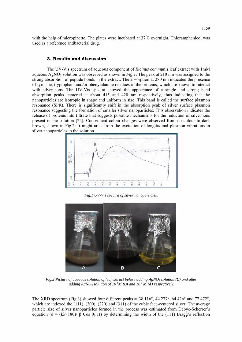
3. Results and discussion The UV-Vis spectrum of aqueous component of Ricinus communis leaf extract with 1mM
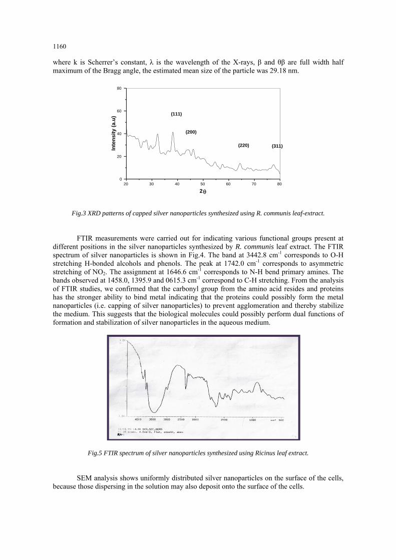
aqueous AgNO3 solution was observed as shown in Fig.1. The peak at 210 nm was assigned to the strong absorption of peptide bonds in the extract. The absorption at 280 nm indicated the presence of tyrosine, tryptophan, and/or phenylalanine residues in the proteins, which are known to interact with silver ions. The UV-Vis spectra showed the appearance of a single and strong band absorption peaks centered at about 415 and 420 nm respectively, thus indicating that the nanoparticles are isotropic in shape and uniform in size. This band is called the surface plasmon resonance (SPR). There is significantly shift in the absorption peak of silver surface plasmon resonance suggesting the formation of smaller silver nanoparticles. This observation indicates the release of proteins into filtrate that suggests possible mechanisms for the reduction of silver ions present in the solution [22]. Consequent colour changes were observed from no colour to dark brown, shown in Fig.2. It might arise from the excitation of longitudinal plasmon vibrations in silver nanoparticles in the solution.
Fig.1 UV-Vis spectra of silver nanoparticles.
Fig.2 Picture of aqueous solution of leaf extract before adding AgNO3 solution (C) and after adding AgNO3 solution of 10-1M (B) and 10-3 M (A) respectively.
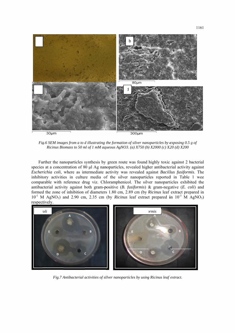
The XRD spectrum (Fig.3) showed four different peaks at 38.116°, 44.277°, 64.426° and 77.472°, which are indexed the (111), (200), (220) and (311) of the cubic face-centered silver. The average particle size of silver nanoparticles formed in the process was estimated from Debye-Scherrer’s equation (d = (kλ×180)/ β Cos θβ Π) by determining the width of the (111) Bragg’s reflection
1160 where k is Scherrer’s constant, λ is the wavelength of the X-rays, β and θβ are full width half maximum of the Bragg angle, the estimated mean size of the particle was 29.18 nm.
Fig.3 XRD patterns of capped silver nanoparticles synthesized using R. communis leaf-extract.
FTIR measurements were carried out for indicating various functional groups present at different positions in the silver nanoparticles synthesized by R. communis leaf extract. The FTIR spectrum of silver nanoparticles is shown in Fig.4. The band at 3442.8 cm-1 corresponds to O-H stretching H-bonded alcohols and phenols. The peak at 1742.0 cm-1 corresponds to asymmetric stretching of NO2. The assignment at 1646.6 cm-1 corresponds to N-H bend primary amines. The bands observed at 1458.0, 1395.9 and 0615.3 cm-1 correspond to C-H stretching. From the analysis of FTIR studies, we confirmed that the carbonyl group from the amino acid resides and proteins has the stronger ability to bind metal indicating that the proteins could possibly form the metal nanoparticles (i.e. capping of silver nanoparticles) to prevent agglomeration and thereby stabilize the medium. This suggests that the biological molecules could possibly perform dual functions of formation and stabilization of silver nanoparticles in the aqueous medium.
Fig.5 FTIR spectrum of silver nanoparticles synthesized using Ricinus leaf extract.
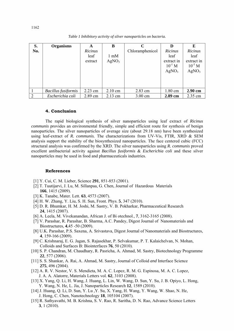
SEM analysis shows uniformly distributed silver nanoparticles on the surface of the cells, because those dispersing in the solution may also deposit onto the surface of the cells.
20 30 40 50 60 70 800
20
40
60
80
(200)
(311)(220)
(111)In
ten
sity
(a.
u)
2
Fig
FuspeciesEscherinhibitcompaantibacformed10-1 Mrespect
a
c
E. c
g.6 SEM imagRicinus Bio
urther the nans at a concenrichia coli, tory activitiearable with cterial activid the zone o
M AgNO3) atively.
Fig.7 A
coli
ges from a to domass to 50 m
noparticles sntration of 8where as ines in culturreference drity against b
of inhibition and 2.90 cm
Antibacterial a
d illustrating tml of 1 mM aqu
synthesis by 0 μl Ag nan
ntermediate are media ofrug viz. Chlboth gram-pof diameters
m, 2.35 cm
activities of si
the formation ueous AgNO3
green routenoparticles, ractivity wasf the silverloramphenic
positive (B. fs 1.80 cm, 2(by Ricinus
ilver nanopart
d
of silver nano3. (a) X750 (b)
was found evealed high revealed ag
r nanoparticol. The silvfusiformis) &.89 cm (by Rleaf extract
ticles by using
d
B. fusifo
b
oparticles by e) X2000 (c) X2
highly toxic her antibactergainst Bacillles reported
ver nanopart& gram-negRicinus leaf t prepared i
g Ricinus leaf
ormis
exposing 0.5 gX20 (d) X200
c against 2 berial activity lus fusiform
d in Table ticles exhibigative (E. co
extract prepin 10-3 M A
f extract.
1161
g of
acterial against is. The 1 wee ted the
oli) and pared in AgNO3)
1162
Table 1 Inhibitory activity of silver nanoparticles on bacteria.
S. No.
Organisms A Ricinus
leaf extract
B
1 mM AgNO3
C Chloramphenicol
D Ricinus
leaf extract in
10-1 M AgNO3
E Ricinus
leaf extract in
10-3 M AgNO3
1 Bacillus fusiformis 2.23 cm 2.10 cm 2.83 cm 1.80 cm 2.90 cm 2 Escherichia coli 2.89 cm 2.13 cm 3.00 cm 2.89 cm 2.35 cm
4. Conclusion The rapid biological synthesis of silver nanoparticles using leaf extract of Ricinus
communis provides an environmental friendly, simple and efficient route for synthesis of benign nanoparticles. The silver nanoparticles of average size (about 29.18 nm) have been synthesized using leaf-extract of R. communis. The characterizations from UV-Vis, FTIR, XRD & SEM analysis support the stability of the biosynthesized nanoparticles. The face centered cubic (FCC) structural analysis was confirmed by the XRD. The silver nanoparticles using R. communis proved excellent antibacterial activity against Bacillus fusiformis & Escherichia coli and these silver nanoparticles may be used in food and pharmaceuticals industries.
References
[1] Y. Cui, C. M. Lieber, Science 291, 851-853 (2001). [2] T. Tuutijarvi, J. Lu, M. Sillanpaa, G. Chen, Journal of Hazardous Materials 166, 1415 (2009). [3] K. Tanabe, Mater. Lett. 63, 4573 (2007). [4] H. W. Zhang, Y. Liu, S. H. Sun, Front. Phys. 5, 347 (2010). [5] D. R. Bhumkar, H. M. Joshi, M. Sastry, V. B. Pokharkar, Pharmaceutical Research 24, 1415 (2007). [6] A. Leela, M. Vivekanandan, African J. of Bi otechnol., 7, 3162-3165 (2008). [7] V. Parashar, R. Parashar, B. Sharma, A.C. Pandey, Digest Journal of Nanomaterials and Biostructures, 4,45 -50 (2009). [8] U.K. Parashar, P.S. Saxena, A. Srivastava, Digest Journal of Nanomaterials and Biostructures, 4, 159-166 (2009). [9] C. Krishnaraj, E. G. Jagan, S. Rajasekhar, P. Selvakumar, P. T. Kalaichelvan, N. Mohan, Colloids and Surfaces B: Biointerfaces 76, 50 (2010). [10] S. P. Chandran, M. Chaudhary, R. Pasricha, A. Ahmad, M. Sastry, Biotechnology Programme 22, 577 (2006). [11] S. S. Shankar, A. Rai, A. Ahmad, M. Sastry, Journal of Colloid and Interface Science 275, 496 (2004). [12] A. R. V. Nestor, V. S. Mendieta, M. A. C. Lopez, R. M. G. Espinosa, M. A. C. Lopez, J. A. A. Alatorre, Materials Letters vol. 62, 3103 (2008). [13] X. Yang, Q. Li, H. Wang, J. Huang, L. Lin, W. Wang, D. Sun, Y. Su, J. B. Opiyo, L. Hong, Y. Wang, N. He, L. Jia, J. Nanoparticles Research 12, 1589 (2010). [14] J. Huang, Q. Li, D. Sun, Y. Lu ,Y. Su, X. Yang, H. Wang, Y. Wang, W. Shao, N. He, J. Hong, C. Chen, Nanotechnology 18, 105104 (2007). [15] R. Sathyavathi, M. B. Krishna, S. V. Rao, R. Saritha, D. N. Rao, Advance Science Letters 3, 1 (2010).
1163
[16] J. Y. Song, H. K. Jang, B. S. Kim, Process Biochemistry 44, 1133 (2009). [17] S. S. Shankar, A. Ahmad, M. Sastry, Biotchnology Programme 19, 1627 (2003). [18] W. R. Rajesh, R. L. Jaya, S. K. Niranjan, D. M. Vijay, B.K. Sahebrao, Curriculum Nanoscience 5, 1117 (2009). [19] S. P. Dubey, M. Lahtinen, M. Sillanpaa, Colloids surface A 364, 34 (2010). [20] S. P. Dubey, M. Lahtinen, H. Sarkka, M. Sillanpaa, Colloid Surface B 80, 26 (2010). [21] N.Prabu, D.T. Raj, Y. Gowri, A. Siddiqua, J. Pushpa, Digest Journal of Nanomaterials and Biostructures, 5, 185-189 (2010). [22] B.D. Sawle, B. Salimath, R. Deshpande, M.D. Bedre, B.K. Prabhakar, A. Venkataraman, Science Technology Advance Materials, 9, 035012 (2008).