Biosynthesis of gold nanoparticles using streptomyces fulvissimus isolate
7
Please cite this paper as: Soltani Nejad M, Shahidi Bonjar Gh, Khaleghi N. Biosynthesis of gold nanoparticles using streptomyces fulvissimus isolate U., Nanomed J, 2015; 2(2): 153 -159. Received: Sep. 18, 2014; Accepted: Dec. 28, 2014 Vol. 2, No. 2, Spring 2015, page 153-159 Online ISSN 2322-5904 http://nmj.mums.ac.ir Original Research Biosynthesis of gold nanoparticles using streptomyces fulvissimus isolate Meysam Soltani Nejad 1* , Gholam Hosein Shahidi Bonjar 1 , Naimeh Khaleghi 2 1 Department of Plant Pathology, College of Agriculture, Shahid Bahonar University of Kerman, Iran 2 Department of Biotechnology, College of Energy Engineering and New Technologies, Shahid Beheshti University, Tehran, Iran Abstract Objective(s): In recent years, the biosynthesis of gold nanoparticles has been the focus of interest because of their emerging application in a number of areas such as biomedicine. In the present study we report the extracellular biosynthesis of gold nanoparticles (AuNPs) by using a positive bacterium named Streptomyces fulvissimus isolate U from rice fields of Guilan Province, Iran. Materials and Methods: From over 20 Streptomyces isolates collected, isolate U showed high AuNPs biosynthesis activity. To determine its taxonomical identity, its morphology was characterized by scanning electron microscope and partial molecular analysis performed by PCR. In this regard, 16S rDNA of isolate U was amplified using universal bacterial primers FD1 and RP2. The PCR products were purified and sequenced. Sequence analysis of 16S rDNA was then conducted using NCBI BLAST method. In biosynthesis of AuNPs by this bacterium, the biomass of bacterium exposed to the HAuCl4 solution. Results: The nanoparticles obtained were characterized by UV-Visible spectroscopy, transmission electron microscopy (TEM) and Energy dispersive X-ray (EDX) spectroscopy and X-ray diffraction spectroscopy (XRD) analyses. Our results indicated that Streptomyces fulvissimus isolate U bio-synthesizes extracellular AuNPs in the range of 20-50 nm. Conclusions: This technique of green synthesis of AuNPs by a microbial source may become a promising method because of its environmental safety. Its optimization may make it a potential procedure for industrial production of gold nanoparticles. Keywords: Biosynthesis, Nanogold, Streptomyces, Green process, 16S rDNA *Corresponding author: Soltani Nejad Meysam, Department of Plant Pathology, College of Agriculture, Shahid Bahonar University of Kerman, Kerman, Iran. Tel: 0098 913 378 2604, Email, [email protected]
-
Upload
nanomedicine-journal-nmj -
Category
Education
-
view
203 -
download
0
Transcript of Biosynthesis of gold nanoparticles using streptomyces fulvissimus isolate
Please cite this paper as:
Soltani Nejad M, Shahidi Bonjar Gh, Khaleghi N. Biosynthesis of gold nanoparticles using streptomyces
fulvissimus isolate U., Nanomed J, 2015; 2(2): 153 -159.
Received: Sep. 18, 2014; Accepted: Dec. 28, 2014
Vol. 2, No. 2, Spring 2015, page 153-159
Online ISSN 2322-5904
http://nmj.mums.ac.ir
Original Research
Biosynthesis of gold nanoparticles using streptomyces fulvissimus isolate
Meysam Soltani Nejad1*
, Gholam Hosein Shahidi Bonjar 1, Naimeh Khaleghi
2
1Department of Plant Pathology, College of Agriculture, Shahid Bahonar University of Kerman, Iran
2Department of Biotechnology, College of Energy Engineering and New Technologies, Shahid Beheshti
University, Tehran, Iran
Abstract
Objective(s): In recent years, the biosynthesis of gold nanoparticles has been the focus of
interest because of their emerging application in a number of areas such as biomedicine. In
the present study we report the extracellular biosynthesis of gold nanoparticles (AuNPs) by
using a positive bacterium named Streptomyces fulvissimus isolate U from rice fields of
Guilan Province, Iran.
Materials and Methods: From over 20 Streptomyces isolates collected, isolate U showed
high AuNPs biosynthesis activity. To determine its taxonomical identity, its morphology was
characterized by scanning electron microscope and partial molecular analysis performed by
PCR. In this regard, 16S rDNA of isolate U was amplified using universal bacterial primers
FD1 and RP2. The PCR products were purified and sequenced. Sequence analysis of 16S
rDNA was then conducted using NCBI BLAST method. In biosynthesis of AuNPs by this
bacterium, the biomass of bacterium exposed to the HAuCl4 solution.
Results: The nanoparticles obtained were characterized by UV-Visible spectroscopy,
transmission electron microscopy (TEM) and Energy dispersive X-ray (EDX) spectroscopy
and X-ray diffraction spectroscopy (XRD) analyses. Our results indicated that Streptomyces
fulvissimus isolate U bio-synthesizes extracellular AuNPs in the range of 20-50 nm.
Conclusions: This technique of green synthesis of AuNPs by a microbial source may become
a promising method because of its environmental safety. Its optimization may make it a
potential procedure for industrial production of gold nanoparticles.
Keywords: Biosynthesis, Nanogold, Streptomyces, Green process, 16S rDNA
*Corresponding author: Soltani Nejad Meysam, Department of Plant Pathology, College of Agriculture,
Shahid Bahonar University of Kerman, Kerman, Iran.
Tel: 0098 913 378 2604, Email, [email protected]

Biosynthesis of gold nanoparticles by Streptomyces fulvissimus
154 Nanomed J, Vol. 2, No. 2, Spring 2015
Introduction Nanotechnology is developed as an
important field of modern research with
potential effects in electronic and medicine
(1,2,3) Nanoparticles can be synthesized
by Physical and chemical methods. The
general chemical method of synthesis of
gold nanoparticles is by Turkevich
method, Frens method, Brust method,
microemulsion method, sonoelectro-
chemical method and lactic acid method
(4). The biosynthesis of gold nanoparticles
would benefit from the development of
clean, nontoxic, and ecofriendly accep-
table procedures concerning micro-
organisms from bacteria to fungi (5). The
simplest method for the production of
nanoparticles is the reduction of their
respective salts (6). Some examples of
nanoparticle formation by organisms are
magnetotactic bacteria synthesizing mag-
netite nanoparticles (7). Bacterium is
always been an organism of choice due to
its inherent properties to produce different
types of enzymes for chemical detox-
ification and energy-dependent ion efflux,
responsible for reduction and stabilization
of metallic nanoparticles (8). Gold
nanoparticles have found many
applications in diagnosis and therapy of
cancers, drug delivery, and gene therapy
(9,3). Nanoparticle synthesis is an
important component of rapidly growing
research efforts in nanoscale science and
engineering. Biotechnology approach tow-
ards the synthesis of nanoparticles has
many advantages, such as ease with which
the process can be scaled up, economic
viability, possibility of easily covering
large surface areas by suitable growth of
the mycelia, and its green chemistry nature
provided the microorganism medium is
safe (10). The aim of the present study was
to optimize the green synthesis of gold
nanoparticles by Streptomyces fulvissimus
isolate U. The biosynthesized gold
nanoparticles were characterized using a
UV-vis spectrophotometer, transmission
electron microscope (TEM), and Energy
dispersive X-ray (EDX) spectroscopy
analysis.
Materials and methods Source of Microorganisms
Soil samples were collected from rice
fields in different localities of Guilan
Province in northern Iran. Several samples
were randomly selected from the
mentioned localities using an open-end
soil borer (20 cm in depth, 2.5 cm in
diameter) as described by Lee and Hwang
(11). Soil samples were taken from a depth
of 10- 20 cm below the soil surface .The
soil of the top region (10 cm from the
surface) was excluded. Samples were air-
dried at room temperature for 10- 15 days
and then passed through a 0.8 mm mesh
sieve. Samples (10 g) of air-dried soil were
mixed with sterile distilled water (100
mL). The mixtures were shaken vigorously
for 1h and then allowed to settle for 1h.
Portions (1 mL) of soil suspensions
(diluted 10-1
) were transferred to 9 mL of
sterile distilled water and subsequently
diluted to 10-2
, 10-3
, 10-4
, 10-5
and 10-6
.
Inocula consisted of adding 1 ml of 10-3
-
10-6
soil dilutions to autoclaved casein
glycerol agar (CGA was prepared by
mixing the following contents; 0.3 g of
casein , 2 g of NaCl, 2 g of KNO3, 2 g of
k2HPO4, 0.5 g of MgSO4, 0.2 g of CaCO3,
10 g of glycerin, 18 g of Agar, in 1000
mL of distilled water ), 1, 25 mL CGA at
50 °C before pouring the 9 cm Petri plates
and solidification (12). Three replicates
were considered for each. Plates were
incubated at 28°C for up to 10 days. From
day 4 on, Streptomyces colonies were
isolated on CGA, incubated at 28°C for
two week and stored refrigerated as pure
cultures before use. Twenty strains of
Streptomyces spp. isolated from herbal rice
fields of Guilan Province.
Synthesis of gold nanoparticles
The bacteria, Streptomyces sp was isolated
and cultured. Culture was grown up in a
conical flask containing 100 mL of
casein glycerol (CG) medium in a shaker

Soltani Nejad M, et al
Nanomed J, Vol. 2, No. 2, Spring 2015 155
incubator at 28 . CG was prepared by
mixing the following contents; 3 g of
casein , 2 g of NaCl, 2 g of KNO3, 2g of
K2HPO4, 0.5 g of MgSO4, 0.2 g of
CaCO3, 10 g of glycerin in 1000 mL of
distilled water. After 5 days of incubation,
colonies developed on the medium. After
incubation time, the biomass was
harvested. The culture was centrifuged at
4000 rpm for 10 minutes and their colonies
were collected and used for further
experiments. 5 mL of 10-3
M aqueous
Auric Chloride (AuCl4) was added into the
colonies and control without the HAuCl4
(only biomass + distilled water) was also
run along with the experimental condition.
Then the reaction mixture was settle for a
further 24-48 h in a shaker incubator at
30 . After 48 h of incubation, red
coloration formed and this absorption
indicate the formation of gold nano-
particles. The synthesized gold
nanoparticles were characterized by UV-
Visible spectroscopy, Transmission
Electron Microscopy (TEM), Energy
Dispersive X-ray (EDX) spectroscopy and
X-ray diffraction spectroscopy (XRD).
Identification of the active Streptomyces
From all active Streptomyces, isolate U
showed high biosynthesis activity and their
colonies were characterized by morpho-
logically and phylogenetic analyses. The
morphological qualities of isolates U as
well as surface ornamentation were
evaluated by scanning electron microscopy
(SEM) of 10-day-old cultures grown on
CGA. Genomic DNA was extracted from
cultured cells with GeneAll® Exgene™
Cell Sv kit (http://www.geneall.com). The
16S rDNA of isolates 5 has been increased
by PCR, using universal bacterial primers
FD1 (5`-AGAGTTTGATCATGGCTCA-
G-3`) and RP2 (5`-ACGGTTACCTTG-
TTACGACTT-3`) following Kim et al
report (13). The PCR products were
purified and sequenced by macrogen
company (Seol, Korea). Sequence analysis
has been done by using BLAST by NCBI
(http://www.ncbi.nlm.nih.gov).
Results and Discussion Extracellular Synthesis of gold
nanoparticle
In this investigation, the gram positive soil
bacterium Streptomyces sp isolate U found
successful in producing gold nanoparticles.
The bacterium incubated with auric
chloride solution at 30 for 48 h. The
auric chloride ions were reduced during
the exposure to bacterial biomass. The
color of the reaction solution changed
from pale yellow color to deep red color
indicating the formation of gold nano-
particle. The result demonstrated those
gold nanoparticles are between 20 to 100
nm. Control experiments without the
HAuCl4 (only biomass + distilled water)
stayed yellow. The color of the solution is
due to the excitation of surface plasmon
vibrations in the gold nanoparticles (10).
Figure 1. Biosynthesized gold nanoparticles in a
colloidal dispersion by Streptomyces fulvissimus
isolate U colonies before (left) and after (right)
exposure to HAuCl4 after 48 h.
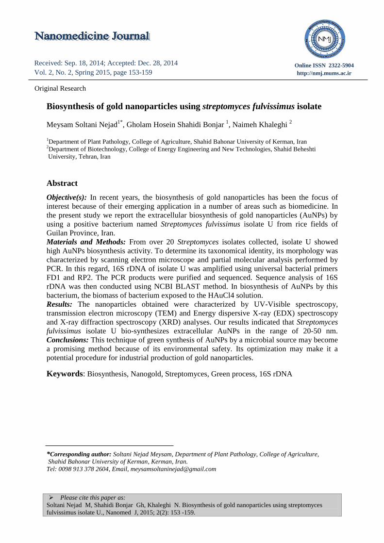
UV-Vis spectroscopy studies
The biosynthesis of gold nanoparticles by
Actinomycetes was performed in this
research. Metallic nanoparticles exhibited
peculiar optical absorption spectra in the
UV–Vis region due to collective osci-
llation of conduction band electrons
around the nanoparticle surface. The
collective oscillation of the conduction
band electrons on absorption of visible
light on the surface of nanoparticles was
known as surface plasmon resonance
(SPR) (14). The SPR indicated the specific
vibration mode according to size and
shape of nanoparticles. The synthesis of
Biosynthesis of gold nanoparticles by Streptomyces fulvissimus
156 Nanomed J, Vol. 2, No. 2, Spring 2015
gold nanoparticles was visually identified
by observing the change in the original
yellow color of gold aqueous solution with
gold cations into red color colloidal gold.
This visible change in color due to SPR
was accurately studied with UV–Vis
absorption spectrophotometer analysis of
colloidal gold solution (8). In this study,
we use UV-Vis spectroscopy to follow up
with the reaction process. After 48 h of
incubation, wine red coloration formed
which has absorption maxima of 550 nm
which clearly indicate the formation of
gold nanoparticles, figure 2.
Figure 2. The UV-vis spectrometer was used to record SPR of gold nanoparticle. After reactions with the
Streptomyces fulvissimus isolate U for 48 h. Presence of a strong peak with maximum absorbance at 550 nm is
prominent.
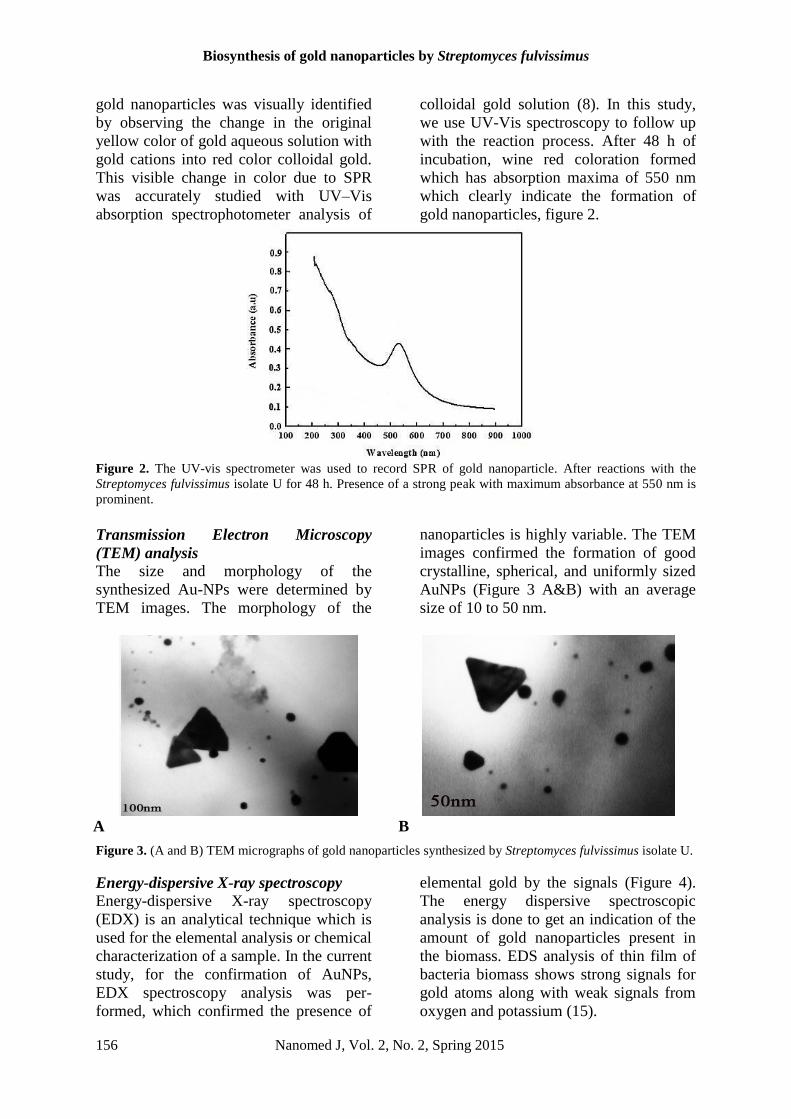
Transmission Electron Microscopy
(TEM) analysis
The size and morphology of the
synthesized Au-NPs were determined by
TEM images. The morphology of the
nanoparticles is highly variable. The TEM
images confirmed the formation of good
crystalline, spherical, and uniformly sized
AuNPs (Figure 3 A&B) with an average
size of 10 to 50 nm.
A B
Figure 3. (A and B) TEM micrographs of gold nanoparticles synthesized by Streptomyces fulvissimus isolate U.
Energy-dispersive X-ray spectroscopy
Energy-dispersive X-ray spectroscopy
(EDX) is an analytical technique which is
used for the elemental analysis or chemical
characterization of a sample. In the current
study, for the confirmation of AuNPs,
EDX spectroscopy analysis was per-
formed, which confirmed the presence of
elemental gold by the signals (Figure 4).
The energy dispersive spectroscopic
analysis is done to get an indication of the
amount of gold nanoparticles present in
the biomass. EDS analysis of thin film of
bacteria biomass shows strong signals for
gold atoms along with weak signals from
oxygen and potassium (15).

Soltani Nejad M, et al
Nanomed J, Vol. 2, No. 2, Spring 2015 157
Figure 4. EDS pattern for Streptomyces fulvissimus isolate U. Showing strong signals for gold nanoparticles at
different places.
XRD Analysis
For detection of Au-NPs used XRD
analysis (Figure 5).
The Au-NPs formed on the surface of
Streptomyces fulvissimus isolate U
have revealed clear peaks at 38.25 (111),
44.46 (200), 64.64 (220), and 77.
20 (311). The slight move in the peak
position may be owing to the presence of
some strain in the crystal structure, which
is a characteristic of nanocrystallites syn-
thesized through bio-method (16, 17). The
XRD result provides strong evidence for
confirming of the UV–Vis spectra and
TEM images for the presence of gold
particles.
Figure 5. X-ray diffraction pattern of gold nanoparticles synthesized by Streptomyces fulvissimus isolate U.
Identification of isolate U
Streptomyces sp isolate U was grown on
CGA (29° C for 14 days, Figure 6A) for
microscopic observations. Spore chain
morphology and spore ornamentation were
observed by scanning electron
microscopes.
Figure 6 shows scanning electron micro-
graph of spore chains of Streptomyces sp
isolate U.

Biosynthesis of gold nanoparticles by Streptomyces fulvissimus
158 Nanomed J, Vol. 2, No. 2, Spring 2015
A B
Figure 6. A: Pure culture of Streptomyces sp isolate U was grown on CGA at 29° C for 14 days, B: Scanning
electron micrograph of Streptomyces sp isolate U, showing spore chains.
The 16S rDNA of isolate U was then
amplified by PCR as presented in Figure 7.
Comparison of the near full length 16S
rDNA sequence of isolate U to GenBank
sequences, showed that it was most similar
to Streptomyces fulvissimus isolate U. (E-
value = 0.0 and max. identity = 99 %).
Figure 7. Amplification of 16S rDNA of isolate U by PCR (A) and the ladder (M).
Conclusion In this investigation, we showed the use of
Streptomyces fulvissimus isolate U in the
extracellular synthesis of gold nano-
particles. Green synthesis of metal nano-
particles using soil Actinomysetes bacteria
is an ecofriendly green process. In this
research, we demonstrated the green
extracellular synthesis of gold nano-
particles when the biomass of the
Streptomyces fulvissimus isolate U was
treated with 1 mM HAuCl4. The gold
nanoparticles were characterized by UV–
Vis spectroscopy, TEM, EDX and XRD.
The particle sizes were in the range of 20-
50 nm. UV–visible absorbance spectral
analysis confirmed the single surface
Plasmon resonance at 550 nm of
biosynthesized AuNPs. According to
previous studies on Actinobacteria, the
production of extracellular enzyme and
nanoparticles in this gram-positive
bacterium is more efficient than other
bacteria (17). It is also shown that
Streptomyces has easier and cheaper
cultivation requirements and higher
growth rates on both industrial and
laboratory scales, thereby having a lower
cost in large-scale production. Thus,
Streptomyces fulvissimus isolate U was
found to be a good candidate for the
production of gold nanoparticles.

Soltani Nejad M, et al
Nanomed J, Vol. 2, No. 2, Spring 2015 159
Acknowledgments This research was supported by Iran
National Science Foundation grant No.
91003706.
References 1. Boisselier E, Astruc D. Gold nanoparticles
in nanomedicine: preparation, imaging,
diagnostics, therapies and toxicity. Chem
Soc Rev. 2009; 38: 1759-1782.
2. Shahidi Bonjar L. Nanogold detoxifying
machine” to remove idle nanogold
particles from blood stream of cancer
patients treated with antibody-nanogold
therapeutics. Med hypotheses. 2013;
80(5): 601–605.
3. Groneberg DA, Giersig M, Welte T, Pison
U. Nanoparticle-based diagnosis and
therapy. Curr Drug Targets. 2006; 7: 643–
648.
4. Duraisamy K, Krishnamoorthy S, PB
Tirupathi P, Youn Soo C, Yang Soo L.
Synthesis, Characterization and In vitro
Cytotoxicity of Gold Nanoparticles Using
Cultural Filtrate of Low Shear Modeled
Microgravity and Normal Gravity
Cultured K. pneumonia. Macromol. Res.
2014; 22(5): 487-493.
5. Khadivi FD, Dehnad A, Mojtaba Salouti.
Extracellular Biosynthesis of Gold
Nanoparticles by Metal Resistance
Bacteria: Streptomyces griseus. synth
react inorg met-org nano-met chem. 2012;
42: 868–871.
6. Pranav V, Batherinarayanan A,
Dilliganesh T, Kumar M, Munusamy C,
Baskar G. Biological synthesis and
characterization of intracellular gold
nanoparticles using biomass of
Aspergillus fumigatus. Bull Mater Sci.
2013; 36(7): 1201–1205.
7. Lowenstam HA. Minerals formed by
organisms. Science. 1981;211:1126–30.
8. Nishant S, Mausumi M. Biosynthesis and
Characterization of Gold Nanoparticles
Using Zooglea ramigera and Assessment
of Its Antibacterial Property. Journal of
Cluster Science. 2014. Available from
URL: http://www.springer.com/chemistry-
/catalysis/journal/10876
9. Huff TB, Tong L, Zhao Y, Hansen MN,
Cheng JX, Wei A. Hyperthermic effects of
gold nanorods on tumor cells. Nano-
medicine. 2007; 2: 125–132.
10. Khabat V, Mansoori GA, Karimi S.
Biosynthesis of Silver Nanoparticles by
Fungus Trichoderma Reesei. Insciences J.
2011; 1(1): 65-79.
11. Lee JY, Hwang BK. Diversity of
antifungal Actinomycetes in various
vegetative soils of Korea. Can J
Microbiol. 2002; 48: 407-417.
12. Shahidi Bonjar GH. New approaches in
screening for antibactrials in plants. Asian
Journal Plant Science. 2004; 3: 55-60.
13. Kim HJ, Lee SC, Hwang BK.
Streptomyces cheonanensis sp. Nov., a
novel streptomycete with antifungal
activity. Int J Syst Evol Microbiol. 2006;
56: 471–475.
14. Pasqua AJD, Mishler RE, Ship YL,
Dabrowiak JC, Asefa T. Preparation of
antibody conjugated gold nanoparticles.
Mater. Lett. 2009; 63:1876.
15. Vasanathi P B, Thangavelu D, Vasanth
KM, Munusamy C, Baskar G. Biological
synthesis and characterization of
intracellular gold nanoparticles using
biomass of Aspergillus fumigatus. Bull
Mat Sci. 2013; 7:1201-1205.
16. Namvar F, Azizi S, Mansour BA, Shameli
K, Rosfarizan, Mahdavi M, Paridah Md,
Tahir. Green synthesis and characterization
of gold nanoparticles using the marine
macroalgae Sargassum muticum. Green
synthesis and characterization of gold
nanoparticles using the marine macroalgae
Sargassum Muticum. Research on Chemical
Intermediates. 2014; Available from URL:
http://springer.libdl.ir/article/10.1007/s11164
-014-1696-4
17. Jannu VG, Thenmozhi M , Kannabiran K ,
Rajakumar G, Velayutham K, Rahuman
AA. Actinobacteria mediated synthesis of
gold nanoparticles using Streptomyces sp.
VITDDK3 and its antifungal activity. Mater.
Lett. 2013; 93: 360–362.